Embed Size (px)
Citation preview

Consensus Micro RNAs Governing the Switch of DormantTumors to the Fast-Growing Angiogenic PhenotypeNava Almog1*, Lili Ma1, Christian Schwager2, Bastian G. Brinkmann3, Afshin Beheshti1, Peter Vajkoczy3,
Judah Folkman4{, Lynn Hlatky1, Amir Abdollahi1,2*
1 Center of Cancer Systems Biology, Steward Research & Specialty Projects Corp., St. Elizabeth’s Medical Center, Tufts University School of Medicine, Boston,
Massachusetts, United States of America, 2 Molecular and Translational Radiation Oncology, Heidelberg Ion Therapy Center, University of Heidelberg Medical School and
National Center for Tumor diseases, German Cancer Research Center, Heidelberg, Germany, 3 Department of Neurosurgery, Charite – Universitaetsmedizin Berlin, Berlin,
Germany, 4 Department of Surgery, Harvard Medical School and Vascular Biology Program, Children’s Hospital, Boston, Massachusetts, United States of America
Abstract
Tumor dormancy refers to a critical stage in cancer development in which tumor cells remain occult for a prolonged periodof time until they eventually progress and become clinically apparent. We previously showed that the switch of dormanttumors to fast-growth is angiogenesis dependent and requires a stable transcriptional reprogramming in tumor cells.Considering microRNAs (miRs) as master regulators of transcriptome, we sought to investigate their role in the control oftumor dormancy. We report here the identification of a consensus set of 19 miRs that govern the phenotypic switch ofhuman dormant breast carcinoma, glioblastoma, osteosarcoma, and liposarcoma tumors to fast-growth. Loss of expressionof dormancy-associated miRs (DmiRs, 16/19) was the prevailing regulation pattern correlating with the switch of dormanttumors to fast-growth. The expression pattern of two DmiRs (miR-580 and 190) was confirmed to correlate with diseasestage in human glioma specimens. Reconstitution of a single DmiR (miR-580, 588 or 190) led to phenotypic reversal of fast-growing angiogenic tumors towards prolonged tumor dormancy. Of note, 60% of angiogenic glioblastoma and 100% ofangiogenic osteosarcoma over-expressing miR190 remained dormant during the entire observation period of , 120 days.Next, the ability of DmiRs to regulate angiogenesis and dormancy-associated genes was evaluated. Transcriptionalreprogramming of tumors via DmiR-580, 588 or 190 over-expression resulted in downregulation of pro-angiogenic factorssuch as TIMP-3, bFGF and TGFalpha. In addition, a G-CSF independent downregulation of Bv8 was found as a commontarget of all three DmiRs and correlated with decreased tumor recruitment of bone marrow-derived CD11b+ Gr-1+ myeloidcells. In contrast, antiangiogenic and dormancy promoting pathways such as EphA5 and Angiomotin were upregulated inDmiR over-expressing tumors. This work suggests novel means to reverse the malignant tumor phenotype into anasymptomatic dormant state and may provide promising targets for early detection or prevention of cancer.
Citation: Almog N, Ma L, Schwager C, Brinkmann BG, Beheshti A, et al. (2012) Consensus Micro RNAs Governing the Switch of Dormant Tumors to the Fast-Growing Angiogenic Phenotype. PLoS ONE 7(8): e44001. doi:10.1371/journal.pone.0044001
Editor: Rossella Rota, Ospedale Pediatrico Bambino Gesu, Italy
Received February 25, 2012; Accepted July 27, 2012; Published August 31, 2012
Copyright: � 2012 Almog et al. This is an open-access article distributed under the terms of the Creative Commons Attribution License, which permitsunrestricted use, distribution, and reproduction in any medium, provided the original author and source are credited.
Funding: This material is based upon work supported by the National Aeronautics and Space Administration Specialized Center of Research (NSCOR, grant no.NNJ06HA28G) issued through the Human Research Program. It was also supported in part by the German Krebshilfe (Deutsche Krebshilfe, Max-Eder 108876),German Research Foundation National Priority Research Program: the Tumor-Vessel Interface ‘‘SPP1190’’, Intramural Grants of the National Center for Tumordiseases (Heidelberg, Germany) and the German Federal Ministry of Research and Technology (Bundesministerium fur Bildung und Forschung – BMBF03NUK004C). The funders had no role in study design, data collection and analysis, decision to publish, or preparation of the manuscript.
Competing Interests: The authors have declared that no competing interests exist.
* E-mail: [email protected] (AA); [email protected] (NA)
{ Deceased.
Introduction
Tumor dormancy is an early stage in cancer progression in
which small cancerous lesions (few millimeters in diameter) remain
occult and asymptomatic until they eventually switch to become
fast-growing, clinically apparent and potentially lethal cancer.
Dormant cancerous lesions are highly prevalent in asymptomatic
normal populations [1]. However, the majority of these lesions
never progress to the stage of exponential tumor growth. This
implies that dormant tumors rarely succeed in overcoming
inherent defense mechanisms against tumor development. These
include cell cycle arrest of disseminated tumor cells (DTCs), tumor
cell senescence, immune response of the host, hormonal control or
the insufficiency of dormant tumors to recruit new blood vessels
[2–14].
Of particular clinical importance is the fact that dormant tumor
cells left after primary tumor removal or treatment may contribute
to tumor relapse and are often refractory to cancer therapies [2].
Although the tumor dormancy phase is a promising therapeutic
target, it is still one of the most neglected areas in cancer biology.
This is mainly due to lack of suitable experimental models and
limited clinical accessibility to dormant tumors.
We have successfully established in-vivo models of vascular
tumor dormancy of human breast cancer, glioblastoma, osteosar-
coma, and liposarcoma in immunocompromised mice [15,16]. In
these models, tumor dormancy is characterized by high prolifer-
ation of tumor cells balanced by apoptosis and impaired tumor
angiogenesis. Tumors remain dormant for a prolonged period of
time until they spontaneously switch to the fast-growing angio-
genic phenotype. These angiogenic tumors retain their ability to
PLOS ONE | www.plosone.org 1 August 2012 | Volume 7 | Issue 8 | e44001
grow fast once injected in new mice. Therefore, we hypothesized
that dormant tumors undergo a stable genetic ‘‘reprogramming’’
during their transition from dormancy to fast-growth [17].
Although the mechanisms triggering the transition from dormancy
to rapid growth remain to be elucidated, pathways promoting
tumor dormancy and the transcriptional switch of dormant tumors
to the fast-growing phenotype was recently deciphered by genome-
wide expression analysis [17].
MicroRNAs are considered potential master regulators’’ of gene
expression [18,19]. In a hierarchical manner, a single microRNA
(miR) could regulate the expression level of multiple target genes.
Therefore, we sought to characterize the ‘master regulators’ of
tumor dormancy by utilizing the in-vivo models established in our
lab.
Here we report the identification of a consensus miR signature
that governs tumor dormancy. A concerted shift in expression of
these miRs correlated with the conversion of all four dormant
tumors to the fast-growing angiogenic phenotype. Expression
patterns of candidate dormancy miRs in tumor cells were
correlated with disease stage in glioma patients. We further show
that reconstitution of single candidate tumor dormancy-associated
miRs (DmiRs) in fast-growing glioblastoma or osteosarcoma cells
resulted in significant inhibition of tumor growth. Over-expression
of the three DmiRs also led to suppression of pro-angiogenic and
upregulation of antiangiogenic genes and correlated with marked
inhibition of tumor growth and prolonged dormancy periods.
Moreover, the expression of DmiRs led to repression of Bv8 and
reduced levels of pro-angiogenic bone marrow-derived CD11b+GR1+ myeloid cells in circulation and in the tumor microenvi-
ronment. These data may provide molecular instructions to
address the unmet medical need for blocking tumor progression in
an early stage and developing novel early tumor dormancy
biomarkers.
Results
Four different human tumor dormancy in-vivo models have been
established and phenotypically characterized in our laboratory.
These include breast carcinoma (MDA-MB-436), glioblastoma
multiforme (GBM, T98G), osteosarcoma (KHOS-24OS) and
liposarcoma (SW872) human tumor cell lines. During the
dormancy period of these tumors, which is characterized by
impaired tumor angiogenesis, tumor cell proliferation is balanced
by a high rate of apoptosis (Figure 1A). From each tumor type, we
isolated and established cell lines that when injected to mice can
form either dormant or fast-growing tumors [15,16]. These cell
lines were shown to maintain their characteristics when cultured
in-vitro. According to the tumor phenotype which the cell lines
generate, we hereinafter use the prefix ‘‘D’’ for dormant and ‘‘A’’
for fast-growing angiogenic tumors.
Identification of the consensus tumor dormancyassociated microRNAs
We sought to identify the consensus set of miRs that govern the
transition of all four dormant tumors to the fast-growing
angiogenic phenotype. Therefore, total RNA was extracted from
dormant (D) and fast-growing angiogenic (A) clones of all four
tumor types and subjected to high-throughput real time qRT-PCR
based miR expression analysis (Figure 1B). Among the 378 miRs
investigated, we found 19 miRs to be differentially regulated in the
same patterns between all dormant vs. fast-growing tumors
(p,0.03). Interestingly, we found a significant enrichment of
upregulated miRs (84% or 16 miRs) in dormant tumors as
compared to their expression levels in switched fast-growing
tumors (p,0.00004 by hypergeometric distribution). These data
suggest that loss of DmiR function is the key regulation pattern
correlating with the ability of dormant tumors to switch to the
angiogenic phenotype and grow rapidly. The 16 DmiRs identified
include the Homo sapiens microRNAs 101, 320, 193b, 218, 151,
19a, 331, 340, 184, 186, 190, 185, 580, 588, 202 and 545. Three
microRNAs (miR-520g, 657 and 92) were found to be down-
regulated in dormant tumors as compared to the fast-growing
tumors (Figure 1B and Information S1). For all subsequent
confirmatory and functional validation studies, the top three
dormancy-associated miRs – miR-580, miR-588 and miR-190–
were used. These three DmiRs were selected based on their
marked differential expression in dormant tumors; at least three
out of four dormant tumors had an average of , 12, 10 and 75-
fold higher expression levels of miR-580, miR-588 and miR-190,
as compared to the fast-growing tumors. The regulation of miR-
580 and miR-190 was further confirmed in a second independent
set of dormant vs. angiogenic tumors (Information S1).
Regulation of DmiRs in glioma patientsTo examine the regulation patterns of the identified DmiRs in
primary human tumor tissue, the expression levels of miR-580,
miR-588 and miR-190 in tissue specimens of glioma patients were
investigated. Total RNA was isolated from three World Health
Organization (WHO) grade I, four grade III and eight grade IV
glioma tumors. Real time qRT-PCR analysis revealed significant
correlation between miR-190 (p,0.0076) and miR-580 (p = 0.05)
expression and tumor grade. As shown in Figure 1 C–D, the
expression levels of miR-580 and miR-190 decreased with
advanced tumor grade. Hence, the direction of DmiR regulation
in primary brain tumors parallels the regulation pattern identified
in the tumor dormancy models.
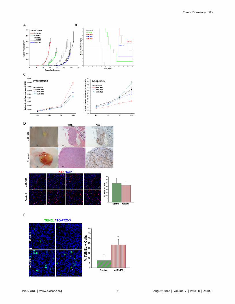
Over-expression of DmiRs in fast-growing glioblastomainhibits tumor growth
To test potential dormancy promoting functional effects of the
identified DmiRs, lentiviral vectors encoding miR-580, miR-588
or miR-190 were used to reconstitute their expression in fast-
growing angiogenic T98G human glioblastoma multiforme cells
(A-GBM). Over-expression of DmiRs in A-GBM cells was
confirmed by qRT-PCR (Information S1), and mice were injected
with miR-580, miR-588, miR-190 or vector control (GFP)
expressing A-GBM cells. In addition non-transfected ‘‘parental’’
A-GBM were used as control.
Consistent with our previous data, the parental fast-growing A-
GBM tumors were detectable between 36–42 days post injection
(Figure 2A). Surprisingly, the infection of A-GBM tumor cells with
negative control vector (expressing all elements as DmiR
expressing vectors but lacking any microRNA sequence) modu-
lated tumor growth kinetic and modestly delayed the time to
establishment of macroscopically detectable tumors. This is
assumed to be a cell type specific effect of GFP expression, as it
was not evident in other tumor cell lines. However, the expression
of each of the DmiRs led to a clear inhibition of tumor growth and
a prolongation of the time required for tumors to become
palpable. The average tumor dormancy period, i.e., the time until
tumors became detectable by gross examination, was increased to
, 100 days by over-expression of a single DmiR (e.g., miR-580 or
miR-190). Inhibition of tumor growth by expression of miR-580,
miR-588 or miR-190 had also been observed in osteosaroma cell
lines.
In line with these data, Kaplan-Meier analysis of the ‘‘tumor
free’’ mice population revealed marked differences in the DmiR
expressing vs. the vector control (GFP only) or parental A-GBM
Tumor Dormancy miRs
PLOS ONE | www.plosone.org 2 August 2012 | Volume 7 | Issue 8 | e44001

bearing mice. ‘‘Tumor free’’ mice were defined as those with no
tumors detectable by gross examination or those with tumors
smaller than 50 mm3. The median ‘‘tumor free’’ period, i.e., the
time until tumors were detected in 50% of the monitored animals
in each group, was markedly prolonged in miR-588 (,78 days)
and miR-580 (,103 days) expressing tumors as compared to
GFP-vector-control (,61 days) or non-transfected parental A-
GBM (40 days) (Figure 2B). Of note, the median survival time of
the mice with miR-190 expressing tumors exceeded the observa-
tion period.
As previously described, tumor ‘‘take’’, i.e., the ability to detect
tumors by gross examination, correlates with the switch of tumors
to the fast-growing angiogenic phenotype, whereas ‘‘no-take’’
indicates the existence of vital but dormant tumors. In the control
groups, all parental A-GBM cell lines developed tumors (11/11,
100% tumor take rate), while , 90% (8/9) of GFP expressing
tumors established detectable tumors within the observation
period (.140 days). In contrast, the tumor take rates were 80%
(4/5) for miR-580, 75% (3/4) for miR-588 and only 40% (2/5) in
miR-190 over-expressing tumors (Table 1). It is intriguing that
over-expression of a single DmiR, e.g., miR-190, was sufficient to
induce a complete phenotypic reversal of fast-growing A-GBM
tumors towards prolonged tumor dormancy in three out of five
transfected tumors. The growth dynamics of single tumors within
the GFP-vector-control and miR-190 expressing tumors are
presented in the Information S1.
DmiRs do not attenuate tumor cell proliferation in-vitroand in-vivo
Next, we aimed to investigate if the dormancy promoting effect
of DmiRs is mediated by direct inhibition of tumor cell
proliferation or enhanced apoptosis. Expression of miR-580,
miR-588, miR-190 as well as the control-GFP-vector neither
blocks cell proliferation nor enhances cell death even in-vitro at
hyperconfluence state (after 144h) as determined by caspase 3/7
activity (Figure 2C). Moreover, miR-580 expressing cells showed a
trend toward enhanced proliferation and reduced apoptosis as
compared to GFP-control. The high proliferative capacity of
DmiR expressing tumors was confirmed by Ki67 immunostaining
of dormant tumors in-vivo. Figure 2D shows the high rate of
proliferative Ki67+ cells in a representative dormant miR-588
expressing A-GBM tumor at day 113 post injection. This tumor
Figure 1. Consensus signature of dormancy associated miRs. Aschematic overview of the in-vivo experimental vascular tumordormancy models employed (A). Dormant tumors are formed afters.c. injection of tumor cells in immunocompromised mice and remainundetectable by gross examination for a prolonged period of time. Thisdormancy period is characterized by impaired tumor angiogenesis andhigh levels of cell turnover, i.e., balanced tumor cell proliferation andapoptosis. Tumors spontaneously exit the dormancy phase and switchto a rapid ‘‘angiogenic’’ growth. Differential regulation of miRs indormant vs. fast-growing tumors (B). 19 miRs were significantlydifferentially regulated during the switch of dormant tumors to fast-growth in all four dormancy models (p,0.03). The prevailing regulationpattern was the loss of dormancy associated miRs (DmiRs) after theswitch of dormant tumors. The heatmap represents fold expressionvalues of miRs in dormant vs. fast-growing tumors according to thescale bar. Red: miRs upregulated in dormant tumors. Green: miRs down-regulated in dormant tumors. MiRs are sorted by hierarchical clustering.The expression level of miR-580, miR-588 and miR-190 was detected inhuman glioma specimens (C–E). In support of the experimental data,the expression of miR-580 and miR-190 was significantly decreased withadvanced tumor grade in glioma specimens. WHO-I (n:3), WHO-III (n:4)and WHO-IV (n:8). *p,0.01, #p = 0.05.doi:10.1371/journal.pone.0044001.g001
Tumor Dormancy miRs
PLOS ONE | www.plosone.org 3 August 2012 | Volume 7 | Issue 8 | e44001

was undetectable by gross examination and could only be detected
by careful examination followed by skin-flip at the site of injection.
The in-vivo proliferation index for miR-588 expressing vs. control
(GFP) A-GBM tumors was assessed by counting the fraction of
Ki67+ tumor cells in 15 (miR-588) and 13 (control) representative
high power fields (20x). This analysis revealed no significant
difference in proliferation between the two groups (p = 0.12) with a
trend toward more proliferation in dormant miR-588 expressing
tumors. These data suggest that the tumor dormancy promoting
effect of DmiRs could not be attributed to the direct blockage of
tumor cell proliferation. The in-vivo apoptosis index for miR-588
expressing vs. control (GFP) A-GBM tumors was assessed by
counting the fraction of TUNEL+ tumor cells in 16 (miR-588) and
18 (control) representative high power fields (20x). In line with our
previous reports in these dormancy models, the fraction of
TUNEL positive apoptotic tumor cells was threefold increased
in-vivo in dormant miR-588 expressing tumors vs. GFP-control
tumors (p,0.0001, Figure 2E).
Complete phenotypic reversal after miR-190 expressionin fast-growing osteosarcoma
MicroRNA-190 was the most effective dormancy promoting
DmiR in our GBM model. The tumor model with the most
significant differential regulation of this DmiR was the KHOS-
24OS osteosarcoma. We detected more than 210-fold higher
expression levels of miR-190 in dormant vs. fast-growing
osteosarcoma tumor cells (Table S1). Over-expression of miR-
190 in fast-growing osteosarcoma cells led to complete inhibition
of osteosarcoma growth (Figure 3). During the observation period
of .120 days, no tumor was detected by gross examination in
miR-190 expressing osteoscaroma (0/5), whereas the vector
control GFP osteosarcoma exhibited 100% tumor take (5/5). In
line with this observation, overall survival was significantly
prolonged in miR-190 vs. control group (p,0.005). Together,
these data support the proposed potent dormancy promoting effect
of miR-190.
Identification of the consensus transcriptional targets ofDmiRs
To identify the consensus target genes of DmiRs, the expression
of a panel of angiogenesis- and dormancy related genes was
profiled by qRT-PCR. These genes were in part selected based on
the consensus transcriptional signature that we recently reported
to participate in the switch of dormant tumors to the fast-growing
angiogenic phenotype [17]. We found that antiangiogenic and
dormancy promoting genes, Angiomotin (AMOT-1) and Eph
receptor A5 (EphA5), were both upregulated in all DmiR
expressing A-GBM tumors as compared to the GFP-vector-
control A-GBM cells (Figure 4). In contrast, genes involved in pro-
angiogenic signaling, including tissue inhibitor of metalloprotei-
nases 3 (TIMP-3), hypoxia-induced factor 1 alpha (HIF-1-alpha),
basic fibroblast growth factor (bFGF, FGF2), and the K-ras tumor
oncogene, were consistently downregulated in DmiR expressing
A-GBM. We found transforming growth factor alpha (TGF-a) as a
common target of all three DmiR expressing A-GBM. Impor-
tantly, we found Bv8 also known as prokineticin 2 (Prok2) to be
markedly downregulated in all three DmiR expressing A-GBM
(Figure 5A). Bv8 has been recently discovered to play a key role in
myeloid cell-dependent tumor angiogenesis and in the angiogenic
switch [20,21]. Therefore, concerted downregulation of Bv8 by all
three DmiRs led us to investigate the contribution of bone
marrow-derived cells (BMDCs) in the reversal of the angiogenic
phenotype observed in DmiR expressing A-GBM tumors.
DmiR induced Bv8 down-regulation correlates withdecreased recruitment of myeloid cells
We sought to investigate the role of Bv8 down-regulation as a
consensus target of DmiR 1-3 expression in A-GBM tumors by
analyzing the mobilization and tumor recruitment of bone marrow-
derived CD11b+ and Gr1+ myeloid cells. Size-matched GFP-
vector-control and miR-588 expressing A-GBM tumors were
harvested at day 35 post injection. Tumors were stained for
Gr1+, CD11b+ (myeloid) or CD31+ (endothelial) cells (Figure 5B).
CD11b+ and Gr1+ myeloid cells were highly abundant in control
A-GBM tumors, whereas their level was markedly reduced in miR-
588 expressing tumors. We found that miR-588 expression and
down-regulation of Bv8 preferentially impaired the recruitment of
the Gr1+ myeloid cell population. Gr1+ myeloid cells were almost
absent in miR-588 expressing tumors (Figure 5B). The reduced
recruitment of Gr1+ myeloid cells also correlated with impaired
angiogenesis in miR-588 expressing tumors as detected by CD31
staining of the tumor microvascular endothelium.
To detect potential effects of DmiRs on bone marrow
mobilization, we examined the circulating levels of CD11b+Gr1+ myeloid cells. In line with the reduced recruitment of these
cells detected in the tumor microenvironment, we found decreased
levels of Gr1+/CD11b+ cells in circulation in miR-588 expressing
tumors as compared to the levels in GFP-vector-control A-GBM
(Figure 5C). Downregulation of Bv8 by DmiRs was clearly
correlated with reduced mobilization and tumor recruitment of
bone marrow derived CD11b+ Gr1+ myeloid cells, as well as with
impaired angiogenesis.
Discussion
We report here the discovery of a consensus set of 19 micro
RNAs that are significantly regulated during the transition of four
Table 1. Numbers of mice with dormant ‘‘undetectable’’ A-GBM tumors.
microRNA Undetectable tumors Total number of mice Percentage (%)
Fast growing ‘parental’ 0 11 0
GFP (Control) 1 9 11
miR-580 1 4 25
miR-588 1 5 20
miR-190 3 5 60
Number of mice bearing undetectable tumors or tumors with volume smaller than 50 mm3 at the end point of experiments: day 64 for parental fast-growing A-GMBtumors, day 107 for control GFP expressing tumors, day 113 for miR-588 tumors, day 118 for miR-190 tumors, and day 127 for miR-580 tumors.doi:10.1371/journal.pone.0044001.t001
Tumor Dormancy miRs
PLOS ONE | www.plosone.org 4 August 2012 | Volume 7 | Issue 8 | e44001
Tumor Dormancy miRs
PLOS ONE | www.plosone.org 5 August 2012 | Volume 7 | Issue 8 | e44001

Figure 2. Over-expression of DmiRs reversed tumor phenotype. Tumor growth kinetics of angiogenic fast-growing glioblastoma cells (A-GBM) was monitored in-vivo (A). Average tumor volume of parental tumor (labeled in red line) n = 11, cells infected with GFP only control vector(n = 9), miR-580 (n = 5), miR-588 (n = 4), and miR-190 (n = 5) expressing A-GBM tumors. Kaplan-Meier analysis of tumor free survival (B). The criterionfor ‘‘tumor free’’ was defined as mice bearing no detectable tumors by gross examination or tumors with volume smaller than 50 mm3. The mediantumor free survival was markedly increased in miR-580 and miR-588 expressing tumors as compared to the GFP control. 60% of miR-190 expressingtumors remained dormant for the whole observation period. Statistical significance was reached for miR-190 (p,0.03) and miR-580 (p = 0.05),respectively, by log-rank test. The dormancy promoting effect of DmiRs was not attributed to impaired proliferation kinetic or enhanced apoptosis ofDmiR expressing tumor cells in-vitro (C). In contrast, there was a trend towards higher proliferation rates and reduced apoptosis in miR-580expressing cells (C). Proliferating tumor cells were also detected by Ki67 immunohistochemistry and representative photomicrographs are shown (D)in dormant miR-588 expressing GBMs (107days post implantation) and fast-growing GFP expressing control tumors (day 113 after implantation). In-vivo proliferation index was determined by counting the fraction of Ki67+ tumor cells. This analysis revealed no significant difference between thedormant miR-588 and the fast growing control (GFP) tumors (p = 0.12). Enhanced apoptosis was detected in-vivo in miR-588 vs. control (GFP) tumorsby TUNEL staining (E). A threefold increase in the fraction of TUNEL+ apoptotic tumor cells was found in dormant miR-588 expressing- vs. controltumors (* p,0.0001).doi:10.1371/journal.pone.0044001.g002
Figure 3. MiR-190 expression in osteosarcoma model. Tumor growth kinetic of vector control (GFP; n = 5) vs. miR-190 (n = 5) expressingangiogenic fast-growing ‘‘A-Osteosarcoma’’ (A). MiR-190 expression led to complete (i.e., 100% inhibition, 5/5) phenotypic reversal of fast-growingosteosarcoma resulting in significant increase of overall survival (p,0.005 by log-rank test) (B). Death event: mouse sacrificed based on large tumorsize.doi:10.1371/journal.pone.0044001.g003
Tumor Dormancy miRs
PLOS ONE | www.plosone.org 6 August 2012 | Volume 7 | Issue 8 | e44001

different human dormant tumors to exponential growth. Sixteen
miR were found with high expression levels in dormant tumors.
Downregulation of these dormancy associated miRs (DmiRs)
correlated with the switch of dormant tumors to the fast-growing
angiogenic phenotype. The top three DmiRs were selected based
on consistent high expression levels in dormant tumors. In support
of the data from the experimental dormancy models, miR-580 and
miR-190 expression was shown to correlate with disease stage in
human glioma specimens. Their expression was gradually
decreased with advanced tumor grade suggesting a tumor
suppressive function for the identified dormancy associated miRs.
Based on the preponderance of upregulated miRs in dormant
vs. fast-growing tumors (16 out of 19 miRs found), we
hypothesized that loss of DmiRs constitutes the prevailing
regulation pattern correlating with the switch of dormant tumors
to a stage of exponential growth. To prove this hypothesis, we
reconstituted the expression of miR-580, miR-588 or miR-190 in
fast-growing A-GBM tumors. Moreover, we tested the expression
of miR-190 in a fast-growing A-Osteosarcoma model. To our
knowledge, this is the first report demonstrating the phenotypic
reversal of fast-growing human tumors back to the dormancy
phase by over-expression of a single miR. It is intriguing that over-
expression of a single miR, in particular miR-190, resulted in
significant delay of time to tumor establishment in 40% and in
sustained dormancy in 60% of GBM tumors, i.e., no detectable
tumors were found by gross examination up to 140 days post
injection. Moreover, miR-190 expression led to complete pheno-
typic reversal in 100% of fast-growing A-Osteosarcoma. These
dramatic effects might be attributed to the unique ability of single
miRs to regulate expression levels of a large number of genes
[22,23]. Unfortunately, the knowledge of the function and
potential gene targets of miR-580, miR-588 and miR-190 is very
limited and their contribution to cancer progression is unknown.
Therefore, identification of potential miR targets in this relatively
new field of gene regulation relies mostly on bioinformatics
prediction.
To dissect the molecular mechanism underlying the phenotypic
reversal of DmiR expressing A-GBM tumors, the expression level
of a selected set of genes was profiled. These genes were either
involved in the angiogenesis process or were recently shown to
participate in the switch of dormant tumors to the angiogenic
phenotype [17]. We further aimed to identify common targets of
all three DmiRs. Among the consensus miR-580, miR-588 and
miR-190 upregulated transcripts, we found two important
dormancy associated genes, i.e., EphA5 and Angiomotin.
Angiostatin, a key endogenous angiogenesis inhibitor and
dormancy promoting factor [24–26], was shown to be preferen-
tially enriched in angiomotin positive dormant tumors [17].
Likewise, EphA5 was previously shown to be expressed in dormant
tumors, downregulated once they switch to the angiogenic
phenotype, and to gradually decrease with advanced tumor stage
in tumor specimens of glioma patients [17]. Moreover, detection
Figure 4. Differential expression of angiogenesis and dormancy related genes in DmiR expressing tumors. qRT-PCR analysis of geneexpression in glioblastoma cells. Expression levels of genes in miR-580, miR-588 and miR-190 over-expressing A-GBM tumors were normalized to GFP-vector control. DmiR expression induced a transcriptional switch towards antiangiogenesis by down-regulation of multiple angiogenesis relatedgenes (e.g., bFGF (FGF2), TIMP-3, HIF1A and TGFalpha). In contrast antiangiogenic and dormancy promoting pathways were upregulated after DmiR-expression (e.g., Amot-1 or EphA5). These experiments were performed in triplicates and repeated at least twice to confirm similar pattern ofdifferential expression. Significant differential expression as compared to GFP-control is indicated with p,0.001, # p#0.01 and 1 p,0.05.doi:10.1371/journal.pone.0044001.g004
Tumor Dormancy miRs
PLOS ONE | www.plosone.org 7 August 2012 | Volume 7 | Issue 8 | e44001

Tumor Dormancy miRs
PLOS ONE | www.plosone.org 8 August 2012 | Volume 7 | Issue 8 | e44001

of circulating EphA5 protein levels correlated with tumor
dormancy phase. The switch of dormant GBM tumors to fast-
growth resulted in a significant decrease of plasma EphA5 levels
suggesting this protein as a novel dormancy biomarker [17].
Therefore, our data reinforce the importance of these two proteins
as common targets of all three tested DmiRs and suggest their
involvement in regulation of the dormancy process.
On the other hand, we found key angiogenic proteins (e.g.,
bFGF, TGFalpha or HIF1-alpha) as common downregulated
targets of the functionally investigated DmiRs. The miR regulation
of HIF-1 is an interesting finding because this key hypoxia sensor
and upstream regulator of a number of angiogenic factors is
believed to be predominantly regulated by post-translational
mechanism [27]. However, a growing body of data indicates
downregulation of HIF1 by endogenous or pharmacologic
antiangiogenesis [28–30]. Inhibition of hypoxia-induced angio-
genesis via DmiRs could provide a novel strategy to control the
expansion of tumor mass beyond the oxygen diffusion limit (100–
200mm). Our data thus support the hypothesis that tumor
dormancy may result from the inability of tumor cells to induce
or sustain tumor angiogenesis [1,12,31]. MiR-580, miR-588 and
miR-190 also downregulated TIMP-3, an endogenous inhibitor of
metalloproteinases, that was recently found to be involved in the
dormancy process. In line with our data, TIMP-3 expression levels
were shown to increase with the progression of dormant tumors
towards fast-growth [17]. These data suggest differential expres-
sion of dormancy- and angiogenesis related genes as relevant
targets of DmiR-induced tumor dormancy.
Congruent with these observations, we found that in-vitro and in-
vivo tumor cell proliferation was not attenuated by over-expression
of DmiRs. Hence, we postulated that tumor-microenvironment
communication could be the principal target of the identified
DmiRs and the perturbation of tumor niche might contribute to
the ‘‘escape’’ of tumors from dormancy. In this context,
identification of Bv8 as a downstream downregulated target of
all three DmiRs was an important finding. Of note, the expression
of the positive regulator of Bv8, the granulocyte colony-stimulating
factor (G-CSF), was undetectable in all GBM cells tested (data not
shown). Hence, our data suggest that that miR-580, miR-588 and
miR-190 downregulate Bv8 in a GCSF independent manner. Bv8
was recently reported as a key mediator of bone marrow-derived
myeloid cell-dependent tumor angiogenesis [20,32]. Therapeuti-
cally, anti-Bv8 treatment was shown to be most efficacious when
initiated in the early stages of tumor development. These data led
us to investigate the contribution of bone marrow-derived cells
(BMDCs) in the reversal of the angiogenic phenotype observed in
DmiR expressing A-GBM tumors. We found that expression of
DmiRs resulted in marked decrease of CD11b+ GR1+ myeloid
BMDCs in the tumor microenvironment. Accordingly, the bone
marrow mobilization of these cells was attenuated in DmiR
expressing tumor bearing mice resulting in reduced circulating
CD11b+ GR1+ myeloid cell levels. The bone marrow-derived
CD11b+ myeloid cells constitute a relatively heterogenous cell
population. Our data indicate that DmiRs might elicit their
dormancy promoting effect via preferential blockage of the Gr1+
positive subpopulation. Together, our data suggest that the
dormancy promoting effect of DmiRs could, at least in part, be
attributed to their ability to block tumor recruitment of BMDCs
via downregulation of Bv8.
Consistent with our findings, a growing body of data suggests a
critical contribution of various BMDC populations in tumor
formation and angiogenesis [33]. It has been recently reported that
tumor recruitment of immature CD11c + dendritic cells correlates
with enhanced angiogenesis and the switch of dormant breast
tumors to the fast-growing phenotype [34]. Further, recruitment of
Lin-/Sca1+/cKit+ bone marrow derived cells was associated with
‘‘instigation’’ of otherwise indolent tumors [35]. Together with our
data, it is conceivable that additional bone marrow derived cell
populations will be identified to participate in the exit of tumors
from tumor dormancy.
Using a phenotypic assisted miR profiling strategy, we could
successfully identify a set of miRs differentially regulated during
the switch of dormant tumors to exponential growth. The
phenotypic reversal of fast-growing tumors by over-expression of
three candidate DmiRs encourages in-depth investigation of
additional dormancy associated miRs discovered in this study.
Our data indicate that development of a tumor ‘‘permissive niche’’
is a critical step for the exit of dormant tumors to the fast-growing
angiogenic phenotype. Generation of a non-permissive tumor
niche via alteration of tumor transcriptome and modulation of
tumor stroma by miRs may constitute a promising strategy to
prevent cancer or to reverse malignant tumors into an asymp-
tomatic dormant stage.
Materials and Methods
Ethics StatementStudies performed on human specimens were approved by the
local ethics committee of the Charite – Universitaetsmedizin
Berlin, Berlin, Germany, and written informed consent was
obtained from all patients. All animal work was approved and
performed in accordance with the standards of the St. Elizabeth’s
Medical Center, Boston, MA, USA, Institutional Animal Care and
Use Committee (IACUC).
Cell lines, tissue culture and surgical specimensHuman breast adenocarcinoma (MDA-MB-436), osteosarcoma
(KHOS-24OS), glioblastoma (T98G), and liposarcoma (SW872)
cell lines were obtained from the American Type Culture
Collection (ATCC, Manassas, VA). Dormant and angiogenic
fast-growing populations were generated and maintained as
previously described [15–17]. Cell proliferation assay was
performed using cellular GFP-expression or CyQUANT NF Cell
Proliferation Assay Kit (Molecular Probes, Invitrogen, CA)
according to manufacturer’s instructions. Apoptosis was detected
by Caspase3/7 activity using Caspase - Glo 3/7 Assay (Promega,
Mannheim, Germany) according to manufacturer’s instructions.
Glioma specimens were obtained from surgical tumor resections.
The diagnosis was made according to the criteria of the WHO.
Figure 5. Reduced recruitment of myeloid BMDCs in DmiR expressing tumors. RT-PCR analysis of Bv8 level in DmiR over-expressing cells(A). All three DmiRs potently inhibit the expression of Bv8. Evaluation of Gr1 and CD11b positive bone marrow-derived myeloid cells in DmiR vs.control (GFP) expressing A-GBM tumors (B). MiR-588-GFP or vector-control- GFP expressing A-GBM tumors were stained for Gr1/CD11b myeloid cell-or CD31 vascular endothelial marker. Images of the same plain with detectors for GFP or antibody staining are shown. Scale bar represent 20 mm.Tumors were collected 73 days following injection to mice. Images were taken as representatives of at least three different tumors per group.Impaired mobilization of Gr1 and CD11b positive myeloid cells in circulation of miR-588 tumor bearing mice (C). Percentage of Gr1 or CD11bexpressing cells among CD45 positive cells in circulation of mice bearing fast-growing glioblastoma expressing either GFP (black bars) or miR-588(dotted bars) was determined using FACS analysis. Error bars represent SEM, ** p,0.02.doi:10.1371/journal.pone.0044001.g005
Tumor Dormancy miRs
PLOS ONE | www.plosone.org 9 August 2012 | Volume 7 | Issue 8 | e44001

Samples were immediately snap frozen in isopentane, precooled
over liquid nitrogen, and stored at 280uC until further processing.
Animals and tumor cell inoculationTumor cells were injected subcutaneously into the lower right
quadrant of the flank of each mouse as previously described [16].
Male SCID mice aged 6–8 weeks (Charles River Laboratories,
MA) were cared for in accordance with the standards of the St.
Elizabeth’s Medical Center Institutional Animal Care and Use
Committee (IACUC). Tumor volume was calculated using the
standard formula: length6width260.52.
Total-RNA isolation and quality controlTotal-RNA, including miRs, was isolated using TRIzol
(Invitrogen) according to the manufacturer’s protocol. RNA
integrity and concentration was determined using RNA 6000
Nano Lab on Chip kits and Agilent 2100 Bioanalyzer (Agilent,
CA).
MicroRNA expression analysisReal-time quantitative miR expression analysis was performed
using TaqMan assays (Applied Biosystems, ABI, CA). First, miRs
were converted into cDNA via sequence specific reverse
transcription (RT) reactions using Multiplex RT MicroRNA Kit
(ABI). C-DNA was then loaded into the low density (384 well)
micro fluidic cards that were pre-loaded with TaqMan probes
(Human MicroRNA Panel v1.0, Applied Biosystems) and micro-
RNA expression level were analyzed using a 7900HT Fast Real-
Time PCR System (ABI) according to the manufacturer’s
protocol.
Stable over-expression of miRs in tumor cellsTarget microRNAs were introduced into tumor cells via the
Lenit-miR system (System Biosciences, CA). Briefly, pre-micro-
RNA constructs were cloned into a lentiviral vector and used
together with pPACKH1 Lentivector Packaging Kit and 293TN
Packaging Cell Line (System Biosciences, CA) to generate virus
particles. Human tumor cells were infected with virus particles
according to manufacturer’s recommendations. Stable expression
of DmiRs in A-GBM cells was monitored via green fluorescence
protein (GFP) expression and GFP positive cells were selected via
fluorescence-activated cell sorting (FACS) prior to in-vitro and in-
vivo experiments. GFP expression was monitored throughout
tumor growth under tissue culture conditions and also validated in-
vivo at the time point of tumor harvest.
Antibodies and FACS analysisAntibodies were purchased from BD Pharmingen (Franklin
Lakes, NJ) and included rat anti-mouse CD11b antibody (clone
m1/70), purified rat anti-mouse Ly-6G /Ly-6C (anti Gr1
antibody, clone RB6-8C5), anti-mouse CD45 (LCA, Ly-5, BO-
F11) and rat anti-mouse CD31 antibody. FACS analysis was
performed using peripheral blood collected by cardiac puncture of
tumor bearing mice, 0.9 ml blood was collected into a 0.1 citrate
buffer tube, and incubated with Lysis buffer (RBC Lysis buffer,
BioLegend). Following centrifugation, supernatant was collected
and resuspended in FACS wash buffer (PBS, 0.1% NaN3, 1% fetal
bovine serum). 1 mL of each primary antibody was added to 50 mL
of blood and incubated for 30 min on ice. Gr1+ and CD11b+ cell
fractions of CD45+ cells were analyzed.
Immunofluorescence staining and microscopyTumor tissues were stored in OCT (Tissue Tek, Sakura Finetek
Europe, The Netherlands). For CD31 antibody, the tissue sections
were rinsed once in 1x PBS and were fixed by first placing them in
80% methanol at 220uC for 10min and then in 4% PFA for 5min
at room temperature (RT). For all other antibodies, tissue was
rinsed once in 1x PBS and fixed in 4% PFA for 5min at RT. First
antibodies were incubated at 4uC over night. Alexa Fluor 555
secondary antibody (Molecular Probes, Carlsbad, CA) was used at
1:300 dilution in 10% goat serum for 1h in dark. For Ki67 studies,
rabbit anti Ki67 (ab833, abcam, Cambridge, UK) and goat anti-
rabbit Alexa Fluor 594 (Invitrogen, A31632) were used. Nuclear
staining was done using DAPI or To-Pro-3 (Molecular Probes).
TUNEL staining was performed using Click-it TUNEL Alexa
Fluor 594 Imagin Assay (Invitrogen) according to manufacturer
instructions. Microscopy was performed using a confocal laser
scanning system (Zeiss LSM 510 Meta, Carl Zeiss, Jena, Germany)
and Zeiss software.
Statistical methodsStatistical analysis of real-time quantitative miR data, Kaplan-
Meier (KM) graphs and pairwise log-rank tests were computed
with Statistical Utility for Microarray and Omics data (SUMO)
software package (HUhttp://www.oncoexpress.org/software/
sumoUH). The time point at which the tumor was first detectable
by gross examination was used to build the KM estimator. Those
animals which did not undergo this switch within the experimental
period, where added as ‘‘censored’’ at the last time point of data
acquisition (day 181). Data are presented as mean 6 SEM unless
otherwise noted. Statistical significance was assessed using
Student’s t test unless otherwise noted. P,0.05 was considered
statistically significant. All statistical tests were two-tailed.
Supporting Information
Information S1 Supporting Information Figure S1. Con-
firmation of miR-580 and miR-190 regulation in an independent
tumor set. Expression levels of tumor dormancy associated
microRNAs were analyzed using real-time qRT-PCR and
compared between dormant and fast-growing tumor cells of each
cancer type. Figure S2. Over-expression of miR-580, miR-588
and miR-190 in fast-growing angiogenic glioblastoma. Using
qRT-PCR the efficacy of DmiR transfection was confirmed by
analyzing their expression levels in miR-580, miR-588 or miR-190
vs. GFP-vector-control infected fast-growing glioblastoma multi-
forme tumors (A-GBM). Figure S3. Tumor growth was
compared between GFP and miR-190 over-expressing clones of
fast growing glioblastoma (A-GBM). Each line represents one
tumor. Green lines represent GFP expressing tumors while blue
lines represent miR-190 over-expressing clones. N = 5 per group.
(PDF)
Table S1 Differential expression of all miRs profiled indormant vs. fast-growing tumors.
(DOC)
Methods S1 Real time quantitative reverse transcrip-tion-PCR (qRT-PCR) analysis of single microRNA tar-gets. All assays were performed according to Applied Biosystems
TaqMan MicroRNA Assays Protocol recommendation. Briefly,
10ng (5ul) of total RNA was taken per 15-ul RT reaction. Reverse
transcription was performed using MultiScribeTM Reverse Tran-
scriptase (Applied Biosystems) and TaqMan MicroRNA Reverse
Transcription Kit (Applied Biosystems). RT reaction products
were diluted 1:10 and 2 microliter were taken for PCR
Tumor Dormancy miRs
PLOS ONE | www.plosone.org 10 August 2012 | Volume 7 | Issue 8 | e44001

amplification reaction together with TaqMan MicroRNA Assay
specific to target microRNA and TaqMan 2x Universal PCR
Master Mix (Applied Biosystems). The following TaqMan
MicroRNA assays were used: has-miR-190 ( Cat #4373110,AB),
has-miR-580 ( Cat #4381024,AB), has-miR-588 ( Cat
#4380952,AB), has-miR-520 ( Cat #4373257,AB) and has-
miR-657( Cat #4380922,AB). The has-miR-RNU6B was used
as endogenous control (Cat #4373381,AB).
(DOCX)
Acknowledgments
The work is dedicated to the memory of Dr. Judah Folkman, who helped
launch, co-designed, analyzed and guided this effort. Authors wish to thank
Janusz Weremowicz and Clare Lamont for assisting with animal work,
Eileen Gartner for histology, Melissa Klumpar for assisting with
manuscript preparation and to Donna Greenberg from Excell laboratories,
St. Elizabeth’s Medical Center, for assistance with FACS analysis of mouse
blood samples.
Author Contributions
Conceived and designed the experiments: NA JF AA. Performed the
experiments: NA LM AB BB PV AA. Analyzed the data: NA CS JF LH
AA. Contributed reagents/materials/analysis tools: CS. Wrote the paper:
NA AA.
References
1. Folkman J, Kalluri R (2004) Cancer without disease. Nature 427: 787.2. Aguirre-Ghiso JA (2007) Models, mechanisms and clinical evidence for cancer
dormancy. Nat Rev Cancer 7: 834–846.3. Favaro E, Amadori A, Indraccolo S (2008) Cellular interactions in the vascular
niche: implications in the regulation of tumor dormancy. APMIS 116: 648–659.
4. Indraccolo S, Minuzzo S, Masiero M, Pusceddu I, Persano L, et al. (2009) Cross-talk between tumor and endothelial cells involving the Notch3-Dll4 interaction
marks escape from tumor dormancy. Cancer Res 69: 1314–1323.5. Tang Y, Wang MT, Chen Y, Yang D, Che M, et al. (2009) Downregulation of
vascular endothelial growth factor and induction of tumor dormancy by 15-lipoxygenase-2 in prostate cancer. Int J Cancer 124: 1545–1551.
6. Gilead A, Meir G, Neeman M (2004) The role of angiogenesis, vascular
maturation, regression and stroma infiltration in dormancy and growth ofimplanted MLS ovarian carcinoma spheroids. Int J Cancer 108: 524–531.
7. Naumov GN, Folkman J, Straume O (2008) Tumor dormancy due to failure ofangiogenesis: role of the microenvironment. Clin Exp Metastasis.
8. Uhr JW, Scheuermann RH, Street NE, Vitetta ES (1997) Cancer dormancy:
opportunities for new therapeutic approaches. Nat Med 3: 505–509.9. Brackstone M, Townson JL, Chambers AF (2007) Tumour dormancy in breast
cancer: an update. Breast Cancer Res 9: 208.10. Gimbrone MA Jr., Leapman SB, Cotran RS, Folkman J (1972) Tumor
dormancy in vivo by prevention of neovascularization. J Exp Med 136: 261–276.
11. Holmgren L, O’Reilly MS, Folkman J (1995) Dormancy of micrometastases:balanced proliferation and apoptosis in the presence of angiogenesis suppression.
Nat Med 1: 149–153.12. Almog N (2010) Molecular mechanisms underlying tumor dormancy. Cancer
Lett.13. Townson JL, Chambers AF (2006) Dormancy of solitary metastatic cells. Cell
Cycle 5: 1744–1750.
14. Uhr JW, Pantel K (2011) Controversies in clinical cancer dormancy. Proc NatlAcad Sci U S A 108: 12396–12400.
15. Almog N, Henke V, Flores L, Hlatky L, Kung AL, et al. (2006) Prolongeddormancy of human liposarcoma is associated with impaired tumor angiogen-
esis. FASEB J 20: 947–949.
16. Naumov GN, Bender E, Zurakowski D, Kang SY, Sampson D, et al. (2006) Amodel of human tumor dormancy: an angiogenic switch from the nonangiogenic
phenotype. J Natl Cancer Inst 98: 316–325.17. Almog N, Ma L, Raychowdhury R, Schwager C, Erber R, et al. (2009)
Transcriptional switch of dormant tumors to fast-growing angiogenic phenotype.Cancer Res 69: 836–844.
18. Bartel DP (2004) MicroRNAs: genomics, biogenesis, mechanism, and function.
Cell 116: 281–297.19. Carthew RW, Sontheimer EJ (2009) Origins and Mechanisms of miRNAs and
siRNAs. Cell 136: 642–655.
20. Shojaei F, Singh M, Thompson JD, Ferrara N (2008) Role of Bv8 in neutrophil-
dependent angiogenesis in a transgenic model of cancer progression. Proc Natl
Acad Sci U S A 105: 2640–2645.
21. Shojaei F, Wu X, Zhong C, Yu L, Liang XH, et al. (2007) Bv8 regulates
myeloid-cell-dependent tumour angiogenesis. Nature 450: 825–831.
22. Zhang B, Pan X, Cobb GP, Anderson TA (2007) microRNAs as oncogenes and
tumor suppressors. Dev Biol 302: 1–12.
23. Ventura A, Jacks T (2009) MicroRNAs and cancer: short RNAs go a long way.
Cell 136: 586–591.
24. Lee TY, Muschal S, Pravda EA, Folkman J, Abdollahi A, et al. (2009)
Angiostatin regulates the expression of antiangiogenic and proapoptotic
pathways via targeted inhibition of mitochondrial proteins. Blood 114: 1987–
1998.
25. O’Reilly MS, Holmgren L, Chen C, Folkman J (1996) Angiostatin induces and
sustains dormancy of human primary tumors in mice. Nat Med 2: 689–692.
26. O’Reilly MS, Holmgren L, Shing Y, Chen C, Rosenthal RA, et al. (1994)
Angiostatin: a novel angiogenesis inhibitor that mediates the suppression of
metastases by a Lewis lung carcinoma. Cell 79: 315–328.
27. Hirota K, Semenza GL (2006) Regulation of angiogenesis by hypoxia-inducible
factor 1. Crit Rev Oncol Hematol 59: 15–26.
28. Abdollahi A, Hahnfeldt P, Maercker C, Grone HJ, Debus J, et al. (2004)
Endostatin’s antiangiogenic signaling network. Mol Cell 13: 649–663.
29. Abdollahi A, Schwager C, Kleeff J, Esposito I, Domhan S, et al. (2007)
Transcriptional network governing the angiogenic switch in human pancreatic
cancer. Proc Natl Acad Sci U S A 104: 12890–12895.
30. Domhan S, Muschal S, Schwager C, Morath C, Wirkner U, et al. (2008)
Molecular mechanisms of the antiangiogenic and antitumor effects of
mycophenolic acid. Mol Cancer Ther 7: 1656–1668.
31. Abdollahi A, Folkman J Evading tumor evasion: current concepts and
perspectives of anti-angiogenic cancer therapy (2010). Drug Resist Updat 13:
16–28.
32. LeCouter J, Zlot C, Tejada M, Peale F, Ferrara N (2004) Bv8 and endocrine
gland-derived vascular endothelial growth factor stimulate hematopoiesis and
hematopoietic cell mobilization. Proc Natl Acad Sci U S A 101: 16813–16818.
33. Shaked Y, Voest EE (2009) Bone marrow derived cells in tumor angiogenesis
and growth: are they the good, the bad or the evil? Biochim Biophys Acta 1796:
1–4.
34. Fainaru O, Almog N, Wing Yung C, Nakai K, Montoya-Zavala M, et al. (2009)
Tumor growth and angiogenesis are dependent on the presence of immature
dendritic cells. FASEB J.
35. McAllister SS, Gifford AM, Greiner AL, Kelleher SP, Saelzler MP, et al. (2008)
Systemic endocrine instigation of indolent tumor growth requires osteopontin.
Cell 133: 994–1005.
Tumor Dormancy miRs
PLOS ONE | www.plosone.org 11 August 2012 | Volume 7 | Issue 8 | e44001