Embed Size (px)
Citation preview

Connective tissue1. Connective tissue –terminology and classification
2. Cells of the connective tissue
3. Extracellular matrix:
� ground substance
� protein fibers
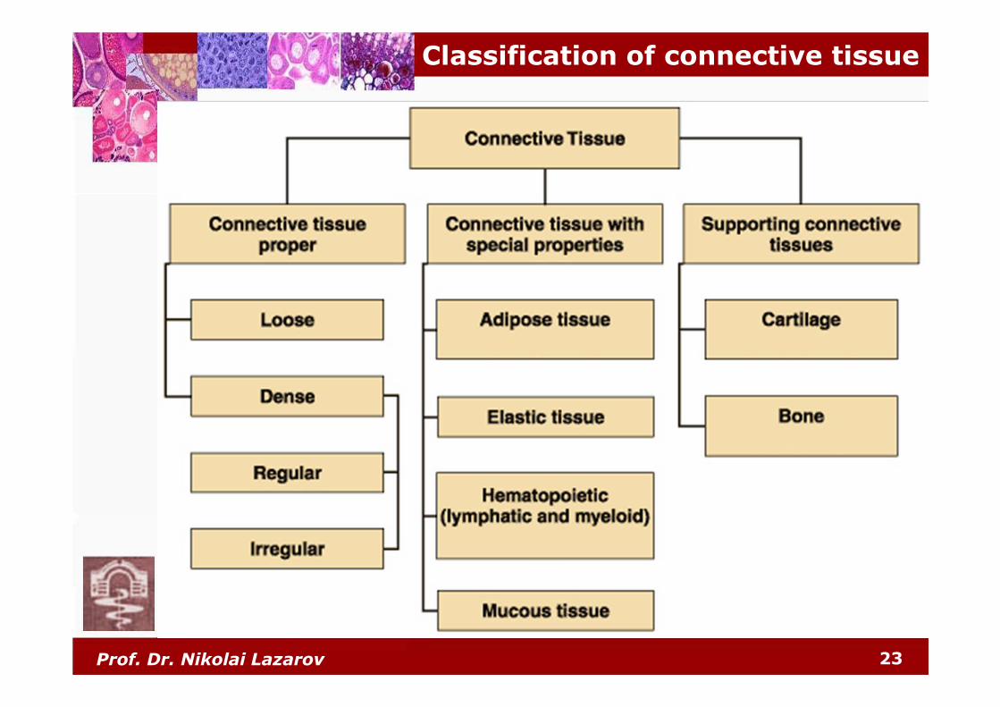
4. Connective tissue proper:
� loose connective tissue
� dense connective tissue
� regular
� irregular
5. Mononuclear phagocyte system

Prof. Dr. Nikolai Lazarov 2
Terminology and peculiarities
� Textus connectivus:� cells of mesenchymal origin� extracellular matrix
� connective tissue features:� interior location –
never found at the surface� cellular polymorphism� abundant amounts of
extracellular matrix –determines the species diversity
� absence of cell polarity� high adaptive and
regenerative capabilities� metaplastic abilities� specialized structures:
� intracellular� extracellular
NB: most abundant of the basic tissues – ½ of the human body mass

Prof. Dr. Nikolai Lazarov 3
� binding together other tissues in the formation of organs – capsules
� structural support (mechanical role) – bones, ligaments and tendons
� nutritive role (homeostasis) – blood� defensive functions (barrier and immunologic –
antibodies)
Functions of connective tissue

Prof. Dr. Nikolai Lazarov 4
Cells of the connective tissue
� productive and nutritive:
� synthesize and secrete
the extracellular matrix
� regenerative and repair
abilities
� defense cells:
� motile and circulatory activity
� pigment cells:
� presence of
specialized structures
Prof. Dr. Nikolai Lazarov 5
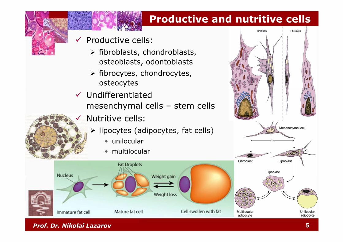
Productive and nutritive cells
� Productive cells:
� fibroblasts, chondroblasts,
osteoblasts, odontoblasts
� fibrocytes, chondrocytes,
osteocytes
� Undifferentiated
mesenchymal cells – stem cells
� Nutritive cells:
� lipocytes (adipocytes, fat cells)
• unilocular
• multilocular
Prof. Dr. Nikolai Lazarov 6
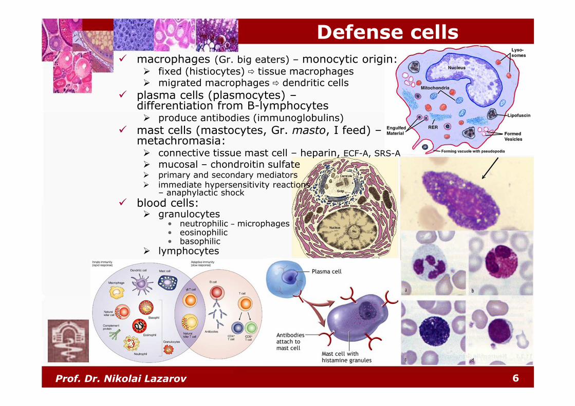
Defense cells
� macrophages (Gr. big eaters) – monocytic origin:� fixed (histiocytes) � tissue macrophages� migrated macrophages � dendritic cells
� plasma cells (plasmocytes) –differentiation from B-lymphocytes� produce antibodies (immunoglobulins)
� mast cells (mastocytes, Gr. masto, I feed) –metachromasia:� connective tissue mast cell – heparin, ECF-A, SRS-A
� mucosal – chondroitin sulfate� primary and secondary mediators� immediate hypersensitivity reactions
– anaphylactic shock
� blood cells:� granulocytes
• neutrophilic – microphages• eosinophilic• basophilic
� lymphocytes

Prof. Dr. Nikolai Lazarov 7
Pigment cells
� melanin-containing cells:
� melanocytes – dermis,
choroidea, meninges
� melanophores – skin
� chromatophores:
� chemosiderophores
� lipochromophores
Prof. Dr. Nikolai Lazarov 8
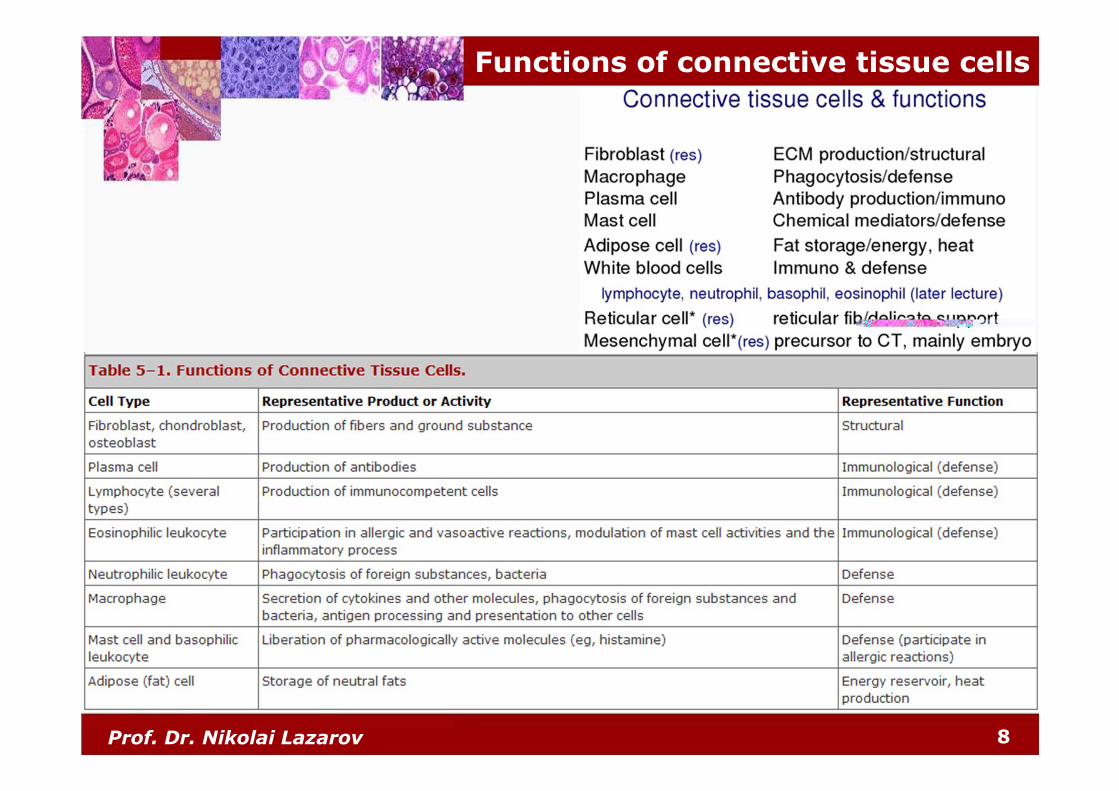
Functions of connective tissue cells
Prof. Dr. Nikolai Lazarov 9
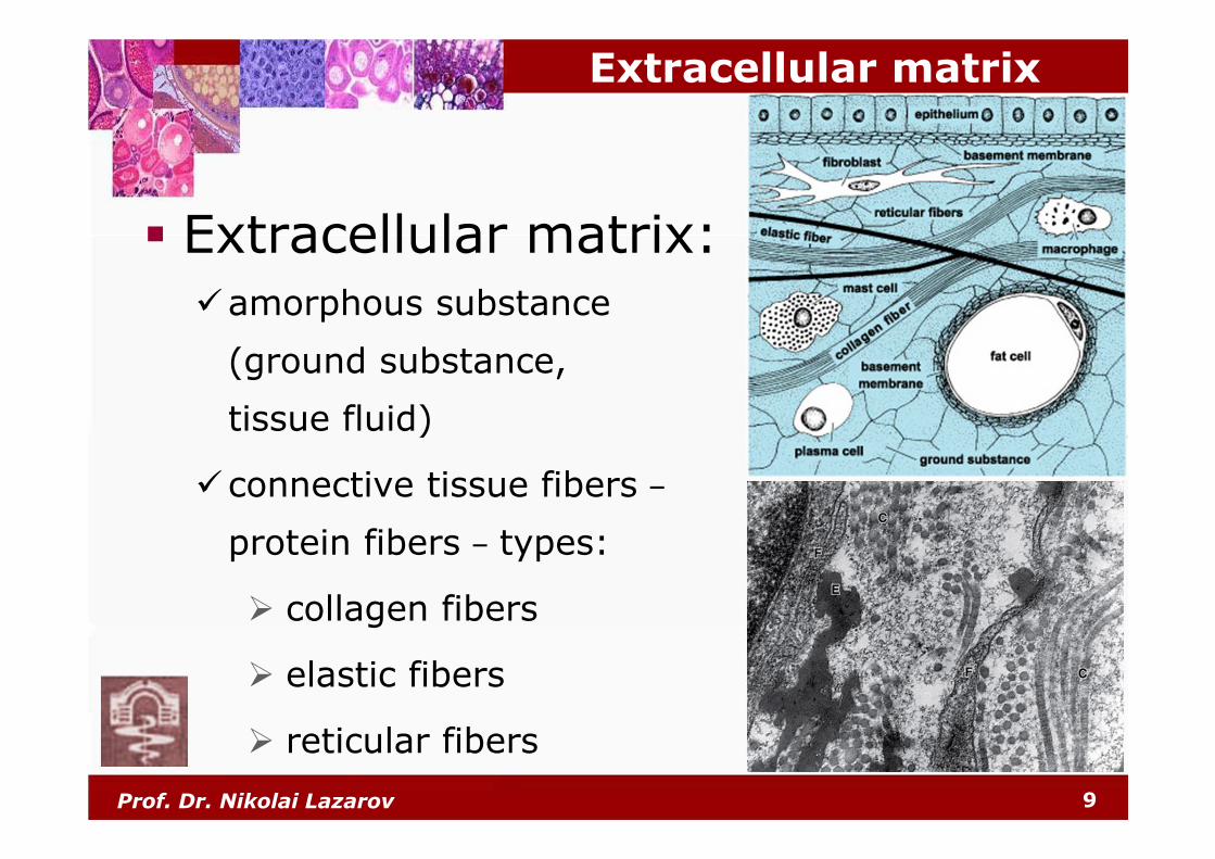
Extracellular matrix
� Extracellular matrix:�amorphous substance
(ground substance,
tissue fluid)
�connective tissue fibers –
protein fibers – types:
� collagen fibers
� elastic fibers
� reticular fibers
Prof. Dr. Nikolai Lazarov 10
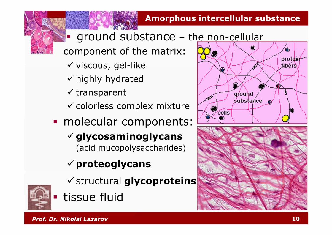
Amorphous intercellular substance
� ground substance – the non-cellular
component of the matrix:
� viscous, gel-like
� highly hydrated
� transparent
� colorless complex mixture
� molecular components:
�glycosaminoglycans(acid mucopolysaccharides)
�proteoglycans
�structural glycoproteins
� tissue fluid
Prof. Dr. Nikolai Lazarov 11
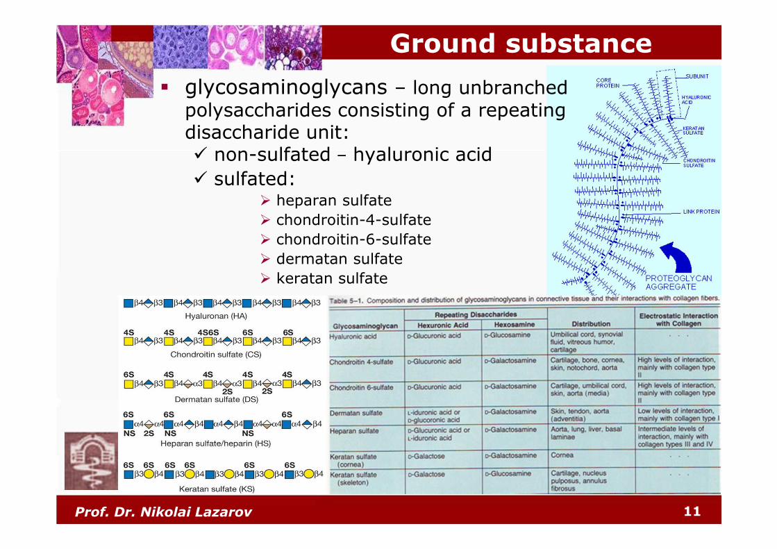
Ground substance
� glycosaminoglycans – long unbranched
polysaccharides consisting of a repeating disaccharide unit:� non-sulfated – hyaluronic acid
� sulfated:� heparan sulfate
� chondroitin-4-sulfate
� chondroitin-6-sulfate
� dermatan sulfate
� keratan sulfate

Prof. Dr. Nikolai Lazarov 12
Ground substance
� proteoglycans – glycoproteins
that are heavily glycosylated:� a core protein
� one or more covalently attached glycosaminoglycan(s) – 80-90%:
� heparan sulfate
� chondroitin-4-sulfate
� chondroitin-6-sulfate
� dermatan sulfate
� keratan sulfate
� extracellular matrix proteoglycans:� aggrecan� perlecan� decorin
� biglycan
� cell-surface proteoglycans:� syndecan� fibroglycan� glypican
Prof. Dr. Nikolai Lazarov 13
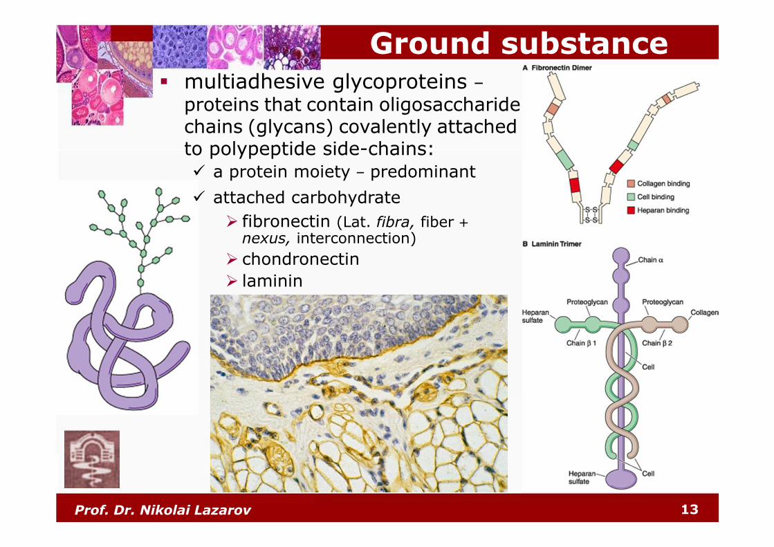
Ground substance� multiadhesive glycoproteins –
proteins that contain oligosaccharidechains (glycans) covalently attached to polypeptide side-chains:� a protein moiety – predominant
� attached carbohydrate
� fibronectin (Lat. fibra, fiber +
nexus, interconnection)
� chondronectin
� laminin
Prof. Dr. Nikolai Lazarov 14
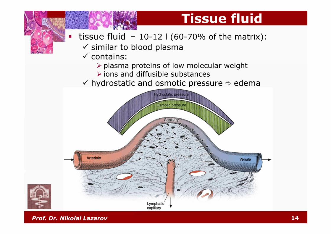
Tissue fluid
� tissue fluid – 10-12 l (60-70% of the matrix):
� similar to blood plasma� contains:
� plasma proteins of low molecular weight� ions and diffusible substances
� hydrostatic and osmotic pressure � edema

Prof. Dr. Nikolai Lazarov 15
� Connective tissue fibers:� collagen� elastic� reticular
Protein fibers
NB: in many cases, the predominant fiber type is responsible
for conferring specific properties on the tissue!

Prof. Dr. Nikolai Lazarov 16
� fibra collagenosa:
� most abundant (30% of dry b.w.)
� low elasticity – 5%
� strength – sustain 500 kg/cm2
� acidophilic collagen fibers – 1-20 µm thick
� collagen fibrils – 0.1-0.5 µm in diameter
� collagen microfibrils – 50-90 nm; 68 nm periodicity
� collagen molecules – molecular mass 360000 Da
Collagen fibers
� collagen:� tropocollagen –
280 nm/1.5 nm
� 3 subunit α-polypeptide
chains – triple helix
� amino acid content:
� glycine (33.5%)
� proline (12%)
� hydroxyproline (10%)
� hydroxylysine
Gr. kolla, glue
Prof. Dr. Nikolai Lazarov 17
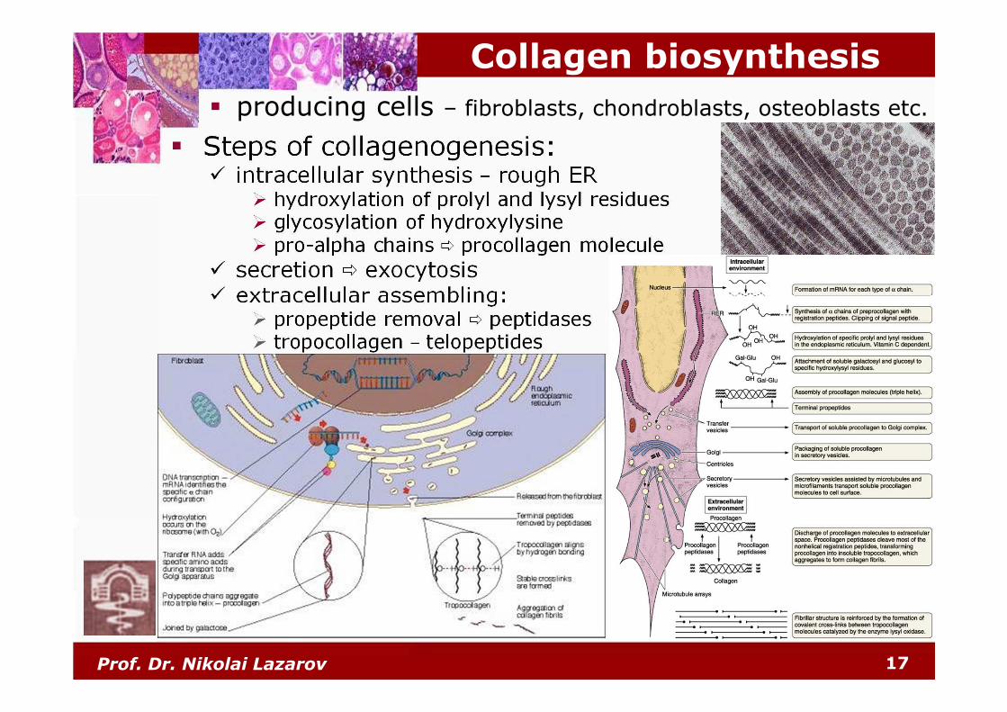
� producing cells – fibroblasts, chondroblasts, osteoblasts etc.
� Steps of collagenogenesis:� intracellular synthesis – rough ER
� hydroxylation of prolyl and lysyl residues� glycosylation of hydroxylysine� pro-alpha chains � procollagen molecule
� secretion � exocytosis� extracellular assembling:
� propeptide removal � peptidases� tropocollagen – telopeptides
Collagen biosynthesis

Prof. Dr. Nikolai Lazarov 18
Collagen types
NB: collagen = 25-30% of all proteins in the human body
� 28 types of collagen:

Prof. Dr. Nikolai Lazarov 19
� fibra reticulares:
� extremely thin – diameter 0.2-2 µm
� form a network (rete = network)
� resistant to acids and trypsin
� PAS-positive – carbohydrates
� argyrophilic – selective affinity for silver
salts (Gr. argyros, silver + philein, to love)
� 68 nm periodicity
� loosely packed thin (45 nm) fibrils� content:
� collagen type ІІІ• rich in cystine,
poor of proline and hydroxyproline
• 6-12% hexoses• glycoproteins• proteoglycans
� build up the reticular tissue:� framework of hemopoietic organs� smooth muscles, endoneurium
Reticular fibers
Prof. Dr. Nikolai Lazarov 20
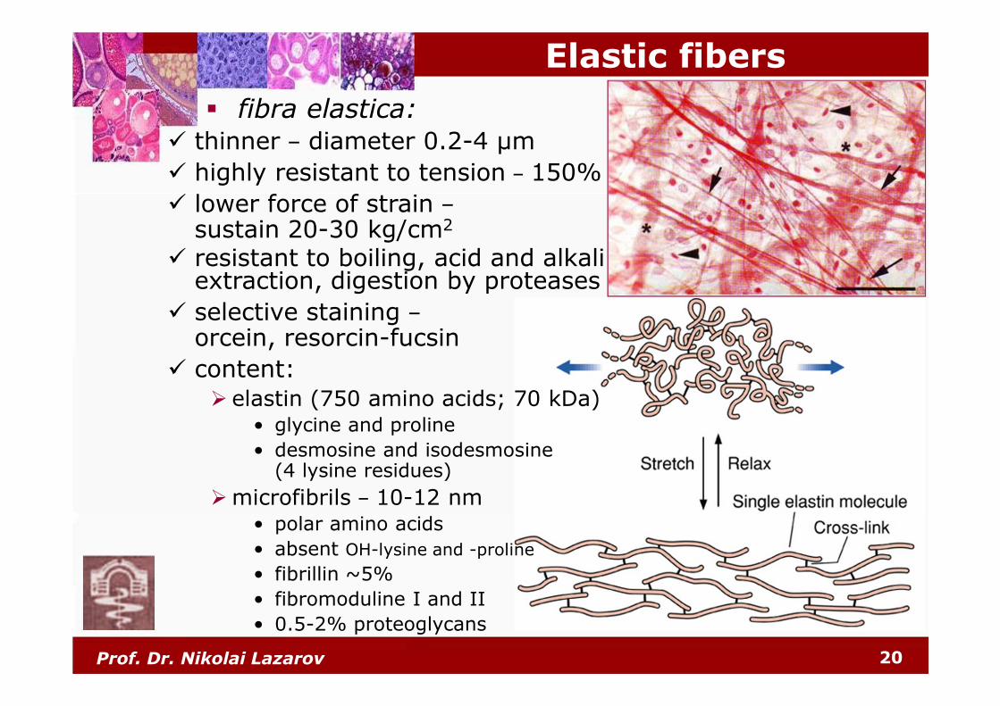
� fibra elastica:
Elastic fibers
� thinner – diameter 0.2-4 µm
� highly resistant to tension – 150%
� lower force of strain –sustain 20-30 kg/cm2
� resistant to boiling, acid and alkaliextraction, digestion by proteases
� selective staining –orcein, resorcin-fucsin
� content:� elastin (750 amino acids; 70 kDa)
• glycine and proline
• desmosine and isodesmosine(4 lysine residues)
�microfibrils – 10-12 nm• polar amino acids
• absent OH-lysine and -proline
• fibrillin ~5%
• fibromoduline І and ІІ
• 0.5-2% proteoglycans
Prof. Dr. Nikolai Lazarov 21
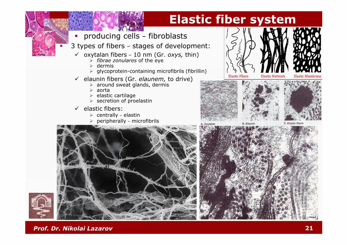
� producing cells – fibroblasts
� 3 types of fibers – stages of development:
� oxytalan fibers – 10 nm (Gr. oxys, thin)� fibrae zonulares of the eye� dermis� glycoprotein-containing microfibrils (fibrillin)
� elaunin fibers (Gr. elaunem, to drive)� around sweat glands, dermis� aorta� elastic cartilage� secretion of proelastin
� elastic fibers:� centrally – elastin� peripherally – microfibrils
Elastic fiber system

Prof. Dr. Nikolai Lazarov 22
Classification of connective tissue
Prof. Dr. Nikolai Lazarov 23
Classification of connective tissue

Prof. Dr. Nikolai Lazarov 24
Loose (areolar) connective tissue
� Cells – productive, nutritive and defense:
� proper (fixed):
� fibroblasts and fibrocytes
� free:
� macrophages (histiocytes) – phagocytosis
� plasma cells – unmature and mature (Russell bodies)
� mast cells
� leukocytes – granular and agranular
� melanocytes
� textus connectivus fibrosus laxus:� most widespread type of connective tissue� cells and extracellular matrix:
� fibers:
� collagen
� elastic
� reticular
� amorphous
ground substance
Prof. Dr. Nikolai Lazarov 25
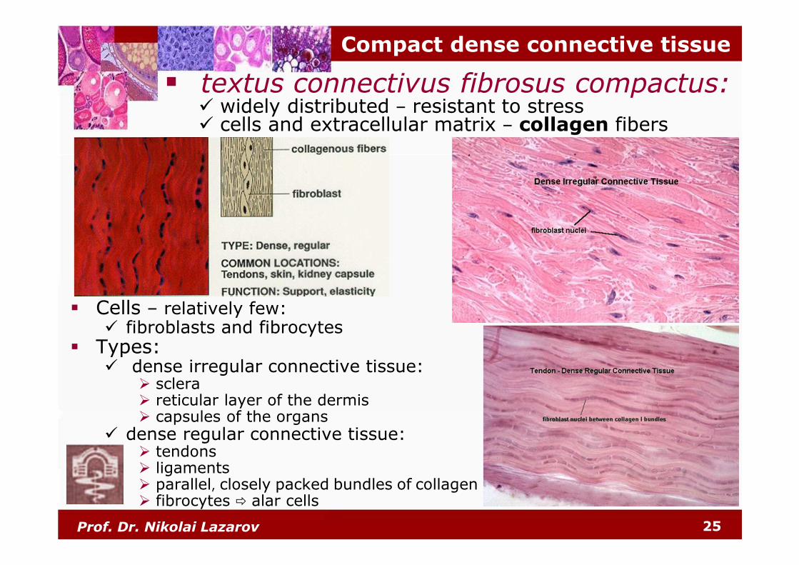
Compact dense connective tissue
� Cells – relatively few:� fibroblasts and fibrocytes
� Types:� dense irregular connective tissue:
� sclera� reticular layer of the dermis� capsules of the organs
� dense regular connective tissue:� tendons� ligaments� parallel, closely packed bundles of collagen� fibrocytes � alar cells
� textus connectivus fibrosus compactus:� widely distributed – resistant to stress� cells and extracellular matrix – collagen fibers

Prof. Dr. Nikolai Lazarov 26
Lamellar dense connective tissue
� Cells – relatively few:� fibroblasts and fibrocytes
� Intercellular matrix:
� numerous collagen fibers
� lesser elastic fibers:
� layers
� lamellae
� textus connectivus fibrosus lamellaris:� widespread distribution – aponeuroses and fascia of
the muscles, dura mater

Prof. Dr. Nikolai Lazarov 27
Reticuloendothelial system
� Systema reticuloendothelialis s. macrophagorum –Aschoff, 1924
� Synonym: Reticulohistiocyte system
Karl Albert Ludwig Aschoff
(1866-1942)

Prof. Dr. Nikolai Lazarov 28
Mononuclear phagocyte system
� Mononuclear phagocyte system – Van Furth, 1969

Prof. Dr. Nikolai Lazarov 29
� Reticuloendothelial System (RES)
� Ludwig Aschoff, 1924
� Reticulohistiocyte System (RHS)
� Mononuclear Phagocyte System (MPS)
� Van Furth, 1969 1866-1942
Mononuclear phagocyte system
Prof. Dr. Nikolai Lazarov 30
to be continuedto be continuedto be continuedto be continued ............Thank you ...