Embed Size (px)
Citation preview
HISTOLOGY –CONNECTIVE TISSUEUNIT 4 – CHAPTER 3
CHARACTERISTICS OF CONNECTIVE TISSUE
• Connective Tissue = CT
• Found everywhere in the body
• Includes the most abundant and widely distributed tissues
• Most connective tissues are well vascularized
• Lots of blood flow
• Exceptions: tendons & ligaments have a poor blood supply and cartilages are avascular
CHARACTERISTICS OF CONNECTIVE TISSUE
• Made up of different types of cells plus varying amounts of a nonlinving
substance
• Extracellular Matrix (EM)
• Produced by the connective tissue cells and secreted
• May be liquid, semisolid/gel-like, or very hard
• Because of its EM, CT is able to bear weight and withstand stretching and
other abuses, such as abrasion, that no other tissue could endure
CHARACTERISTICS OF CONNECTIVE TISSUE
• Extremes
• Fat tissue is composed mostly of cells, and the matrix is soft
• Bone and cartilage have very few cells and large amounts of hard martrix, which makes
them extremely strong
• Types of Fibers in EM
• Collagen (white) – tensile strength
• Elastic (yellow) – stretch with recoil
• Reticular (fine collagen) – support
FUNCTIONS OF CONNECTIVE TISSUES
1. Binds body tissues together
2. Supports the body
3. Provides protection
4. Insulates to maintain body temperature
5. Transportation of other molecules
TYPES OF CONNECTIVE TISSUE
• Types of connective tissues differ in filler types and the
number of fibers in the matrix
• Types
1. Bone
2. Cartilage
3. Dense connective tissue
4. Loose connective tissue
5. Blood
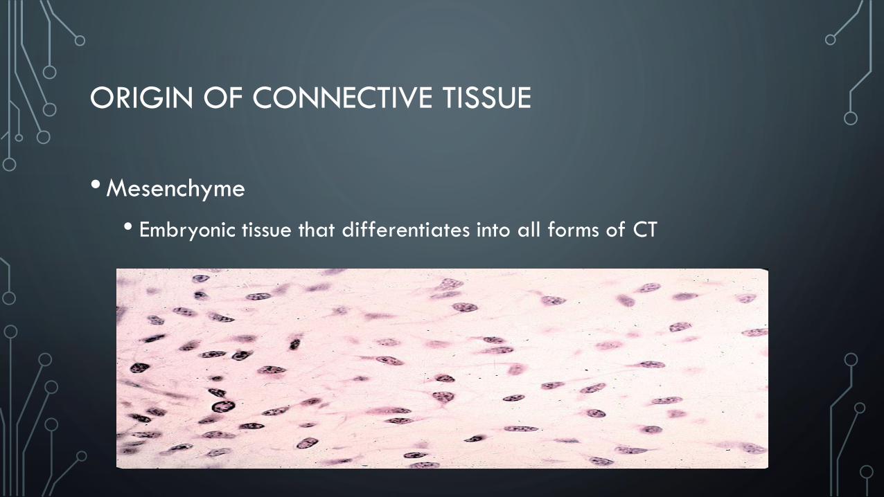
ORIGIN OF CONNECTIVE TISSUE
•Mesenchyme
• Embryonic tissue that differentiates into all forms of CT
CONNECTIVE TISSUE CELLS
• Prefixes
• Fibro
• Osteo
• Chondro
• Hemo(cyto)
• Suffixes
• Blast – build the cells
• Cyte – cell
• Clast – breadown the cells
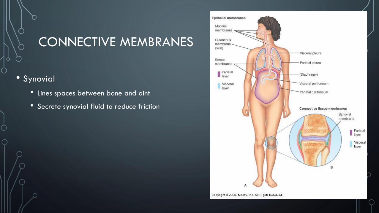
CONNECTIVE MEMBRANES
• Synovial
• Lines spaces between bone and oint
• Secrete synovial fluid to reduce friction

AREOLAR OR LOOSE
• Structure
• Cells (fibroblasts, macrophages, & lymphocytes) within a fine network of
mostly collagen fibers
• Often merges with denser connective tissue
• Location
• Widely distributed throughout the body
• Substance on which epithelial basement membranes rest
• Packing between glands, muscles, and nerves
• Attaches the skin to underlying tissues
• Function
• Loose packing
• Support
• Nourishment for the structures with which it’s associated
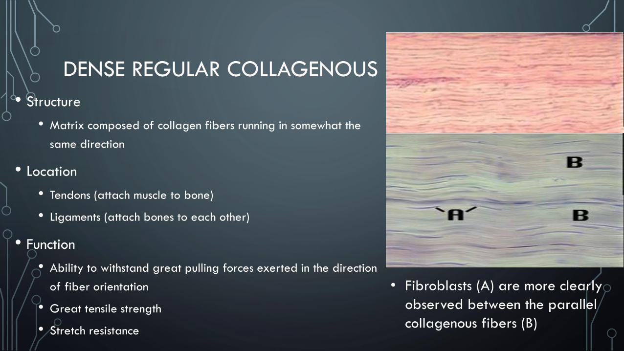
DENSE REGULAR COLLAGENOUS
• Structure
• Matrix composed of collagen fibers running in somewhat the
same direction
• Location
• Tendons (attach muscle to bone)
• Ligaments (attach bones to each other)
• Function
• Ability to withstand great pulling forces exerted in the direction
of fiber orientation
• Great tensile strength
• Stretch resistance
• Fibroblasts (A) are more clearly
observed between the parallel
collagenous fibers (B)
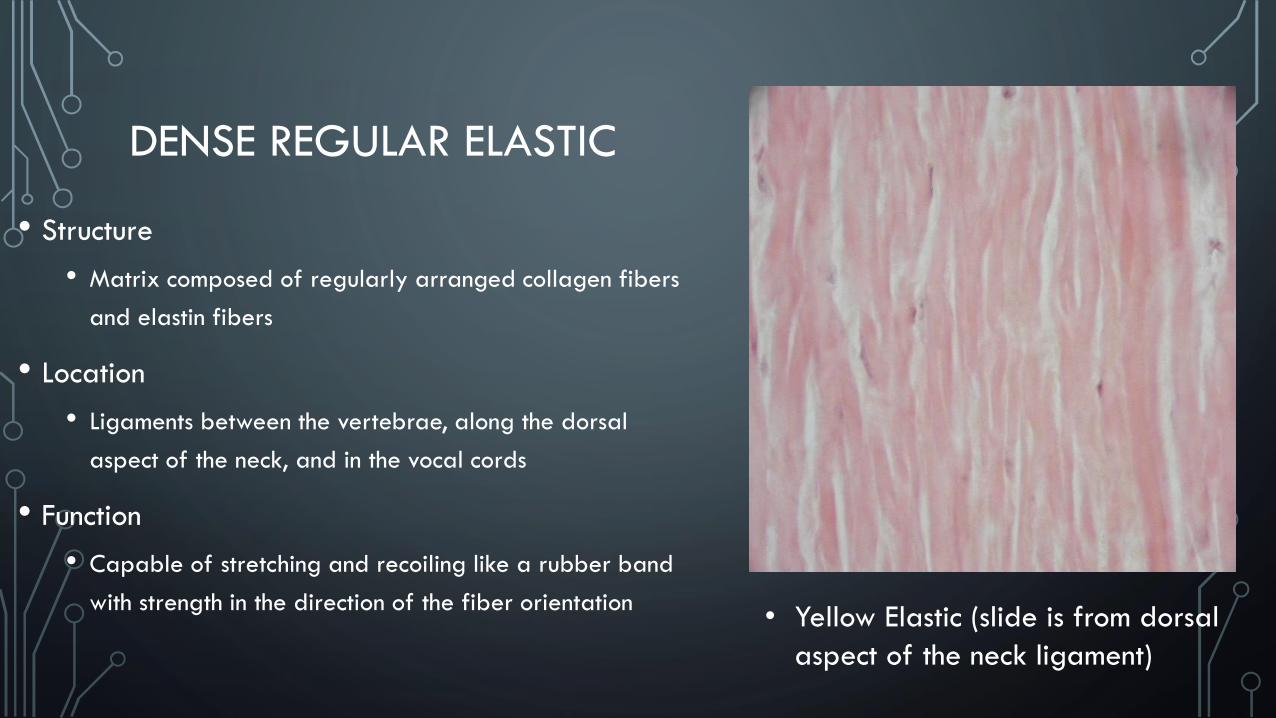
DENSE REGULAR ELASTIC
• Structure
• Matrix composed of regularly arranged collagen fibers
and elastin fibers
• Location
• Ligaments between the vertebrae, along the dorsal
aspect of the neck, and in the vocal cords
• Function
• Capable of stretching and recoiling like a rubber band
with strength in the direction of the fiber orientation• Yellow Elastic (slide is from dorsal
aspect of the neck ligament)
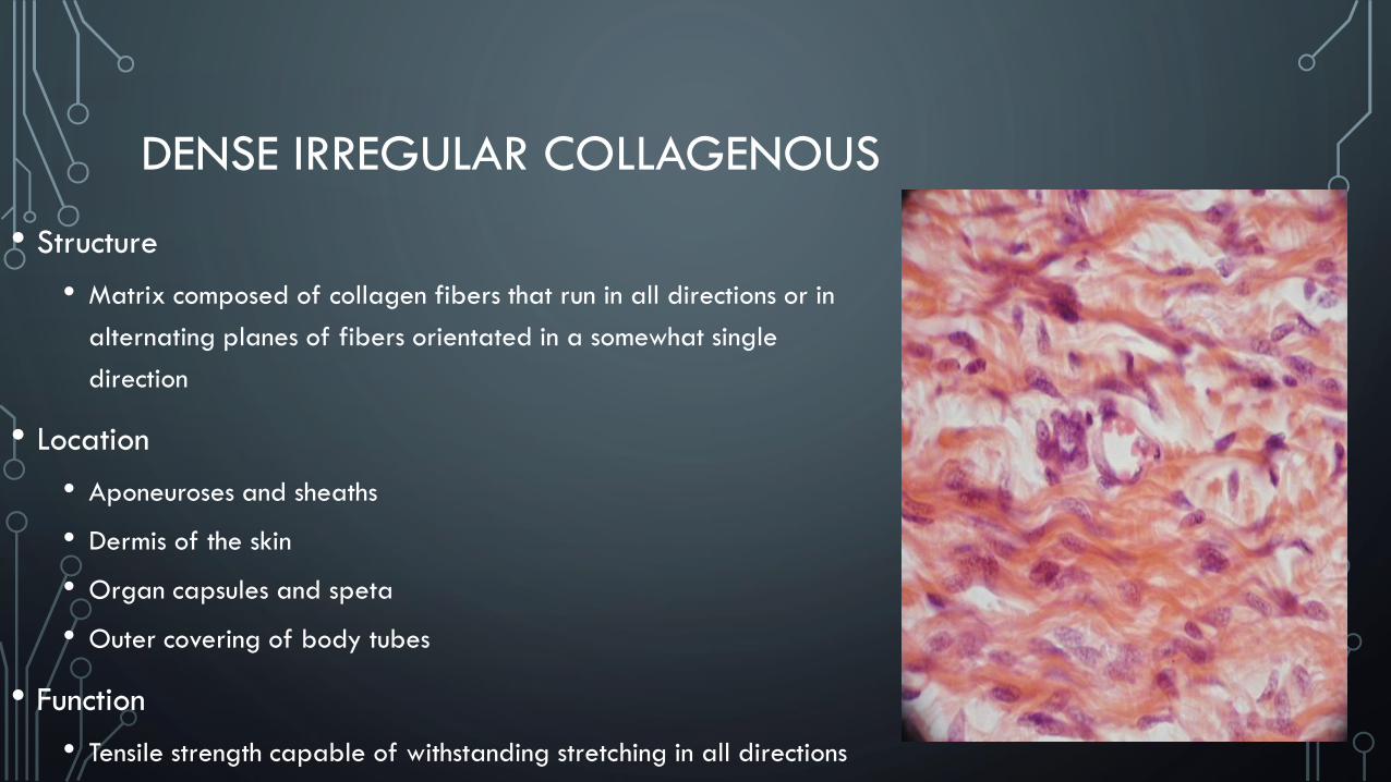
DENSE IRREGULAR COLLAGENOUS
• Structure
• Matrix composed of collagen fibers that run in all directions or in
alternating planes of fibers orientated in a somewhat single
direction
• Location
• Aponeuroses and sheaths
• Dermis of the skin
• Organ capsules and speta
• Outer covering of body tubes
• Function
• Tensile strength capable of withstanding stretching in all directions
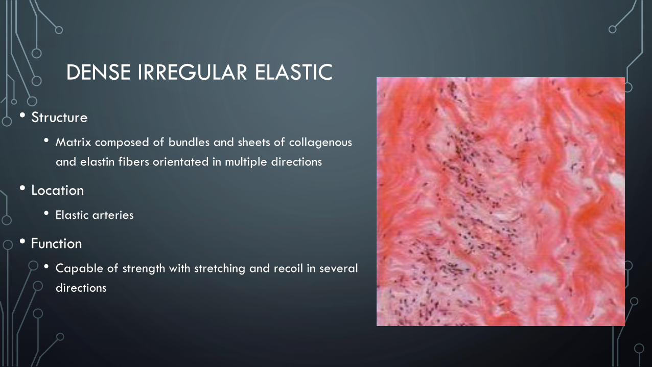
DENSE IRREGULAR ELASTIC
• Structure
• Matrix composed of bundles and sheets of collagenous
and elastin fibers orientated in multiple directions
• Location
• Elastic arteries
• Function
• Capable of strength with stretching and recoil in several
directions
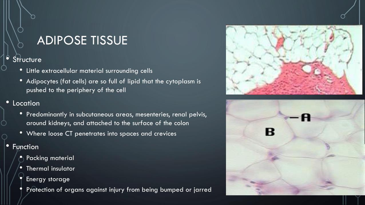
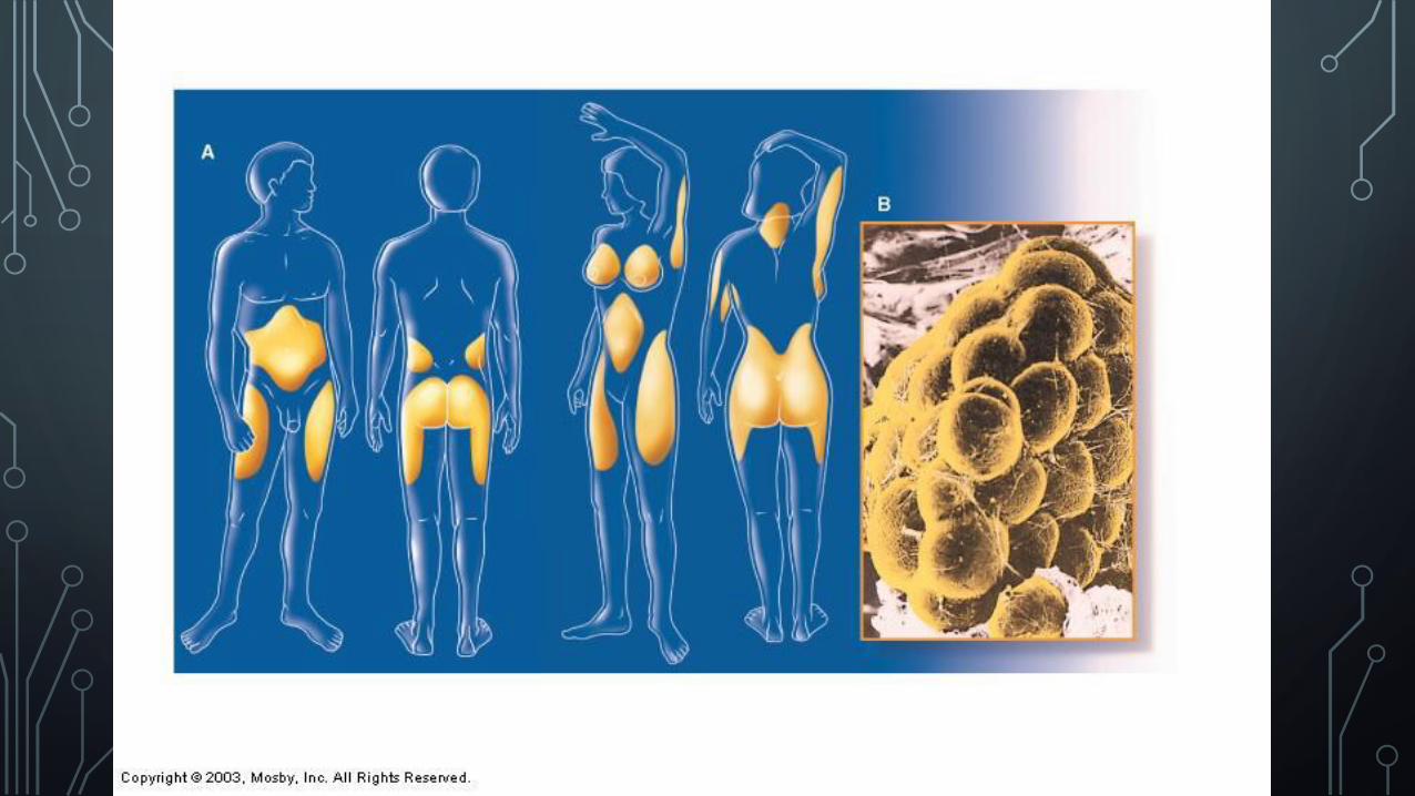
ADIPOSE TISSUE
• Structure
• Little extracellular material surrounding cells
• Adipocytes (fat cells) are so full of lipid that the cytoplasm is
pushed to the periphery of the cell
• Location
• Predominantly in subcutaneous areas, mesenteries, renal pelvis,
around kidneys, and attached to the surface of the colon
• Where loose CT penetrates into spaces and crevices
• Function
• Packing material
• Thermal insulator
• Energy storage
• Protection of organs against injury from being bumped or jarred
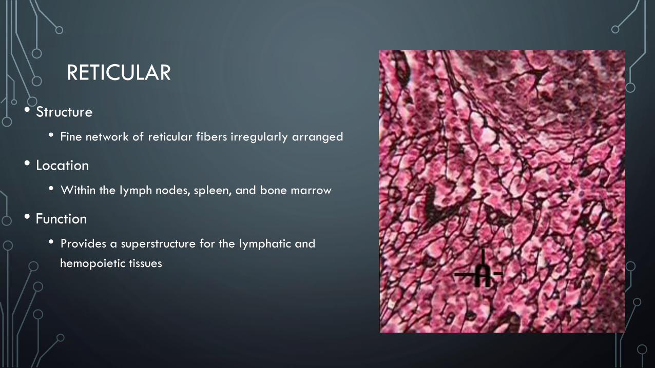
RETICULAR
• Structure
• Fine network of reticular fibers irregularly arranged
• Location
• Within the lymph nodes, spleen, and bone marrow
• Function
• Provides a superstructure for the lymphatic and
hemopoietic tissues
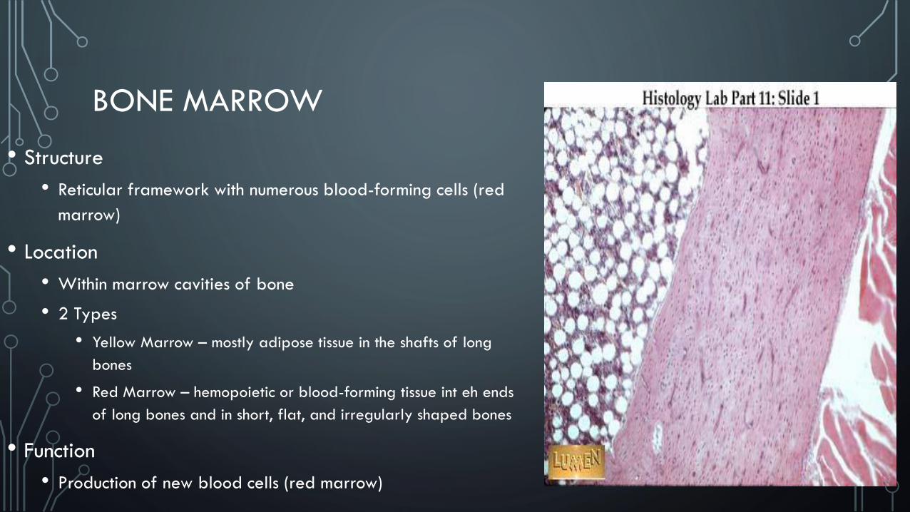
BONE MARROW
• Structure
• Reticular framework with numerous blood-forming cells (red
marrow)
• Location
• Within marrow cavities of bone
• 2 Types
• Yellow Marrow – mostly adipose tissue in the shafts of long
bones
• Red Marrow – hemopoietic or blood-forming tissue int eh ends
of long bones and in short, flat, and irregularly shaped bones
• Function
• Production of new blood cells (red marrow)
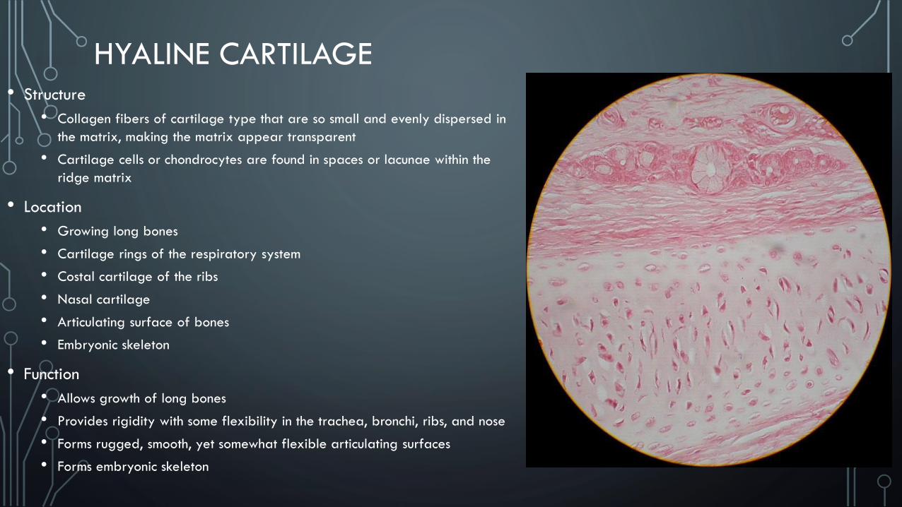
HYALINE CARTILAGE• Structure
• Collagen fibers of cartilage type that are so small and evenly dispersed in
the matrix, making the matrix appear transparent
• Cartilage cells or chondrocytes are found in spaces or lacunae within the
ridge matrix
• Location
• Growing long bones
• Cartilage rings of the respiratory system
• Costal cartilage of the ribs
• Nasal cartilage
• Articulating surface of bones
• Embryonic skeleton
• Function
• Allows growth of long bones
• Provides rigidity with some flexibility in the trachea, bronchi, ribs, and nose
• Forms rugged, smooth, yet somewhat flexible articulating surfaces
• Forms embryonic skeleton
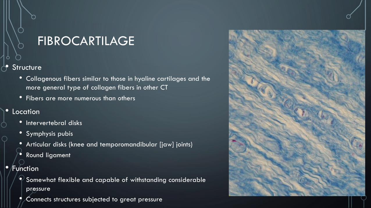
FIBROCARTILAGE
• Structure
• Collagenous fibers similar to those in hyaline cartilages and the
more general type of collagen fibers in other CT
• Fibers are more numerous than others
• Location
• Intervertebral disks
• Symphysis pubis
• Articular disks (knee and temporomandibular [jaw] joints)
• Round ligament
• Function
• Somewhat flexible and capable of withstanding considerable
pressure
• Connects structures subjected to great pressure
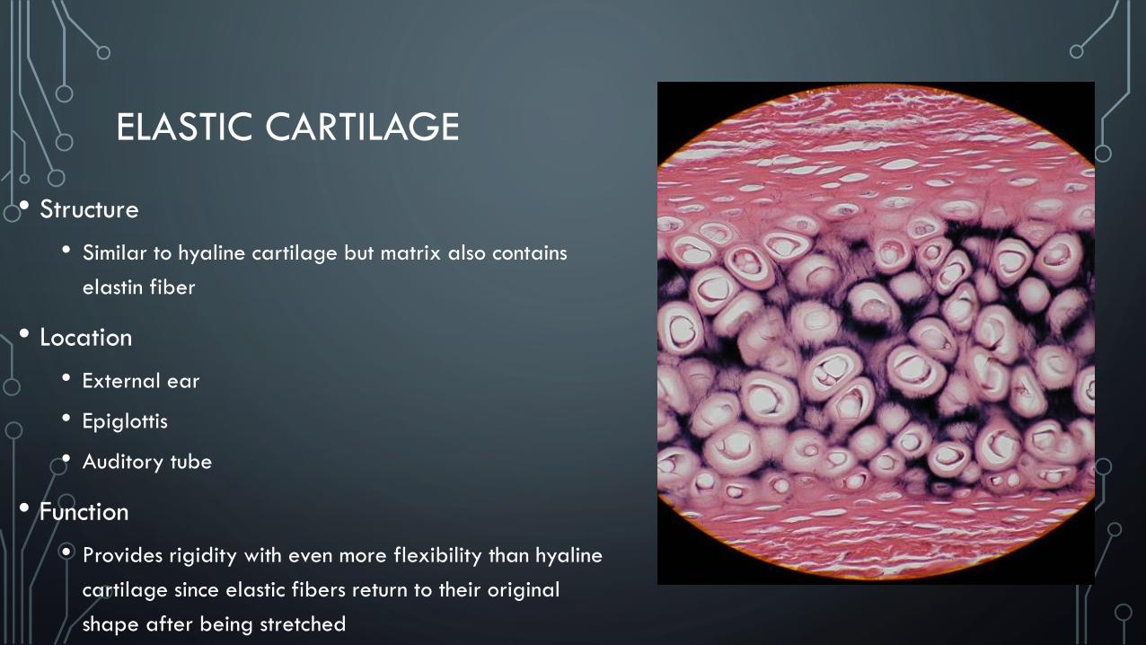
ELASTIC CARTILAGE
• Structure
• Similar to hyaline cartilage but matrix also contains
elastin fiber
• Location
• External ear
• Epiglottis
• Auditory tube
• Function
• Provides rigidity with even more flexibility than hyaline
cartilage since elastic fibers return to their original
shape after being stretched

CANCELLOUS BONE
• Structure
• Lattice-like network of scaffolding characterized by trabeculae with
large spaces between them
• Osteocytes or bone cells are located within lacunae in the trabeculae
• Location
• In the interior of the bones of the skull, sternum, and pelvis
• Found in the ends of the long bones
• Function
• Acts as a scaffolding to provide strength and support without the
greater weight of solid bone
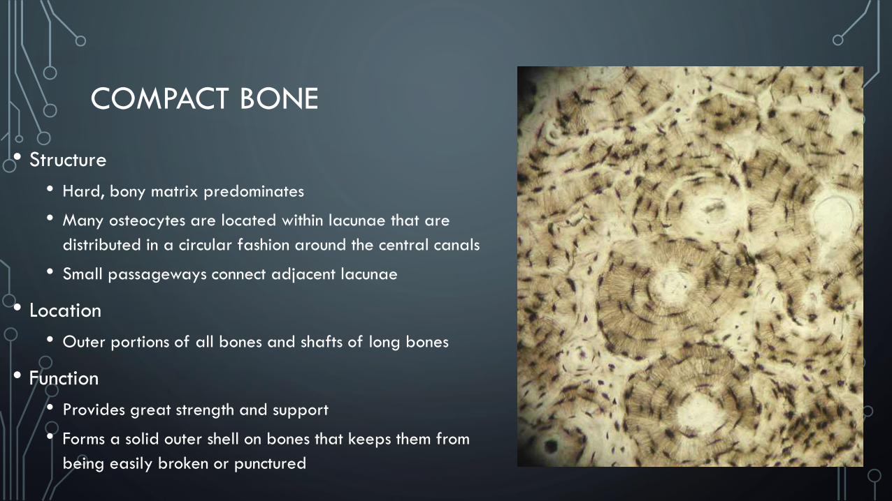
COMPACT BONE
• Structure
• Hard, bony matrix predominates
• Many osteocytes are located within lacunae that are
distributed in a circular fashion around the central canals
• Small passageways connect adjacent lacunae
• Location
• Outer portions of all bones and shafts of long bones
• Function
• Provides great strength and support
• Forms a solid outer shell on bones that keeps them from
being easily broken or punctured
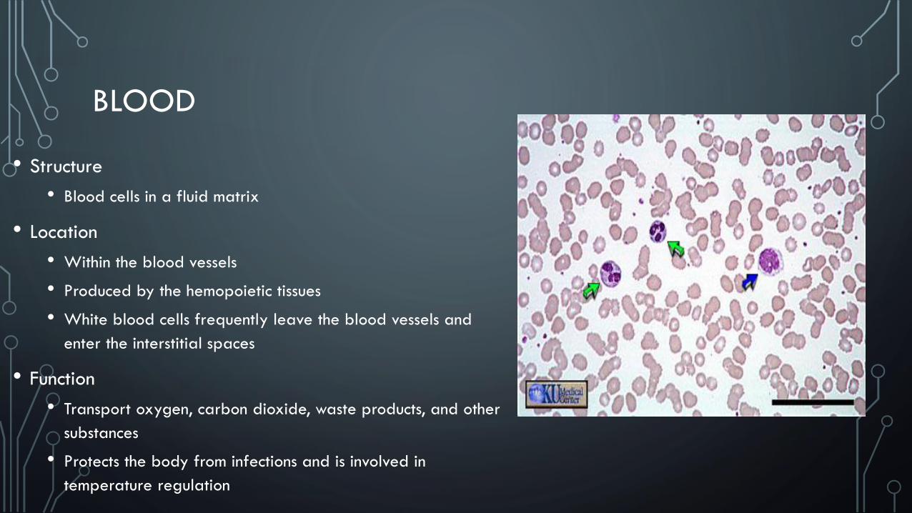
BLOOD
• Structure
• Blood cells in a fluid matrix
• Location
• Within the blood vessels
• Produced by the hemopoietic tissues
• White blood cells frequently leave the blood vessels and
enter the interstitial spaces
• Function
• Transport oxygen, carbon dioxide, waste products, and other
substances
• Protects the body from infections and is involved in
temperature regulation