Embed Size (px)
Citation preview
C RSE
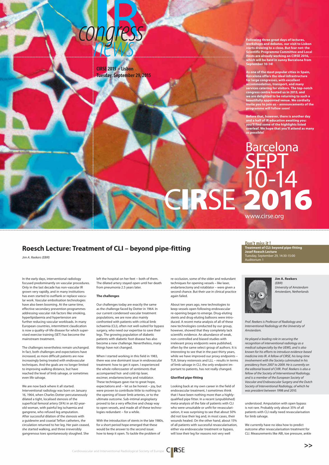
CIRSE 2015 – Lisbon
Tuesday, September 29, 2015
Cardiovascular and Interventional Radiological Society of Europe
Rnewscongress
Don’t miss it !
re-occlusion, some of the older and redundant
techniques for opening vessels – like laser,
endarterectomy and rotablator – were given a
second chance. But their use in clinical practice
again failed.
About ten years ago, new technologies to
keep vessels open following endovascular
re- opening began to emerge. Drug-eluting
stents and drug-eluting balloons were intro-
duced. A recent meta-analysis of all of these
new technologies conducted by our group,
however, showed that they completely lack
scientific evidence. An abundance of weak,
non-controlled and biased studies with
irrelevant proxy endpoints were published,
often by the same select group of authors. It is
interesting to see that in the past thirty years,
while we have improved our proxy endpoints –
TLR, binary restenosis and LLL – results in terms
of limb salvage in CLI, the only endpoint im-
portant to patients, has not really changed.
Glorified pipe-fitting
Looking back at my own career in the field of
endovascular treatment, I sometimes think
that I have been nothing more than a highly-
qualified pipe fitter. In a recent (unpublished)
meta-analysis of the fate of patients with CLI
who were unsuitable or unfit for revasculari-
sation, it was surprising to see that about 50%
did not lose their leg and, in most cases, their
wounds healed. On the other hand, about 15%
of all patients with successful revascularisation,
either via endovascular treatment or bypass,
will lose their leg for reasons not very well
>>
Prof. Reekers is Professor of Radiology and
Interventional Radiology at the University of
Amsterdam.
He played a leading role in securing the
recognition of interventional radiology as a
medical subspecialty by the UEMS, and is also
known for his efforts to introduce evidence-based
medicine into IR. A fellow of CIRSE, his long-time
involvement with the Society culminated in his
presidency from 2007 to 2009. He is currently on
the editorial board of CVIR. Prof. Reekers is also a
fellow of the Society of Interventional Radiology,
and is a member of the European Society of
Vascular and Endovascular Surgery and the Dutch
Society of Interventional Radiology, of which he
was president between 1998 and 2010.
Jim A. Reekers
(EBIR)
University of Amsterdam
Amsterdam, Netherlands
In the early days, interventional radiology
focused predominantly on vascular procedures.
Only in the last decade has non-vascular IR
grown very rapidly, and in many institutions
has even started to outflank or replace vascu-
lar work. Vascular embolisation technologies
have also been booming. At the same time,
effective secondary prevention programmes
addressing vascular risk factors like smoking,
hyperlipi daemia and hypertension are
further re ducing vascular workloads. In many
European countries, intermittent claudication
is now a quality-of-life disease for which super-
vised exercise training (SET) has become the
mainstream treatment.
The challenges nevertheless remain unchanged.
In fact, both challenges and expectations have
increased, as more difficult patients are now
increasingly being treated with endo vascular
techniques. And the goals are no longer limited
to improving walking distance, but have
reached the level of limb salvage, or sometimes
even life salvage.
We are now back where it all started.
Interventional radiology was born on January
16, 1964, when Charles Dotter percutaneously
dilated a tight, localised stenosis of the
superficial femoral artery (SFA) in an 82-year-
old woman, with painful leg ischaemia and
gangrene, who refused leg amputation.
After successful dilation of the stenosis with
a guidewire and coaxial Teflon catheters, the
circulation returned to her leg. Her pain ceased,
she started walking, and three irreversibly
gangrenous toes spontaneously sloughed. She
left the hospital on her feet − both of them.
The dilated artery stayed open until her death
from pneumonia 2.5 years later.
The challenges
Our challenges today are exactly the same
as the challenge faced by Dotter in 1964. In
our current condensed vascular treatment
populations, we are now also mainly
con fronted with patients with critical limb
ischaemia (CLI), often not well-suited for bypass
surgery, who need our expertise to save their
legs. The growing population of diabetic
patients with diabetic foot disease has also
become a new challenge. Nevertheless, many
things have not changed.
When I started working in this field in 1983,
there was one dominant issue in endo vascular
treatment: how to get it open. I experienced
the whole rollercoaster of sentiments that
accompanied hot- and cold-tip laser,
excimer, endarterectomy and rota blator.
These techniques gave rise to great hope,
expectations and − let us be honest − joy, but
were proven to contribute little to nothing to
the opening of lower limb arteries, or to the
ultimate outcome. Sub-intimal angioplasty
proved to be a very effective and cheap way
to open vessels, and made all of these techno-
logies redundant − for a while.
With the introduction of stents in the late 1980s,
for a short period hope emerged that these
would be the answer to the second issue:
how to keep it open. To tackle the problem of
Treatment of CLI: beyond pipe-fitting
Josef Roesch Lecture
Tuesday, September 29, 14:30-15:00
Auditorium 1
Roesch Lecture: Treatment of CLI – beyond pipe-fittingJim A. Reekers (EBIR)
understood. Amputation with open bypass
is not rare. Probably only about 35% of all
patients with CLI really need revascularisation
for limb salvage.
We currently have no idea how to predict
outcome after revascularisation treatment for
CLI. Measurements like ABI, toe pressure, ankle
CIRSE 201610-14
Barcelona
www.cirse.org
SEPT
Following three great days of lectures,
workshops and debates, our visit to Lisbon
starts drawing to a close. But fear not: the
Scientific Programme Committee and Local
Hosts are already working on CIRSE 2016,
which will be held in sunny Barcelona from
September 10-14!
As one of the most popular cities in Spain,
Barcelona offers the ideal infrastructure
for large congresses, with excellent
accommodation, transport, and many
services catering for visitors. The top-notch
congress centre hosted us in 2013, and
we are delighted to be returning to such a
beautifully appointed venue. We cordially
invite you to join us – announcements of the
programme will follow soon!
Before that, however, there is another day
and a half of IR education awaiting you:
you’ll find some of the highlights listed
overleaf. We hope that you’ll attend as many
as possible!
Tuesday, September 29, 2015Josef Roesch Lecture2
Special Edition / CIRSE 2015 – Lisbon
pressure, TcPo2 and observation of straight
flow to the foot or improved vascularisation
of the foot have not been shown to have any
predictive value. Because we have no tools to
select those patients who will benefit from
revascularisation, current practice in the endo-
vascular treatment of CLI is opportunistic: we
treat all, based on a debatable definition of CLI,
and hope for the best.
Have we identified the correct problem?
Another very interesting observation is that,
during the past 20 years, limb salvage of
80-85% after a successful endovascular pro-
cedure is consistently reported, irrespective of
the technique used. How far can we push the
technology? A very recent paper published
in the NEJM about drug-eluting balloons in
the SFA showed that DEB had better patency
at 12 months (still only a disappointing 65%
versus 52%), but that this better patency did
not translate into a better clinical outcome.
Although the patient in this study had (90%)
intermittent claudication, a BMI > 30 and no
previous lifestyle intervention (supervised
exercise training), this shows again that there
is no direct relation between patency and
outcome, as we often also observe in patients
with CLI.
What is the clinical problem we are actually
trying to treat? Nobody would treat renal artery
stenosis in a patient on dialysis due to chronic
kidney function failure in order to improve
renal function, or open up a carotid artery to
improve flow to the areas of old ischaemia in
order to regain brain function. What seems so
obvious in other situations has never been a
topic of much interest in the context of revas-
cularisation of patients with CLI. If we also take
into account that the vast majority of lower
limb amputations start with local problems in
the foot, the conclusion should be that the
foot is the end-organ for patients with CLI.
Therefore, the condition of the foot − its
remaining functionality − might play a crucial
role in outcome after revascularisation. It is
probably not just the amount of blood flow to
the foot, but also the capability of the foot to
use that extra blood to restore tissue nutrition
in the foot, that is vital.
Oxygen, needed to prevent change from
aerobe metabolism in the tissue of the foot
to anaerobe metabolism (causing the symp-
toms of ischaemia) plays a crucial role. Oxygen
needs no trans-membrane pressure gradient
to be transported to the tissue, so oxygen
diffusion is independent of blood pressure. It
only depends on volume flow. The equation
for oxygen concentration in the tissue of the
foot depends on two parameters: the amount
of oxygen locally available in the blood, and
the functionality of microcirculation. There are
many ways to improve the amount of oxygen
in the foot, such as by increasing blood flow
to the foot (revascularisation) or via angio-
genesis (stem cell). Increasing oxygen levels in
the blood itself by way of hyperbaric oxygen
treatment is also a well-known treatment
method for ischaemia. But blood oxygen levels
can also be increased with simple measures
such as improvement of the cardio-pulmonary
condition (supervised exercise training and
stopping smoking), and treatment of anaemia,
which is often seen in patients with chronic
kidney failure.
Microcirculation
The other parameter in the equation for tissue
oxygen in the foot is the condition of the
microcirculation. Although a lot of research
has been carried out, especially in the field
of diabetic foot disease and ulcer healing,
this knowledge has never matured into a
clinical tool. Explanations for this are that all
of these techniques are very cumbersome,
often research-based, and have primarily fo-
cused on the skin as target organ for wound
healing. Atherosclerosis and diabetic vascular
foot disease are an overlapping entity with
some disease-specific differences. Impaired
wound healing due to the dysfunctional local
glucose metabolism in diabetes, and the
decreased collateral formation, especially make
a difference. Nevertheless, atherosclerosis and
diabetic vascular foot disease are both part of
generalised vascular disease. In chronic foot
disease, it is not only the area of the ulcer,
but the whole vasculature of the foot, that is
diseased; the area of the ulcer is just the spot
acutely in need of an extra supply of oxygen to
support ulcer healing.
Here the angiosome theory has misguided us
by focusing on the segment where the ulcer is.
Of course, increasing the blood flow specifically
to that segment is probably good, but it is
only part of the story. We need to have more
information about the functionality of the
micro circulation of the whole foot to be able
to judge the burden of vascular disease, and to
better be able to predict outcome, regarding
both wound healing and limb salvage.
When we know who will benefit from our
procedures, we can improve the outcome of
revascularisation by way of better patient
selection. In a normal microcirculation of a
foot, the vast majority of the blood is shunted.
With CLI, the shunting in the microcircula-
tion is reduced to retain as much oxygen as
possible to be able to maintain a normal citric
acid cycle, which takes place in the mitochon-
dria. Only when there is a shortage of oxygen
in the tissue will this cycle become anaerobe,
producing lactate. Lactate is responsible for
some of the clinical features of CLI, such as pain
and loss of motoric functioning.
This ability to adapt to decreasing oxygen
levels by reducing the shunting results from
vasoconstriction of the arterioles in the micro-
circulation. This adaptation by way of vasocon-
striction can only be seen in a foot that still has
some reserve functionality. In a foot unable
to adapt with vasoconstriction, any increased
inflow will be shunted away and not be used
for tissue perfusion. In diabetic foot disease
shunting is well known.
How does perfusion angiography play
a role here?
Perfusion angiography is a new technique
that measures the change in contrast density
in the whole foot over time. This time-density
curve consists of three elements: 1) the con-
trast transported through the macrovessels
(remai ning crural vessels and collaterals) to
the foot; 2) the contrast in the microcirculation
(arterioles and capillaries); and 3) the contrast
in the tissue of the foot.
If there is more inflow, this is represented as
a higher curve with a higher peak. Secondly,
more blood transported through the crural
vessels and collaterals shifts the whole curve
to the left. With this information from per-
fusion angiography, which is instantly avail-
able during angioplasty as a post-processing
modality, one can calculate the increase in
blood flow after angioplasty. Quantification
of this blood flow improvement might be a
good endpoint for discriminating between
an optimal and a sub-optimal PTA procedure.
This hypothesis is currently being tested in
a European multi-centre trial with clinical
endpoints.
An interesting observation we made is that
sometimes revascularisation of a tibial artery
with poor run-off might steal blood from
collaterals, and so decrease actual perfusion
of the foot. The ultimate consequence in this
situation: refraining from the obvious −open-
ing a crural vessel− might sometimes be even
better for the final perfusion result.
The second part of the curve is the represen-
tation of the contrast in the tissue. Therefore
this is a horizontal line, which will only
gradually decrease. This is probably mostly
determined by the amount, density and type of
contrast medium. Standardisation of contrast
medium for perfusion angiography is therefore
mandatory to permit data comparisons.
Perfusion angiography can also be used to
test the functionality of the microcirculation.
This is really greatly promising because this
can be used as a direct test to predict outcome
and to select patients. Selective vasodilation
of the microcirculation will lead to increased
microcirculation flow by shunting at the
microcirculation level. Quantification of this
response to local stimulation is a parameter for
the functionality of the microcirculation. This
could be termed "capillary flow resistance" of
the foot. This option is also currently under
investi gation.
Moving beyond pipe-fitting
Perfusion angiography paves the way for
developing a new way to predict outcome
and to look beyond the simple re-opening of
vessels. It will teach us about the functionality
of the diseased foot and about the disease
burden in the microcirculation. It will guide
us in making essential decisions about our
intervention. It can help in selecting patients
who will benefit from revascularisation.
However, to make perfusion angiography a
success, we need to apply lessons learned
from mistakes made with CT-perfusion for
stroke, which never matured into a clinical
tool because of a lack of standardisation, lack
of proper clinical testing, and opportunistic
industry introduction.
Perfusion angiography holds huge promise,
but the technology still needs more time to
become robust enough to be appropriate for
use in everyday clinical practice. This is when
the pipe fitter will finally become a true clinical
doctor.
>>
CIRSE Info PointQuestions about the programme?
Having trouble with the app?
Need help finding a lecture room?
Our friendly staff are on hand to help you with all your congress queries.
Call by the CIRSE Info Point, conveniently located between
Auditoria 1 and 2.
MR1.100
um 22Auditorium
rium 1Auditor 1
Coffee Bar
LCiAngio-o
Dynyy ammicg
scc
LC Straraub CdicaMedic l
Room 55.CRo C
Room 55.B
om 55Roo .A
toCCVIR Lounge
WC
WC
CCIRSEInnInfo Pointt
ud
en
ts'
Stu
dLo
ng
eu
ng
Me
mb
ers
' Lo
un
ge
alleryGal
to Exhibition Hall
Poster Area I
ditoriuAud um 3
CVIRCVIR
CCVIR
S13 S112S14 S 414 S11 S10 S9 S8 S7 S6 S5 S4 S3 S2 S1
Auditorium 2
Auditorium 1
LCAngiDyna
LC StMedi
Ro
to CVIR Lounge
WC
CIRSE Info Point
m 3
CVIR
CVIR
C RSE
3IRnewscongress Interventional Oncology
With the release of data from the SIRFLOX study
of radioembolisation (or selective internal
radiation therapy; SIRT) with yttrium-90 (Y-90)-
labelled resin microspheres, we now have high-
quality evidence on the efficacy and safety of
this treatment in a large cohort of patients with
metastatic colorectal cancer (mCRC) (showing
a 31% lower risk of tumours in the liver pro-
gressing when SIRT was administered).
Several more large-scale trials of SIRT for mCRC
and hepatocellular carcinoma (HCC) will be
completed in the next few years, and this will
allow for a more evidence-based approach to
the use of SIRT. Increasingly, therefore, inter-
ventional radiologists will need to analyse
this new evidence as it emerges, and must be
intimately involved in tumour board de cision-
making to ensure that SIRT is targeted to
the most appropriate patients with mCRC and
HCC.
Building the evidence
Involvement of a multidisciplinary team (MDT)
is essential in the management of mCRC and
HCC. Within these teams, and on tumour
boards, the role of the interventional radio-
logist is becoming increasingly important, and
thus, the need to generate level 1 evidence in
interventional radiology is growing. In turn, the
emergence of data from randomised controlled
trials will help define evidence-based practice
in interventional oncology.
Because the liver is the predominant site of
metastatic disease in CRC [1], radioembolisa-
tion is a treatment modality that warrants
careful study. SIRT could have potential benefits
in a broad group of patients with mCRC and
HCC, or when used earlier in the treatment
algorithm. SIRT with Y-90-labelled spheres are
available as resin microspheres (SIR-Spheres®,
Sirtex Medical Limited) or glass microspheres
(TheraSphere®, MDS Nordion). SIRT has been
administered to thousands of patients over the
last two decades, and some small but impor-
tant trials have been published. However with
the presentation of the SIRFLOX study results in
2015, and looking forward to the next few years,
the volume of evidence for this treatment will
increase dramatically (Fig. 1).
mCRC
Three previously-published randomised studies
[2-4] provided the basis of our knowledge on
the use of SIRT with Y-90 resin microspheres to
treat mCRC. These studies indicated that SIRT
has a role in chemotherapy-refractory mCRC,
but also delays liver progression and possibly
improves overall survival (OS) when added to
first-line chemotherapy regimens (Table 1).
A fourth, and considerably larger randomised
controlled trial has now been reported,
SIRFLOX [5], which greatly enhances our
knowledge of the use of SIRT with Y-90 resin
microspheres (SIR-Spheres®) in combination
with first-line chemotherapy for patients
with liver-dominant mCRC. In SIRFLOX,
patients were recruited with non-resectable,
liver-only or liver-dominant mCRC with
no previous chemotherapy for advanced
disease. After screening, 530 patients were
randomised to receive mFOLFOX chemotherapy
(+/- bevacizumab) or mFOLFOX chemotherapy
(+/- bevacizumab), plus a single session of SIRT
with Y-90 resin microspheres.
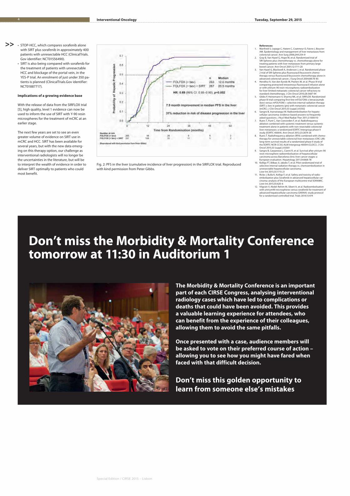
The primary endpoint was progression-free
survival (PFS) at any site, and there was no
significant difference between the groups
(median PFS 10.7 months and 10.2 months in
the SIRT group and non-SIRT group, respec-
tively). However, and importantly, assessment of
PFS in the liver with a competing risks analysis
showed that patients whose treatment included
SIRT had a 7.9 month improvement in PFS in the
liver from 12.6 to 20.5 months (p=0.002) and
a 31% reduced risk (HR=0.69) of the tumours
in their liver progressing (Fig. 2). Similar liver
resection rates were observed in the two arms
of the study.
Y-90 resin microspheres have an average
diameter of 30-35 μm (smaller than the
particles of other liver-directed therapies such
as transarterial chemoembolisation), which
allows them to reach intra-tumoural vessels
to deliver short-range irradiation [6]. Thus,
SIRT has a localised effect that is reflected in
the results of the SIRFLOX study. However,
why the striking improvements in PFS in the
liver did not translate into overall PFS benefits
is still intriguing. One possibility (which is
currently being investigated) is that the high
pro portion of patients with synchronous dis-
ease (approximately 90%) and primary tumour
in-situ (45%) in the SIRFLOX study resulted in
extra-hepatic progression irrespective of the
progression-status in the liver.
The SIRFLOX study also showed no significant
difference in the objective response rate (ORR)
between groups, but the complete response
rate (6.0% versus 1.9%, p=0.020) and complete
response plus partial response rate (78.7% ver-
sus 68.8%, p=0.042) in the liver was significantly
improved with the addition of SIRT.
The addition of SIRT in the SIRFLOX study had
no impact on the duration of chemotherapy
and the safety profile was acceptable as
predicted from previous data.
OS from the SIRLOX study will be evaluated
as part of a pre-planned combined analysis of
SIRFLOX and two studies that have recently
completed recruitment, FOXFIRE and FOXFIRE
Global. These studies combined will include
over 1,100 patients and data are expected in
mid-2017. The absence of overall PFS benefit in
SIRFLOX does not exclude the possibility that
OS benefits may be observed in the longer
term – for example, the CLOCC study of radio-
frequency ablation for patients with colorectal
liver metastases showed no OS benefit at 30
months [7], but recently reported long-term
data that showed a significant difference in
OS in favour of RFA plus chemotherapy versus
chemotherapy alone [8]. The combined OS data
from SIRFLOX/FOXFIRE/FOXFIRE Global are
eagerly anticipated and will further enhance
our knowledge of the potential benefits
of SIRT.
Another randomised study involving ap-
proximately 350 patients with mCRC (EPOCH)
is assessing the use of SIRT when added to
second-line chemotherapy versus second-line
chemotherapy alone. The primary endpoint
is PFS (Fig. 1; ClinicalTrials.Gov identifier:
NCT01483027).
HCC
A similar growth in the evidence base and
drive to a greater evidence-based approach is
occurring with SIRT for HCC.
ENRY provided survival data in a large popu-
lation (n=325) who had received Y-90 resin
microspheres, and showed that factors such as
ECOG performance status and tumour burden
influenced survival after treatment with SIRT [9].
Recently, a pilot randomised trial (SIRTACE)
suggested that SIRT may be an alternative
to TACE for patients with unresectable HCC,
as a single session of SIRT with Y-90 resin
microspheres had a similar impact on ORR and
HRQoL as multiple sessions of TACE [10].
The full reporting of much larger randomised
controlled trials of SIRT in HCC (possibly as early
as 2016) will greatly increase the evidence base
for this treatment. Ongoing studies (all with sur-
vival as the primary endpoint) include (Fig. 1):
• The SORAMIC trial, which compares sorafenib
alone with SIRT followed by sorafenib in 375
patients with advanced HCC [11].
• The SARAH trial compares sorafenib with
SIRT in at least 400 patients with advanced
HCC [12].
• The SIRveNIB trial, which also compares
sorafenib with SIRT in approximately
360 patients with locally-advanced HCC
(ClinicalTrials.Gov identifier: NCT01135056).
Radioembolisation trials: what is the current evidence? Trials and current evidence in IO
Special Session
Tuesday, September 29, 10:00-11:00
Room 5.A
Don’t miss it !
José Ignacio Bilbao (EBIR)
Prof. José Ignacio Bilbao heads the University
Clinic of Navarra’s Interventional Radiology
Department, which he co-established. He has been
an active member of CIRSE since its beginnings,
and has attended every congress, often presenting
award-winning posters and oral presentations or
giving invited lectures.
Prof. Bilbao was awarded a Gold Medal at CIRSE
2013. He was the Josef Roesch Lecturer for CIRSE
2010, speaking on TIPS. That same year, he was
also awarded the CVIR Editor’s Medal for a paper
on non-radioactive resin microspheres, of which
he was the primary author.
He has served on a number of CIRSE committees
over the years, and currently leads CIRSE’s Registry
for SIR-Spheres Therapy (CIRT).
José Ignacio Bilbao
(EBIR)
University Clinic of Navarra
Pamplona, Spain
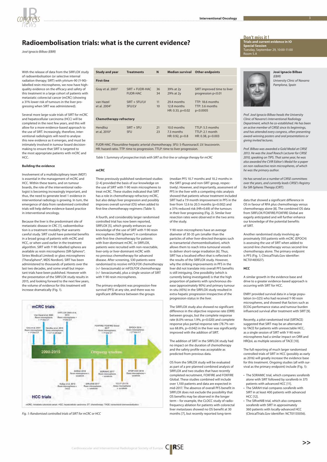
Fig. 1: Randomised controlled trials of SIRT for mCRC or HCC
Table 1: Summary of prospective trials with SIRT as first-line or salvage therapy for mCRC
Study and year Treatments N Median survival Other endpoints
First-line
Gray et al. 20012 SIRT + FUDR-HAC 36 39% at 2y SIRT improved time to liver
FUDR-HAC 34 29% at 2y progression p<0.01
van Hazel SIRT + 5FU/LV 11 29.4 months TTP: 18.6 months
et al. 20043 5FU/LV 10 12.8 months TTP: 3.6 months
HR: 0.33, p=0.02 p<0.0005
Chemotherapy-refractory
Hendlisz SIRT + 5FU 21 10.0 months TTLP: 5.5 months
et al, 20104 5FU 23 7.3 months TTLP: 2.1 month
HR: 0.92, p=0.8 HR: 0.38, p=0.003
FUDR-HAC: Floxuridine-hepatic arterial chemotherapy. 5FU: 5-fluorouracil. LV: leucovorin.
HR: hazard ratio. TTP: time to progression. TTLP: time to liver progression.
>>Cardiovascular and Interventional Radiological Society of Europe
Special Edition / CIRSE 2015 – Lisbon
Tuesday, September 29, 2015Interventional Oncology4
• STOP-HCC, which compares sorafenib alone
with SIRT plus sorafenib in approximately 400
patients with unresectable HCC (ClinicalTrials.
Gov identifier: NCT01556490).
• SIRT is also being compared with sorafenib for
the treatment of patients with unresectable
HCC and blockage of the portal vein, in the
YES-P trial. An enrolment of just under 350 pa-
tients is planned (ClinicalTrials.Gov identifier:
NCT01887717).
Implications of a growing evidence base
With the release of data from the SIRFLOX trial
[5], high quality, level 1 evidence can now be
used to inform the use of SIRT with Y-90 resin
microspheres for the treatment of mCRC at an
earlier stage.
The next few years are set to see an even
greater volume of evidence on SIRT use in
mCRC and HCC. SIRT has been available for
several years, but with the new data emerg-
ing on this therapy option, our challenge as
interventional radiologists will no longer be
the uncertainties in the literature, but will be
to interpret the wealth of evidence in order to
deliver SIRT optimally to patients who could
most benefit.
Fig. 2: PFS in the liver (cumulative incidence of liver progression) in the SIRFLOX trial. Reproduced
with kind permission from Peter Gibbs.
References:
1. Manfredi S, Lepage C, Hatem C, Coatmeur O, Faivre J, Bouvier
AM. Epidemiology and management of liver metastases from
colorectal cancer. Ann Surg 2006;244:254-9
2. Gray B, Van Hazel G, Hope M, et al. Randomised trial of
SIR-Spheres plus chemotherapy vs. chemotherapy alone for
treating patients with liver metastases from primary large
bowel cancer. Ann Oncol 2001;12:1711-20
3. Van Hazel G, Blackwell A, Anderson J, et al. Randomised phase
2 trial of SIR-Spheres plus fluorouracil/leucovorin chemo-
therapy versus fluorouracil/leucovorin chemotherapy alone in
advanced colorectal cancer. J Surg Oncol 2004;88:78-85
4. Hendlisz A, Van den Eynde M, Peeters M, et al. Phase III trial
comparing protracted intravenous fluorouracil infusion alone
or with yttrium-90 resin microspheres radioembolization
for liver-limited metastatic colorectal cancer refractory to
standard chemotherapy. J Clin Oncol 2010;28:3687-94
5. Gibbs P, Heinemann V, Sharma NK, et al. SIRFLOX: Randomized
phase III trial comparing first-line mFOLFOX6 ± bevacizumab
(bev) versus mFOLFOX6 + selective internal radiation therapy
(SIRT) ± bev in patients (pts) with metastatic colorectal cancer
(mCRC). J Clin Oncol 2015;33 (suppl.):A3502
6. Sangro B, Inarrairaegui M. Radioembolization for hepato-
cellular carcinoma: evidence-based answers to frequently
asked questions. J Nucl Med Radiat Ther 2011;2:1000110
7. Ruers T, Punt C, Van Coevorden F, et al. Radiofrequency
ablation combined with systemic treatment versus systemic
treatment alone in patients with non-resectable colorectal
liver metastases: a randomized EORTC Intergroup phase II
study (EORTC 40004). Ann Oncol 2012;23:2619-26
8. Ruers T. Radiofrequency ablation (RFA) combined with chemo-
therapy for unresectable colorectal liver metastases (CRC LM):
long-term survival results of a randomized phase II study of
the EORTC-NCRI CCSG-ALM Intergroup 40004 (CLOCC). J Clin
Oncol 2015;33 (suppl.):A3501
9. Sangro B, Carpanese L, Cianni R, et al. Survival after yttrium-90
resin microsphere radioembolization of hepatocellular
carcinoma across Barcelona clinic liver cancer stages: a
European evaluation. Hepatology 2011;54:868-78
10. Kolligs FT, Bilbao JI, Jakobs T, et al. Pilot randomized trial of
selective internal radiation therapy vs. chemoembolization in
unresectable hepatocellular carcinoma.
Liver Int 2015;35:1715-21
11. Ricke J, Bulla K, Kolligs F, et al. Safety and toxicity of radio-
embolization plus Sorafenib in advanced hepatocellular car-
cinoma: analysis of the European multicentre trial SORAMIC.
Liver Int 2015;35:620-6
12. Vilgrain V, Abdel-Rehim M, Sibert A, et al. Radioembolisation
with yttrium90 microspheres versus sorafenib for treatment of
advanced hepatocellular carcinoma (SARAH): study protocol
for a randomised controlled trial. Trials 2014;15:474
>>
Don’t miss the Morbidity & Mortality Conference tomorrow at 11:30 in Auditorium 1
The Morbidity & Mortality Conference is an important
part of each CIRSE Congress, analysing interventional
radiology cases which have led to complications or
deaths that could have been avoided. This provides
a valuable learning experience for attendees, who
can benefit from the experience of their colleagues,
allowing them to avoid the same pitfalls.
Once presented with a case, audience members will
be asked to vote on their preferred course of action –
allowing you to see how you might have fared when
faced with that difficult decision.
Don’t miss this golden opportunity to
learn from someone else’s mistakes
Cardiovascular and Interventional Radiological Society of Europe C RSE
5IRnewscongress IR Management
Sclerotherapy is well recognised as a first-line
treatment for most lymphatic malformations
(LMs). These slow-flow malformations occur
most commonly in early childhood, usually
presenting as subcutaneous cysts in the neck,
trunk or less frequently in the extremities.
Rarely, they occur in locations such as the
mesentery, genitourinary tract and orbit. As
those radiologists with an interest in vascular
anomalies will know, such lesions are often
poorly understood by the specialists to whom
they present and patients can struggle to get a
definitive diagnosis or appropriate treatment.
LMs of the orbit are rare. They are known to
oculoplastic surgeons as ‘lymphangiomas’ and
are recognised as lesions that are difficult to
resect and highly prone to complications and
recurrence. Sclerotherapy has led to a paradigm
shift in the management of this condition, with
percutaneous sclerotherapy replacing high-risk
surgical intervention entirely and showing a
dramatic improvement in patient outcomes.
As with other vascular malformations, orbital
LMs usually present in childhood, although
small lesions can present late after a spon-
taneous intralesional bleed. They tend to be
isolated, unilateral lesions, though a subset
is associated with micro-cystic lymphatic
malformations of the face. Orbital LMs are
typically macro-cystic and have a characteristic
imaging appearance, with large thin-walled
cysts, fluid-fluid levels and little enhancement.
They are usually centred in the posterior
orbit with both intraconal and extraconal
components. They tend to wrap around the
optic nerve complex, which explains why
oculoplastic surgeons are often reluctant to
intervene.
Orbital LMs cause mass effect within the orbit,
leading to orbital expansion if they are present
from an early age, deformity of the globe and
proptosis. Proptosis causes stretching of the
optic nerve, which is extremely painful when an
acute bleed causes rapid expansion of the cysts.
Stretching of the optic nerve and raised orbital
pressures lead to optic neuropathy and visual
loss over time. In young children, vision may fail
to develop normally at all. The condition is ex-
acerbated by recurrent intralesional bleeds and
infection. In severe cases, patients are plagued
by severe chemosis (inflammation and swelling
of the conjunctiva), disfigurement and pain, as
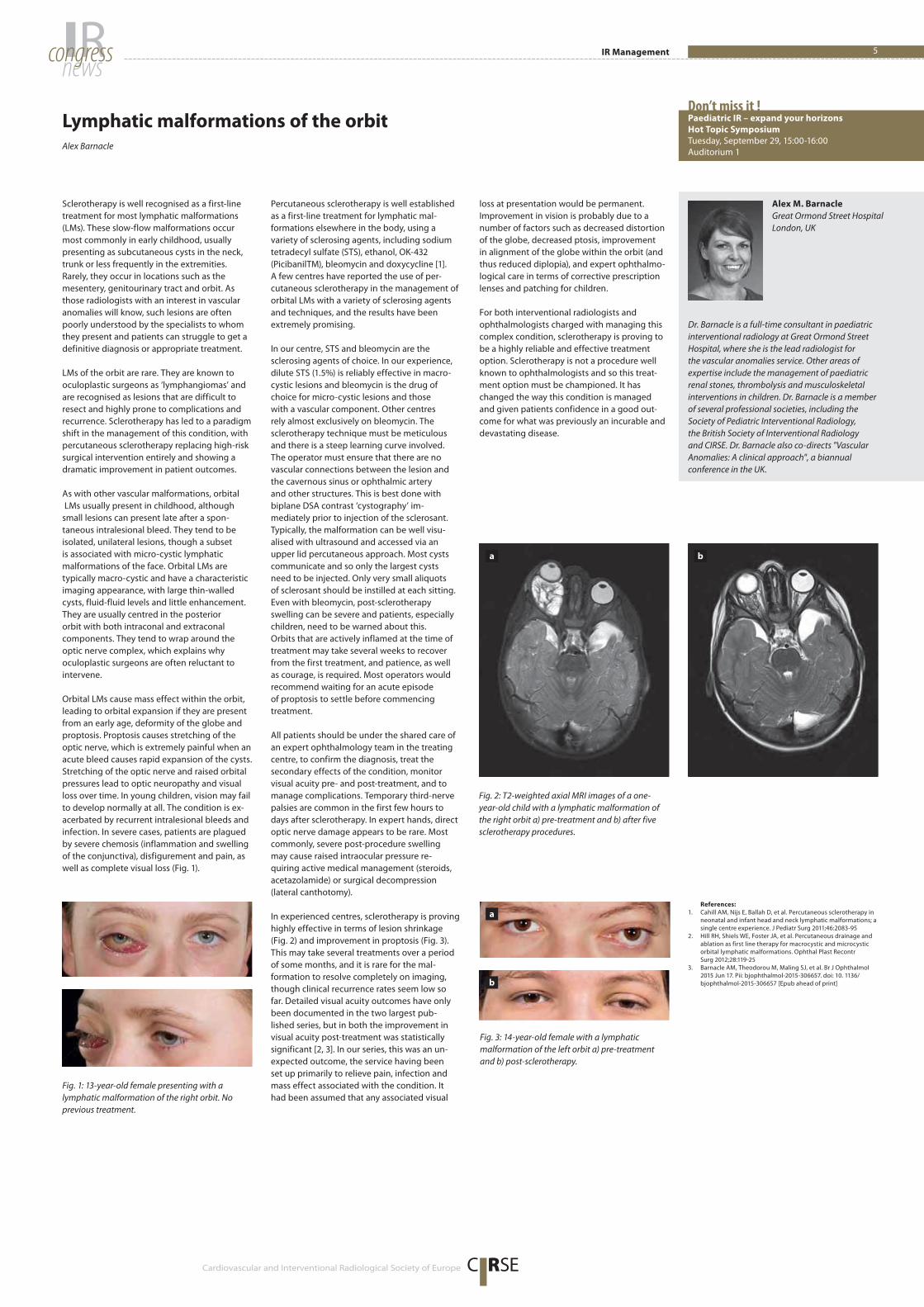
well as complete visual loss (Fig. 1).
Percutaneous sclerotherapy is well established
as a first-line treatment for lymphatic mal-
formations elsewhere in the body, using a
variety of sclerosing agents, including sodium
tetradecyl sulfate (STS), ethanol, OK-432
(PicibanilTM), bleomycin and doxycycline [1].
A few centres have reported the use of per-
cutaneous sclerotherapy in the management of
orbital LMs with a variety of sclerosing agents
and techniques, and the results have been
extremely promising.
In our centre, STS and bleomycin are the
sclerosing agents of choice. In our experience,
dilute STS (1.5%) is reliably effective in macro-
cystic lesions and bleomycin is the drug of
choice for micro-cystic lesions and those
with a vascular component. Other centres
rely almost exclusively on bleomycin. The
sclerotherapy technique must be meticulous
and there is a steep learning curve involved.
The operator must ensure that there are no
vascular connections between the lesion and
the cavernous sinus or ophthalmic artery
and other structures. This is best done with
biplane DSA contrast ‘cystography’ im-
mediately prior to injection of the sclerosant.
Typically, the mal formation can be well visu-
alised with ultrasound and accessed via an
upper lid percutaneous approach. Most cysts
com municate and so only the largest cysts
need to be injected. Only very small aliquots
of sclerosant should be instilled at each sitting.
Even with bleomycin, post-sclerotherapy
swelling can be severe and patients, especially
children, need to be warned about this.
Orbits that are actively inflamed at the time of
treatment may take several weeks to recover
from the first treatment, and patience, as well
as courage, is required. Most operators would
recommend waiting for an acute episode
of proptosis to settle before commencing
treatment.
All patients should be under the shared care of
an expert ophthalmology team in the treating
centre, to confirm the diagnosis, treat the
secondary effects of the condition, monitor
visual acuity pre- and post-treatment, and to
manage complications. Temporary third-nerve
palsies are common in the first few hours to
days after sclerotherapy. In expert hands, direct
optic nerve damage appears to be rare. Most
commonly, severe post-procedure swelling
may cause raised intraocular pressure re-
quiring active medical management (steroids,
acetazolamide) or surgical decompression
(lateral canthotomy).
In experienced centres, sclerotherapy is proving
highly effective in terms of lesion shrinkage
(Fig. 2) and improvement in proptosis (Fig. 3).
This may take several treatments over a period
of some months, and it is rare for the mal-
formation to resolve completely on imaging,
though clinical recurrence rates seem low so
far. Detailed visual acuity outcomes have only
been documented in the two largest pub-
lished series, but in both the improvement in
visual acuity post-treatment was statistically
significant [2, 3]. In our series, this was an un-
expected outcome, the service having been
set up primarily to relieve pain, infection and
mass effect associated with the condition. It
had been assumed that any associated visual
loss at presentation would be permanent.
Improvement in vision is probably due to a
number of factors such as decreased distortion
of the globe, decreased ptosis, improvement
in alignment of the globe within the orbit (and
thus reduced diplopia), and expert ophthalmo-
logical care in terms of corrective prescription
lenses and patching for children.
For both interventional radiologists and
ophthalmologists charged with managing this
complex condition, sclerotherapy is proving to
be a highly reliable and effective treatment
option. Sclerotherapy is not a procedure well
known to ophthalmologists and so this treat-
ment option must be championed. It has
changed the way this condition is managed
and given patients confidence in a good out-
come for what was previously an incurable and
devastating disease.
Lymphatic malformations of the orbit Paediatric IR – expand your horizons
Hot Topic Symposium
Tuesday, September 29, 15:00-16:00
Auditorium 1
Don’t miss it !
Alex Barnacle
Dr. Barnacle is a full-time consultant in paediatric
interventional radiology at Great Ormond Street
Hospital, where she is the lead radiologist for
the vascular anomalies service. Other areas of
expertise include the management of paediatric
renal stones, thrombolysis and musculoskeletal
interventions in children. Dr. Barnacle is a member
of several professional societies, including the
Society of Pediatric Interventional Radiology,
the British Society of Interventional Radiology
and CIRSE. Dr. Barnacle also co-directs "Vascular
Anomalies: A clinical approach", a biannual
conference in the UK.
Alex M. Barnacle
Great Ormond Street Hospital
London, UK
References:
1. Cahill AM, Nijs E, Ballah D, et al. Percutaneous sclerotherapy in
neonatal and infant head and neck lymphatic malformations; a
single centre experience. J Pediatr Surg 2011;46:2083-95
2. Hill RH, Shiels WE, Foster JA, et al. Percutaneous drainage and
ablation as first line therapy for macrocystic and microcystic
orbital lymphatic malformations. Ophthal Plast Recontr
Surg 2012;28:119-25
3. Barnacle AM, Theodorou M, Maling SJ, et al. Br J Ophthalmol
2015 Jun 17. Pii: bjophthalmol-2015-306657. doi: 10. 1136/
bjophthalmol-2015-306657 [Epub ahead of print]
Fig. 2: T2-weighted axial MRI images of a one-
year-old child with a lymphatic malformation of
the right orbit a) pre-treatment and b) after five
sclerotherapy procedures.
Fig. 1: 13-year-old female presenting with a
lymphatic malformation of the right orbit. No
previous treatment.
Fig. 3: 14-year-old female with a lymphatic
malformation of the left orbit a) pre-treatment
and b) post-sclerotherapy.
a
a
b
b
Special Edition / CIRSE 2015 – Lisbon
The CIRSE meets. . . programme has proved to
be an important platform for establishing and
strengthening the relations between CIRSE and
its distinguished Group Members – the national
societies in the field of interventional radiology.
Joining us this year is China. This vast and
dynamic country has acquired a wealth of
clinical data on a number of promising IR pro-
cedures, and representatives of the Chinese
Society of Interventional Radiolgoy (CSIR) will
join us today to share their experiences.
Background
Interventional radiology (IR) was introduced
to China during the 1980s in conjunction
with China’s policy of economic reform and
opening up. IR was immediately accepted
and welcomed by most radiologists after
its intro duction. The first national inter-
ventional radiology meeting was held in
1986 in Weifang City, Shangdong Province,
and more than 100 radiologists, residents
and graduate students participated. This was
a landmark event announcing the advent
of IR in China. Four years later, the Chinese
Society of Interventional Radiology (CSIR)
was founded, and Dr. Lin was elected as the
first CSIR president at the first national CSIR
meeting in Hangzhou, Zhejiang Province, in
1990. The national meeting of CSIR was held
every 4 years in the early years, and then every
2 years, and will become annual meeting from
2015 onwards. A total of eleven national CSIR
meetings have been held. The most recent CSIR
biennial meeting was held in Changsha in 2014,
with more than 3,000 IRs participating.
Currently, there are approximately 5,000
full-time interventional radiologists across the
country, meaning that CSIR has become the
third largest IR society in the world, after SIR
and CIRSE. Most non-coronary IR procedures
are performed by radiologists, including
various vascular and non-vascular interven-
tions, neurointerventions, etc. However, turf
battles have become intense since the 1990s,
especially in the field of vascular inter ventions
and neurointerventions. Nevertheless, we have
not only survived, but also won the battles
in many hospitals. One of major reasons we
manage to hold our ground is that CSIR has
been a strong advocate for inter ventional
radiology to be a more clinical specialty.
Currently, over 70% of IR departments in
China have their own dedicated inpatient
wards. Some of them have become a hybrid
department of interventional radiology with
other specialties, such as vascular surgery.
Many IR pioneers have contributed to the
growth of CSIR, including overseas inter-
ventional radiologists, who made important
contributions to the practice of IR in China,
especially during the early years. To commend
such great contributions, the awards of CSIR
Lifetime Achievement and CSIR Honorable
Member have been established, respectively.
So far, twelve Chinese IR pioneers and eight
foreign interventional radiologists have been
awarded.
Today, three representatives, including
Distinguished Fellow Gao-Jun Teng, will
describe their findings in their respective fields.
Join us in Auditorium 8 for this exciting
event!
CIRSE meets
China
Tuesday, September 29, 2015CIRSE meets China / Autumn ESIR Courses6
Tuesday, September 29
10:00-11:00
CM 2605 CIRSE meets China
Moderators: A.-M. Belli (London/UK), W.-J. Jiang (Beijing/CN)
2605.1 Percutaneous transhepatic portosystemic shunt
H. Wang (Guangzhou/CN)
2605.2 Stent loaded with 125I seeds in malignancies – from bench to bedside
G.-J. Teng (Nanjing/CN)
2605.3 Hybrid intervention for complex cerebrovascular disease
W.-J. Jiang (Beijing/CN)
Register now for the ESIR autumn courses!
Don’t miss your chance to benefit from the first-rate educational programme
offered by the European School of Interventional Radiology.
Critical Limb Ischaemia – Diagnosis, Treatment and Parameters for Success
Expert Course
Amsterdam (NL), October 16-17
Prostate Embolisation
Expert Course
Milan (IT), October 29-30
Effective Hepatocellular Carcinoma (HCC) Treatments – Advanced Local Therapies
Expert Course
Lausanne (CH), November 13-14
DVT & Pulmonary Embolus
Fundamental Course
Dublin (IE), November 27-28
The Future of Image-Guided Tumour Ablation – Targeting Techniques and
High-End Clinical Strategies
Expert Course
Innsbruck (AT), December 11-12
CoursesESIR 2015
For more information, please visit
www.cirse.org/esir2015
Fundamental courses are suitable for
preparing for EBIR (European Board of
Interventional Radiology)
C RSE f o u n d a t i o n
Cardiovascular and Interventional Radiological Society of Europe
7IRnewscongress
C RSE
Vascular Interventions
Transjugular intrahepatic portosystemic shunt
(TIPS) is one of the treatments available to
control the complications of portal hyper-
tension. Portal hypertension is defined as
portal venous pressure >10 mmHg, or a portal
venous pressure gradient of >5 mmHg, and
may lead to complications such as variceal
bleeding and ascites. Liver cirrhosis is the most
common cause of portal hypertension.
TIPS, by creating a low-resistance shunt
between the hepatic vein and the intrahepatic
portion of the portal vein, and keeping it
patent with an expandable covered metal
stent, allows the blood to return to the
systemic circulation at low pressure. The ability
of TIPS to function like a surgical side-to-side
portacaval shunt without requiring general
anaesthesia and major surgery led to its rapid
acceptance into clinical practice [1]. TIPS is a
procedure performed under fluoroscopy and/
or ultrasound guidance, by an interventional
radiologist (IR), and it has a fairly steep learning
curve.
The only curative treatment for portal hyper-
tension and cirrhosis is liver transplant. We
believe that this type of procedure should
be performed in reference centres where a
multidisciplinary approach is possible, and
where experienced IRs can train others.
What is refractory ascites?
Diuretic-resistant ascites is usually associated
with advanced cirrhosis, marked neurohumoral
activation and low urinary excretion of
sodium, despite maximal tolerated doses
of diuretics. The development of diuretic
resistance is most often due to progression
of liver disease, however, it can also be due
to other complications of cirrhosis, such as
hepatocellular carcinoma and portal vein
thrombosis, which must be ruled out.
The 2-year survival rate of all patients with
cirrhosis after the development of ascites is
approximately 50% [2, 3]. By comparison, sur-
vival in patients with diuretic-resistant ascites is
50% at 6 months and 25% at 1 year [4].
Diuretic-resistant ascites in patients with
cirrhosis is present if one of the following
criteria is fulfilled in the absence of therapy
with NSAIDs [5](which can interfere with
diuretic responsiveness):
• inability to reduce ascites despite confirmed
adherence to the dietary sodium restriction
and the administration of maximum tolerable
doses of diuretics;
• rapid re-accumulation of fluid after
therapeutic paracentesis, despite adherence
to a sodium restricted diet; and
• the development of diuretic-related com-
plications such as progressive azotemia,
hepatic encephalopathy, or progressive
electrolyte imbalances.
Diuretic-resistant ascites in patients with
cirrhosis must be differentiated from malignant
ascites due to peritoneal carcinomatosis, Budd-
Chiari syndrome or malignant chylous ascites.
Liver transplantation is the only definitive
treatment for refractory ascites.
A guideline from the American Association for
the Study of Liver Diseases (2009) recommends
that TIPS should be considered only in patients
who are intolerant of repeated large volume
paracentesis [6].
TIPS has also been validated as an effective and
safe treatment for ascites secondary to portal
hypertension [7].
A recently published case-control study [8]
concluded that TIPS appears to be bene-
ficial compared to serial paracentesis in terms
of intermediate/long-term overall survival,
without significantly worsening short-term
mortality, suggesting a potential earlier role in
properly selected patients, when conservative
medical managements fails.
Acute variceal haemorrhage
As a consequence of portal hypertension,
this can be recurrent or refractory to therapy.
Variceal bleeding is one of the leading causes
of death in patients with cirrhosis. After the
first episode, the risk or recurrent bleeding in
the next 2 years is 50-80% [9].
Several treatments are available for the
management of acute variceal haemorrhage.
These can be broadly grouped into treatments
that address the local bleeding site, and those
that reduce portal pressure. Neither treatment
available for variceal haemorrhage is optimal.
Firstly, because these fail to uniformly achieve
homeostasis, and secondly, due to the inability
to arrest progression of cirrhosis or prevent
liver failure. The goal of treatment of active
variceal haemorrhage is to stop initial bleeding,
prevent recurrent bleeding and minimise
treatment-associated morbidity and mortality.
Endoscopic and pharmacologic treatments are
the first-line therapies.
The early placement of TIPS can lead to good
outcomes in select patients when performed
by experienced IRs, in institutions where TIPS is
readily available [10].
The definition of "failure of endoscopic
treatment" remains controversial; we consider
failure to be the recurrence of variceal
haemorrhage despite at least two sessions of
endoscopic treatment performed no more than
two weeks apart.
Multiple series have demonstrated the efficacy
of TIPS for uncontrolled oesophageal variceal
haemorrhage despite emergent endoscopic
and pharmacologic treatment in patients who
are poor candidates for urgent surgery [11-12].
Few studies have compared TIPS to surgery in
the management of refractory haemorrhage in
patients who are good surgical candidates.
A 2009 guideline issued by the American
Association for the Study of Liver Diseases
concluded that TIPS or a distal splenorenal
shunt were similarly effective in the prevention
of re-bleeding, in patients who failed medical
therapy, based upon the results of a controlled
trial [6, 13]. Thus, it may be reasonable to
consider the available expertise and the
patient’s individual circumstances. TIPS is also
a useful temporising measure in those awaiting
liver transplantation.
Contraindications to TIPS
Absolut contraindications to TIPS include heart
failure, severe tricuspid regurgitation, severe
pulmonary hypertension, multiple hepatic
cysts, uncontrolled systemic infection or sepsis
or unrelieved biliary obstruction. Relative
contraindications are central hepatoma, portal
vein thrombosis and severe coagulopathy.
Complications
TIPS insertion can be associated with a
number of complications, some of which may
be fatal. It is therefore essential for all who
use this procedure to be aware of the clinical
spectrum of TIPS-related complications and
their management. There are technical compli-
cations, complications related to creation of the
shunt, and unique TIPS-related complications.
Liver failure and encephalopathy (30%) are the
more frequently expected.
The future
Since it was proven that TIPS is beneficial
in cases of refractory ascites and variceal
bleeding, the next step is to proceed to better
selection of patients and optimising the timing
of the procedure’s execution. In fact, studies
have shown that TIPS must be performed
earlier in case of variceal bleeding [14].
But what about refractory ascites?
The benefits of TIPS in other clinical situations,
such as hepatorenal syndrome, Budd-Chiari
syndrome, hepatic hydrothorax, portal hyper-
tensive gastropathy and hepatic sinusoidal
obstruction syndrome, remain unclear. Studies
predicting post-TIPS encephalopathy are
important in order to help guide patient se-
lection [15].
TIPS for refractory ascites and variceal bleeding Venous Forum IV: Portal hypertension
Special Session
Wednesday, September 30, 08:30-09:30
Auditorium 6
Don’t miss it !
Élia Coimbra
Dr. Élia Coimbra is the head of the Interventional
Radiology Department in the Hepato-Biliary
Centre of Hospital Curry-Cabral CHLC Lisbon.
She is the Chairperson of the Local Host
Committee for CIRSE 2015, and previously served
on the Local Host Committee for CIRSE 2012.
Dr. Coimbra has contributed to several scientific
posters exhibited at CIRSE events, including
on hepatic vein embolisation after portal vein
embolisation, and on IVC filters above renal veins.
Élia Coimbra
Centro Hospitalar
Lisboa Central
Lisbon, Portugal
References:
1. Boyer TD. Transjugular intrahepatic portosystemic shunt:
current status. Gastroenterology 2003;124:1700
2. Ginés P, Quintero E, Arroyo V et al. Compensated cirrhosis:
natural history and prognostic factors. Hepatology 1987; 7:122
3. D’Amico G, Morabito A, Pagliaro L,Marubini E. Survival and
prognostic indicators in compensated and decompensated
cirrhosis. Dig Dis Sci 1986;31:468
4. Bories P, Garcia Compean D, Michel H et al. The treatment
of refractory ascites by the LeVeen shunt. A multi-centre
controlled trial. J.Hepatol 1986;3:212
5. Runyon BA. Refractory ascites. Semin Liver Dis 1993; 13:343
6. Boyer TD, Haskal ZJ, American Association for the Study of
Liver. The Role of Transjugular Intrahepatic portosystemic
shunt (TIPS) in the Management of Portal Hypertension: up-
date 2009. Hepatology 2010;51.306
7. Rossle M, Gerbes AL. TIPS for the treatment of refractory
ascites, hepatorenal syndrome and hepatic hydrothorax: a
critical update. Gut 2010;59:988-1000
8. Gaba RC, Parvinian A, Casadaban LC, Couture PM, Zivin SP,
Lakhoo J, Minocha J, Ray JR CE, Knuttinen MG, Bui JT, Survival
benefits of TIPS versus serial paracentesis in patients with
refractory ascites: a single institution case-control propensity
score analysis. Clinical Radiology 2015; 70, 51-57
9. Monescillo A, Martínez-Lagares F, Ruiz-del-Arbol L,et al.
Influence of portal hypertension and its early decom pression
by TIPS placement on the outcome of variceal bleeding.
Hepatology 2004; 40:793
10. Pagliaro I, D’Amico G, Luca A, et al. Portal Hypertension:
diagnosis and treatment. J Hepatology 1995; 23 (suppl I):36-44
11. Jalan R, John TG, Redhead DN, et al. A comparative study
of emergency transjugular intrahepatic portosystemic
stent-shunt and esophageal transection in the management
of uncontrolled variceal hemorrhage. Am J Gastroenterol 1
995; 90:1932
12. Bañares AJ, Casado M, Rodriguez-Láiz JM, et al. Urgent
transjugular intrahepatic portosystemic shunt for control of
acute variceal bleeding. Am J Gastroenterol 1998;93:75
13. Henderson JM, Boyer TD, Kutner MH, et al. Distal splenorenal
shunt versus transjugular intrahepatic portal systematic shunt
for variceal bleeding: a randomized trial. Gastroenteroly
1996;111:138
14. Garcia-Pagan JC, Pascoli M, Caca K, Laleman W, Bureau C,
Appenrodt B, Luca a, Zipprich A, Abraldes JG, Nevens F,
Vinel JP, Saverbrunch T, Bosh J. Use of early-TIPS for high risk
Variceal Bleeding: results of a pos-RCT surveillance study.
J of Hepatology 2013; 58
15. Berlioux P, Robic MA, Poirson H, Metivier S, Otal P, Barret
C, Lopez F, Péron JM, Vinel JP, Bureau C. Pre-Transjugular
Intrahepatic Portosystemic shunts(TIPS) Prediction of
Post-TIPS Overt Hepatic Encephalopathy: The Critical Flicker
Frequency is More Accurate than Psychometric Tests.
Hepatology 2014;59,2
Don’t forget you can submityour session evaluations online!The CIRSE Society app offers paperless session evaluation –
and not just during the session itself!
Session evaluation is a pre-requisite for obtaining
CMEs for congress attendance. It also helps us
refine the programme for next year.
Be sure to submit in time!
Cardiovascular and Interventional Radiological Society of Europe
Available for iOS
and Android
I N N O V A T I O N | E D U C A T I O N | I N T E R V E N T I O N
C RSENew! Use the CIRSE App
to search for exhibitors
by product category
CIRSE is more than just an annual congress – the society
strives to facilitate and encourage continuing medical
education all year round.
One such service is ESIRonline – the world’s largest
educational database for IR.
Its 8,000 presentations will soon be expanded
to include the presentations from CIRSE 2015!
To further ongoing IR learning, all congress delegates
benefit from full access for a whole year.
A W E A L T H O F I R L E A R N I N G
www.esir.orgCardiovascular and Interventional Radiological Society of Europe
Special Edition / CIRSE 2015 – Lisbon
C RSE
9IRnewscongress Non-Vascular Interventions
Cardiovascular and Interventional Radiological Society of Europe
The definition of the end-point(s) is the
very first step of the management of spinal
metastases in cancer patients, and must be
discussed by a multidisciplinary board. The
end-points can be both pain palliation and/
or prevention of complications related to the
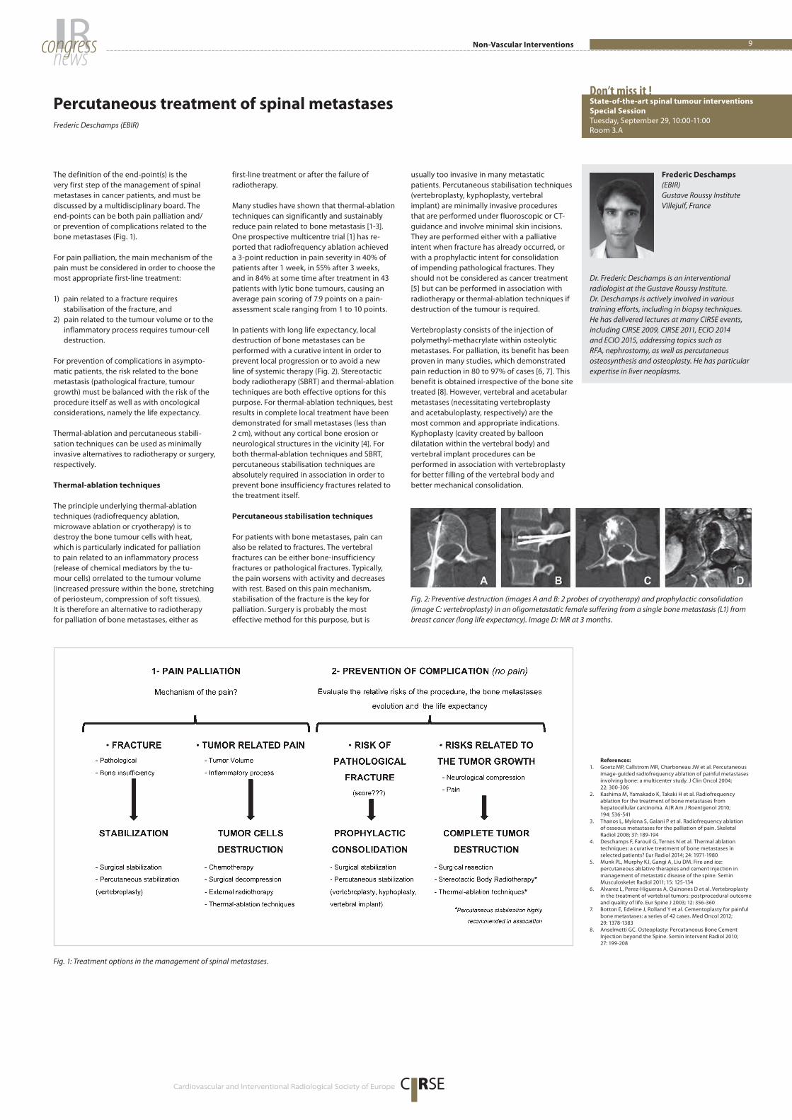
bone metastases (Fig. 1).
For pain palliation, the main mechanism of the
pain must be considered in order to choose the
most appropriate first-line treatment:
1) pain related to a fracture requires
stabilisation of the fracture, and
2) pain related to the tumour volume or to the
inflammatory process requires tumour-cell
destruction.
For prevention of complications in asympto-
matic patients, the risk related to the bone
metastasis (pathological fracture, tumour
growth) must be balanced with the risk of the
pro cedure itself as well as with oncological
considerations, namely the life expectancy.
Thermal-ablation and percutaneous stabili-
sation techniques can be used as minimally
invasive alternatives to radiotherapy or surgery,
respectively.
Thermal-ablation techniques
The principle underlying thermal-ablation
techniques (radiofrequency ablation,
microwave ablation or cryotherapy) is to
destroy the bone tumour cells with heat,
which is particularly indicated for palliation
to pain related to an inflammatory process
(release of chemical mediators by the tu-
mour cells) or related to the tumour volume
(increased pressure within the bone, stretching
of periosteum, compression of soft tissues).
It is therefore an alternative to radiotherapy
for palliation of bone metastases, either as
first-line treatment or after the failure of
radiotherapy.
Many studies have shown that thermal-ablation
techniques can significantly and sustainably
reduce pain related to bone metastasis [1-3].
One prospective multicentre trial [1] has re-
ported that radiofrequency ablation achieved
a 3-point reduction in pain severity in 40% of
patients after 1 week, in 55% after 3 weeks,
and in 84% at some time after treatment in 43
patients with lytic bone tumours, causing an
average pain scoring of 7.9 points on a pain-
assessment scale ranging from 1 to 10 points.
In patients with long life expectancy, local
destruction of bone metastases can be
performed with a curative intent in order to
prevent local progression or to avoid a new
line of systemic therapy (Fig. 2). Stereotactic
body radiotherapy (SBRT) and thermal-ablation
techniques are both effective options for this
purpose. For thermal-ablation techniques, best
results in complete local treatment have been
demonstrated for small metastases (less than
2 cm), without any cortical bone erosion or
neurological structures in the vicinity [4]. For
both thermal-ablation techniques and SBRT,
percutaneous stabilisation techniques are
absolutely required in association in order to
prevent bone insufficiency fractures related to
the treatment itself.
Percutaneous stabilisation techniques
For patients with bone metastases, pain can
also be related to fractures. The vertebral
fractures can be either bone-insufficiency
fractures or pathological fractures. Typically,
the pain worsens with activity and decreases
with rest. Based on this pain mechanism,
stabilisation of the fracture is the key for
palliation. Surgery is probably the most
effective method for this purpose, but is
usually too invasive in many metastatic
patients. Percutaneous stabilisation techniques
(vertebroplasty, kyphoplasty, vertebral
implant) are minimally invasive procedures
that are performed under fluoroscopic or CT-
guidance and involve minimal skin incisions.
They are performed either with a palliative
intent when fracture has already occurred, or
with a prophylactic intent for consolidation
of impending pathological fractures. They
should not be considered as cancer treatment
[5] but can be performed in association with
radiotherapy or thermal-ablation techniques if
destruction of the tumour is required.
Vertebroplasty consists of the injection of
polymethyl-methacrylate within osteolytic
metastases. For palliation, its benefit has been
proven in many studies, which demonstrated
pain reduction in 80 to 97% of cases [6, 7]. This
benefit is obtained irrespective of the bone site
treated [8]. However, vertebral and aceta bular
metastases (necessitating vertebroplasty
and acetabuloplasty, respectively) are the
most common and appropriate indications.
Kyphoplasty (cavity created by balloon
dilatation within the vertebral body) and
vertebral implant procedures can be
performed in association with vertebroplasty
for better filling of the vertebral body and
better mechanical consolidation.
Percutaneous treatment of spinal metastases State-of-the-art spinal tumour interventions
Special Session
Tuesday, September 29, 10:00-11:00
Room 3.A
Don’t miss it !
Frederic Deschamps (EBIR)
Dr. Frederic Deschamps is an interventional
radiologist at the Gustave Roussy Institute.
Dr. Deschamps is actively involved in various
training efforts, including in biopsy techniques.
He has delivered lectures at many CIRSE events,
including CIRSE 2009, CIRSE 2011, ECIO 2014
and ECIO 2015, addressing topics such as
RFA, nephrostomy, as well as percutaneous
osteosynthesis and osteoplasty. He has particular
expertise in liver neoplasms.
Frederic Deschamps
(EBIR)
Gustave Roussy Institute
Villejuif, France
References:
1. Goetz MP, Callstrom MR, Charboneau JW et al. Percutaneous
image-guided radiofrequency ablation of painful metastases
involving bone: a multicenter study. J Clin Oncol 2004;
22: 300-306
2. Kashima M, Yamakado K, Takaki H et al. Radiofrequency
ablation for the treatment of bone metastases from
hepatocellular carcinoma. AJR Am J Roentgenol 2010;
194: 536-541
3. Thanos L, Mylona S, Galani P et al. Radiofrequency ablation
of osseous metastases for the palliation of pain. Skeletal
Radiol 2008; 37: 189-194
4. Deschamps F, Farouil G, Ternes N et al. Thermal ablation
techniques: a curative treatment of bone metastases in
selected patients? Eur Radiol 2014; 24: 1971-1980
5. Munk PL, Murphy KJ, Gangi A, Liu DM. Fire and ice:
percutaneous ablative therapies and cement injection in
management of metastatic disease of the spine. Semin
Musculoskelet Radiol 2011; 15: 125-134
6. Alvarez L, Perez-Higueras A, Quinones D et al. Vertebroplasty
in the treatment of vertebral tumors: postprocedural outcome
and quality of life. Eur Spine J 2003; 12: 356-360
7. Botton E, Edeline J, Rolland Y et al. Cementoplasty for painful
bone metastases: a series of 42 cases. Med Oncol 2012;
29: 1378-1383
8. Anselmetti GC. Osteoplasty: Percutaneous Bone Cement
Injection beyond the Spine. Semin Intervent Radiol 2010;
27: 199-208
Fig. 1: Treatment options in the management of spinal metastases.
Fig. 2: Preventive destruction (images A and B: 2 probes of cryotherapy) and prophylactic consolidation
(image C: vertebroplasty) in an oligometastatic female suffering from a single bone metastasis (L1) from
breast cancer (long life expectancy). Image D: MR at 3 months.
Tuesday, September 29, 2015Morbidity and Mortality Conference / ESIR 201510
Cardiovascular and Interventional Radiological Society of Europe
globe: designed by freepik.com
RCVCardioVascular and Inter vent ional Radio lo g yThe official journal of the Cardiovascular and Interventional Radiological Society of Europe
Send your manuscript around the world
by submitting it to CVIR – the international
platform for Interventional Radiology!
· Official journal of 19 national and regional societies
· Prominent Editors from 12 countries
· Editorial Board Members from over 24 countries
· Contributions from over 40 countries
· Truly international readership
C RSECardiovascular and Interventional Radiological Society of Europe
11Featured Papers / ESIRonlineIRnewscongress
Today’s Featured Paperswill be presented in the Free Paper sessions, taking place from 16:15-17:15 and from 17:30-18:30
16:15 – 17:15
FP 3005 Vascular: aorta
Room 5.A
Long-term follow-up results of endovas-
cular repair in the management of arterial
stenosis caused by Takayasu arteritis
A. Gülcü, N.S. Gezer, S. Akar, N. Akkoç, F. Önen,
A.Y. Göktay; İzmir/TR
FP 3006 Liver TACE: experimental/
new frontier
Room 5.B
Does DEB-TACE enhance the local effect
of IRE? Imaging and histopathological
evaluation in a porcine model
P. Isfort1, P. Rauen1, H.-S. Na1, N. Ito2, C. Wilkmann1,
C.K. Kuhl1, P. Bruners1;1Aachen/DE, 2Tokyo/JP
FP 3007 IVC filters
Room 3.A
The impact of a prospectively maintained IR
database on IVC filter retrieval rates
J.G. McGarry, K.A. Pennycooke, E. Ryan,
P. Tharanatnam, S. Awaiyhan, M.F. Given,
F. McGrath, A. Keeling, M.J. Lee; Dublin/IE
FP 3008 Lung ablation
Room 1.15
Radiofrequency ablation versus surgery
for the treatment of lung metastases:
a comparative study
L. Tselikas1, S. Yevich1, T. de Baère1, A. Auperin1,
O. Mercier2, A. Hakimé1, C. Teriitehau1, E. Fadel2,
F. Deschamps1; 1Villejuif/FR, 2Plessis Robinson/FR
17:30 – 18:30
FP 3104 Venous intervention
Room 5.A
Five-year outcome after catheter-directed
thrombolysis for upper femoral and/or iliac
vein thrombosis: results of a randomized
controlled trial (the CaVenT study)
Y. Haig1, T.R. Enden1, O. Grøtta1, P.M. Sandset1,
C.-E. Slagsvold1, G. Sandbæk1, G. Hafsahl1,
L.O. Holmen2, N.-E. Kløw1; 1Oslo/NO, 2Fredrikstad/NO
FP 3105 IR in liver transplant
Room 5.B
Identification of cofactors influencing
hypertrophy of the future liver remnant
after portal vein embolization: the effect
of newly developing portal collaterals on
embolized liver volume
M. Zeile, G.A. Stavrou, A. Bakal, J.E. Volkmer,
P. Dautel, J. Hoeltje, A. Stang, K.J. Oldhafer,
R. Bruening; Hamburg/DE
FP 3106 Kidney and ureter
Room 3.A
Remote ischemic preconditioning to
reduce contrast-induced nephropathy:
a randomized controlled trial
T. Sterenborg1, T. Menting1, Y. de Waal1,
R. Donders1, K. Wever1, L. Suzan2, D. van der Vliet1,
J. Wetzels1, L.J. Schultze Kool1, M. Warlé1; 1Nijmegen/NL, 2Doetinchem/NL
FP 3107 Portal vein (TIPS)
Room 3.B
Single-centre experience of extending
indications for percutaneous intra-portal
islet auto-transplantation (PIPIAT) after
pancreatic surgery to prevent type 1
diabetes (T1D): feasibility, technical aspects,
complications, and clinical outcome
C. Sallemi, M. Venturini, L. Piemonti, G. Balzano,
P. Maffi, A. Del Maschio, F. De Cobelli; Milan/IT
FP 3108 Miscellaneous
Room 1.15
Severe gastrointestinal bleeding: a national
confidential enquiry into the quality of care
in patients requiring blood transfusion of 4
or more units
S.J. McPherson, N. Smith, M. Sinclair; London/UK
Expert Course
The Future of Image-Guided Tumour Ablation – Targeting Techniques and High-End Clinical Strategies
Innsbruck (AT), December 11-12, 2015
This course brings together seasoned interventional radiologists to
scrutinise the latest image-guidance systems and ablation methods.
Featuring a live case, product overviews and workshops examining
individual imaging systems and discussing ablation devices, as well as
forward-looking round-table discussions focusing on improving outcomes,
the course promises to appeal to experienced practitioners dedicated to
both shaping and further developing the future of interventional oncology.
Early Bird Fee available until October 16!
www.cirse.org/esir2015
C RSE f o u n d a t i o n
ESIR 2015 Course
Special Edition / CIRSE 2015 – Lisbon
Tuesday, September 29, 2015CIRT / ICCIR 201612
CIRSE is pleased to announce that its pivotal registry on radioembolisation with
SIR-Spheres microspheres, the CIRSE Registry for SIR-Spheres Therapy (CIRT) has
enrolled its 100th patient!
Just nine months after the launch of the registry in January 2015, CIRT is the largest
observational study on the real-life application of SIR-Spheres in Europe, with
hospitals enrolled from eight different countries, painting a diverse picture of the
clinical use of SIR-Spheres microspheres in Europe.
For more information on CIRT, please visit www.cirse.org/cirt
or contact Niels de Jong at [email protected].
CIRT enrolls its 100th patient!CIRT
CIRSE Registry for SIR-Spheres Therapy
Cardiovascular and Interventional Radiological Society of Europe
ICCIR 2016
June 9-11
Poertschach | Austria
www.iccir.eu
International Conference on Complications in Interventional Radiology
C RSE f o u n d a t i o n
Save the Date!
As always, ICCIR 2016 will offer a discreet forum for doctors with
diverse experience levels to explore the difficult but necessary subject
of procedural complications.
Open Discussion
This unique congress allows participants to openly discuss cases that did
not go as planned, and gives young doctors the opportunity to interact
directly with colleagues who experienced such situations. The event attracts
physicians who are strongly committed to keep perfecting their work. As a
result, discussions are exceptionally frank, engaging and informative.
Unique Format
The event’s discreet and professional environment is vital. The faculty is
carefully selected and overall participation is limited. Hand-picked case
reports constitute another essential element. This unusual approach has
been tremendously popular, and ICCIR has quickly established itself as the
main complications meeting in Europe. Don’t miss your chance to take part
in this exceptional event – mark your calendars now!
For more information, please visit: www.iccir.eu
C RSECardiovascular and Interventional Radiological Society of Europe
Upper gastro-intestinal (GI) bleeding is defined
as acute bleeding from a source located above
Treitz’s angle. A distinction should be made
between chronic and acute upper GI bleeding
as their consequences and management are
quite different. Fifty to 60% of acute upper GI
bleeding is related to ulcers. Other causes of
upper GI bleeding include tumours, Mallory-
Weiss syndrome and oeso-gastric varices due
to portal hypertension. A distinction has to be
made between cases where the bleeding is
arterial in origin and cases where the bleeding
is venous in origin, as their management will be
totally different.
In the setting of acute GI bleeding there is
usually no room for diagnostic imaging such
as contrast-enhanced CT. Diagnostic imaging
is reserved for the investigation of chronic GI
bleeding. Acute upper GI bleeding must first
be assessed via endoscopy after appropriate
fluid resuscitation. Endoscopy allows for the
identification of the bleeding source in most
cases, and therapeutic haemostasis can be
performed in the same session [1]. In case of
arterial bleeding, injection of epinephrine and
application of haemostatic clips are usually
performed. The role of endoscopy in case
of bleeding from a tumoural origin is often
limited to diagnosis, as there are few efficient
ways to control bleeding from these kinds
of lesions endoscopically. In case of variceal
bleeding, endoscopic band ligation (EBL) is the
treatment of choice. Injection of sclerosants
(aethoxysclerol) or cyanoacrylate can be an
option in some cases. Gastric varices represent
a challenging subset of variceal bleeding, with
endoscopic treatment more difficult and fewer
data available in the literature.
Interventional radiology plays an important
role in case of a failure to control bleeding
endoscopically. In case of arterial bleeding,
transcatheter arterial embolisation (TAE) is the
treatment of choice. Before engaging in this
procedure, the interventional radiologist (IR)
should get important information from the
endoscopist, such as the nature and location
of the bleeding source. Haemostatic clips
placed in the vicinity of the bleeding artery can
provide important help to the IR seeking the
culprit vessel. Whenever possible, clips should
be placed onto or near the bleeding vessel
as radiologic tags, even if they are deemed
useless. After gaining access to the arterial
system at the level of the common femoral
artery (CFA), a selective digitally subtracted
angiogram (DSA) of both the celiac axis (CA)
and superior mesenteric artery (SMA) should be
obtained. In most cases of arterial bleeding, the
culprit vessel can be identified by the presence
of vascular anomalies such as irregular
vessel walls, pseudo-aneurysm, contrast
blush or active extravasation. Provocative
angiography or pharmaco-angiography,
where vasodilatative or thrombolytic drugs
are administered intra-arterially to provoke
bleeding, is not recommended.
In the vast majority of cases, the gastro-
duodenal artery (GDA) or one of its branches
is responsible for the bleeding. Some authors
even advocate systemic embolisation of the
GDA when performing TAE for acute upper
GI bleeding if there is no evidence of vascular
anomaly [2]. A microcatheter should be
used to reach the bleeding vessel, then coils
should be used as the embolic material of
choice. Because of the redundant vascular
supply and numerous arterial anastomoses
in the supramesocolic abdominal compart-
ment, care should be taken to embolise the
bleeding vessel on both sides of the vascular
lesion to avoid re-bleeding (i.e. front door &
back door technique). Particles can be used
in case of bleeding from tumoural origin, but
should never be used in any other setting
as they will induce ischaemia, necrosis and,
ultimately, perforation. Glue should only be
used in severely bleeding, unstable patients,
and carries higher risks of complications, such
as wall necrosis and perforation. Nowadays,
there is no room for intra-arterial vasopressin
infusion.
Venous bleeding from portal vein hyper tension
(PVH) is a totally different story: patients with
PVH will develop venous porto-systemic shunts
in form of oesophageal or gastric varices.
These fragile vessels are prone to bleeding
either from mechanical stress of just from PVH
itself. As for bleeding from arterial sources,
endoscopy should be performed first, as it
allows diagnosis and treatment in most cases.
When endoscopy fails to control bleeding,
emergency placement of a transjugular
intrahepatic porto-systemic shunt (TIPSS)
should be considered. TIPSS is an artificially-
created conduit between the portal venous
circulation and one hepatic vein. Briefly, the
right hepatic vein is catheterised from a right
jugular venous access, and from there, a special
trocart is used to puncture the right branch of
the portal vein. Once access to the portal vein
is obtained, the intrahepatic tract is dilated
with a balloon catheter and a covered stent is
put in place between the portal and hepatic
venous systems.
This procedure allows the immediate lower-
ing of the portal vein pressure and ,in most
cases, control of the bleeding. This procedure
has the advantage of addressing the cause
of bleeding, which is PVH. However, this
procedure carries the risk of precipitating
encephalopathy or causing acute liver failure
by diverting all the portal blood flow away
from the liver. It is always better to make the
decision to perform this procedure in light
of the general health status of the patient
and the presence of comorbidities such as
cirrhosis or cardiac failure. Early TIPSS (i.e.
TIPSS performed within 72 hours of the initial
bleeding episode) has shown good results in
terms of bleeding control, with lower morbidity
than emergent TIPSS [3]. In case of bleeding
from gastric varices, balloon-assisted retro-
grade transvenous obliteration (B-RTO) of the
varices is an interesting alternative [4]. Briefly,
B-RTO consists of the retrograde occlusion of a
spleno-renal vein. Through a jugular or femoral
access, a balloon catheter is used to occlude
the spleno-renal shunt, and cyanoacrylate
or a sclerosant agent is injected to occlude
the varices. Additional coils can be used to
prevent migration of the occluding agent. This
technique carries the advantage of limiting
the risks of consecutive encephalopathy and
preserving hepatic function. However, it does
not address the problem of PVH.
In summary, we strongly believe that endos-
copists and IRs are playing on the same
team when dealing with patients with a
condition as challenging as upper GI bleeding.
If endoscopists can be regarded as the
equivalent of defensive linemen on a football
team, interventional radiologists should be
seen as their linebackers.
Treatment and outcome: acute upper GI bleeding GI tract haemorrhage
Special Session
Tuesday, September 29, 11:30-12:30
Auditorium 6
Don’t miss it !
Pierre Bize (EBIR)
Dr. Pierre Bize is a senior physician in the
Department of Medical Imaging at CHUV. He
received his medical degree from the University
of Geneva in 1993, and trained in general surgery,
cardiac surgery and neurosurgery before focusing
on radiology and interventional radiology. As an
interventional radiologist, he specialises in tumour
ablation and embolisation techniques, including
chemoembolisation, and has particular expertise
in the management of VX2 animal model trials. Dr.
Bize has authored 24 peer-reviewed articles and 6
book chapters. Dr. Bize would like to acknowledge
the contributions of Dr. Michel H. Maillard and Prof.
Alban Denys, both of Lausanne, Switzerland.
Pierre Bize (EBIR)
University Hospital of Lausanne
Lausanne, Switzerland
References:
1. Hwang JH, Fisher DA, Ben-Menachem T, et al. The role of
endoscopy in the management of acute non-variceal upper GI
bleeding. Gastrointestinal endoscopy. 2012;75(6):1132-8
2. Loffroy R, Favelier S, Pottecher P, et al. Transcatheter arterial
embolization for acute nonvariceal upper gastrointestinal
bleeding: Indications, techniques and outcomes.
Diagn Interv Imaging. 2015;96(7-8):731-44
3. Garcia-Pagan JC, Caca K, Bureau C, et al. Early use of TIPS in
patients with cirrhosis and variceal bleeding.
N Engl J Med. 2010;362(25):2370-9
4. Saad WE, Darcy MD. Transjugular Intrahepatic Portosystemic
Shunt (TIPS) versus Balloon-occluded Retrograde Transvenous
Obliteration (BRTO) for the Management of Gastric Varices.
Semin Intervent Radiol. 2011;28(3):339-49
13IRnewscongress Transcatheter Embolisation
Expert CourseEffective Hepatocellular Carcinoma (HCC) Treatments – Advanced Local TherapiesLausanne (CH), November 13-14
Minimally invasive treatments of HCC are of ever-growing importance, and
ESIR’s decision to offer an educational course structured around the condition
itself rather than a single therapy reflects the complex variety of IR options
that are currently available.
This course will address the whole spectrum of radiological interventions,
from ablation to TACE and radioembolisation, as well as patient triage and
indications and classifications.
www.cirse.org/esir2015
ESIR 2015 Course
C RSE f o u n d a t i o n
Special Edition / CIRSE 2015 – Lisbon
C RSE
EB IR
Branching out beyond Europe Places now available for corresponding members!
Register for the EBIR examinations to be held in Europe in 2016:
VIENNA, March 2-3, 2016
BARCELONA, September 9-10, 2016
CERTIFY YOUR EXPERTISE!
Colleagues from Australia and New Zealand can also register for the EBIR examination held in cooperation with IRSA:
QUEENSTOWN, August 1-2, 2016
For more information, please visit www.cirse.org/ebir
CIRSE’s new app contains essential information on pharmacological
agents used by interventional radiologists in everyday clinical practice.
The app:
• Covers over 60 commonly encountered agents
• Information is divided into eight main categories: cardiovascular, contrast,
embolic/thrombotic agents, GI-hepatic-pancreatic, haematologic, infection
control, oncology and pain management
• Entries outline the agent’s pharmacological properties, indications and
contraindications; its proper administration; and, where applicable,
available reversal agents
• Available for iPhone, iPad and Android
Prepared by experienced CIRSE members, IR Drugs and Doses serves
as a handy guide for medical practitioners, trainees and students alike –
COMING SOON!
IR Drugs and Doses – crucial pharmacology details at your fingertips!
I N N O V A T I O N | E D U C A T I O N | I N T E R V E N T I O N
Available for iOS
and Android
Cardiovascular and Interventional Radiological Society of Europe
C RSE
Interventional Radiology
Drugs and Doses
C RSECardiovascular and Interventional Radiological Society of Europe
15ECIO 2016IRnewscongressnews
IR Congress News is published as an additional source of information for all CIRSE 2015
participants. The articles and advertorials in this newspaper reflect the authors‘ opinion.
CIRSE does not accept any responsibility regarding their content.
If you have any questions about this publication, please contact us at [email protected].
Editors-in-Chief: P. Haage, C. Binkert
Managing Editor: Ciara Madden, CIRSE Office
Graphics/Artwork: LO O P. EN T ER PR I SE S media / www.loop-enterprises.com
The annual ECIO meeting offers all
physicians interested in image-guided
oncological therapies an opportunity to
learn about new developments and discuss
best practice. Since the early days of image-
guided biopsies and palliative stenting, the
field has expended to cover a staggering
range of clinical options.
Next year’s meeting, to be held Dublin, Ireland,
will endeavour to cover a broad cross-section
of these therapies, focusing on the most recent
advances. Under the new chairperson, Prof.
Thomas Helmberger, the Scientific Programme
Committee has already devised an exciting and
varied programme.
Colorectal liver metastases
A core theme will be metastatic colorectal
liver cancer. More than one million new
colorectal patients are seen each year
worldwide: approximately 15% of these have
liver metastases at diagnosis and around
60% develop these during follow-up. Recent
interventional oncology data demonstrate
some promising adjuvant therapies, as well
as increased survival time and improved
quality of life in unresectable patients.
These treatments and their clinical
application will be thoroughly
examined in number of Clinical Focus
Sessions and a Multidisciplinary
Tumour Board.
A varied programme
Other topics to be discussed include staples
such as imaging, HCC, lung cancers, new
developments and the clinical management
of patients. 2016 will also feature a dedicated
immunotherapy session – an exciting field
which deserves the attention of the oncology
community. The conference will also address
newer clinical territories such as neuroendo-
crine tumours and cholangiocarcinoma, as well
as hosting a discussion on quality assurance in
the IO field.
The full scientific programme is already
available online – visit the ECIO website for
more details (www.ecio.org/2016).
Join us in Dublin!
These exciting sessions will take place in
the Convention Centre Dublin, the first
carbon-neutral convention centre in the world,
whose striking glass frontage and curved walls
offer the perfect backdrop to a field as dynamic
and forward-thinking as interventional
oncology. We hope to see you there!
To browse the Scientific Programme,
please visit www.ecio.org/2016 or scan
the QR code:
Bridging the gap between research and practice – ECIO 2016
General Information
Congress Date
April 17-20, 2016
Venue
The Convention Centre Dublin
Spencer Dock
Dublin 1
Ireland
Contact
CIRSE Central Office
Neutorgasse 9, Vienna
Email: [email protected]
C RSE Cardiovascular and Interventional Radiological Society of Europe
LEADERS IN ONCOLOGICINTERVENTIONS
www.ecio.org
ECIO 2016
April 17-20Dublin, Ireland
European Conference on Interventional Oncology