Embed Size (px)
Citation preview
JOURNAL OF CLINICAL MICROBIOLOGY, May 1988, p. 827-8300095-1137/88/050827-04$02.00/0Copyright C 1988, American Society for Microbiology
Congo Red as a Fluorochrome for the Rapid Detection of FungiMALCOLM SLIFKIN* AND RICHARD CUMBIE
Microbiology Section, Department of Laboratory Medicine, Allegheny General Hospital, Pittsburgh, Pennsylvania 15212
Received 17 September 1987/Accepted 28 January 1988
Congo red may be applied as a fluorochrome to rapidly detect fungi in clinical specimens, tissue, and fungalculture preparations. This generally available stain is cost effective and simple to prepare. The stain may beprepared with potassium permanganate as a counterstain or with Formalin or glutaraldehyde as a fungicide.
Various fluorescent stains with a high affinity for celluloseor chitin have been applied to stain fungal elements. Accord-ingly, the fluorescent brightener Calcofluor (Cellofluor, Cal-cofluor White M2R New, and Fungi-fluor) (6, 8, 11, 16, 17,24; P. G. Jones, M. Hammer, and R. J. Zabransky, Clin.Microbiol. Newsl. 9:6-7, 1987) and primulin (direct yellow)(3) have been reported to stain the cell walls of plants andfungal elements in fresh or fixed histopathologic sectionsfrom human specimens. Recently, other fluorescent bright-eners were demonstrated to be effective in the staining offungi in frozen and paraffin-embedded tissues (10). Congored was demonstrated to have an affinity for the cellulose-associated sites of various plants (8, 24), showing an intensered fluorescence.
This report discusses the use of Congo red as a nonspecificfluorochrome stain for the rapid examination and detectionof fungi in fresh and paraffin-embedded tissues and of fungalisolates.
MATERIALS AND METHODS
Organisms (i) Fungi. The following fungi were obtainedfrom our stock culture collection and were cultured onSabouraud dextrose agar slants at 25°C: Aspergillus fumi-gatus, Paecilomyces sp., Fusarium sp., Mucor sp., Rhi-zopus sp., Cladosporium sp., Penicillium sp., Phialophoraverrucosa, Acremonium sp., Candida albicans, and Crypto-coccus neoformans. Phialophora richardsiae ATCC 26465was obtained from the American Type Culture Collection,Rockville, Md. Cultures of Exophialajeanselmei, Wangielladermatitidis, Fonsecaea pedrosoi, and Aureobasidium pul-lulans were kindly provided by D. H. Pincus, AnalytabProducts, Plainview, N.Y. Teased preparations, slide cul-tures, and cellophane tape preparations were made withthese organisms by standard methods (15). Congo red stainprepared with Formalin or glutaraldehyde was used toexamine these fungal preparations.
(i) Bacteria. One strain of each of the following bacteriawas obtained from our stock cultures and grown on sheepblood agar, brain heart infusion agar, and chocolate agar inair at 35°C for 25 h: Staphylococcus aureus, Staphylococcusepidermidis, group A streptococcus, Escherichia coli, Kleb-siella pneumoniae, Enterobacter agglomerans, Pseudomo-nas aeruginosa, Streptococcus pneumoniae, Pasteurellamultocida, Citrobacterfreundii, Proteus mirabilis, and No-cardia asteroides. The staining response with Congo red wasdetermined for each of these organisms.
Stain reagents. (i) A 0.1% Congo red (Fisher Scientific Co.,Pittsburgh, Pa.) stain was prepared in distilled water (pH
* Corresponding author.
5.2). (ii) For the glutaraldehyde-Congo red stain (pH 5.0),glutaraldehyde (70%) (Polysciences, Inc., Warrington, Pa.)was diluted in distilled water to yield a 3.0% solution. Congored was added to produce a 0.1% solution of the stain. (iii)For the Formalin-Congo red stain (pH 7.1), a 10% Formalinsolution was diluted to 1% and Congo red was added to yielda 0.1% solution. (iv) For the potassium permanganate coun-
terstain, a 0.5% solution of potassium permanganate (J. T.Baker Chemical Co., Phillipsburg, N.J.) was prepared indistilled water. One set of these stains was stored at room
temperature, and a second set was maintained at 8°C.In one experiment, 10 ml of an aqueous preparation of
0.1% Congo red in a capped test tube was placed 3 in. (ca. 8cm) from a 100-W light source. After a 7-day exposure to thelight, the stain was examined to determine its ability to reactwith fungal elements.
Fluorescence microscopy. The microscope used was aLeitz Ortholux with a 75-W xenon lamp and a Ploemsilluminator. Specimens were examined with a 40x or 63 x oilimmersion lens with two combinations of Leitz filters: (i) anarrow green band excitation filter (BP530-560) and an LP580 barrier filter that yielded an excitation maximum of 546nm or (ii) a narrow blue band excitation filter (BP450-490)and an LP 515 barrier filter that yielded an excitationmaximum of 470 nm.
Clinical specimens. Fresh clinical specimens of 40 sputumsamples, 1 bile fluid sample, 11 skin scrapings, and 18bronchial washings were each smeared on two microscopeslides. After air drying, one slide was Gram stained (BBLMicrobiology Systems, Cockeysville, Md.), using acetone asa decolorizer, and the other slide was stained with the Congored stain and the potassium permanganate counterstain.These specimens were also cultured for fungi by a standardmethod (15).A retrospective microscopic examination of a cerebrospi-
nal fluid sample that contained Cryptocococcus neoformanswas performed by mixing several drops of spinal fluid withthe Formalin-Congo red stain or the glutaraldehyde stain.
Paraffin-embedded tissues. Paraffin-embedded tissues con-
taining specific fungi were selected for a retrospective ex-
amination with Congo red stain. These tissues were sec-tioned, deparaffinized, hydrated, and stained with thepotassium permanganate and Congo red stains. Histologicstains including hematoxylin and eosin (H&E), periodicacid-Schiff (PAS), and Gomori methanamine silver (GMS)were concurrently performed with other sections of thesetissues by standard procedures. The various tissues chosencontained C. albicans, Aspergillus fumigatus, Coccidioidesimmitis, or Lecythophora mutabilis (9, 21).
Staining procedure. The teased mounts and cellophanetape preparations were placed in a few drops of the glutaral-
827
Vol. 26, No. 5
on May 7, 2018 by guest
http://jcm.asm
.org/D
ownloaded from
828 SLIFKIN AND CUMBIE
dehyde-Congo red or Formalin-Congo red stain, placedunder cover slips, and then examined by fluorescence mi-croscopy. The deparaffinized tissue sections were stainedwith 0.1% Congo red for 1 min, rinsed in tap water, coun-terstained with the potassium permanganate reagent forabout 10 to 20 s, and then washed in tap water. The slideswere drained and dried by light blotting. A drop of nonfluo-rescent immersion oil (Stephens Scientific, Denville, N.J.)was placed on the stained section, a cover slip was added,and the section was microscopically examined. In someexperiments, Congo red and counterstained preparations oftissue sections were dehydrated through successively in-creasing concentrations of ethanol, cleared in xylene, andplaced under cover slips with the histological mountingmedium Permount (Fisher Scientific).
In one experiment, the H&E-, PAS-, and GMS-stainedsections were restained with potassium permanganate andCongo red. For these preparations, the cover slips wereremoved with xylene and the sections were dehydratedthrough xylene and decreasing concentrations of ethanol todistilled water and stained with the Congo red reagent for 30 s.
RESULTSSlide cultures and teased and cellophane tape preparations
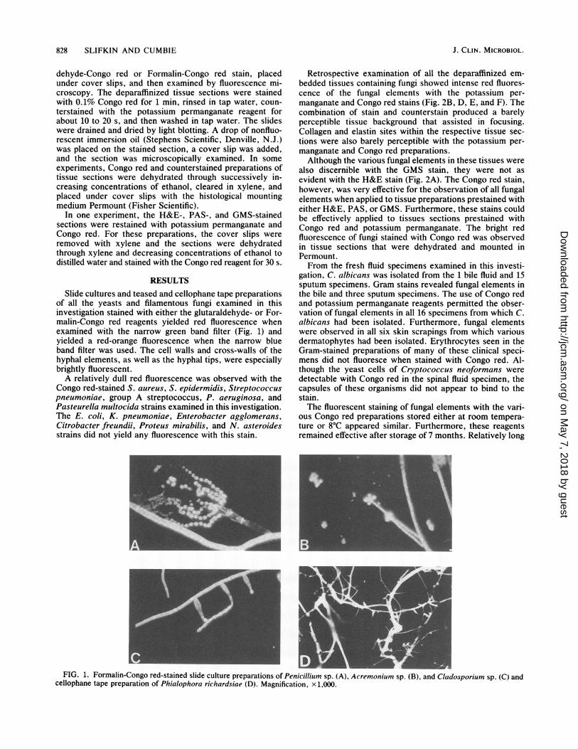
of all the yeasts and filamentous fungi examined in thisinvestigation stained with either the glutaraldehyde- or For-malin-Congo red reagents yielded red fluorescence whenexamined with the narrow green band filter (Fig. 1) andyielded a red-orange fluorescence when the narrow blueband filter was used. The cell walls and cross-walls of thehyphal elements, as well as the hyphal tips, were especiallybrightly fluorescent.A relatively dull red fluorescence was observed with the
Congo red-stained S. aureus, S. epidermidis, Streptococcuspneumoniae, group A streptococcus, P. aeruginosa, andPasteurella multocida strains examined in this investigation.The E. coli, K. pneumoniae, Enterobacter agglomerans,Citrobacter freundii, Proteus mirabilis, and N. asteroidesstrains did not yield any fluorescence with this stain.
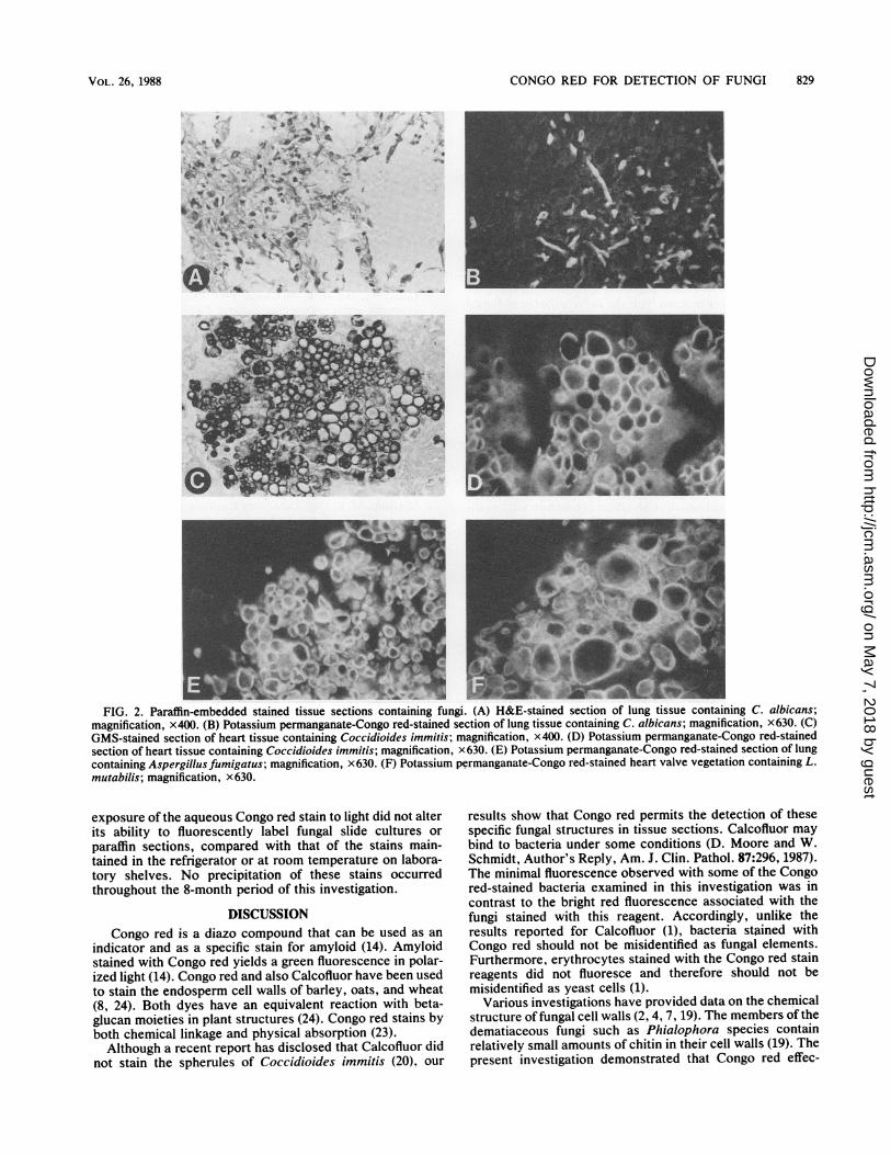
Retrospective examination of all the deparaffinized em-bedded tissues containing fungi showed intense red fluores-cence of the fungal elements with the potassium per-manganate and Congo red stains (Fig. 2B, D, E, and F). Thecombination of stain and counterstain produced a barelyperceptible tissue background that assisted in focusing.Collagen and elastin sites within the respective tissue sec-tions were also barely perceptible with the potassium per-manganate and Congo red preparations.Although the various fungal elements in these tissues were
also discernible with the GMS stain, they were not asevident with the H&E stain (Fig. 2A). The Congo red stain,however, was very effective for the observation of all fungalelements when applied to tissue preparations prestained witheither H&E, PAS, or GMS. Furthermore, these stains couldbe effectively applied to tissues sections prestained withCongo red and potassium permanganate. The bright redfluorescence of fungi stained with Congo red was observedin tissue sections that were dehydrated and mounted inPermount.From the fresh fluid specimens examined in this investi-
gation, C. albicans was isolated from the 1 bile fluid and 15sputum specimens. Gram stains revealed fungal elements inthe bile and three sputum specimens. The use of Congo redand potassium permanganate reagents permitted the obser-vation of fungal elements in all 16 specimens from which C.albicans had been isolated. Furthermore, fungal elementswere observed in all six skin scrapings from which variousdermatophytes had been isolated. Erythrocytes seen in theGram-stained preparations of many of these clinical speci-mens did not fluoresce when stained with Congo red. Al-though the yeast cells of Cryptococcus neoformans weredetectable with Congo red in the spinal fluid specimen, thecapsules of these organisms did not appear to bind to thestain.The fluorescent staining of fungal elements with the vari-
ous Congo red preparations stored either at room tempera-ture or 8°C appeared similar. Furthermore, these reagentsremained effective after storage of 7 months. Relatively long
FIG. 1. Formalin-Congo red-stained slide culture preparations of Penicillium sp. (A), Acremonium sp. (B), and Cladosporium sp. (C) andcellophane tape preparation of Phialophora richardsiae (D). Magnification, x 1,000.
J. CLIN. MICROBIOL.
on May 7, 2018 by guest
http://jcm.asm
.org/D
ownloaded from
CONGO RED FOR DETECTION OF FUNGI 829
0.l
*1 .0a
i..,* 4.
0
4
dg
FIG. 2. Paraffin-embedded stained tissue sections containing fungi. (A) H&E-stained section of lung tissue containing C. albicans;magnification, x400. (B) Potassium permanganate-Congo red-stained section of lung tissue containing C. albicans; magnification, x630. (C)GMS-stained section of heart tissue containing Coccidioides immitis; magnification, x400. (D) Potassium permanganate-Congo red-stainedsection of heart tissue containing Coccidioides immitis; magnification, x630. (E) Potassium permanganate-Congo red-stained section of lungcontaining Aspergillus fumigatus; magnification, x 630. (F) Potassium permanganate-Congo red-stained heart valve vegetation containing L.mutabilis; magnification, x630.
exposure of the aqueous Congo red stain to light did not alterits ability to fluorescently label fungal slide cultures or
paraffin sections, compared with that of the stains main-tained in the refrigerator or at room temperature on labora-tory shelves. No precipitation of these stains occurredthroughout the 8-month period of this investigation.
DISCUSSIONCongo red is a diazo compound that can be used as an
indicator and as a specific stain for amyloid (14). Amyloidstained with Congo red yields a green fluorescence in polar-ized light (14). Congo red and also Calcofluor have been usedto stain the endosperm cell walls of barley, oats, and wheat(8, 24). Both dyes have an equivalent reaction with beta-glucan moieties in plant structures (24). Congo red stains byboth chemical linkage and physical absorption (23).Although a recent report has disclosed that Calcofluor did
not stain the spherules of Coccidioides immitis (20), our
results show that Congo red permits the detection of thesespecific fungal structures in tissue sections. Calcofluor maybind to bacteria under some conditions (D. Moore and W.Schmidt, Author's Reply, Am. J. Clin. Pathol. 87:296, 1987).The minimal fluorescence observed with some of the Congored-stained bacteria examined in this investigation was incontrast to the bright red fluorescence associated with thefungi stained with this reagent. Accordingly, unlike theresults reported for Calcofluor (1), bacteria stained withCongo red should not be misidentified as fungal elements.Furthermore, erythrocytes stained with the Congo red stainreagents did not fluoresce and therefore should not bemisidentified as yeast cells (1).
Various investigations have provided data on the chemicalstructure of fungal cell walls (2, 4, 7, 19). The members of thedematiaceous fungi such as Phialophora species containrelatively small amounts of chitin in their cell walls (19). Thepresent investigation demonstrated that Congo red effec-
VOL. 26, 1988
on May 7, 2018 by guest
http://jcm.asm
.org/D
ownloaded from
830 SLIFKIN AND CUMBIE
tively produces bright fluorescent labeling of the cell wallstructures of various Phialophora species and other relatedorganisms. Although the cell wall polysaccharides of Cryp-tococcus neoformans contain chitin and glucan complexes(19), the capsular material of this organism contains man-
nose-associated polysaccharides (19). Accordingly, Congored, like Calcofluor (16), did not stain these structures.
Potassium permanganate is an established counterstain influorescence microscopy of acid-fast bacilli (12, 22). Thiscounterstain produced an essentially nonfluorescent back-ground in contrast to the bright red fluorescent sites ob-served with Congo red. The use of Evans blue as a counter-stain (11, 16) would have imparted a reddish fluorescencethat would have interfered with the differentiation of fungalelements with Congo red.
This investigation demonstrated that Congo red may beused as an alternative to Calcofluor for the examination offungi in paraffin-embedded tissues, as well as of fungalisolates and fungi in slide cultures, cellophane tape prepara-tions, and fresh clinical specimens. Both Congo red- andCalcofluor (17)-stained preparations can be prepared in a
mounting medium that consists of a synthetic resin such as
Permount.Congo red has several distinct advantages beyond those
reported for Calcofluor and other fluorescent compounds(10, 24). Congo red is a cost-effective reagent, since 25 gcosts about $4.00; the same amount of Calcofluor costsabout $138.00. At present, Calcofluor is commercially avail-able in diagnostic kits. Congo red, in contrast to Calcofluor,is generally available in a histological laboratory, particularyfor its applications in the microscopic examination of amy-loid in tissue sections (18). The Congo red reagents de-scribed in this investigation have a shelf life of at least 8months at room temperature or in a refrigerator. The variousCongo red reagents used in this investigation were effectiveat pH 5 to pH 7 and appear not to be light sensitive. Unlikethe commercially available Calcofluor preparations, the glu-taraldehyde- or Formalin-Congo red reagents afford thetechnologist a less biohazardous means to examine fungalpreparations derived from cultures or fresh tissues. This isanalogous to the fungicidal aspect of lactophenol cotton bluestain (5, 13).
LITERATURE CITED
1. Al-Doory, Y., R. J. Yankey, and M. L. Elgart. 1985. A rapidmicroscopic method for fungi in clinical specimens. Lab. Med.23:63-68.
2. Bartnicki-Garcia, S. 1968. Cell wall chemistry, morphogenesis,and taxonomy of fungi. Annu. Rev. Microbiol. 22:87-184.
3. Berstein, M. E., H. M. Howard, and G. C. Carroll. 1973.
Fluorescence microscopy of Douglas fir foliage epiflora. Can. J.Microbiol. 19:1129-1130.
4. Cabib, E. 1975. Molecular aspects of yeast morphogenesis.Annu. Rev. Microbiol. 29:191-214.
5. Campbell, M. C., and J. L. Stewart. 1980. The medical mycol-ogy handbook, p. 173. John Wiley & Sons, Inc., New York.
6. Darken, M. A. 1962. Absorption and transport of fluorescentbrighteners by microorganisms. Appl. Microbiol. 10:387-393.
7. Farkas, V. 1979. Biosynthesis of cell walls of fungi. Microbiol.Rev. 43:117-144.
8. Fulcher, R. G., and S. I. Wong. 1980. Inside cereals-a fluores-cence microchemical view, p. 1-27. In G. E. Inglett and L.Munck (ed.), Cereals for food and beverages: recent advancesin chemistry and technology. Academic Press, Inc., New York.
9. Gams, W., and M. R. McGinnis. 1983. Phialemonium, a newanamorph genus intermediate between Phialophora and Acre-monium. Mycologia 75:977-987.
10. Green, L. K., and D. G. Moore. 1987. Fluorescent compoundsthat nonspecifically stain fungi. Lab. Med. 18:456-458.
11. Hageage, G. J., and B. J. Harrington. 1984. Use of Calcofluorwhite in clinical mycology. Lab. Med. 15:109-112.
12. Kuper, S. W. A., and J. R. May. 1960. Detection of acid-fastorganisms in tissue sections by fluorescence microscopy. J.Pathol. Bacteriol. 79:59-68.
13. Larone, D. H. 1987. Medically important fungi: a guide toidentification, 2nd ed., p. 189. Elsevier Science Publishing, Inc.,New York.
14. Lillie, R. D. 1977. Diazo and polyazo dyes, p. 147-148. In E. H.Stotz and V. M. Emmel (ed.), H. J. Conn's biological stains,9th ed. The Williams & Wilkins Co., Baltimore.
15. McGinnis, M. R. 1980. Laboratory handbook of medical mycol-ogy, p. 121-144. Academic Press, Inc., New York.
16. Monheit, J. E., D. F. Cowan, and D. G. Moore. 1984. Rapiddetection of fungi in tissues using calcofluor white and fluo-rescence microscopy. Arch. Pathol. Lab. Med. 108:616-618.
17. Monheit, J. G., G. Brown, M. M. Kott, W. A. Schmidt, andP. G. Moore. 1986. Calcofluor white detection of fungi incytopathology. Am. J. Clin. Pathol. 85:222-225.
18. Pearse, A. G. E. 1975. Histochemistry. Theoretical and applied,3rd ed., vol. 1, p. 305-388. Churchill Livingstone, Ltd., Edin-burgh.
19. San-Blas, G. 1982. The cell wall of fungal human pathogens: itspossible role in host-parasite relationships. A review. Myco-pathologia 79:159-184.
20. Sauter, R. L., and H. G. Kwee. 1987. Correspondence andcorrections. Am. J. Clin. Pathol. 87:295-296.
21. Sliflin, M., and H. M. Bowers. 1975. Phialophora mutabilisendocarditis. Am. J. Clin. Pathol. 63:120-130.
22. Truant, J. P., W. A. Brett, and W. Thomas, Jr. 1962. Fluores-cence microscopy of tubercle bacilli stained with auramine andrhodamine. Henry Ford Hosp. Med. Bull. 10:207-296.
23. Van Brummelen, J. 1981. The operculate ascus and allied forms,p. 29. In D. R. Reynolds (ed.), Ascomycete systematics. TheLuttrellian concept. Springer-Verlag, New York.
24. Wood, P. J., and R. G. Fulcher. 1978. Interaction of some dyeswith cereal ,-glucans. Cereal Chem. 55:952-966.
J. CLIN. MICROBIOL.
on May 7, 2018 by guest
http://jcm.asm
.org/D
ownloaded from


![Vol. in U.S.A. Effect ofIron and Salt Prodigiosin Synthesis · culture on TS slants (1.0% ion agar no. 2 [Colab], 3.0% Trypticase soy broth [BBL]) or on slants of Brain Heart Infusion](https://img.pdfslide.us/doc/110x75/5f6410a6530e2f494935985b/vol-in-usa-effect-ofiron-and-salt-prodigiosin-synthesis-culture-on-ts-slants.jpg)