Embed Size (px)
Citation preview
122 Case Reports
neoplastic disease. Leukaemia has been reported in2 patients with D group chromosomal aberrations(Schade et al, 1962; Zuelzer et al, 1968).The present cases of D-trisomy each had, at
necropsy, a tumour of an adrenal gland. In case 1,the tumour was regarded as a probable carcinomabecause of its size, cellularity, and irregular histo-logical arrangement, because the cells were baso-philic and arranged in cords and acini, it was con-sidered to have originated from the definitivecortex. Marin-Padilla, Hoefnagel, and Benirschke(1964) described 2 cases of D-trisomy in which theadrenal glands were enlarged and morphologicallyabnormal. However, the association of D-trisomywith an adrenal cortical carcinoma is most unusual.A 5-year-old boy with an anaplastic adrenal carci-noma had, on chromosome analysis of the peripherallymphocytes, an extra large sub-metacentric chro-mosome similar to group B (Pascasio et al, 1967).Ellwood and Pearson (1968) found normal karyo-types in 2 girls with adrenal cortical carcinomas.
In case 2, a microscopic neuroblastoma was pre-sent in the adrenal medulla. This tumour is fre-quently seen as an incidental finding in infants whodie before the age of 3 months (Beckwith andPerrin, 1963). The child with neuroblastomadescribed by Mittelbach and Szekely (1934/1935)had a cleft lip and palate, microcephaly, cerebralatrophy, absence of the corpus callosum, widelypatent ductus arteriosus, and patent foramen ovale-features suggestive of D-trisomy. Cytogeneticfindings in children with neuroblastoma have beenvariable; Nichols (1968) did not observe any abnor-mality but recently Wakonig-Vaartaja et al (1971)who studied 21 children with neuroblastoma notedan increased number of abnormal metaphases inpretreatment samples of peripheral blood and bonemarrow.
SummaryTwo unrelated infants with D-trisomy and
adrenal tumours are reported. The first patientwho died aged 15 days had a large adrenal corticalcarcinoma; the second who died aged 5 days had amicroscopic neuroblastoma. The relationship ofchromosome changes and the development ofmalignant tumours is discussed.
N. C. NEVIN, J. A. DODGE, and INGRID V. ALLEN
Human Genetics Unit, Department of MedicalStatistics, and Departments of Child Health and
Pathology, The Queen's University of Belfast
REFERENCES
Beckwith, J. B. and Perrin, E. V. (1963). In situ neuroblastomas.A contribution to the natural history of neural crest tumors.American Journal of Pathology, 43, 1089-1100.
Ellwood, L. C. and Pearson, H. A. (1968). Normal lymphocytekaryotypes in adrenocortical carcinoma. Lancet, 1, 302.
Geiser, C. F. and Schindler, A. M. (1969). Long survival in a malewith 18-trisomy syndrome and Wilm's tumour. Pediatrics, 44,111-116.
Harnden, D. G. (1970). Genetic Concepts and Neoplasia, p. 31.Williams and Wilkins, Baltimore.
Marin-Padilla, M., Hoefnagel, D., and Benirschke, K. (1964).Anatomic and histopathologic study of two cases of D, (13-15)trisomy. Cytogenetics, 3, 258-284.
Miller, R. W. (1966). Relation between cancer and congenitaldefects in man. New EnglandJournal of Medicine, 275, 87-93.
Mittelbach, M. and Szekely, P. (1934/1935). Ein Fall von Neuro-blastom des Nebennierenmarkes mit mehreren Missbildungen.Frankfurt Zeitschrift fiur Pathologie, 47, 517-521.
Nichols, W. W. (1968). Cytogenetic aspects of neuroblastoma.Journal of Pediatric Surgery, 3, 143-146.
Pascasio, F. M., Jesalva, P. S., Cruz, E. P., and Alikpala, S. (1967).Extra large chromosome in adrenocortical carcinoma. Lancet, 1,1107.
Schade, H., Schoeller, L., and Schultze, K. W. (1962). D-Trisomie(Patau-Syndrom) mit kongenitaler myeloischer Leukamie.Medizinische Welt, 2, 2690-2692.
Stemberg, W. H., Barclay, D. L., and Kloepfer, H. W. (1968).Familial XY gonadal dysgenesis. NVew England Journal of Medi-cine, 278, 695-700.
Wald, N., Borges, W. H., Li, C. C., Turner, J. H., and Harnois,M. C. (1961). Leukaemia associated with mongolism. Lancet, 1,1228.
Wakonig-Vaartaja, T., Helson, L., Baren, A., Koss, L. G., andMurphy, M. L. (1971). Cytogenetic observations in childrenwith neuroblastoma. Pediatrics, 47, 839-843.
Zuelzer, W. W., Thompson, R. I., and Mastrangelo, R. (1968).Evidence for a genetic factor related to leukaemogenesis and con-genital anomalies: chromosomal aberrations in pedigree of aninfant with partial D trisomy and leukaemia. Journal of Pediatrics,72, 367-376.
Congenital Hypothyroidism inAssociation with a
Ring Chromosome 18Previous reports of hypothyroidism in association
with short arm deficiency of chromosome 18(Buhler, Buhler, and Stalder, 1964; Uchida et al,1965) have suggested a possible role for this chro-mosome in the embryogenesis of the thyroid gland.The present finding of a ring chromosome 18 in agirl with congenital hypothyroidism lends furthersupport to this hypothesis.
Case ReportThis girl presented at 10 years 9 months with a com-
plaint of visual hallucinations of 3 weeks' duration. Shehad been the product of a 38-week uneventful pregnancyand normal delivery. Her birth weight was 318 kg,
Received 24 March 1971.
copyright. on D
ecember 29, 2019 by guest. P
rotected byhttp://jm
g.bmj.com
/J M
ed Genet: first published as 10.1136/jm
g.9.1.122 on 1 March 1972. D
ownloaded from
Case Reports
length 44-4 cm, and head circumference 31-7 cm. Atbirth, 'clubbed feet' and a 'mongolian' appearance werenoted, but no other anomalies. On the 4th day of lifeshe was mildly jaundiced; no umbilical hemia was ob-served. Her developmental milestones were all re-tarded: she rolled over at 8 months, sat at 11 months,spoke at 18 months, and walked at 24 months. She wasconstipated from birth.On physical examination at presentation, the patient's
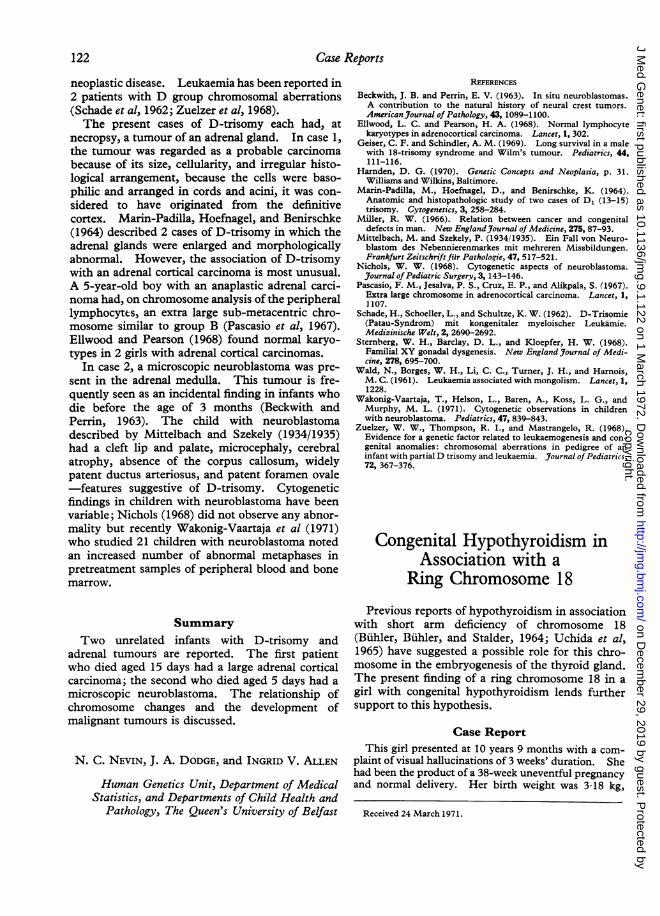
height was 116-2 cm, weight 22-8 kg, and head circum-ference 47-6 cm, all below the 3rd centile. She was aquiet, lethargic child with a rounded face and low hair-line who showed obvious signs of hypothyroidism, in-cluding carotenaemia, a depressed nasal bridge, cool dryskin, and pallor (Fig. 1). She had normal eyes and ears,
a high arched palate, and a short, but not webbed neck.There was a small, but palpable thyroid gland, with apalpable pyramidal lobe. Her blood pressure was 100/80 and pulse 88 per minute. Neurological examinationwas within normal limits except for apparent mentalretardation and a marked delay in relaxation of tendonreflexes. She had minimal clinodactyly and normalpalmar creases.
Complete blood count, urinalysis, serum electrolytes,blood sugar, and urea nitrogen were normal. Serumcarotene was 192 pg/100 ml and cholesterol 374 mg/100ml. Total serum proteins were 8 3 g/100 ml, withnormal pattems on electrophoresis and immuno-electrophoresis. Urine amino acids were normal bypaper chromatography. Her protein-bound iodine was
FIG. 1. The proposita at age 10 (left) and after 2 years' thyroid replacement (right).
123
copyright. on D
ecember 29, 2019 by guest. P
rotected byhttp://jm
g.bmj.com
/J M
ed Genet: first published as 10.1136/jm
g.9.1.122 on 1 March 1972. D
ownloaded from
5-1 lAg/100 ml, and her serum thyroxine 0 3 pg/100 ml.The thyroid 24-hour 131I uptake was 8%; after 2 days ofTSH stimulation the uptake was 6%. There were nocirculating antibodies to thyroglobulin demonstrated bytanned red blood cell agglutination. Plasma cortisol was21-1 iLgIlOO ml at 8 am and 111 iLg/lOO ml at 4 pm.Urinary 17-ketosteroids and 17-hydroxysteroids were1-5 and 11 mg/24 hr respectively. Following theadministration of intravenous crystalline insulin (01units/kg), there was a fall in blood sugar from 60 to32 mg/100 ml with a rise in circulating growth hormonefrom 2-0 to a peak of 22-7 mpug/ml. An electrocardio-gram and an electroencephalogram both showedgeneralized low voltage activity, but no other abnor-mality. A skull x-ray showed a small symmetricalskull with no intracranial calcification. Chest x-ray wasnormal. Her bone age at the wrist was 8 years with noepiphyseal dysgenesis. Psychological testing whilehypothyroid showed a mental age of 4 years 6 months.With thyroid replacement therapy, her myxoedema
disappeared, her thyroid gland ceased to be palpable,and she grew 18-3 cm in 2 years. At 12 years 2 months,her mental age was 6-7 years. Pubic hair and breastdevelopment began at 12j years.
Family HistoryBoth parents are of French-Canadian extraction. At
the time of the patient's birth, the father was 32 years oldand the mother 26. Both were in good health andneither had received significant amounts of radiation.The patient was the 5th of 7 children-all the sibs arewell. There is no family history of thyroid disease ormental retardation. The parents and sibs all showednormal levels of protein-bound iodine and serum thy-roxine, and none had circulating anti-thyroglobulinagglutinins.
CytologyThe patient's buccal smear was sex-chromatin positive.
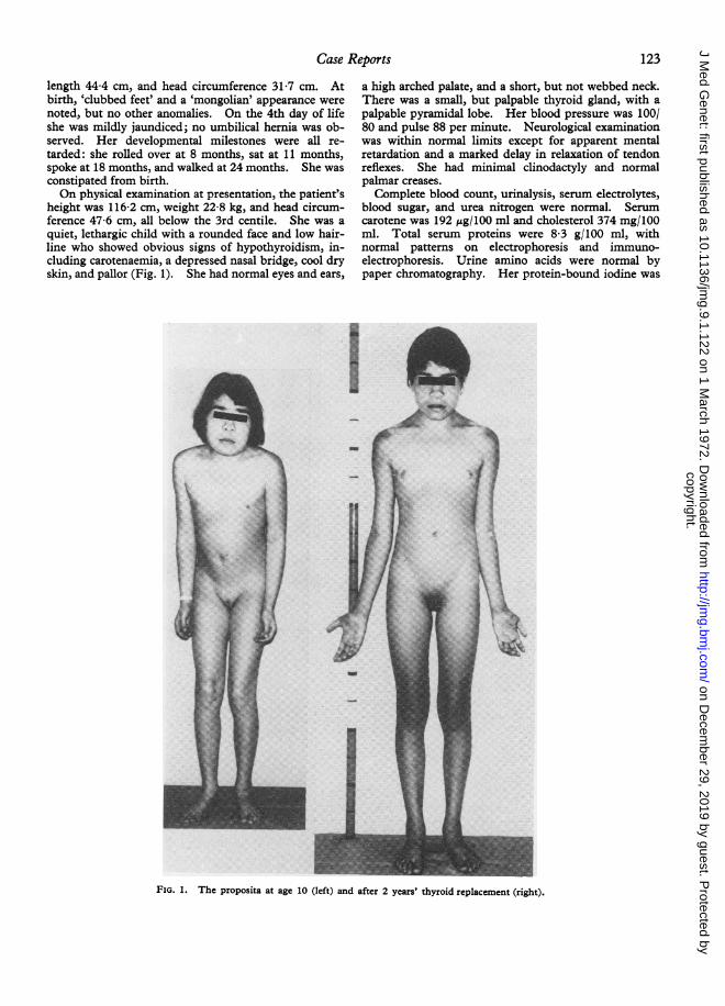
Metaphase preparations from leucocyte and skin fibro-blast cultures showed a majority of cells with 46 chro-mosomes including a ring; a few cells showed either 45chromosomes and no ring, or 47 chromosomes and tworings (Table I). Autoradiographic analysis followingthe incorporation of H3-thymidine (specific activity1-9 C/mM) into leucocyte cultures (1 puC/ml of mediumfor 6 hours incubation) showed that the ring and onemember of the 17-18 group were most heavily labelled
A^
.$
.f!.j :.M'.
A.
.....
I
:~~TV
16 17 18
E
FIG. 2. Chromosomes of the E group from proposita with auto-radiographs following incubation with H3-thymidine shown beloweach chromosome.
TABLE ISUMMARY OF CHROMOSOME COUNTS AT METAPHASE
124 Case Reports
copyright. on D
ecember 29, 2019 by guest. P
rotected byhttp://jm
g.bmj.com
/J M
ed Genet: first published as 10.1136/jm
g.9.1.122 on 1 March 1972. D
ownloaded from
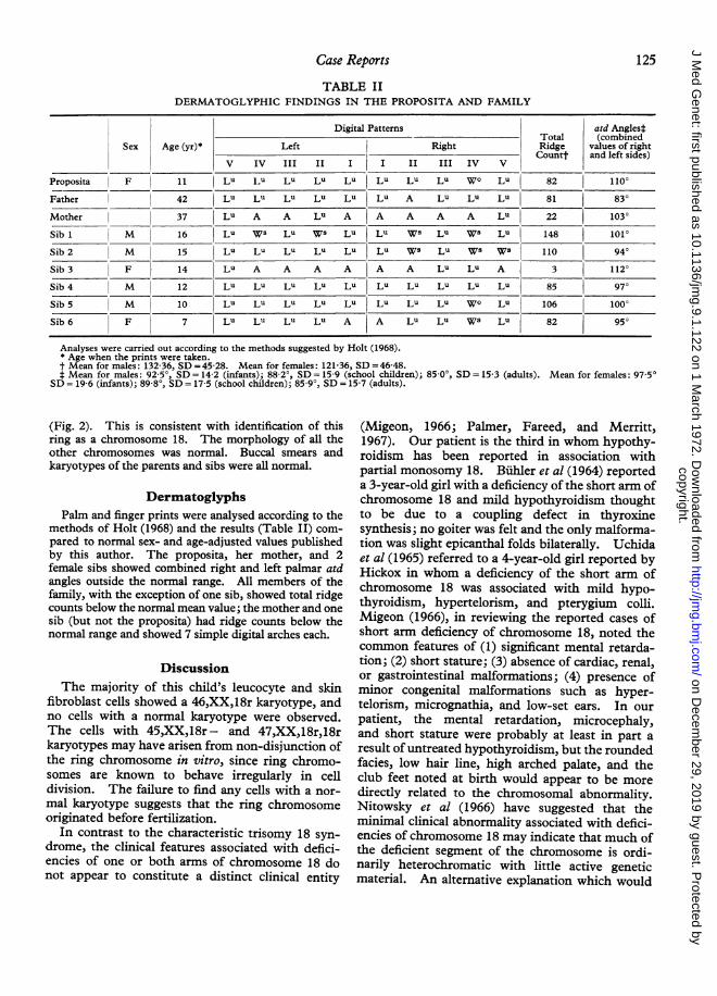
TABLE IIDERMATOGLYPHIC FINDINGS IN THE PROPOSITA AND FAMILY
Digital Pattems atd AnglestTotal (combined
Sex Age (yr)* Left Right Ridge values of rightV ~ ~ ~ ~~ ~ ~ ~ Countt and left sides)
V IV III II I I II III IV V
Proposita F 11 LU Lu Lu LU Lu Lu Lu LU WO Lu 82 110°Father I 42 Lu LU Lu Lu Lu Lu A Lu LU LU 81 830Mother 37 LU A A Lu A A A A A Lu 22 1030Sib 1 M 16 Lu Ws Lu Ws Lu Lu Ws LU Ws Lu 148 101°Sib 2 M 15 Lu Lu Lu Lu Lu Lu Ws Lu Ws Ws 110 940Sib 3 F 14 Lu A A A A A A Lu Lu A 3 112°Sib 4 M 12 Lu LU LU Lu Lu Lu Lu LU LU Lu 85 970Sib 5 M 10 Lu LU LU Lu Lu Lu Lu LU W° Lu 106 100°Sib 6 F 7 Lu LU Lu Lu A A Lu LU Ws Lu 82 950
Analyses were carried out according to the methods suggested by Holt (1968).* Age when the prints were taken.t Mean for males: 132-36, SD =45-28. Mean for females: 121-36, SD =46-48.t Mean for males: 92-5°, SD = 14-2 (infants); 88 2°, SD = 15 9 (school children); 85-0', SD = 15-3 (adults). Mean for females: 97-50SD = 19 6 (infants); 89 8', SD = 17 5 (school children); 85 9', SD = 15-7 (adults).
(Fig. 2). This is consistent with identification of thisring as a chromosome 18. The morphology of all theother chromosomes was normal. Buccal smears andkaryotypes of the parents and sibs were all normal.
DermatoglyphsPalm and finger prints were analysed according to the
methods of Holt (1968) and the results (Table II) com-pared to normal sex- and age-adjusted values publishedby this author. The proposita, her mother, and 2female sibs showed combined right and left palmar atdangles outside the normal range. All members of thefamily, with the exception of one sib, showed total ridgecounts below the normal mean value; the mother and onesib (but not the proposita) had ridge counts below thenormal range and showed 7 simple digital arches each.
DiscussionThe majority of this child's leucocyte and skin
fibroblast cells showed a 46,XX,18r karyotype, andno cells with a normal karyotype were observed.The cells with 45,XX,18r - and 47,XX,18r,18rkaryotypes may have arisen from non-disjunction ofthe ring chromosome in vitro, since ring chromo-somes are known to behave irregularly in celldivision. The failure to find any cells with a nor-mal karyotype suggests that the ring chromosomeoriginated before fertilization.
In contrast to the characteristic trisomy 18 syn-drome, the clinical features associated with defici-encies of one or both arms of chromosome 18 donot appear to constitute a distinct clinical entity
(Migeon, 1966; Palmer, Fareed, and Merritt,1967). Our patient is the third in whom hypothy-roidism has been reported in association withpartial monosomy 18. Biihler et al (1964) reporteda 3-year-old girl with a deficiency of the short arm ofchromosome 18 and mild hypothyroidism thoughtto be due to a coupling defect in thyroxinesynthesis; no goiter was felt and the only malforma-tion was slight epicanthal folds bilaterally. Uchidaet al (1965) referred to a 4-year-old girl reported byHickox in whom a deficiency of the short arm ofchromosome 18 was associated with mild hypo-thyroidism, hypertelorism, and pterygium colli.Migeon (1966), in reviewing the reported cases ofshort arm deficiency of chromosome 18, noted thecommon features of (1) significant mental retarda-tion; (2) short stature; (3) absence of cardiac, renal,or gastrointestinal malformations; (4) presence ofminor congenital malformations such as hyper-telorism, micrognathia, and low-set ears. In ourpatient, the mental retardation, microcephaly,and short stature were probably at least in part aresult of untreated hypothyroidism, but the roundedfacies, low hair line, high arched palate, and theclub feet noted at birth would appear to be moredirectly related to the chromosomal abnormality.Nitowsky et al (1966) have suggested that theminimal clinical abnormality associated with defici-encies of chromosome 18 may indicate that much ofthe deficient segment of the chromosome is ordi-narily heterochromatic with little active geneticmaterial. An alternative explanation which would
Case Reports 125
copyright. on D
ecember 29, 2019 by guest. P
rotected byhttp://jm
g.bmj.com
/J M
ed Genet: first published as 10.1136/jm
g.9.1.122 on 1 March 1972. D
ownloaded from
126 Case Reports
account for the phenotypic variability in these caseswould be the expression of recessive genes presenton the hemizygous segments of the normal chromo-some 18.Our patient clearly had some thyroid tissue, as
indicated by physical examination and the presenceof some 13"I accumulation in the neck. Butwhether the hypothyroidism is a result of a struc-tural malformation in the gland or a defect in thy-roxine synthesis was unfortunately not completelyresolved before therapy was initiated. However,the presence of a striking difference between herprotein-bound iodine (51 Htg/1OO ml) and herserum thyroxine (0-3 p,g/100 ml) suggests that shewas synthesizing an abnormal iodinated peptide.Such a defect in thyroxine synthesis might resultfrom the action of a recessive gene on the hemi-zygous portion of her normal chromosome 18.The coincidence in 3 patients, including the presentcase, of hypothyroidism with loss of geneticmaterial from chromosome 18 suggests that at leastone gene responsible for thyroid function may belocated on this chromosome. The significance ofthe abnormal dermatoglyphics in several membersof this family is not clear.
SummaryA girl with apparent congenital hypothyroidism
and minimal visible anomalies was found to have aring chromosome, identified by autoradiography aschromosome 18. The hypothyroidism appeared tobe related to a defect in thyroxine synthesis with theproduction of abnormal iodinated protein. It issuggested that at least one gene responsible forthyroid function may be located on chromosome 18.
The authors would like to express their thanks to DrG. E. C. Ducasse for the dermatoglyphic analysis andDr J. L. Hamerton for advice and encouragement.This work was supported by grants from the MedicalResearch Council and the Children's Hospital ResearchFoundation.
J. S. D. WINTER, K. AHLUWALIA, and M. RAY
The Winnipeg Children's Hospital ResearchFoundation and the Department of Paediatrics,
University of Manitoba, Canada
REFERENCESBiihler, E. M., Biihler, U. K., and Stalder, G. R. (1964). Partialmonosomy 18 and anomaly of thyroxine synthesis. Lancet, 1,170-171.
Holt, S. B. (1968). The Genetics of Dermal Ridges. Thomas,Springfield, Illinois.
Migeon, B. R. (1966). Short arm deletions in group E and chro-mosomal 'deletion' syndromes. Journal of Pediatrics, 69, 432-438.
Nitowsky, H. M., Sindhvananda, N., Konigsberg, U. R., and Wein-berg, T. (1966). Partial 18 monosomy in the cyclops malforma-tion. Pediatrics, 37, 260-269.
Palmer, C. G., Fareed, N., and Merritt, A. D. (1967). Ringchromosome 18 in a patient with multiple anomalies. J'ournal ofMedical Genetics, 4, 117-123.
Uchida, I. A., McRae, K. N., Wang, H. C., and Ray, M. (1965).Familial short arm deficiency of chromosome 18 concomitant witharhinencephaly and alopecia congenita. American Jrournal ofHuman Genetics, 17, 410-419.
Nystagmus in a Female Carrier ofOcular Albinism
Ocular albinism is a rare hereditary disorder longconsidered by pedigree analysis to be due to anabnormal gene on the X chromosome. This waseventually confirmed by the demonstration of closelinkage between the loci for ocular albinism and forthe Xga blood group (Fialkow, Giblett, andMotulsky, 1967; Pearce, Sanger, and Race, 1968).The condition as found in affected males is charac-terized by a deficiency of pigment in the retinal pig-ment epithelium and in the pigment epitheliallayer of the iris. The most striking clinical featureis the nystagmus with accompanying photophobiaand visual impairment which probably result fromthe deficiency of retinal pigment. So distinctive afeature is this nystagmus that before the initial re-cognition of ocular albinism as a separate entity byVogt (1925), it had been included in the group ofdisorders termed congenital nystagmus. Laterheterozygous females were noted to have minorfundus abnormalities characterized by stripe-like areas of depigmentation alternating with nor-mally pigmented patches in the periphery of theretina (Vogt, 1942; Falls, 1951). Similarly irregu-lar diaphanous areas were visible in the iris onretro-illumination (Waardenburg and van denBosch, 1956). No visual complaints, however-were associated with these changes.
In this communication a female heterozygousfor ocular albinism is described who displays thecongenital nystagmus, photophobia, and visual im-pairment one associates with the hemizygous male.
Received 7 June 1971.
copyright. on D
ecember 29, 2019 by guest. P
rotected byhttp://jm
g.bmj.com
/J M
ed Genet: first published as 10.1136/jm
g.9.1.122 on 1 March 1972. D
ownloaded from
![OVALE accessories - SANTI :: Vybavenie kúpeľní · OVALE accessories OVALE accessories [181] 25344 Porta scopino d'appoggio in ceramica bianca. White ceramic standing brush holder](https://img.pdfslide.us/doc/110x75/5be3b83709d3f281048c23e8/ovale-accessories-santi-vybavenie-kupelni-ovale-accessories-ovale-accessories.jpg)