Embed Size (px)
Citation preview

Congenital hyperinsulinism
SWISS SOCIETY OF NEONATOLOGY
FEBRUARY 2012
Winner of the
Case of the Year
Award 2012

2
Morgillo D, Berger TM, Caduff JH, Barthlen W, Mohnike K,
Mohnike W, Neonatal and Pediatric Intensive Care Unit
(MD, BTM), Department of Pediatric Radiology (CJH),
Children‘s Hospital of Lucerne, Lucerne, Switzerland,
Department of Pediatric Surgery, University Medicine
Greifswald (BW), Greifswald, Germany, Department of
Pediatrics, University Hospital of Magdeburg (MK),
Magdeburg, Germany, Diagnostic Therapeutic Centre
Frankfurter Tor (MW), Berlin, Germany
© Swiss Society of Neonatology, Thomas M Berger, Webmaster

3
Congenital hyperinsulinism (CHI) is characterized by
inappropriate secretion of insulin by the ß cells of the
islets of Langerhans and is an extremely heterogene-
ous condition in terms of clinical presentation, histo-
logical subgroups and underlying molecular biology.
Histologically, CHI has been classified into two major
subgroups: diffuse (affecting the whole pancreas)
and focal (being localized to a single region of the
pancreas) disease. Advances in molecular genetics,
radiological imaging techniques (such as fluorine-18
L-3,4-dihydroxyphenylalanine-PET-CT (18FDOPA-PET-CT)
scanning) and surgical techniques have completely
changed the clinical approach to infants with severe
congenital forms of hyperinsulinemic hypoglycemia.
INTRODUCTION
This male infant was born to a healthy 35-year-old G3/P3
by spontaneous vaginal delivery at 38 4/7 weeks. His
birth weight was 3530 g (P 50-75), his head circum-
ference was 35 cm (P 25) and his length was 50 cm
(P 25-50). Postnatal adaptation was normal with an
arterial cord pH of 7.28 and Apgar scores of 8, 9, and
9 at 1, 5, and 10 minutes, respectively. Pregnancy had
been uneventful without any evidence of gestational
diabetes.
On the second day of life, he was noted to have a
grayish skin color and poor muscle tone. A POCT
CASE REPORT

4
blood glucose measurement indicated a glucose con-
centration of 0.1 mmol/l, which only increased to 0.4
mmol/l after the administration of oral glucose solu-
tion. At that point, our neonatal transport team was
called. On arrival, intravenous access was established,
blood cultures were obtained and a bolus of 2 ml/kg
of a 10% dextrose solution was given, followed by a
continuous glucose infusion at a rate of 5 mg/kg/min.
He was started on antibiotics and transferred to our
neonatal intensive care unit.
On transport, there was focal tonic-clonic seizure ac-
tivity involving the right arm. Blood glucose concen-
tration at that time was 3 mmol/l. Phenobarbital was
started and the seizures did not reoccur. Antibiotics
were discontinued after 72 hours. The further hospital
course was remarkable for recurrent hypoglycemic epi-
sodes despite increasing rates of enteral and parente-
ral glucose administration (up to 18 mg/kg/min). High
insulin concentrations were documented repetitively
during episodes of hypoglycemia without concurrent
increase in free fatty acids or ketone bodies. Cortisol
and growth hormone responses, however, were ade-
quate. Thus, a diagnosis of hyperinsulinemic hypo-
glycemia was made.
At the age of one month, the patient was transferred
to the University Children‘s Hospital of Zurich for fur-
ther management. The patient did not respond to a
trial with the potassium channel activator diazoxide.

5
Fig. 1
Enhanced activity in the head of the pancreas
(A, B: frontal view, C: coronal view); in addition,
there is enhancement over the kidneys and bladder.
A
C
B

6
One week later, he was started on octreotide, initi-
ally by bolus injections but eventually by continuous
subcutaneous infusion. At the age of seven weeks, he
was discharged home fully breastfed on octreotide at
a rate of 17 mcg/kg/day.
At the age of 4 months, MR studies of the head and
abdomen were normal. Shortly thereafter, an 18FDOPA-
PET-CT scan was obtained. This study revealed increa-
sed focal activity in the region of the pancreatic head
(Fig. 1, 2). At this time, a curative resection of the
focal abnormality in the region of the pancreatic head
was not scheduled because the parents were satisfied
with the medical management and because of the
considerable risks involved with a surgical approach.
However, after several episodes of gastroenteritis
with concurrent hypoglycemia, and after obtaining
a second opinion at the University of Greifswald in
collaboration with the University of Magdeburg and
the Diagnostic Therapeutic Centre Frankfurter Tor in
Berlin, Germany (a team that specializes in congenital
hyperinsulinism) the parents opted for the operation.
At the age of 16 months, a resection of the pancre-
atic head with preservation of the duodenum using
a Roux-en-Y approach was performed (Fig. 3). Histo-
logy revealed hyperplasia of the islet cells without
signs of malignancy (Fig. 4, 5).
Following the intervention octreotide was no longer
required. No further episodes of hypoglycemia were

7
noted and regular glucose measurements were no
longer necessary. Today, at the age of three years, the
patient is cured without neurological deficits (Fig.6).
Fig. 2
18FDOPA-PET-CT: Focus located in the head of the
pancreas adjacent to the superior mesenteric vein
(measuring 13.5 mm in diameter).

8
Fig. 3
Schematic drawing of the surgical procedure with
excision of the focal lesion and reconstruction using a
Roux-en-Y loop (note: in our patient, the excision was
located in the head of the pancreas).

9
Focal adenomatous hyperplasia with hyperplastic but
normally structured islet and a peripheral rim of
non-ß cells (HE stain).
Fig. 4
10
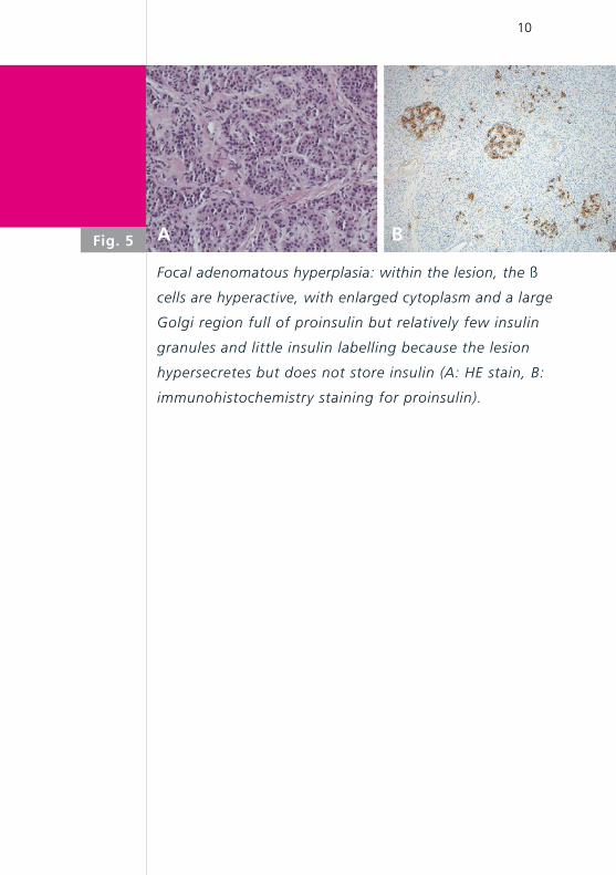
Focal adenomatous hyperplasia: within the lesion, the ß
cells are hyperactive, with enlarged cytoplasm and a large
Golgi region full of proinsulin but relatively few insulin
granules and little insulin labelling because the lesion
hypersecretes but does not store insulin (A: HE stain, B:
immunohistochemistry staining for proinsulin).
Fig. 5 A B

11
Patient at the age of 3 years: normal psychomotor
and cognitive development.
Fig. 6

12
Hyperinsulinemic hypoglycemia (HH) occurs as a conse-
quence of unregulated insulin secretion from pancreatic
ß cells. This is the major cause of persistent and recurrent
hypoglycemia in the neonatal and infancy period. Rapid
diagnosis and appropriate management of these patients
is essential to prevent brain injury, as HH is associated
with a high risk of epilepsy, cerebral palsy and neurologi-
cal handicap. Inappropriate insulin secretion drives gluco-
se into insulin-sensitive tissues (such as skeletal muscle,
adipose tissue and the liver) and simultaneously inhibits
glucose production via glycolysis and gluconeogenesis,
suppresses fatty acid release and ketone body synthesis
(i.e., inhibition of lipolysis and ketogenesis). This meta-
bolic „footprint“ of insulin action (hypoglycemia with in-
appropriately low fatty acid and ketone body formation)
explains why patients with HH have an increased risk of
brain injury. The brain is not only deprived of its most im-
portant substrate (i.e., glucose) but also ketone bodies,
which form an alternative source of fuel (1).
HH may be congenital (CHI, congenital hyperinsulinism),
secondary to certain risk factors (such as maternal diabe-
tes, perinatal asphyxia or intrauterine growth restriction)
or it can be associated with developmental syndromes
(such as Beckwith-Wiedemann syndrome).
CHI is a genetically heterogeneous disease with muta-
tions having been described in 8 different genes (ABCC8,
KCNJ11, GLUD1, GCK, HADH, HNF4A, UCP2 and SL-
C16A1) (2, 3). Although dominant mutations have been
DISCUSSION

13
reported in a number of these genes, recessively inheri-
ted CHI is more common. The estimated incidence of CHI
in the general population is 1:30‘000 to 1:50‘000 but it
increases to 1:2500 in communities with high rates of
consanguinity (1). Mutations in the ABCC8 (ATP-binding
cassette, sub-family C, member 8) and KCNJ11 (potas-
sium inwardly rectifying channel, sub-family J, mem-
ber 11) genes that encode the ATP-sensitive potassium
channels (KATP channels) in the pancreatic ß cells are by
far the most common cause of CHI and are estimated
to account for 40-45% of all cases, whereas mutations
in the remaining 5 genes are identified in approximately
5-10% of cases. The genetic etiology for the remaining
45-55% of patients remains unknown (2).
KATP channels play a central role in the regulation of in-
sulin secretion in the pancreatic ß cells. The channels
couple glucose metabolism to membrane electrical ac-
tivity and insulin release. When glucose is metabolized
by the ß cells the intracellular ratio of ATP/ADP increases
and leads to closure of the channels; this results in cell
membrane depolarization, Ca2+ influx via voltage-gated
calcium channels and insulin exocytosis (1) (Fig. 7). CHI
is associated with loss-of-function KATP channel muta-
tions.
There are two main histologic subtypes of CHI: diffuse
(60-70% of patients) and focal (30-40% of patients)
(Fig. 8). Focal pancreatic lesions appear as small regions
of islet adenomatosis measuring 2-10 mm in diameter,
14
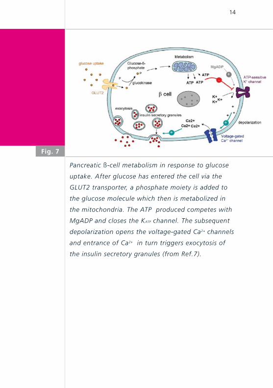
Fig. 7
Pancreatic ß-cell metabolism in response to glucose
uptake. After glucose has entered the cell via the
GLUT2 transporter, a phosphate moiety is added to
the glucose molecule which then is metabolized in
the mitochondria. The ATP produced competes with
MgADP and closes the KATP channel. The subsequent
depolarization opens the voltage-gated Ca2+ channels
and entrance of Ca2+ in turn triggers exocytosis of
the insulin secretory granules (from Ref.7).
15
which are characterized by ß cells with enlarged nuclei
surrounded by normal tissue. In contrast, diffuse pan-
creatic disease affects all the ß cells within the islets of
Langerhans (1). The focal form of CHI exhibits a parti-
cular genetic pattern with a paternally inherited mutati-
on on chromosome 11p15.1 and a loss of the maternal
allele specifically in the cells of the focal lesion (4). The
majority of patients with diffuse disease have homozy-
gous or compound heterozygous mutations in ABCC8
and KCNJ11.
Advances in diagnostic imaging have revolutionized the
ability to localize lesions in the pancreas by the introduc-
tion of integrated 18FDOPA-PET-CT that merges anatomi-
cal and functional data. L-DOPA is adsorbed by neuro-
endocrine and pancreas islet cells and metabolized into
dopamine. Beta cells of the pancreas possess dopamine
receptors. The uptake of 18FDOPA is considerably incre-
ased in foci with high insulin synthesis rates. It is not
only possible to differentiate between diffuse and focal
forms with high sensitivity and specificity, but localiza-
tion of the focus can also be provided with a formerly
unthinkable precision of up to a few millimetres (5).
The goal of treatment in infants with CHI is to maintain
plasma glucose levels > 4 mmol/l. Long-term treatment
with diazoxide has dramatically reduced the need for
extensive surgical procedures. Diazoxide acts by keeping
the KATP channel open, thereby preventing depolarisati-
on of the ß cell membrane and insulin secretion.

16
In patients unresponsive to diazoxide, it is essential to
differentiate focal from diffuse disease, as the surgical
approaches are radically different. In patients with focal
disease, precise preoperative localization and limited
surgical excision „cures“ the patient. In contrast, pati-
ents with diffuse disease may require a near-total pan-
createctomy, which will have lifelong implications (high
risk of diabetes mellitus and/or pancreatic exocrine in-
sufficiency).
Other medical treatments that can be used while awai-
ting surgical treatment include octreotide, glucagon,
and continuous intragastric dextrose administration. Oc-
treotide is the second line of medical therapy for infants
with CHI who are unresponsive to diazoxide. Octreotide
is a long-acting somatostatin analogue that inhibits in-
sulin secretion by inducing hyperpolarization of ß cells
and by direct inhibition of voltage-dependent calcium
channels. Long-term medical management of diffuse
disease with subcutaneous octreotide administration
should not be taken lightly as it may impose a huge bur-
den and is extremely stressful on the family. Glucagon
can be given as a continuous intravenous infusion to
help maintain euglycemia in infants who are awaiting
surgery. Unfortunately, glucagon is too unstable in solu-
tion to be useful for chronic management (1, 6). Fig. 9
outlines various treatment options in patients with CHI.

17
Diffuse CHI (A) involves the entire pancreas while the
focal form (B) is localized to a single region of the
pancreas (from MediVisuals.inc).
Fig. 8A B

18
Fig. 9
Flow chart outlining the management cascade of
neonates with hyperinsulinemic hypoglycemia (CHI).
Clinically, CHI can be classified into diazoxide-respon-
sive and diazoxide-unresponsive disease. A 18FDOPA-
PET-CT scan is currently only indicated in neonates
who are unresponsive to diazoxide and do not have
genetically confirmed diffuse disease.

19
1. Kapoor R, Flanagan S, Hussain K. et al. Hyperinsulinemic
hypoglycemia. Arch Dis Child 2009;94:450-457
2. Flanagan S, Kapoor R, Hussain K. Genetics of congenital
hyperinsulinemic hypoglycemia. Semin Pediatr Surg
2011;20:13-17
3. Glaser B, Thornton P, Otonkoski T, et al. Genetics of neonatal
hyperinsulinism. Arch Dis Child Fetal Neonat Ed 2000;
82:F79-F86
4. Rahier J, Guiot Y, Sempoux C. Morphologic analysis of focal
and diffuse forms of congenital hyperinsulinism. Semin
Pediatr Surg Ed 2011;20:3-12
5. Mohnike W, Barthlen W, Mohnike K, Blankenstein O. Positron
emission tomography and computed tomography diagnostics
by means of fluorine-18-L-dihydroxy-phenylalanine in
congenital hyperinsulinism. Semin Pediatr Surg 2011;20:23-27
6. Palladino A, Stanley C. A specialized team approach to
diagnosis and medical versus surgical treatment of infants
with congenital hyperinsulinism. Semin Pediatr Surg
2011;20:32-37
7. Saint-Martin C, Arnoux JB, De Lonlay P, Bellanné-Chantelot
C. KATP channel mutations in congenital hyperinsulinism.
Semin Pediatr Surg 2011;20:18-22
REFERENCES

![Dopa decarboxylaseactivity of the living human · nine (L-dopa). We measured regional dopa decarboxylase activity in brains ofsix healthy volunteers with 6-[18F]fluoro-L-dopaandpositron](https://img.pdfslide.us/doc/110x75/5fd3ff72add4681c6146e1fc/dopa-decarboxylaseactivity-of-the-living-human-nine-l-dopa-we-measured-regional.jpg)