Embed Size (px)
Citation preview
Vol. 11 • No. 41 (3/2015)148
Gineco.eu[11] 148-153 [2015]DOI: 10.18643/gieu.2015.148@ 2015 Romanian Society of Ultrasonography in Obstetrics and Gynecology
Congenital heart disease and the role of genetic factors
in cardiac morphogenesisIoana Roșca1,
Marcela Şerban1,
Anca Ristea2, Mariana
Nanea1,3, Raluca Tocariu1,
Mihai Mitran1,3
1. ”Prof. Dr. Panait Sîrbu” Obstetrics-Gynecology
Clinical Hospital, Bucharest, Romania
2. IOMC „Prof. Dr. Alfred Rusescu”,
Bucharest, Romania 3. ”Carol Davila” UMF
Bucharest, Romania
Correspondence: Dr. Ioana Rosca
e-mail: ioanarosca76@ yahoo.com
Cardiovascular malformations are the most common types of birth malformations, causing a significant increase in mortality worldwide. The etiology of most of these abnormalities remains unknown, but the genetic factors involved are considered to have an increasingly important role. Advances in understanding normal molecular cardiac development have led to the identification of numerous genes required in cardiac morphogenesis and drove to the discovery of a growing number of monogenic causes of human cardiac malformations. Sequencing of the human genome and advances in molecular techniques have led to an increase in proving the crucial role of the genetic factors.Keywords: congenital malformations, genetic factors, newborn
Abstract
Received: April 09, 2015
Revised: June 12, 2015
Accepted: July 18, 2015
IntroductionCardiac malformations are structural or functional
diseases of the fetal heart which manifest immediately after birth or later. These anomalies affect the heart and the intrathoracic great vessels and determine poor functioning or predisposition to malfunctioning(1-3). The clinical signs are:n cianosis;n cardiac murmurs;n tachypnea;n respiratory distress syndromes;n heart failure;n low pulse, petechiae, hypotension, metabolic aci-
dosis, circulatory failure;n abnormal heart rhythm (i.e. tachycardia/bradycar-
dia, atrio-ventricular block);n abnormal location, size and shape(4).
History and general presentationWhen a newborn baby presents with cianosis, repira-
tory distress syndrome and/or shock, the clinician must choose the right diagnosis, taking into consideration a pulmonary disease, a cardiac disease, a neurological disease or an infection.
Family history and pregnancy medication a) the existence of a brother or a sister with congenital
heart disease triples the risk of recurrence. If the mother suffers from a congenital heart disease, the risk for the child to be affected is higher;
b) alcohol and drugs (i.e. amphetamines, anticonvul-sants, lythium, progesterone, estrogen) intake during the first trimester of pregnancy increases the risk of heart malformations. Viral infections (like rubella, enterovirus, coxsakie B), diabetes, systemic lupus erythematosus, ma-ternal age of over 40, phenylketonuria, may be maternal conditions that predispose to heart malformations;
c) Pregnancy, labour and birth complications, may be considered risk factors for congenital heart diseases. For example, intrauterine and perinatal hypoxia is a risk factor for developing myocardial dysfunctioning and persistent pulmonary hypertension;
d) Assisted Reproductive Technologies: the risk of cardi-ac malformations in babies conceived by in vitro fertiliza-tion appears to be a little higher than naturally conceived babies. The most frequent congenital heart diseases are atrial and ventricular septal defects. There is no proven relation between in vitro fertilization and the incidence of cardiac malformations.
Neonatal history: gestational age, onset signs and severity(4,5,6). If, during the first week of life, the new-born baby presents with general cyanosis, murmurs, heart failure and/or vasculary collapse, the possibility of a congenital heart disease must be evaluated. Clinical history and detection of onset clinical signs are very important(3).
Prognostical classification of the fetal cardiac diseasesn associated pathology:� isolated 50-60%;� associated extracardiac malformation 10-20%;� genetic syndromes 30%.n survival:� malformations incompatible with life;� malformations that can benefit from palliative treat-
ment;� malformations that can be entirely treated(7).On the basis of the prognosis and the possible, genetic
or structural, associated pregnancy pathologies may be continued or aborted. Heart defects detected during intrauterine life may be more severe than those detected after birth(8,9).
Vol. 11 • Nr. 41 (3/2015)149
ginecoeu
During the last decades a multitude of studies on the genetic factors and molecular pathways that interfere with the development of the cardiac system have been made. Various structural genes, critical in normal car-diac morphogenesis, have been discovered as the result of these studies and helped the identification of the genetical ethiology of congenital heart diseases(10,11).
In most of the children born with a congenital heart malformation, their condition is not associated with other inborn errors, but in 25-40% of the cases, heart malformation is associated with another anomaly or is a part of a genetic syndrome(12). In approximately
30% of the affected children, a cromosomial defect is associated with a heart malformation(13).
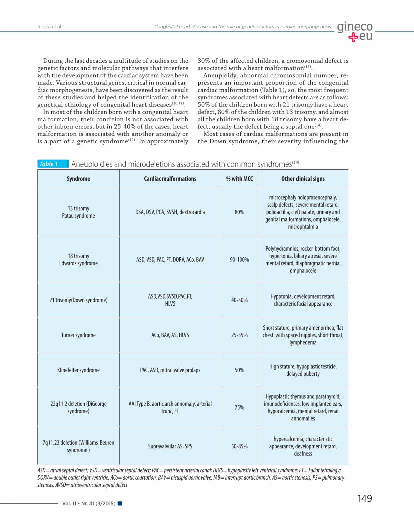
Aneuploidy, abnormal chromosomial number, re-presents an important proportion of the congenital cardiac malformation (Table 1), so, the most frequent syndromes associated with heart defects are as follows: 50% of the children born with 21 trisomy have a heart defect, 80% of the children with 13 trisomy, and almost all the children born with 18 trisomy have a heart de-fect, usually the defect being a septal one(14).
Most cases of cardiac malformations are present in the Down syndrome, their severity influencing the
Aneuploidies and microdeletions associated with common syndromes(19)Table 1
Syndrome Cardiac malformations % with MCC Other clinical signs
13 trisomy Patau syndrome DSA, DSV, PCA, SVSH, dextrocardia 80%
microcephaly holoprosencephaly, scalp defects, severe mental retard,
polidactilia, cleft palate, urinary and genital malformations, omphalocele,
microphtalmia
18 trisomyEdwards syndrome ASD, VSD, PAC, FT, DORV, ACo, BAV 90-100%
Polyhydramnios, rocker-bottom foot, hypertonia, biliary atresia, severe
mental retard, diaphragmatic hernia, omphalocele
21 trisomy(Down syndrome) ASD,VSD,SVSD,PAC,FT,HLVS 40-50% Hypotonia, development retard,
characteric facial appearance
Turner syndrome ACo, BAV, AS, HLVS 25-35%Short stature, primary amenorrhea, flat chest with spaced nipples, short throat,
lymphedema
Klinefelter syndrome PAC, ASD, mitral valve prolaps 50% High stature, hypoplastic testicle, delayed puberty
22q11.2 deletion (DiGeorge syndrome)
AAI Type B, aortic arch annomaly, arterial trunc, FT 75%
Hypoplastic thymus and parathyroid, imunodeficiences, low implanted ears,
hypocalcemia, mental retard, renal annomalies
7q11.23 deletion (Williams-Beuren syndrome ) Supravalvular AS, SPS 50-85%
hypercalcemia, characteristic appearance, development retard,
deafness
ASD= atrial septal defect; VSD= ventricular septal defect; PAC= persistent arterial canal; HLVS= hypoplastiv left ventrical syndrome; FT= Fallot tetrallogy; DORV= double outlet right ventricle; ACo= aortic coartation; BAV= bicuspid aortic valve; IAB= interrupt aortic branch; AS= aortic stenosis; PS= pulmonary stenosis; AVSD= atrioventricular septal defect
Roşca et al. Congenital heart disease and the role of genetic factors in cardiac morphogenesis
Vol. 11 • No. 41 (3/2015)150
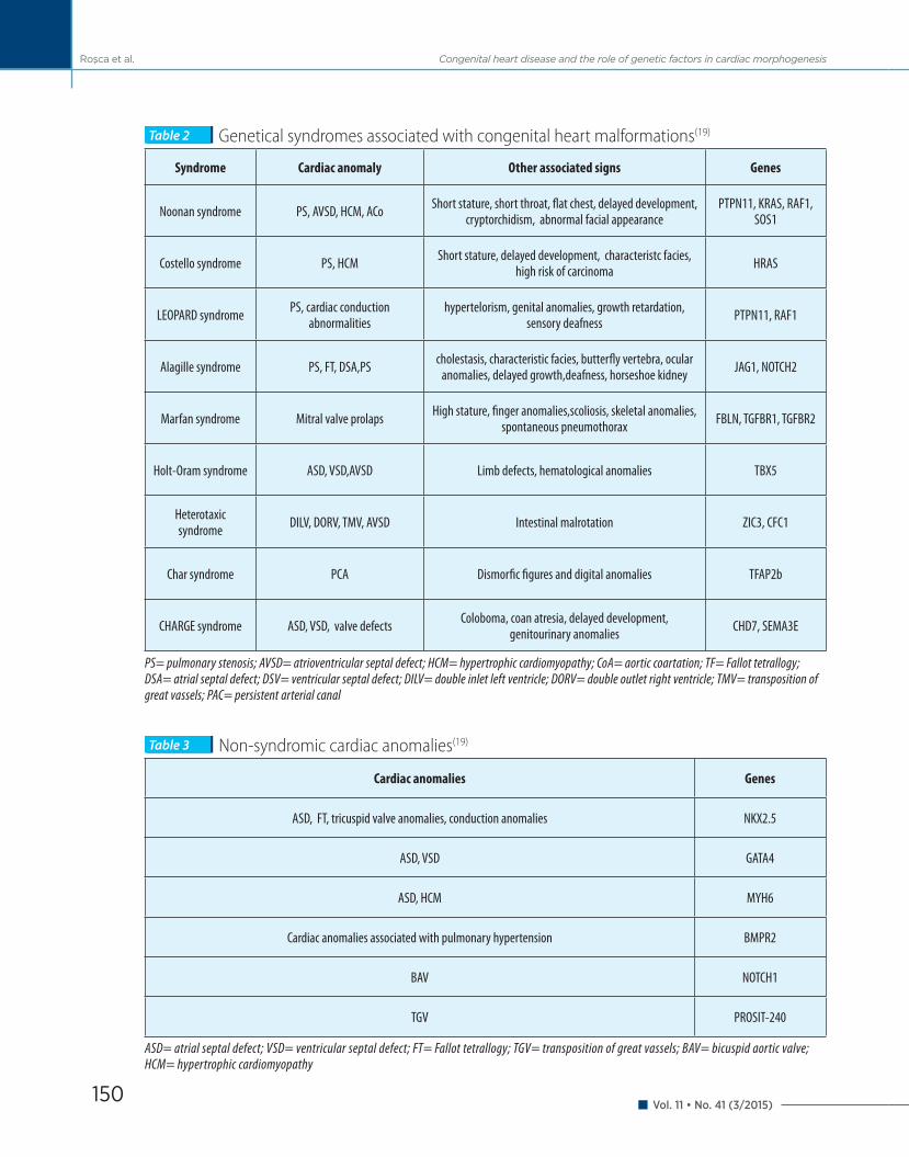
Genetical syndromes associated with congenital heart malformations(19)Table 2
Non-syndromic cardiac anomalies(19)Table 3
Syndrome Cardiac anomaly Other associated signs Genes
Noonan syndrome PS, AVSD, HCM, ACo Short stature, short throat, flat chest, delayed development, cryptorchidism, abnormal facial appearance
PTPN11, KRAS, RAF1, SOS1
Costello syndrome PS, HCM Short stature, delayed development, characteristc facies, high risk of carcinoma HRAS
LEOPARD syndrome PS, cardiac conduction abnormalities
hypertelorism, genital anomalies, growth retardation, sensory deafness PTPN11, RAF1
Alagille syndrome PS, FT, DSA,PS cholestasis, characteristic facies, butterfly vertebra, ocular anomalies, delayed growth,deafness, horseshoe kidney JAG1, NOTCH2
Marfan syndrome Mitral valve prolaps High stature, finger anomalies,scoliosis, skeletal anomalies, spontaneous pneumothorax FBLN, TGFBR1, TGFBR2
Holt-Oram syndrome ASD, VSD,AVSD Limb defects, hematological anomalies TBX5
Heterotaxic syndrome DILV, DORV, TMV, AVSD Intestinal malrotation ZIC3, CFC1
Char syndrome PCA Dismorfic figures and digital anomalies TFAP2b
CHARGE syndrome ASD, VSD, valve defects Coloboma, coan atresia, delayed development, genitourinary anomalies CHD7, SEMA3E
PS= pulmonary stenosis; AVSD= atrioventricular septal defect; HCM= hypertrophic cardiomyopathy; CoA= aortic coartation; TF= Fallot tetrallogy; DSA= atrial septal defect; DSV= ventricular septal defect; DILV= double inlet left ventricle; DORV= double outlet right ventricle; TMV= transposition of great vassels; PAC= persistent arterial canal
Cardiac anomalies Genes
ASD, FT, tricuspid valve anomalies, conduction anomalies NKX2.5
ASD, VSD GATA4
ASD, HCM MYH6
Cardiac anomalies associated with pulmonary hypertension BMPR2
BAV NOTCH1
TGV PROSIT-240
ASD= atrial septal defect; VSD= ventricular septal defect; FT= Fallot tetrallogy; TGV= transposition of great vassels; BAV= bicuspid aortic valve; HCM= hypertrophic cardiomyopathy
Roşca et al. Congenital heart disease and the role of genetic factors in cardiac morphogenesis
Vol. 11 • Nr. 41 (3/2015)151
ginecoeu
prognosis of these children. These heart defects vary from simple to complex ones, determining various signs and symptoms. The most common malformations are: atroventricular canal defect, ventricular and atrial septal defects, persistent arterial canal or cyanogenic cardiac malformation such as Fallot tetralogy or the hypoplastic left ventricle(13).
Edwards syndrome or 18 trisomy is characterized by low birth weight, intrauterine growth retardati-on, mental retardation. About 80% of the pacients are female. The incidence is 1/3500-8000 births. Life expectancy is of approximately 2-4 months, as these pacients present with severe heart malformations, central nervous system defects and other anomalies that lead to premature death. The karyotype features chromosome 18 in triple dose, commonly citogene-tic, as it is homogenous. Only 10% of the cases have mosaicism. It has been observed that in 10% of the babies born with Edward syndrome, this is associated with 13 or 21 trisomy. Affected females survive longer. The most frequent cardiac malformations are: septal defects, persistent arterial canal, and complex defects as double outlet right ventricle, Fallot tetrallogy, aortic coarctation(13).
Patau syndrome or 13 trisomy, is characterized by a plurimalformative syndrome with neurological ano-malies (mental retardation, apnea, seizures etc.), cra-nio-facial defects (ear malformations, microcephaly, cleft palate, micrognatia etc.), oculoorbital defects (microphthalmia, coloboma etc.), cervical, cardio-vas-culary, gastro-intestinal, urinary and genital defects, skeletal anomalies (polidactilia)(13). Multiple and severe malformations associated with this syndrome cause death during the first month of life in 50% of the cases. Only 3% of the patients survive over a year with severe mental retardation. Commonly, the condition of these patients is associated with septal defects, persistent arterial canal, dextrocardia, hypoplastic left ventricle(15).
One third of the females with Turner syndrome or X monosomy have a congenital heart malformation (most frequent are bicuspid aortic valve, aortic ste-nosis, hypoplastic left ventricle, aortic coarctation), 50% of the males with Klinefelter syndrome or 47XXY associate a heart defect (persistent arterial canal, septal defects)(13). Furthermore, there are many other, less frequent, chromosomial defects detected in patients with a congenital cardiac malformation.
The development of fluorescence in situ hybridization technique (FISH), a procedure in which marked floures-cent probes are hybridized to metaphase chromosomes, detecting small submicroscopic chromosomal deletions, various syndromes caused by chromosomial defects have been elucidated (Table 1), such as 22q11deletion known as DiGeorge syndrome and Williams-beuren syndrome. DiGeorge syndrome is caused by the micro-scopic deletion on chromosome 22q11.2 and leads to defects of the heart, thymus and parathyroid glands, dysmorphic facies due to the abnormal development of the pharyngeal arch(14). The most frequent cardiac
malformations are common arterial trunk and Fallot tetrallogy. Genetic tests are required in patients with cardiac defects(15). William-Beuren syndrome caused by the microdeletions on the 7p11.23 chromosome, is characterized by cardiac defects, associates typical elf facial appearance, hypercalcemia, renal impairment and cognitive disability(16). Loss of elastin is thought to be the cause of cardiac defects in this syndrome(17,18).
Along with the progress în genetic technology and the discovery of the human genome, singular genetic de-fects that lead to syndromes associated with congenital cardiac malformations have been elucidated (Table 2).
Most recent studies have made possible the disco-very of the mutation of fibrillin 1, the cause of Marfan syndrome characterized by the progressive dilatation of the aortic root with a high risk of dissection, patho-logy of the lens and skeletal anomalies(20). Holt-Oram syndrome characterized by atrial and ventricular septal defects, a progressive disease of the atrioventricular conduction system and limb anomalies, is associated with mutations of the transcription factor Tbx5(21). Alagille syndrom, that encodes a ligand in signaling pathway Notch is characterized by intrahepatic billiary atresia and cardiac malformations (pulmonary stenosis, mpulmonary valve stenosis and Fallot tetrallogy)(22,23). In Noonan syndrome phenotip that includes cardiac defects, pulmonary stenosis and hypertrophic cardio-miopathy as well as mental retardation, characteristic figure, hemorrhagic disorders, mutations of protein tyrosine phosphatase non-receptor type 11 are invol-ved in 50% of the cases(24). Heterotaxic syndrome that associates cardiac, pulmonary and gastrointestinal positional defects is commonly complicated with con-genital heart diseases (atrioventricular septal defects, great vassels transposition)(25).
Singular genic defects associated with isolated/non-syndromic congenital heart malformations (Table 3) have also been discovered.
The progress in this field also proves that singular genic defects can lead to congenital heart diseases and reveal more information about the molecular pathways in cardiac morphogenesis.
In spite of the ultimate discoveries, the great majo-rity of patients with mutated in colorectal cancers do not have singular genic defects(26-30). Along with the sequencing of the human genome, new information about the genetic variety has been reported.
One type of genetic variation, changes in children number, leads to a change in the gene dose and affects aproximatly 12% of the human genome(31). These va-riations are considered to be polymorphisms when present in more than 1% of population and are more susceptible in presenting associated diseases when present in less than 1% of the patients.
The variation in the number of copies is associated with cardiac malformations.
CHARGE syndrome, a constellation of anomalies that includes coloboma, cardiac defects, coan atresia, growth retard and anomalies of the ear, has been re-
Roşca et al. Congenital heart disease and the role of genetic factors in cardiac morphogenesis
Vol. 11 • No. 41 (3/2015)152
cently associated with a microdeletion on the 8p21 chromosome(32). Later sequencing of the critical region showed heterozygous mutations in chromodomain he-licase deoxyribonucleic acid (DNA)-binding 7 (CHD7), a gene that encodes a binding protein in DNA(32). Further studies have identified pathogenic mutations in CHD7 in over 50% of the pacients with CHARGE syndro-me(33). Array and contributors helped with detecting the gene responsible for the great majority of cases of this syndrome, proving the importance of this method in discivering the gene, especially when studying rare diseases(33).
Thien other authors studied patients with congenital heart malformations and other inborn anomalies and detected a variation in the number of copies in 30%(34). The relation cause-effect was supported when variation în number of copies include important genes in cardiac development when they appeared by novo.
Variations in the number of copies were also identi-fied in pacients with isolated congenital heart malfor-mations. They appear also in parents with no proof of congenital heart malformation, thus showing probably the fact that variation in number of copies increases the sensitivity in developing heart anomalies, also needing another factors.
The decrease in sequencing DNA price, the rate of discovering the gene in cardiac malformations may be higher.
At the same time with the discovery of new genetic anomalies associated with heart malformations, the development of a phenotipic information international database, one can study clinical results and prognosis on the basis of a genetic average(34).
Yet, the high cost of these genetic tests and the impossiblity of performing them in many countries, makes evaluation of children with congenital heart malformations syndromic or non-syndromic almost impossible.
When a new patient is diagnosed with heart patho-logy, the examination of the relatives to exclude ge-netic involvement is recommended as well as detailed evaluation of the newborn.
The clinical examination must search for dismorphic appearance, eye and ear anomalies, skeletal and limb anomalies, gastrointestinal and genitourinary patho-logy, a change in the neurologic status.
Multidisciplinary consults (neurological, ophtalmo-logical, orthopedical, ORL, genetical, imagistic) and adjuvant tests (cardiac ultrasound, abdominal ultra-sound, brain imaging) in establishing the diagnosis are recommended when necessary.
Genetic tests should be performed in the following situations:n every newborn/child with suggestive phenotypen every newborn with dysmorphic features, multiple
anomalies, growth retard without a clear causen children with a family history of genetic anomalyn when anomalies are discovered in fetal ultrasound
examination
In case of a normal cariotype, when the child presents with sugestive clinical features for a genetic disorder, detailed genetic tests (i.e. FISH) are recommended for detecting a singular inborn anomaly(32).
Discovering a genetic anomaly in a child with con-genital heart malformation is beneficial for the pati-ent and his/her familly, alerting the specialist on the possible associated asymptomatic anomalies, on one hand, and conducting a family investigation which can detect genetic defects in other members, on the other hand, offering important data for genetic counselling of the familly(33).
The most important congenital defectsCongenital heart malformations are the most impor-
tant congenital defects in a newborn(34):1. In Romania there is no efficient functional network
for approaching this pathology, there is no national screening program for congenital heart defects to en-sure the diagnosis, treatment and follow-up of these children. In our country the number of cardiovascular surgery centers is insufficient to treat all the newborns with cardiac malformations.
2. The obstetrician has an important role in de-tecting cardiac malformation by fetal 3-dimensional ultrasound. The neonatologist should be informed if there is a high suspicion of a cardiac malformation, as there are many that represent an immediate medical and surgical postnatal emergency.
3. If the diagnosis is made before birth, the pregnant woman can be sent to a high level of neonatal unit or to a specialized local cardio-vascular surgery center or abroad. Thus, mortality decreases and also neurological sequelae and the quality of life increases.
4. The family has an important role in detecting the patients with congenital heart malformations in time, through periodic antenatal and postnatal consults by the paediatrician or familly practitioner when discove-ring certain abnormalities in the infant’s state (growth retardation, central or peripheric cyanosis, feeding difficulties, pale skin, respiratory effort, hypotonia).
5. The newborn should be examined in detail by the neonatologist and if there is suspicion of a heart disease he/she should be monitored in order to esta-blish a rapid diagnosis. Many of the newborn babies with a congenital heart disease may have a real chance if diagnosis is quickly established and treatment is started immediately.
6. Neonatologists should be familiar with cardiac ultrasound as they are the main factor in the precocious detection of congenital heart diseases, thus contribu-ting to a decrease in neonatal mortality and morbidity. If the newborn is in a critical state, neonatal transport to another unit for cardiac ultrasound is made with difficulty and agravates the state.
7. Unless immediately treated, congenital heart malformations lead to existus or irreversible and severe complications such as persistent pulmonary hypertension, systemic arterial hypertension, hyper-
Roşca et al. Congenital heart disease and the role of genetic factors in cardiac morphogenesis
Vol. 11 • Nr. 41 (3/2015)153
ginecoeu
tophic cardiomiopathy, cardiac failure. All of these create a physical and psychological impairment for the patient. Patients diagnosed at a later time need a more laborious surgical technique or the surgery can not be performed anymore, leading to a decrease of the quality of life.
8. We also need to mention here long and short term cardiac complications of prematurity: persistent arterial canal and later right ventricular hypertrophy.
ConclusionsGenetic factors have a much greater influence on
determining various types of cardiac malformations than it was established previous to the multitude of studies in this area. All accomplished knowledge re-garding genetic influence over heart malformations can develop early detecting or preventing strategies, of great importance for future generations of affected children with isolated forms or genetic syndromes. n
1. Lacour -Gayet. Congenital Heart Surgery Nomenclature and Database Project, 2000, 234-32.
2. Socoteanu I, Tratat de Cardiopatii congenitale, vol.I, II, III, 2010.3. Karlsen A. Kristine, Tani Y. Lloyd, S.T.A.B.L.E. Cardiac Module 2003, 13(23),
29-31.4. Flanagan MF, Yeager SB. Cardiac disease. In: Avery GB, Fletcher MA,
MacDonald MG. Neonatology: Pathophysiology and Management of the Newborn 1999, 577-646.
5. Gardner SL, Johnson JL. Initial nursery care. In: Merenstein GB, Gardner SL. Handbook of Neonatal Intensive Care, 2002, 5th ed, 725-53.
6. Chameides L, Hazinski MF. Pediatric advanced life support, 1997-99: Emergency Cardiovascular Care Programs 1999, 9, 7-9.
7. Gewitz MH. Cardiac disease in the newborn infant. In:Polin RA, Yoder MC Burg FD. Workbook in practical neonatology, 2001, 3rd ed:251-98
8. Glass SM. Routine care. In: Thureen PJ, Deacon J, ONeil P, Hernandez J. Assessment and Care of the Well Newborn 1999, 188-93.
9. Kourembanas S. Shock. In: Cloherty JP, Stark AR. Manual of Neonatal Care, 1998, 4th ed, 171-3
10. Martin RJ. Sosenko II. Respratory problems. In: Klaus MH, Fanaroff AA. Care of the High Risk Neonate 2001, 5th ed, 243-76.
11. Lyons Jones K, Crandall Jones M, Del Campo Casanelles M. Smith’s Recongnizable Patternes of Human Malformation 2006, 5th ed, 8-64.
12. Bernstein D. In: Evaluation of the cardiovascular system. Behrman RE, Kliegman RM, Jenson HB, editors. Nelson Textbook of Pediatrics 2004, 1481-8.
13. Pierpont ME, Basson CT, Benson DW Jr. et al. Genetic basis for congenital heart defects: current knowledge: a scientific statement from the American Heart Association Congenital Cardiac Defects Committee, Council on Cardiovascular Disease in the Young: endorsed by the American Academy of Pediatrics. Circulation 2007, 115(23), 3015-38.
14. Scambler PJ. The 22q11 deletion syndromes. Hum Mol Genet 2000, 9(16), 2421-6.
15. Goldmuntz E, Clark BJ, Mitchell LE. et al. Frequency of 22q11 deletions in patients with conotruncal defects. J Am Coll Cardiol 1998, 32(2), 492-8.
16. Ewart AK, Morris CA, Atkinson D. et al. Hemizygosity at the elastin locus in a developmental disorder, Williams syndrome. Nat Genet 1993, 5(1), 11-6.
17. Ewart AK, Jin W, Atkinson D, Morris CA, Keating MT. Supravalvular aortic stenosis associated with a deletion disrupting the elastin gene. J Clin Invest 1994, 93(3), 1071-7.
18. Li DY, Toland AE, Boak BB. et al. Elastin point mutations cause an obstructive vascular disease, supravalvular aortic stenosis. Hum Mol Genet 1997, 6(7), 1021-8.
19. Garg V, Richards A. Genetics of Congenital Heart Disease. Curr Cardiol Rev 2010, 6(2), 91-7.
20. Dietz HC, Cutting GR, Pyeritz RE. et al. Marfan syndrome caused by a recurrent de novo missense mutation in the fibrillin gene. Nature 1991, 352(6333), 337-9.
21. Basson CT, Bachinsky DR, Lin RC. et al. Mutations in human TBX5 cause limb and cardiac malformation in Holt-Oram syndrome. Nat Genet 1997, 15(1), 30-5.
22. Li L, Krantz ID, Deng Y, et al. Alagille syndrome is caused by mutations in human Jagged1, which encodes a ligand for Notch1. Nat Genet 1997, 16(3), 243-51.
23. Oda T, Elkahloun AG, Pike BL, et al. Mutations in the human Jagged1 gene are responsible for Alagille syndrome. Nat Genet 1997, 16(3), 235-42.
24. Tartaglia M, Mehler EL, Goldberg R. et al. Mutations in PTPN11, encoding the protein tyrosine phosphatase SHP-2, cause Noonan syndrome. Nat Genet 2001, 29(4), 465-8.
25. Zhu L, Belmont JW, Ware SM. Genetics of human heterotaxias. Eur J Hum Genet 2006, 14(1), 17-25.
26. Schott JJ, Benson DW, Basson CT. et al. Congenital heart disease caused by mutations in the transcription factor NKX2-5. Science 1998, 281(5373), 108-11.
27. Robinson SW, Morris CD, Goldmuntz E. et al. Missense mutations in CRELD1 are associated with cardiac atrioventricular septal defects. Am J Hum Genet 2003, 72(4), 1047-52.
28. Roberts KE, McElroy JJ, Wong WP. et al. BMPR2 mutations in pulmonary arterial hypertension with congenital heart disease. Eur Respir J 2004, 24(3), 371-4.
29. Smith KA, Joziasse IC, Chocron S. et al. Dominant-negative ALK2 allele associates with congenital heart defects. Circulation 2009, 119(24), 3062-9.
30. Garg V, Muth AN, Ransom JF. et al. Mutations in NOTCH1 cause aortic valve disease. Nature 2005, 437(7056), 270-4.
31. Redon R, Ishikawa S, Fitch KR. et al. Global variation in copy number in the human genome. Nature 2006, 444(7118), 444-54.
32. Vissers LE, van Ravenswaaij CM, Admiraal R. et al. Mutations in a new member of the chromodomain gene family cause CHARGE syndrome. Nat Genet 2004, 36(9), 955-7.
33. Lalani SR, Safiullah AM, Fernbach SD. et al. Spectrum of CHD7 mutations in 110 individuals with CHARGE syndrome and genotype-phenotype correlation. Am J Hum Genet 2006, 78(2), 303-14.
34. Thienpont B, Mertens L, de Ravel T. et al. Submicroscopic chromosomal imbalances detected by array-CGH are a frequent cause of congenital heart defects in selected patients. Eur Heart J 2007, 28(22), 2778-84.
Refe
renc
es
Roşca et al. Congenital heart disease and the role of genetic factors in cardiac morphogenesis