Embed Size (px)
Citation preview
Congenital Heart Disease
An Approach for Simple and Complex Anomalies
Michael D. Pettersen, MDDirector, EchocardiographyRocky Mountain Hospital for ChildrenDenver, CO
Disclosures
• Consultant to Fuji Medical Imaging
ASCeXAM• Contains questions on general congenital heart disease, not “adult” CHD• Study guide contain all of the information in this talk plus addition topics that will be helpful for the exam• There have been a few questions on fetal echo which have appeared on the ASCeXAM which are covered in the handout• Insider information provided in study guide –topics that have appeared on prior exams (last page of study guide)
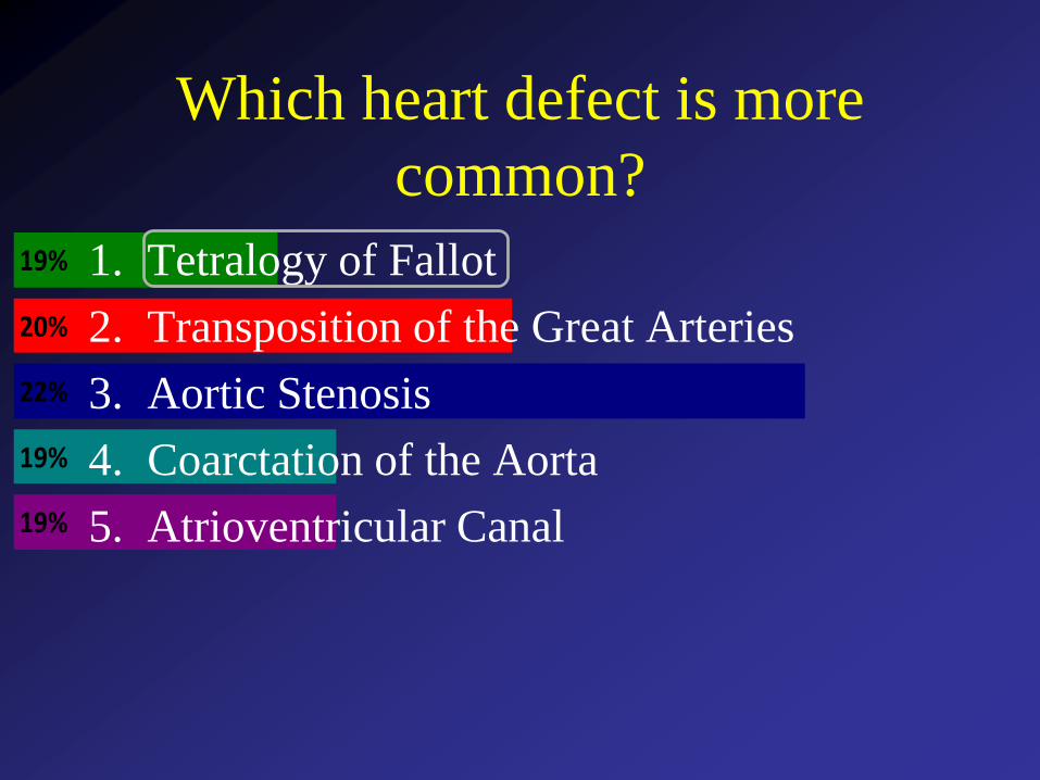
Which heart defect is more
common?
19%
19%
22%
20%
19% 1. Tetralogy of Fallot
2. Transposition of the Great Arteries
3. Aortic Stenosis
4. Coarctation of the Aorta
5. Atrioventricular Canal
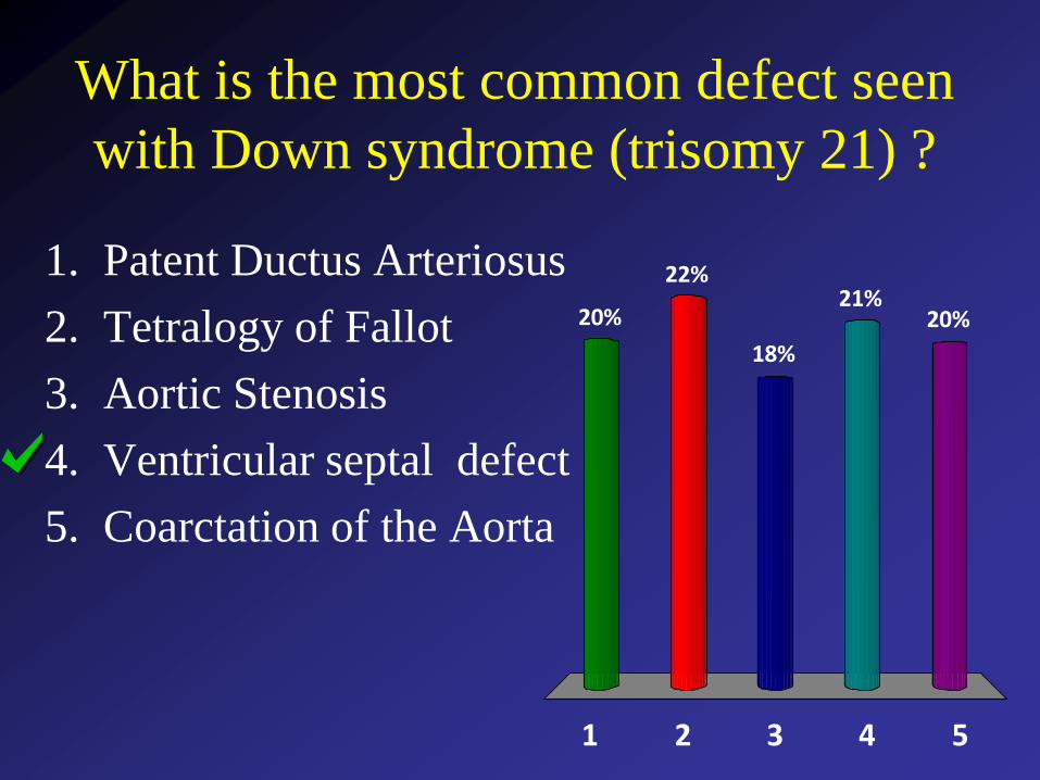
What is the most common defect seen
with Down syndrome (trisomy 21) ?
1 2 3 4 5
20%
22%
20%21%
18%
1. Patent Ductus Arteriosus
2. Tetralogy of Fallot
3. Aortic Stenosis
4. Ventricular septal defect
5. Coarctation of the Aorta
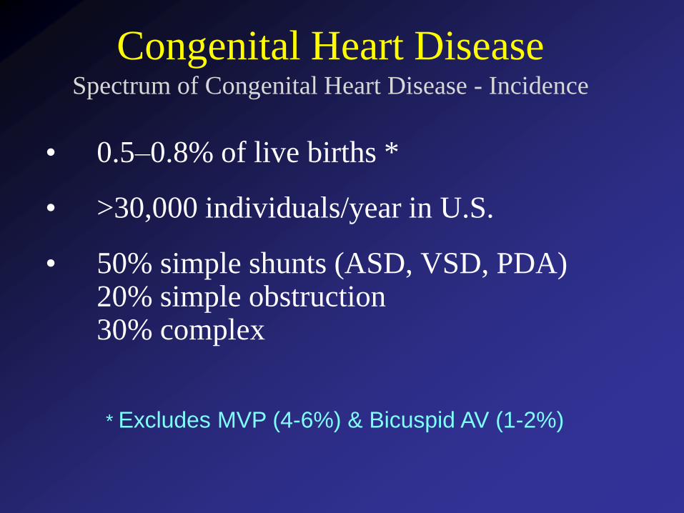
Congenital Heart DiseaseSpectrum of Congenital Heart Disease - Incidence
• 0.5–0.8% of live births *
• >30,000 individuals/year in U.S.
• 50% simple shunts (ASD, VSD, PDA)20% simple obstruction30% complex
* Excludes MVP (4-6%) & Bicuspid AV (1-2%)
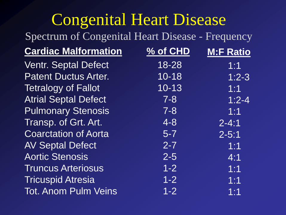
Congenital Heart DiseaseSpectrum of Congenital Heart Disease - Frequency
Cardiac Malformation % of CHD M:F Ratio
Ventr. Septal Defect 18-28 1:1Patent Ductus Arter. 10-18 1:2-3Tetralogy of Fallot 10-13 1:1Atrial Septal Defect 7-8 1:2-4Pulmonary Stenosis 7-8 1:1Transp. of Grt. Art. 4-8 2-4:1Coarctation of Aorta 5-7 2-5:1AV Septal Defect 2-7 1:1Aortic Stenosis 2-5 4:1Truncus Arteriosus 1-2 1:1Tricuspid Atresia 1-2 1:1Tot. Anom Pulm Veins 1-2 1:1
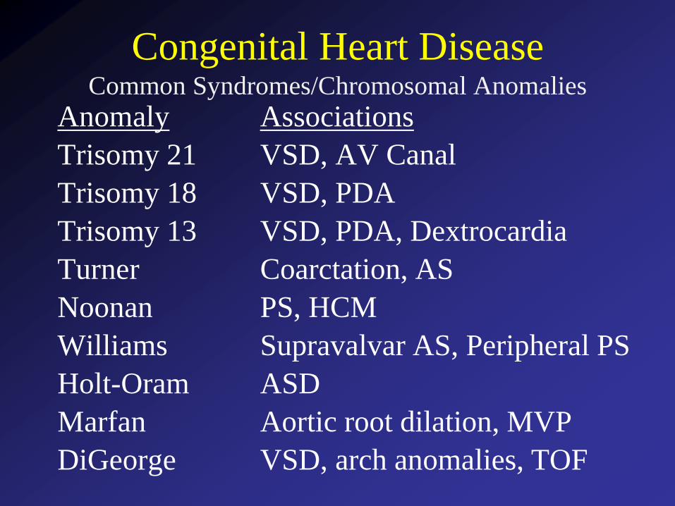
Congenital Heart DiseaseCommon Syndromes/Chromosomal Anomalies
Anomaly Associations
Trisomy 21 VSD, AV Canal
Trisomy 18 VSD, PDA
Trisomy 13 VSD, PDA, Dextrocardia
Turner Coarctation, AS
Noonan PS, HCM
Williams Supravalvar AS, Peripheral PS
Holt-Oram ASD
Marfan Aortic root dilation, MVP
DiGeorge VSD, arch anomalies, TOF
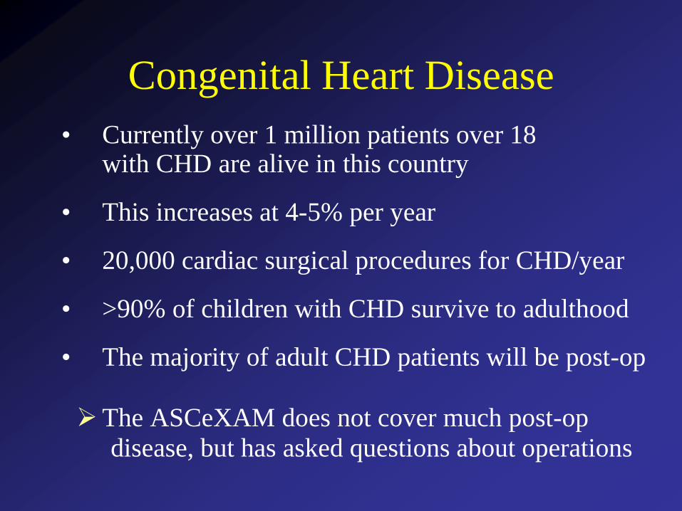
Congenital Heart Disease
• Currently over 1 million patients over 18with CHD are alive in this country
• This increases at 4-5% per year
• 20,000 cardiac surgical procedures for CHD/year
• >90% of children with CHD survive to adulthood
• The majority of adult CHD patients will be post-op
The ASCeXAM does not cover much post-op
disease, but has asked questions about operations
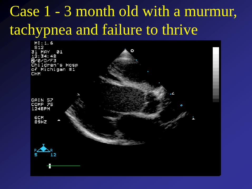
Case 1 - 3 month old with a murmur,
tachypnea and failure to thrive
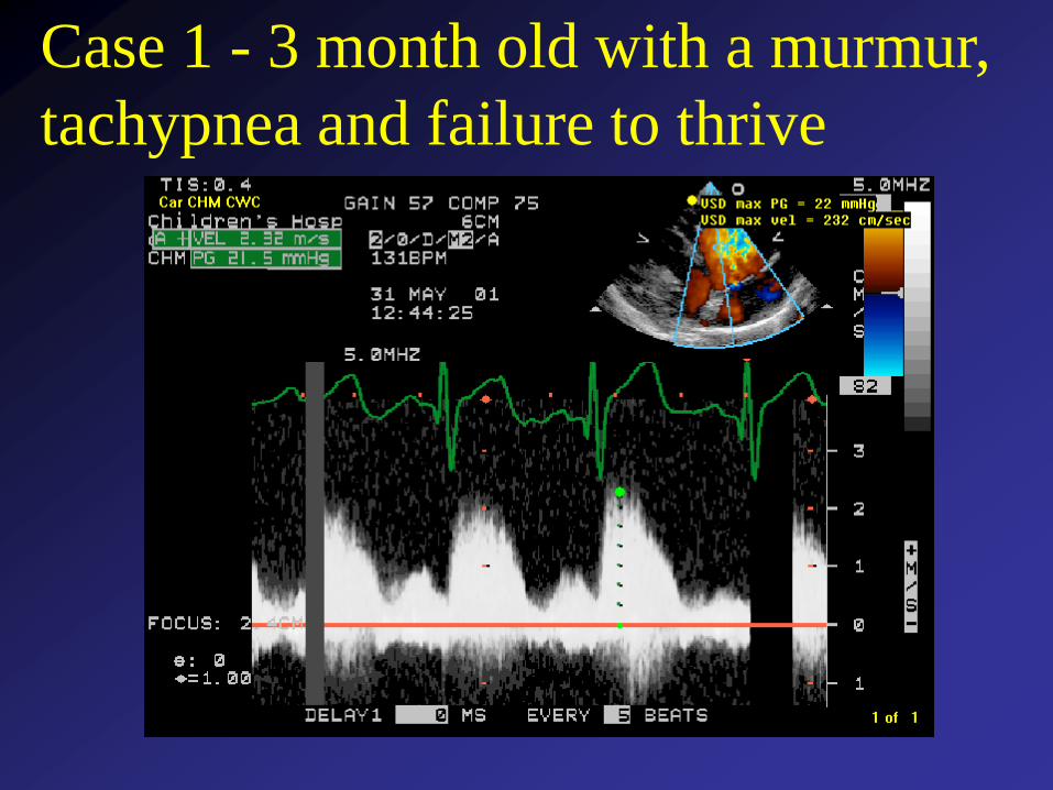
Case 1 - 3 month old with a murmur,
tachypnea and failure to thrive
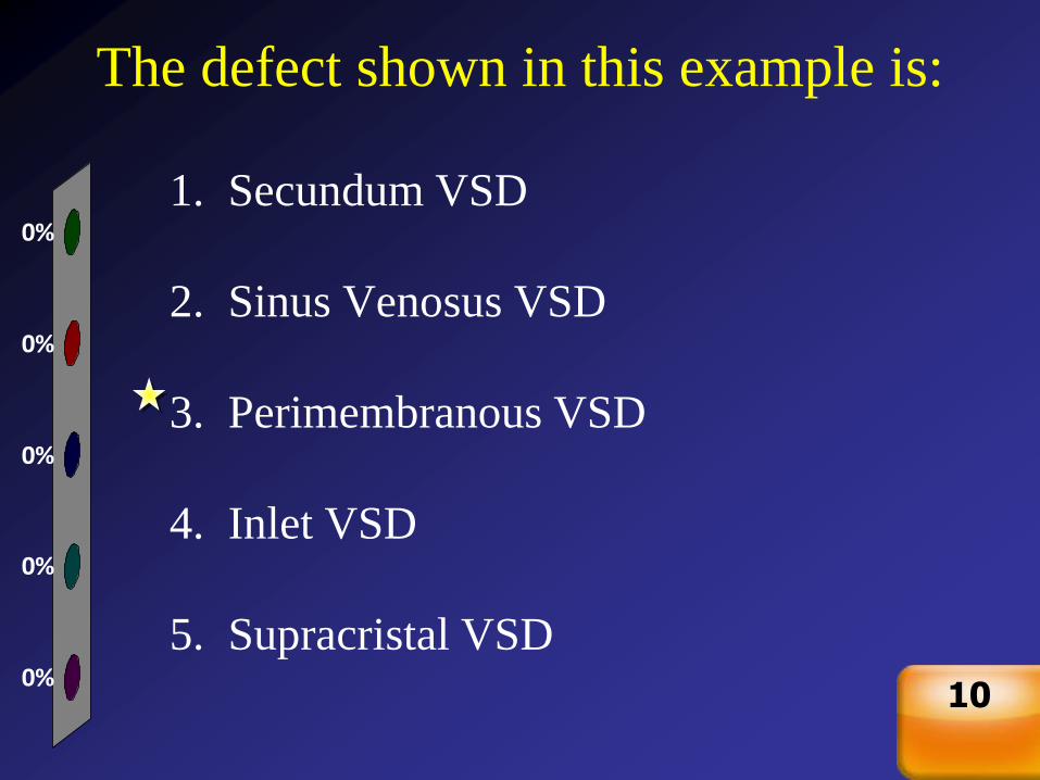
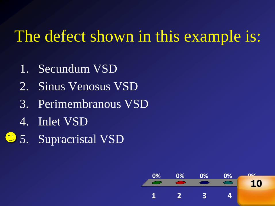
The defect shown in this example is:
0%
0%
0%
0%
0%
1. Secundum VSD
2. Sinus Venosus VSD
3. Perimembranous VSD
4. Inlet VSD
5. Supracristal VSD
Countdown
10
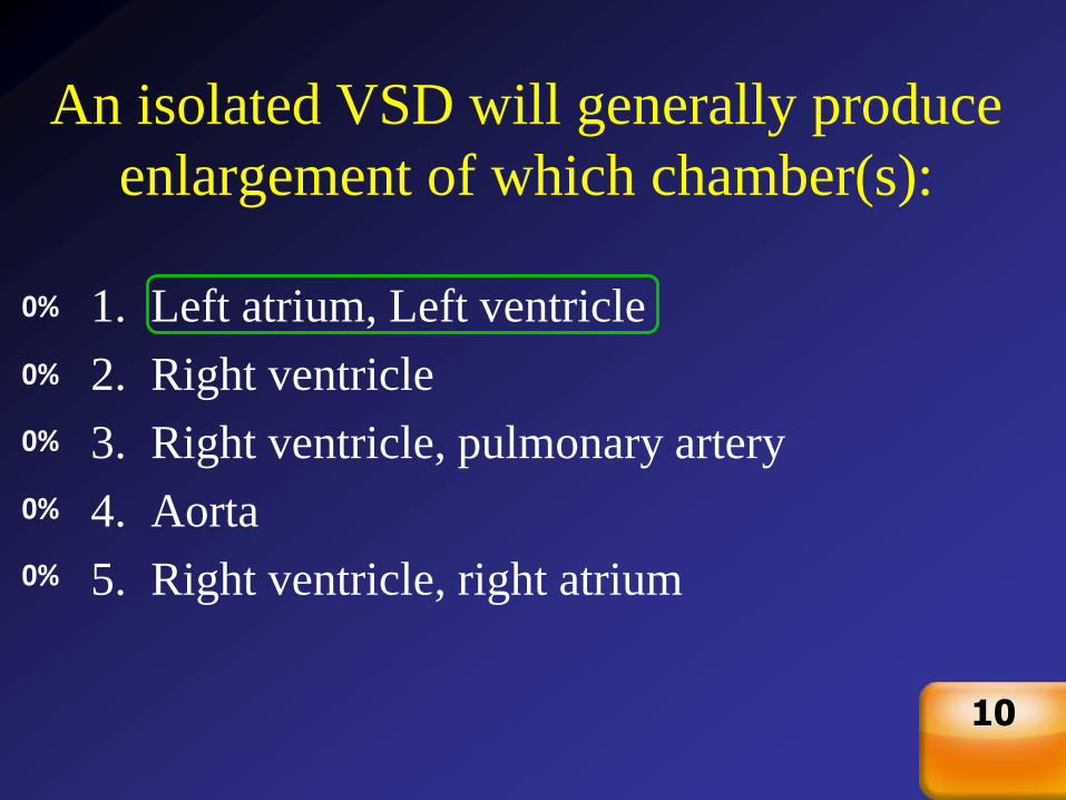
An isolated VSD will generally produce
enlargement of which chamber(s):
0%
0%
0%
0%
0% 1. Left atrium, Left ventricle
2. Right ventricle
3. Right ventricle, pulmonary artery
4. Aorta
5. Right ventricle, right atrium
Countdown
10
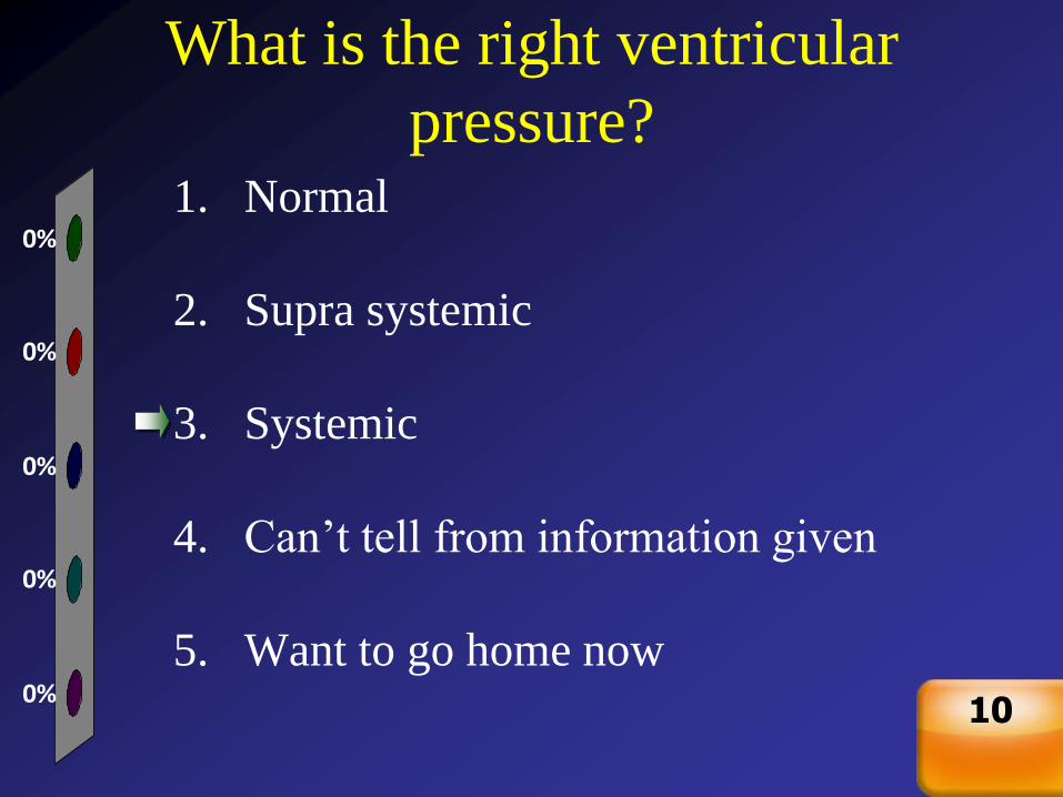
What is the right ventricular
pressure?
0%
0%
0%
0%
0%
1. Normal
2. Supra systemic
3. Systemic
4. Can’t tell from information given
5. Want to go home now
Countdown
10

Ventricular Septal DefectClinical
• Most common defect, 25% of CHD
• Shunt flow should be left to right
• Symptoms depend on the size of the holeLarge - >50% of aortic annulus sizeMedium - 25-50% of annulus sizeSmall - <25% of annulus size
• Large VSDs result in pulmonary edema → tachypnea, poor feeding, failure to thrive in infants
• In un-operated patients with large defects pulmonary vascular disease develops → shunt reversal and cyanosis (Eisenmenger’s complex)
1
1
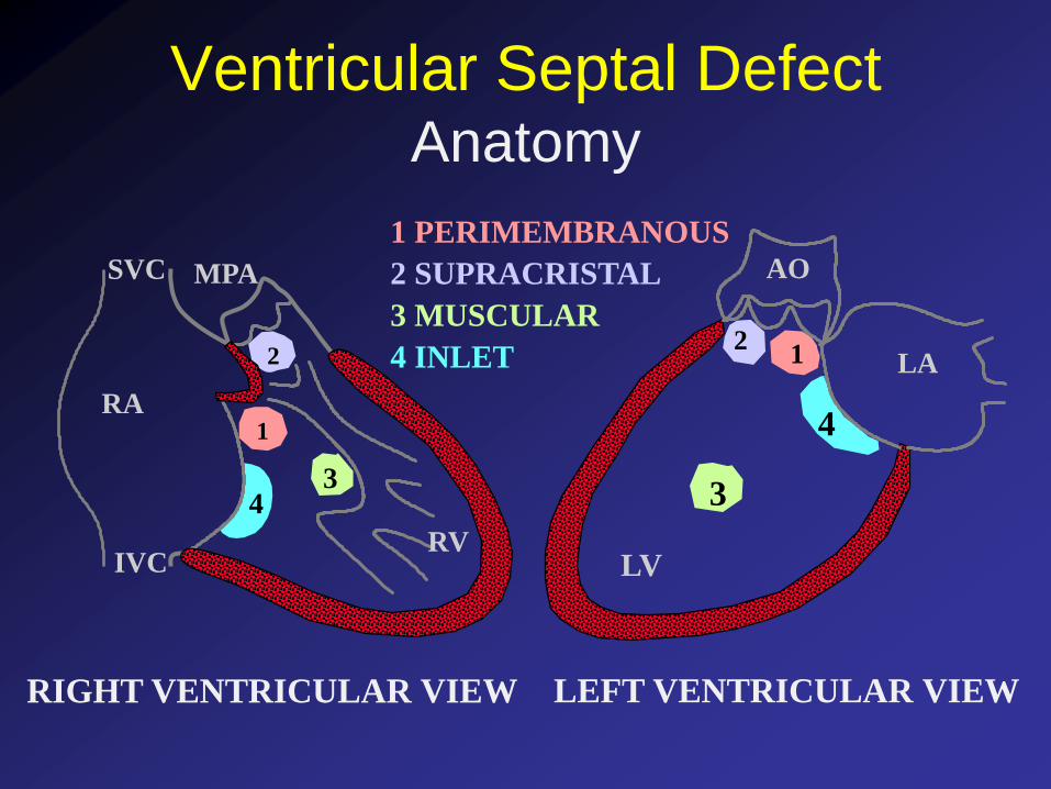
1 PERIMEMBRANOUS
22
2 SUPRACRISTAL
33
3 MUSCULAR
4
4
4 INLET
Ventricular Septal DefectAnatomy
RIGHT VENTRICULAR VIEW
AO
LA
LV
LEFT VENTRICULAR VIEW
IVC
SVC
RV
MPA
RA
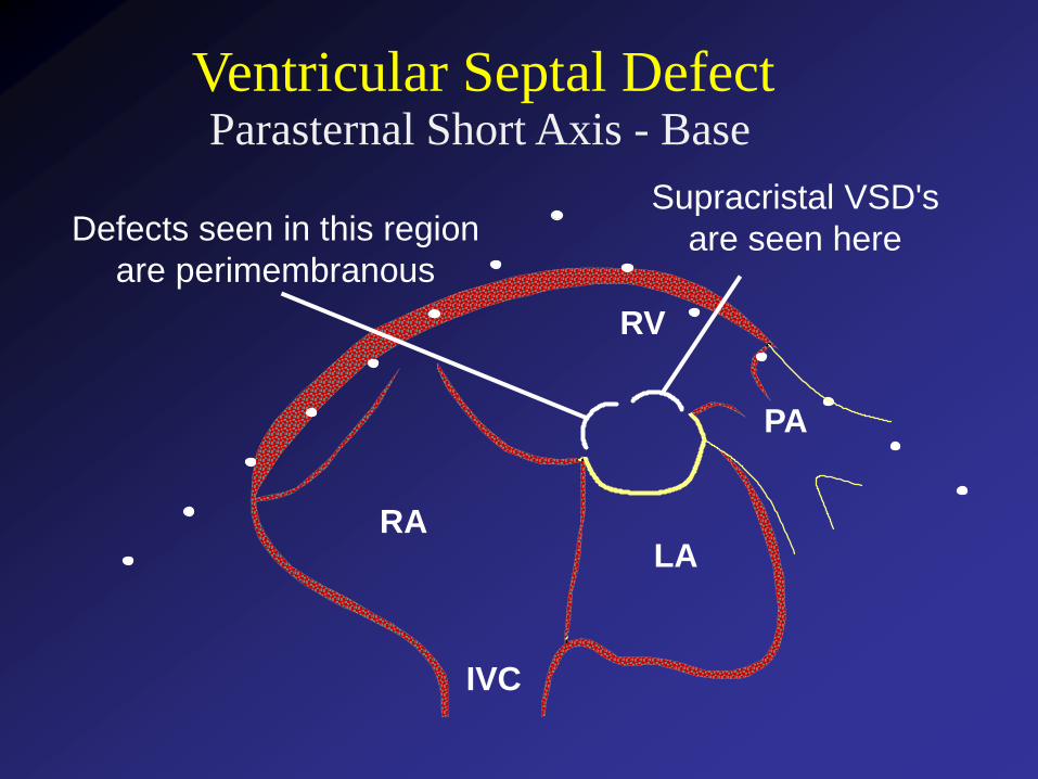
Parasternal Short Axis - Base
RV
LARA
IVC
PA
Defects seen in this region
are perimembranous
Supracristal VSD's
are seen here
Ventricular Septal Defect
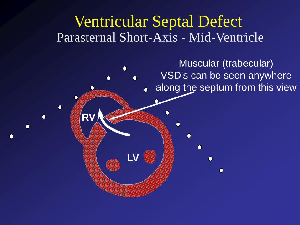
Parasternal Short-Axis - Mid-Ventricle
LV
RV
Ventricular Septal Defect
Muscular (trabecular)
VSD's can be seen anywhere
along the septum from this view
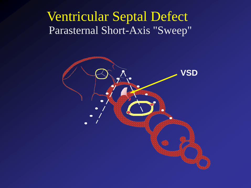
Ventricular Septal DefectParasternal Short-Axis "Sweep"
VSD
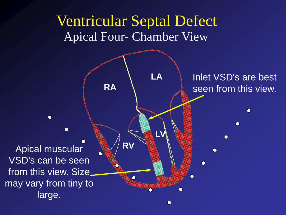
Apical Four- Chamber View
Ventricular Septal Defect
Apical muscular
VSD's can be seen
from this view. Size
may vary from tiny to
large.
RA
LA
LV
RV
Inlet VSD's are best
seen from this view.
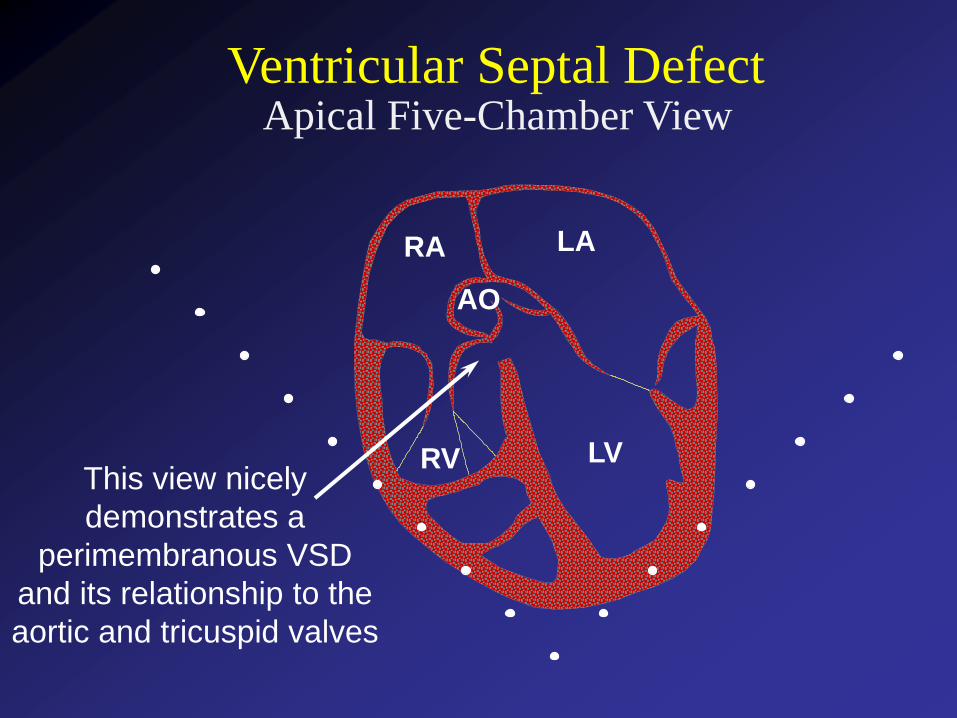
Apical Five-Chamber ViewVentricular Septal Defect
This view nicely
demonstrates a
perimembranous VSD
and its relationship to the
aortic and tricuspid valves
LA
LVRV
RA
AO
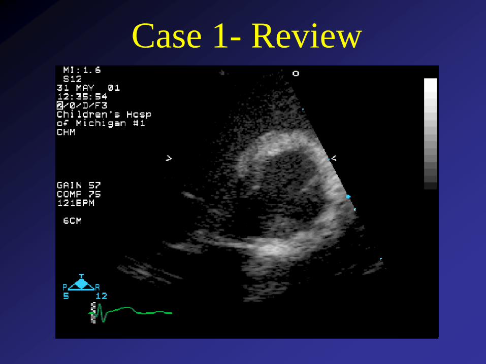
Case 1- Review
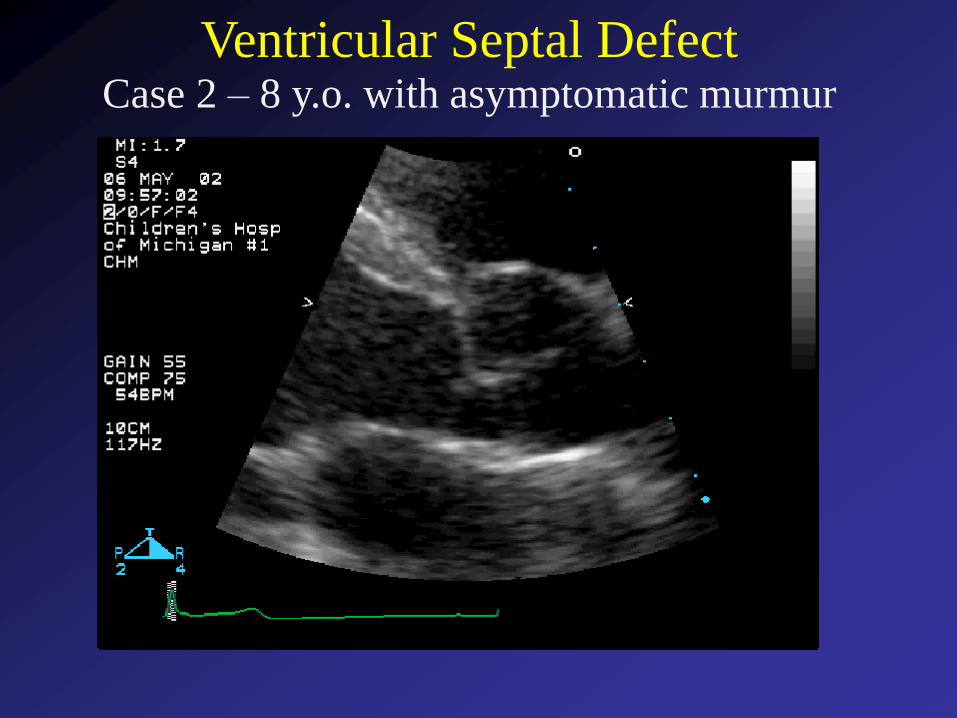
Ventricular Septal DefectCase 2 – 8 y.o. with asymptomatic murmur
The defect shown in this example is:
1 2 3 4 5
0% 0% 0%0%0%
1. Secundum VSD
2. Sinus Venosus VSD
3. Perimembranous VSD
4. Inlet VSD
5. Supracristal VSD
Countdown
10
Question 10 - A common
complication of this defect is:
0%
0%
0%
0%
0%
1. Pulmonary valve endocarditis
2. Aortic regurgitation
3. Aortic dissection
4. Tricuspid regurgitation
5. Right ventricular enlargement
Countdown
10
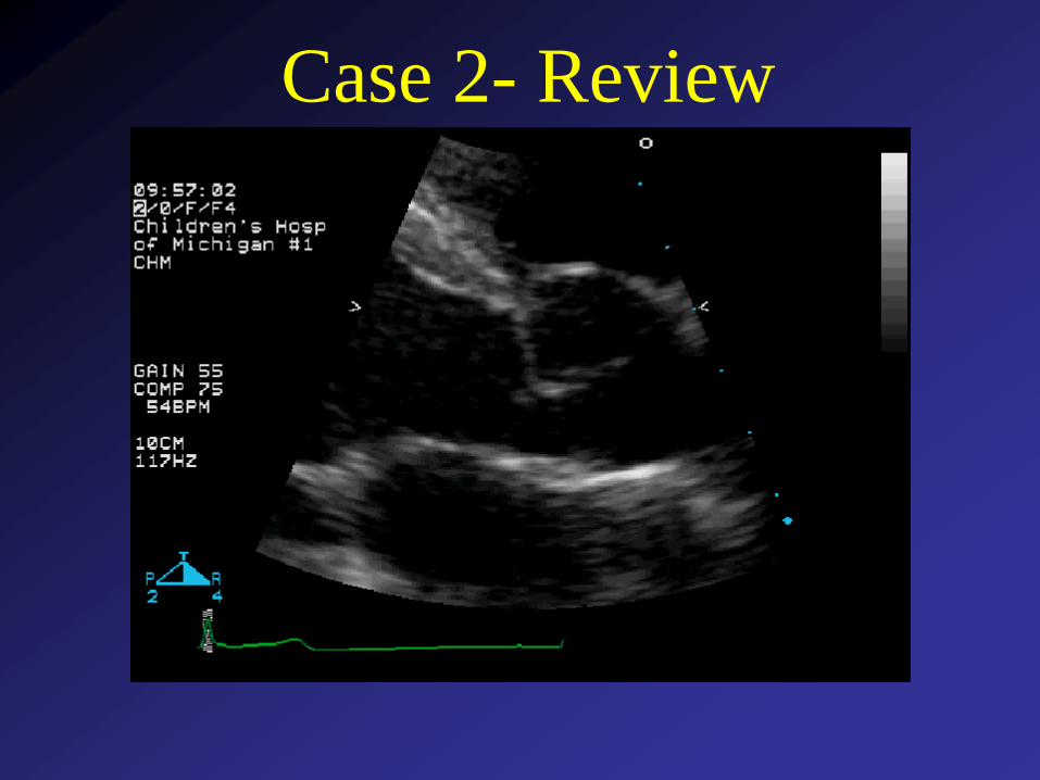
Case 2- Review
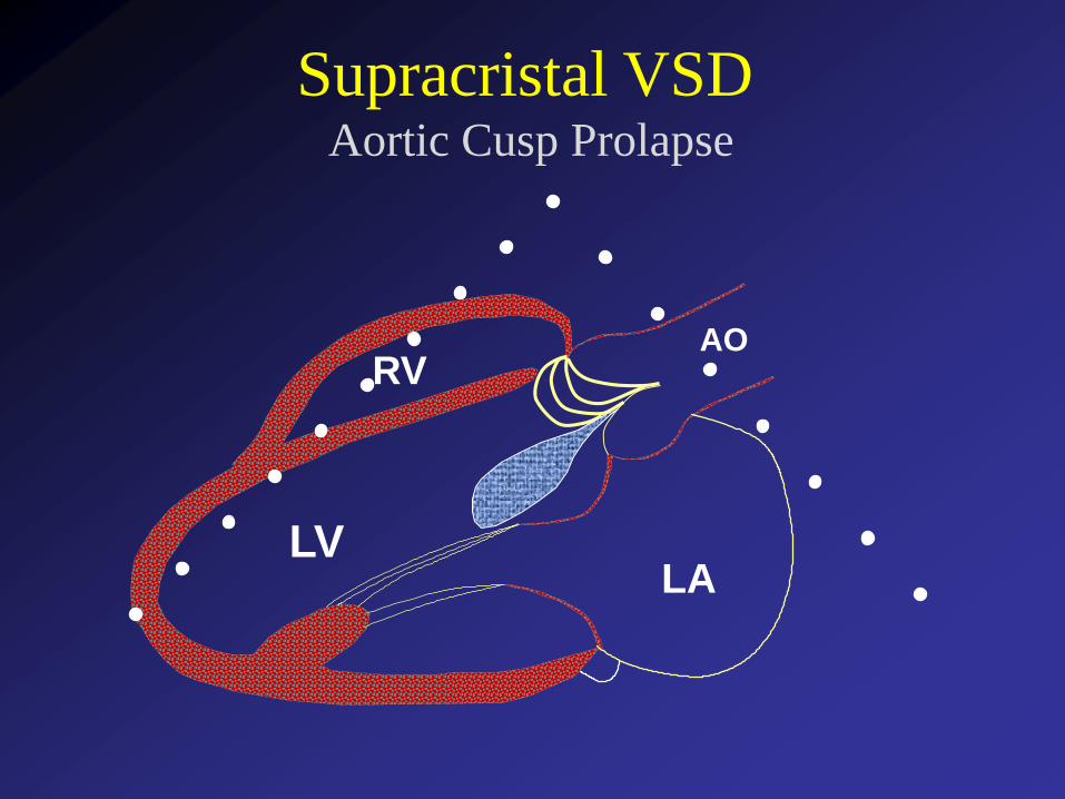
Aortic Cusp Prolapse
Supracristal VSD
LVLA
AORV
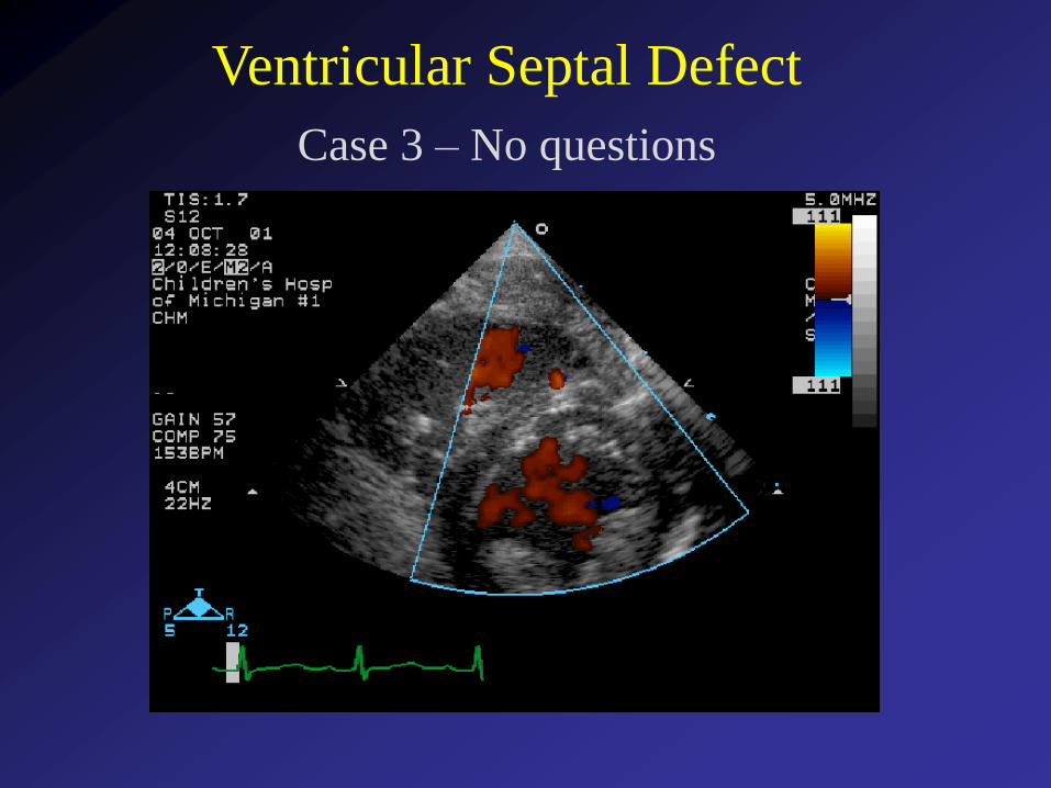
Ventricular Septal Defect
Case 3 – No questions
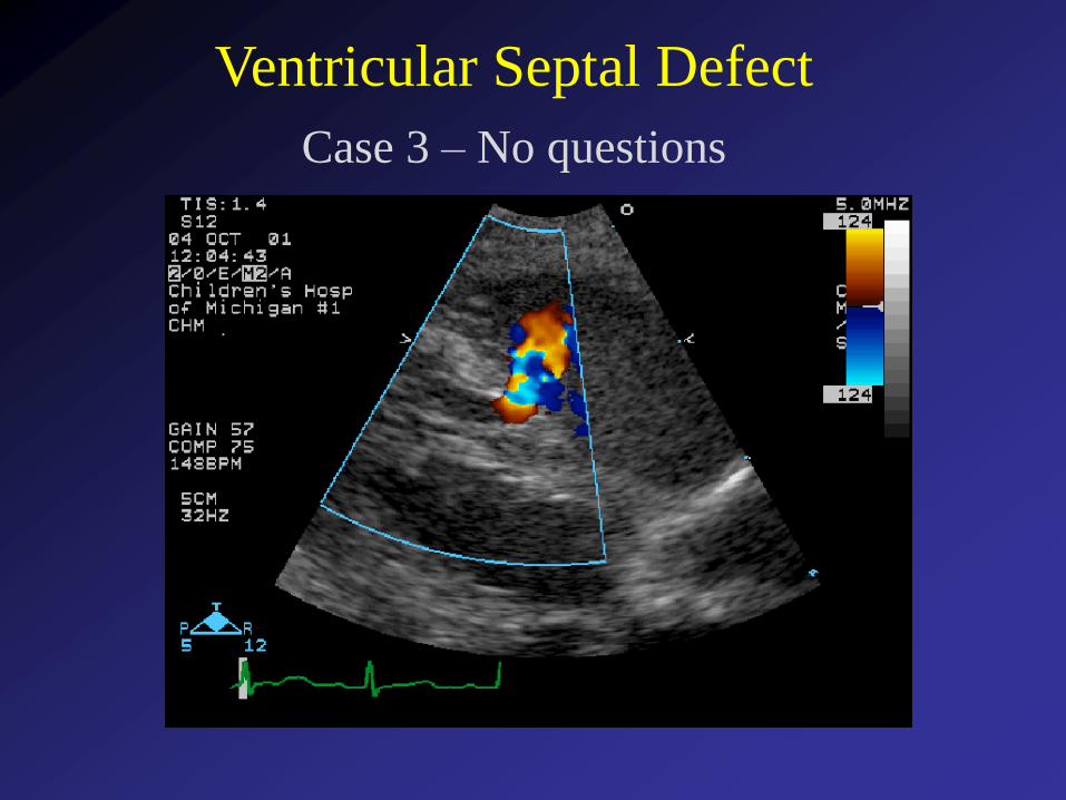
Ventricular Septal Defect
Case 3 – No questions
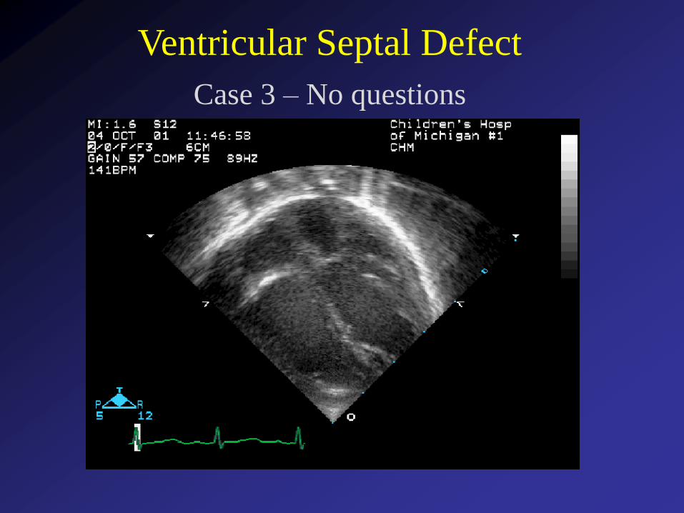
Ventricular Septal Defect
Case 3 – No questions
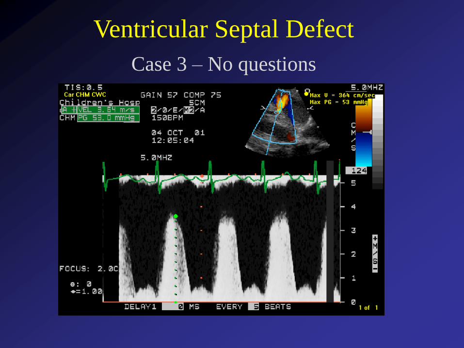
Ventricular Septal Defect
Case 3 – No questions

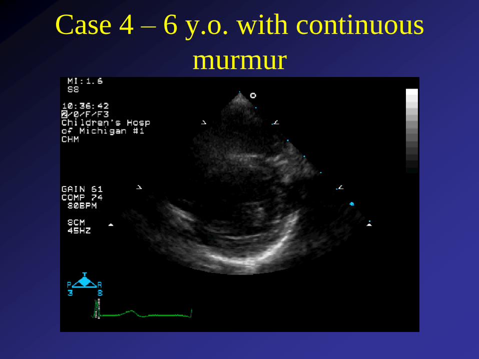
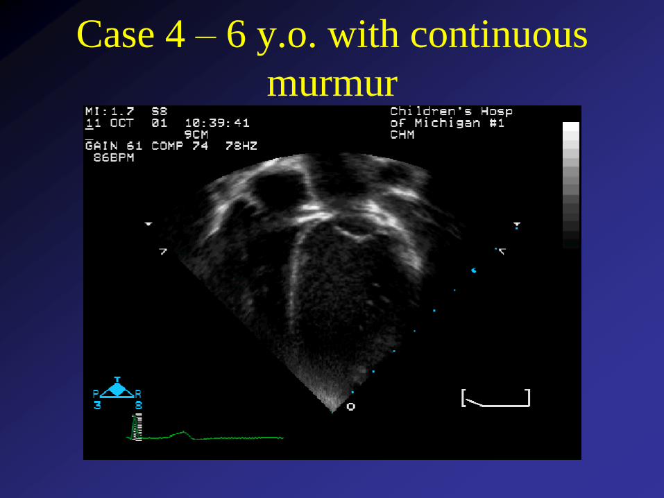
Case 4 – 6 y.o. with continuous
murmur
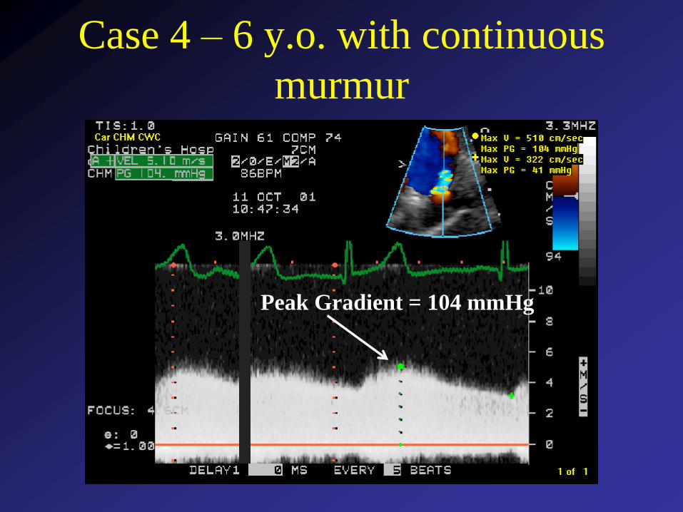
Case 4 – 6 y.o. with continuous
murmur
Case 4 – 6 y.o. with continuous
murmur
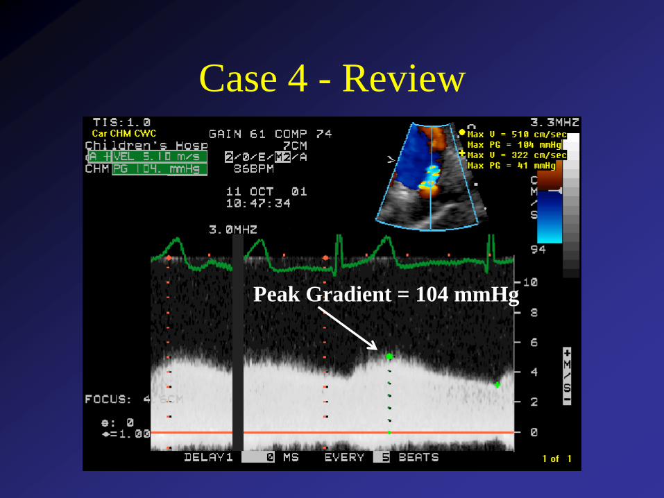
Peak Gradient = 104 mmHg
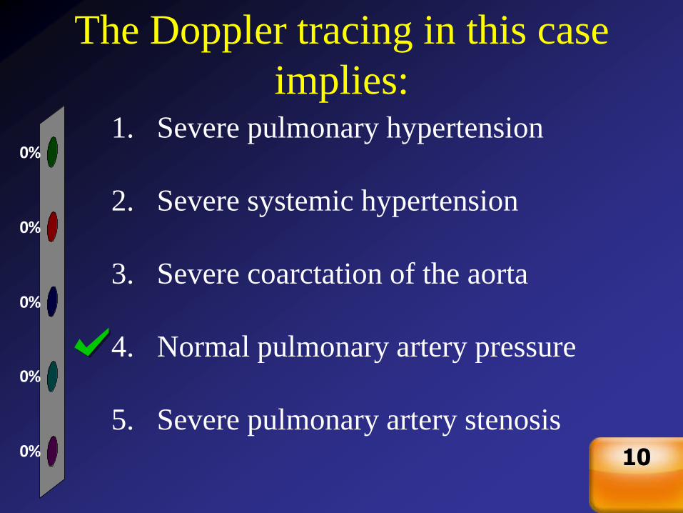
The Doppler tracing in this case
implies:
0%
0%
0%
0%
0%1. Severe pulmonary hypertension
2. Severe systemic hypertension
3. Severe coarctation of the aorta
4. Normal pulmonary artery pressure
5. Severe pulmonary artery stenosis
Countdown
10
Patent Ductus ArteriosusClinical
• Continuous murmur in older patients
• Bounding pulses, wide pulse pressure, respiratory symptoms in neonates with a large PDA
• Large PDA will act much like a large VSD, producing pulmonary over-circulation and signs/symptoms of congestive heart failure
• A small PDA is generally hemodynamically insignificant but is at risk for endarteritis
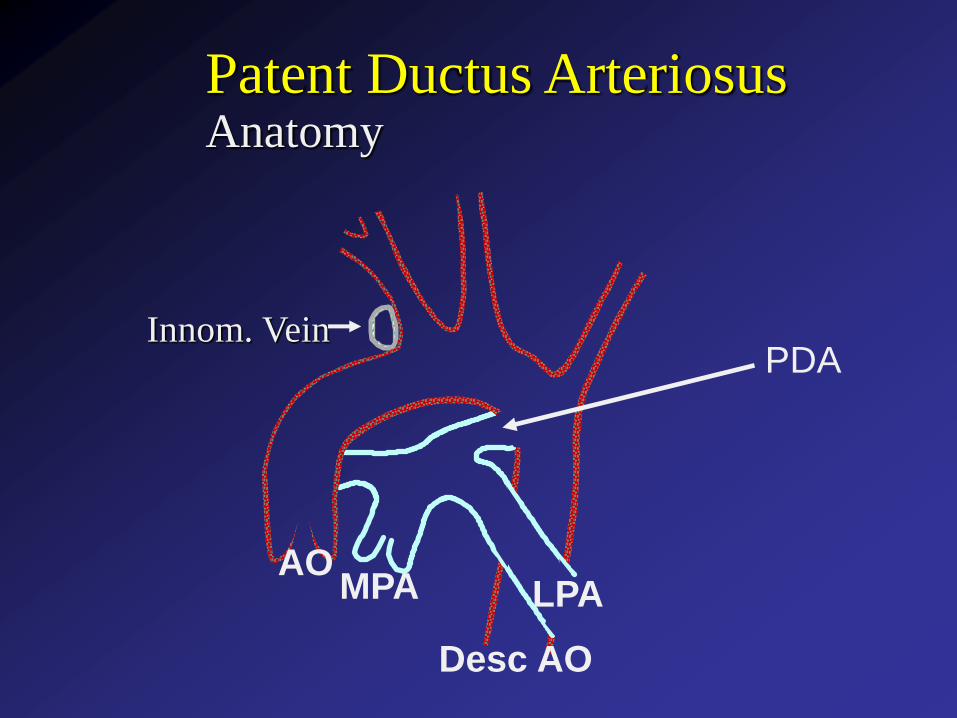
PDA
AOMPA LPA
Desc AO
Patent Ductus ArteriosusAnatomy
Innom. Vein
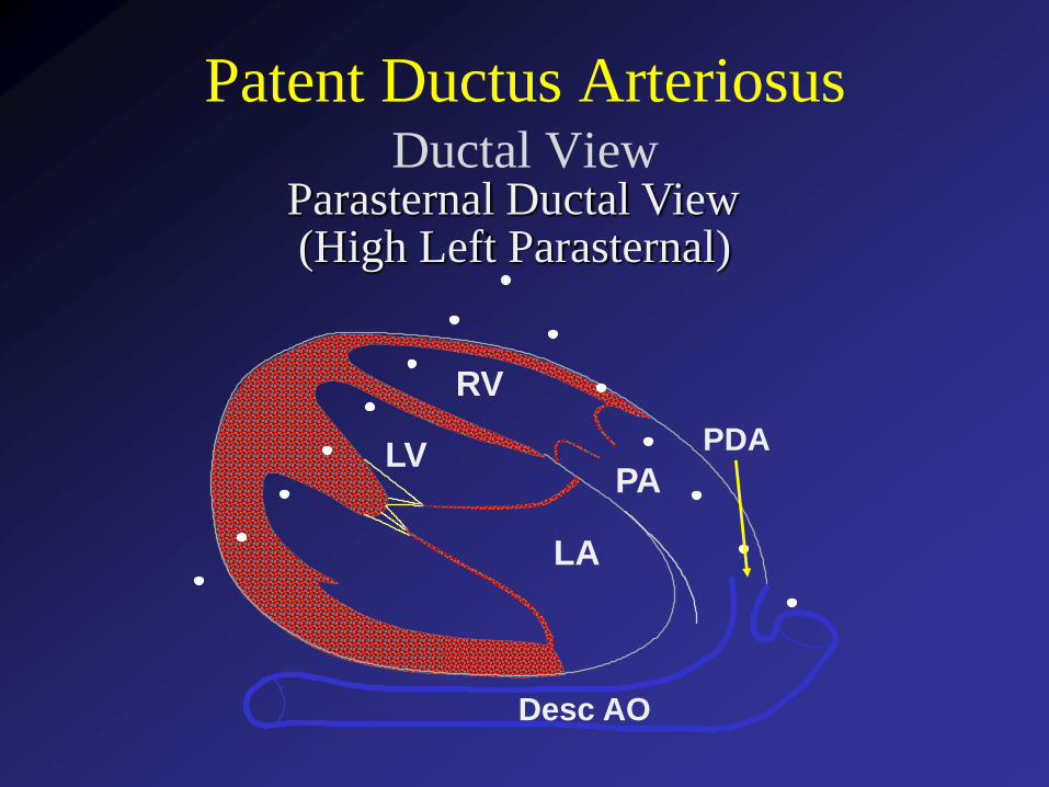
Patent Ductus ArteriosusDuctal View
Parasternal Ductal View
PA
LA
LV
RV
Desc AO
PDA
(High Left Parasternal)
PA
LA
LV
RV
AO
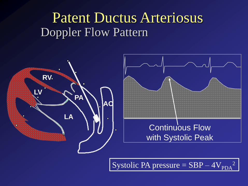
Doppler Flow PatternPatent Ductus Arteriosus
Continuous Flow
with Systolic Peak
Systolic PA pressure = SBP – 4VPDA2
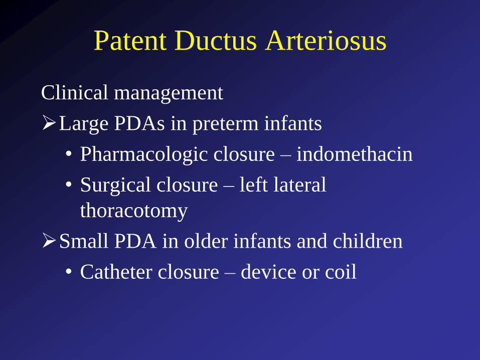
Patent Ductus Arteriosus
Clinical management
Large PDAs in preterm infants
• Pharmacologic closure – indomethacin
• Surgical closure – left lateral
thoracotomy
Small PDA in older infants and children
• Catheter closure – device or coil
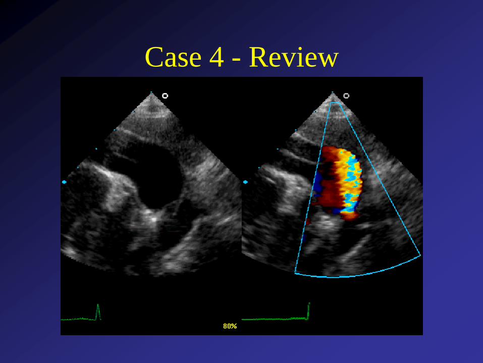
Case 4 - Review
Case 4 - Review
Peak Gradient = 104 mmHg
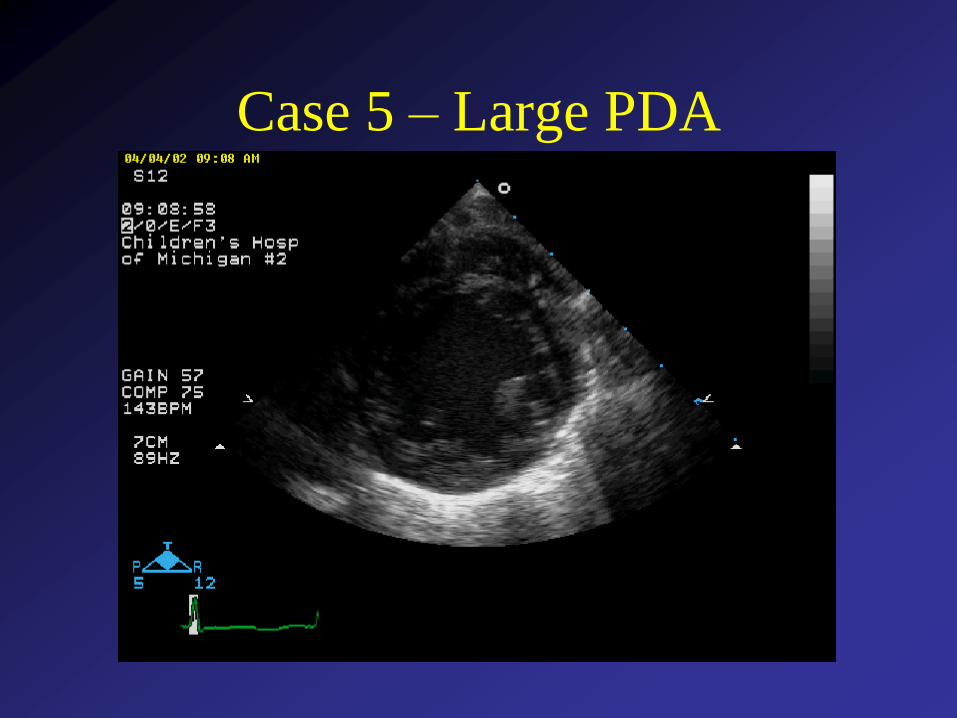
Case 5 – Large PDA

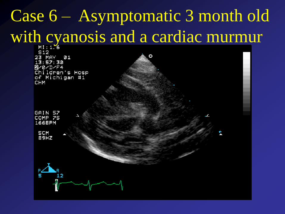
Case 6 – Asymptomatic 3 month old
with cyanosis and a cardiac murmur
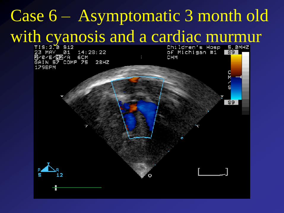
Case 6 – Asymptomatic 3 month old
with cyanosis and a cardiac murmur

The defect shown in this example is:
1 2 3 4 5
0% 0% 0%0%0%
1. Single ventricle
2. Transposition of the great
arteries
3. Perimembranous VSD
4. Tetralogy of Fallot
5. Complete atrioventricular
canal
Countdown
10
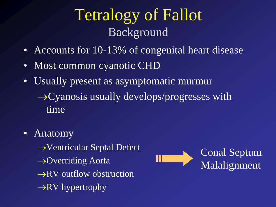
Tetralogy of FallotBackground
• Accounts for 10-13% of congenital heart disease
• Most common cyanotic CHD
• Usually present as asymptomatic murmur
Cyanosis usually develops/progresses with
time
• Anatomy
Ventricular Septal Defect
Overriding Aorta
RV outflow obstruction
RV hypertrophy
Conal Septum
Malalignment
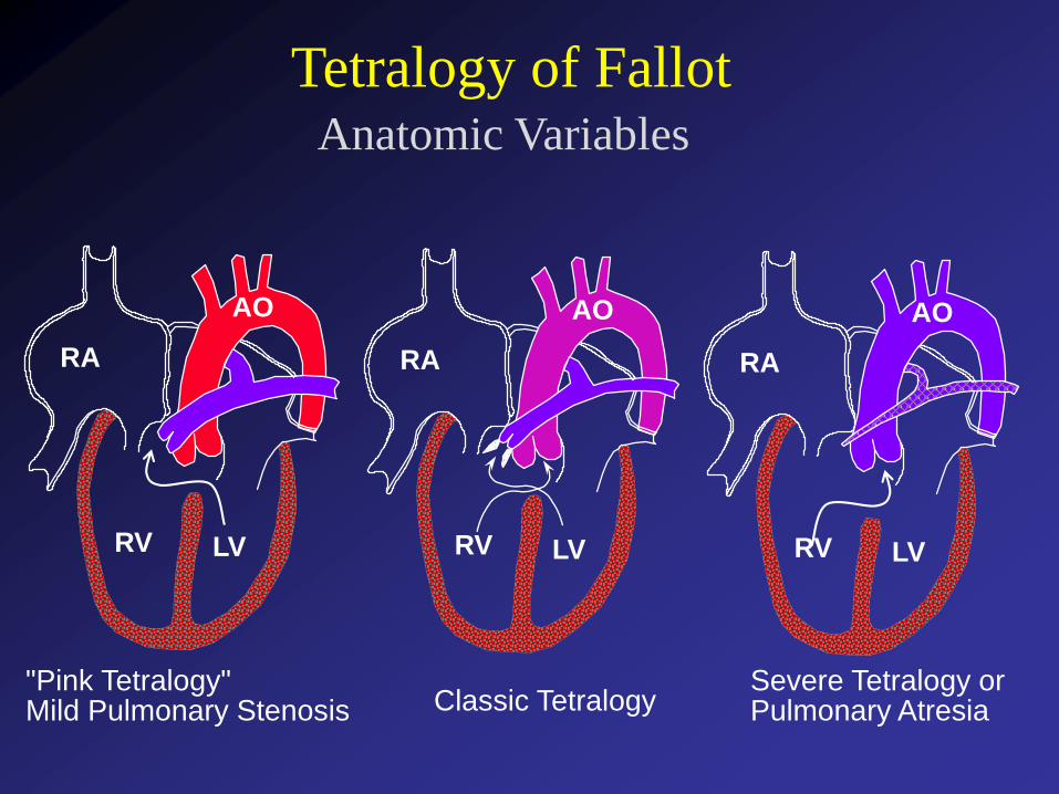
RA
RV LV
AO
RA
RV LV
AO
RA
RV LV
AO
"Pink Tetralogy"Mild Pulmonary Stenosis Classic Tetralogy
Severe Tetralogy orPulmonary Atresia
Anatomic Variables
Tetralogy of Fallot
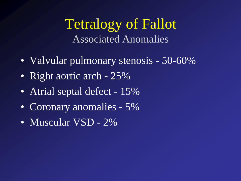
Tetralogy of FallotAssociated Anomalies
• Valvular pulmonary stenosis - 50-60%
• Right aortic arch - 25%
• Atrial septal defect - 15%
• Coronary anomalies - 5%
• Muscular VSD - 2%
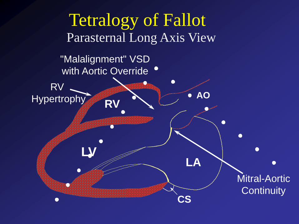
Parasternal Long Axis View
Tetralogy of Fallot
LVLA
AO
CS
RV
"Malalignment" VSD
with Aortic Override
Mitral-Aortic
Continuity
RV
Hypertrophy
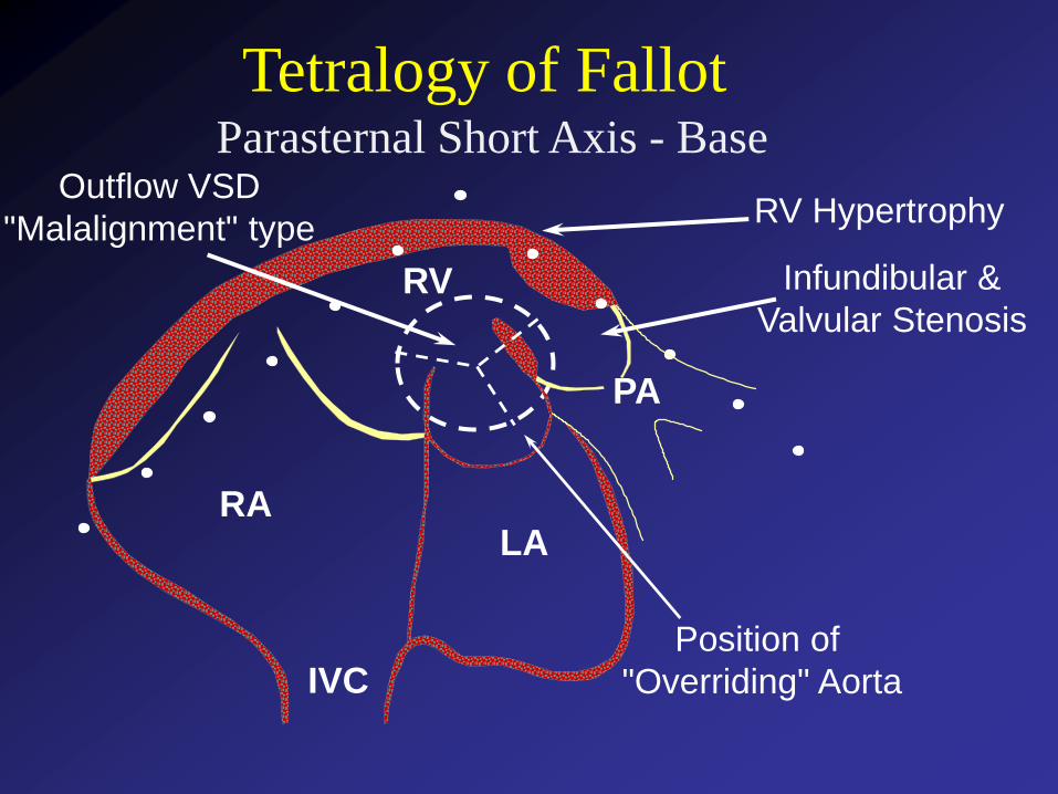
Parasternal Short Axis - Base
Tetralogy of Fallot
RV
LARA
IVC
PA
Outflow VSD
"Malalignment" typeRV Hypertrophy
Infundibular &
Valvular Stenosis
Position of
"Overriding" Aorta
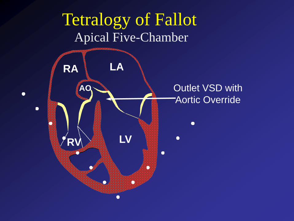
Apical Five-Chamber
Tetralogy of Fallot
LA
LVRV
RA
AO Outlet VSD with
Aortic Override
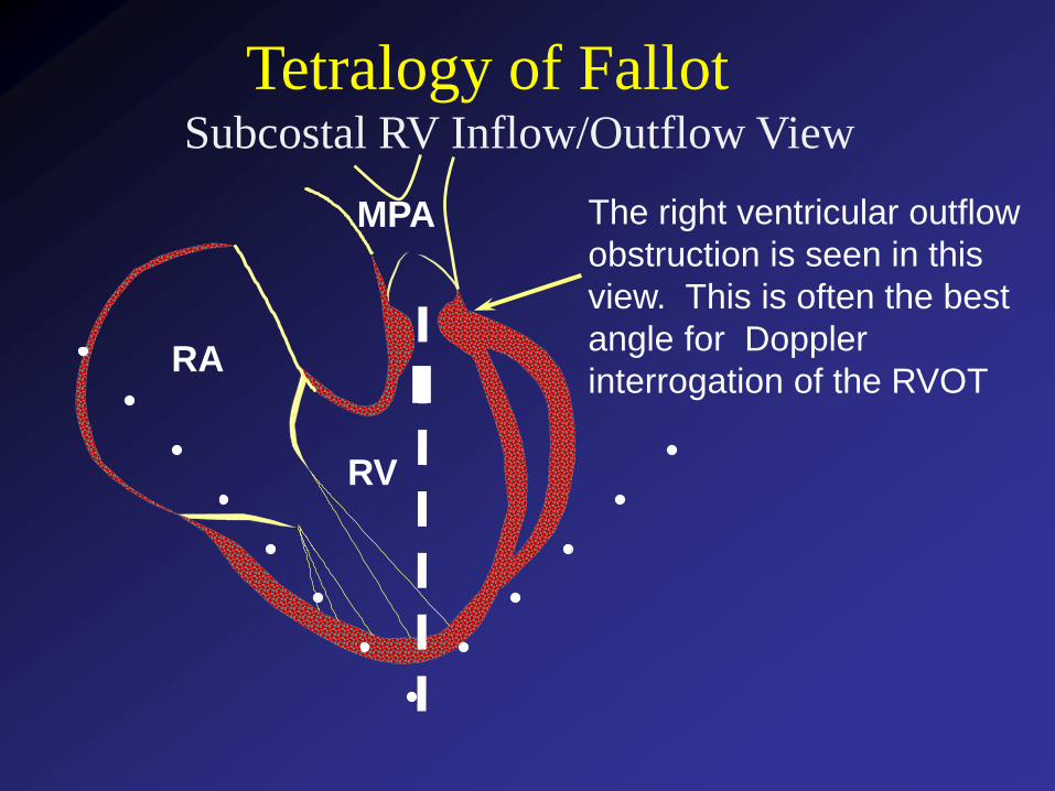
Subcostal RV Inflow/Outflow View
Tetralogy of Fallot
RA
RV
MPA The right ventricular outflow
obstruction is seen in this
view. This is often the best
angle for Doppler
interrogation of the RVOT
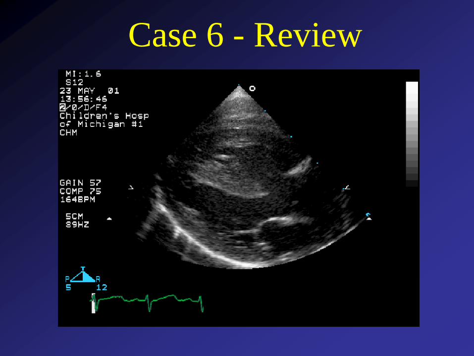
Case 6 - Review
Tetralogy of FallotSurgical Intervention
• Timing – usually during first 6 months
• VSD closure, relief of RVOTO obstruction
• Many repairs require a trans annular RV
outflow patch with results in chronic severe
pulmonary regurgitationLikely need for late pulmonary valve replacement
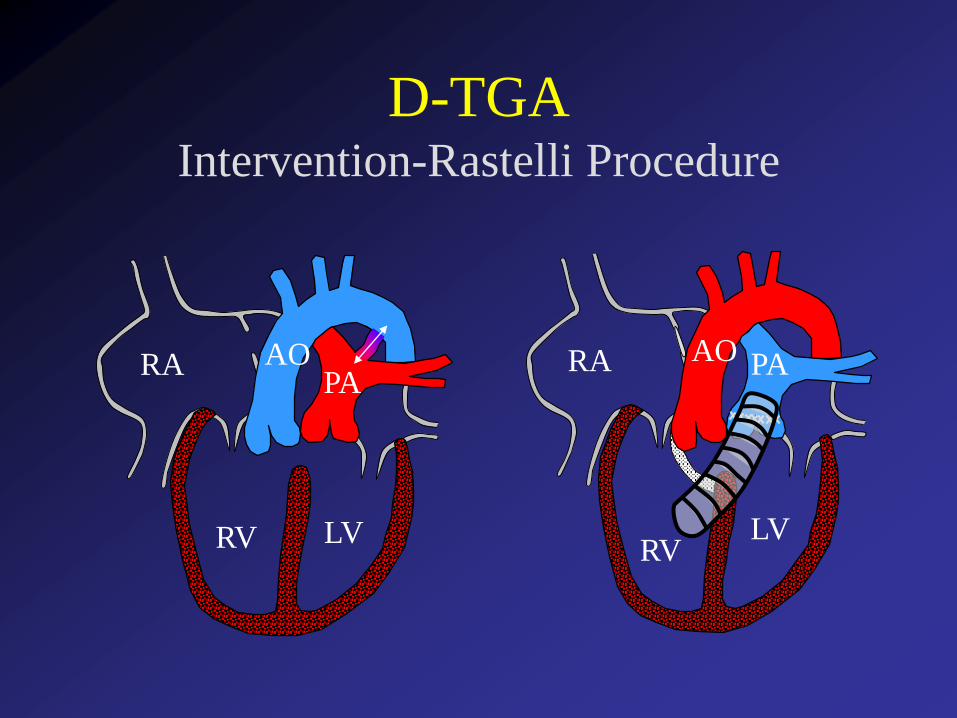
• Rastelli type repair (VSD closure + RV to
pulmonary artery conduit) may be required for
complex anatomy – pulmonary atresia,
coronary anomalies

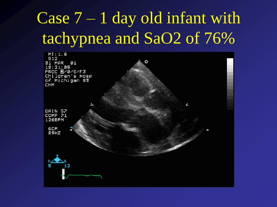
Case 7 – 1 day old infant with
tachypnea and SaO2 of 76%
What congenital heart defect is shown:
0%
0%
0%
0%
0% 1. Perimembranous VSD
2. Truncus arteriosus
3. Corrected transposition of the great
arteries (L-TGA)
4. Complete transposition of the great
arteries (D-TGA)
5. Tetralogy of Fallot
Countdown
10
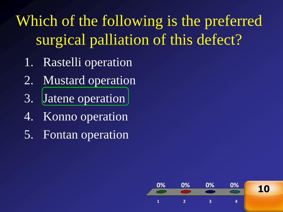
Which of the following is the preferred
surgical palliation of this defect?
1 2 3 4 5
0% 0% 0%0%0%
1. Rastelli operation
2. Mustard operation
3. Jatene operation
4. Konno operation
5. Fontan operation
Countdown
10
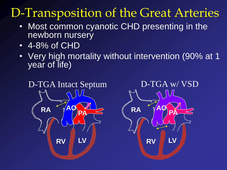
D-Transposition of the Great Arteries• Most common cyanotic CHD presenting in the
newborn nursery
• 4-8% of CHD
• Very high mortality without intervention (90% at 1 year of life)
D-TGA Intact Septum D-TGA w/ VSD
RV LV
RA AOPA
RV LV
RA AOPA
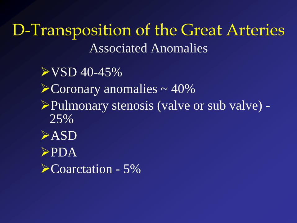
D-Transposition of the Great Arteries Associated Anomalies
VSD 40-45%
Coronary anomalies ~ 40%
Pulmonary stenosis (valve or sub valve) -25%
ASD
PDA
Coarctation - 5%
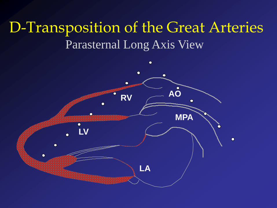
LA
LV
RV
MPA
AO
D-Transposition of the Great ArteriesParasternal Long Axis View
RV
LARA
IVC
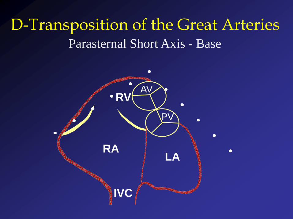
D-Transposition of the Great ArteriesParasternal Short Axis - Base
AV
PV
RA
AO
PA
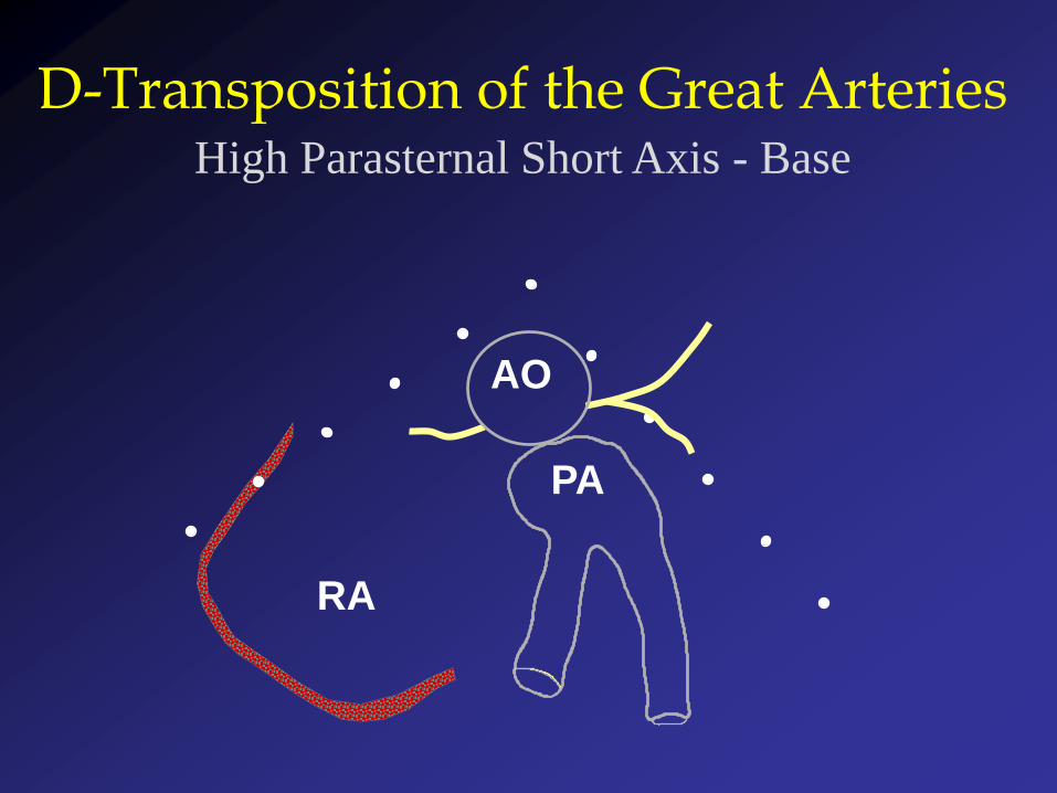
D-Transposition of the Great ArteriesHigh Parasternal Short Axis - Base
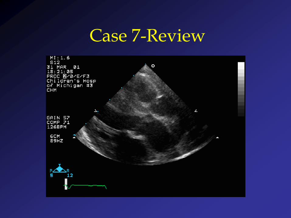
Case 7-Review
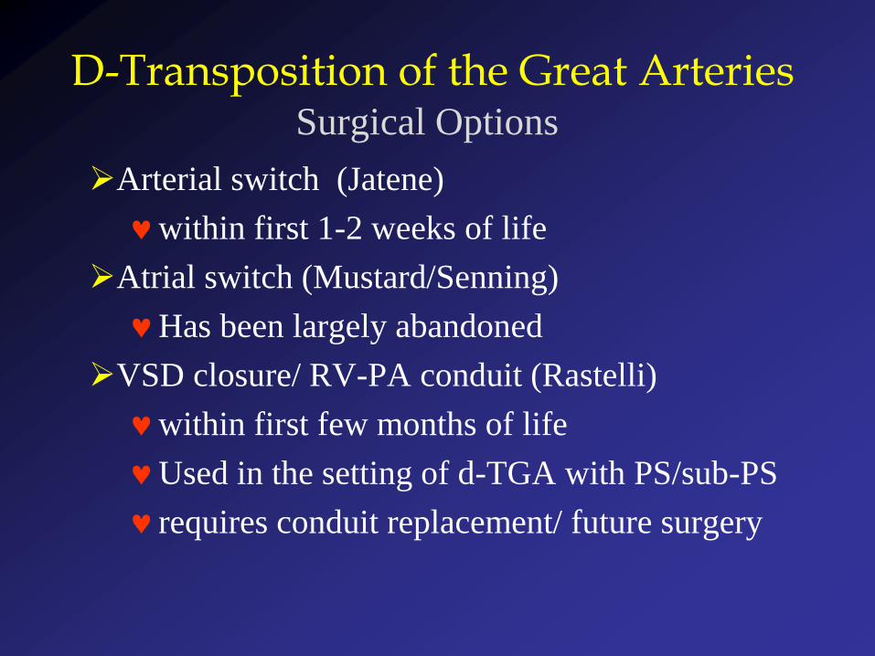
Arterial switch (Jatene)
within first 1-2 weeks of life
Atrial switch (Mustard/Senning)
Has been largely abandoned
VSD closure/ RV-PA conduit (Rastelli)
within first few months of life
Used in the setting of d-TGA with PS/sub-PS
requires conduit replacement/ future surgery
D-Transposition of the Great ArteriesSurgical Options
RV LV
RA AOPA
D-TGAIntervention-Rastelli Procedure
RVLV
RA AO PA
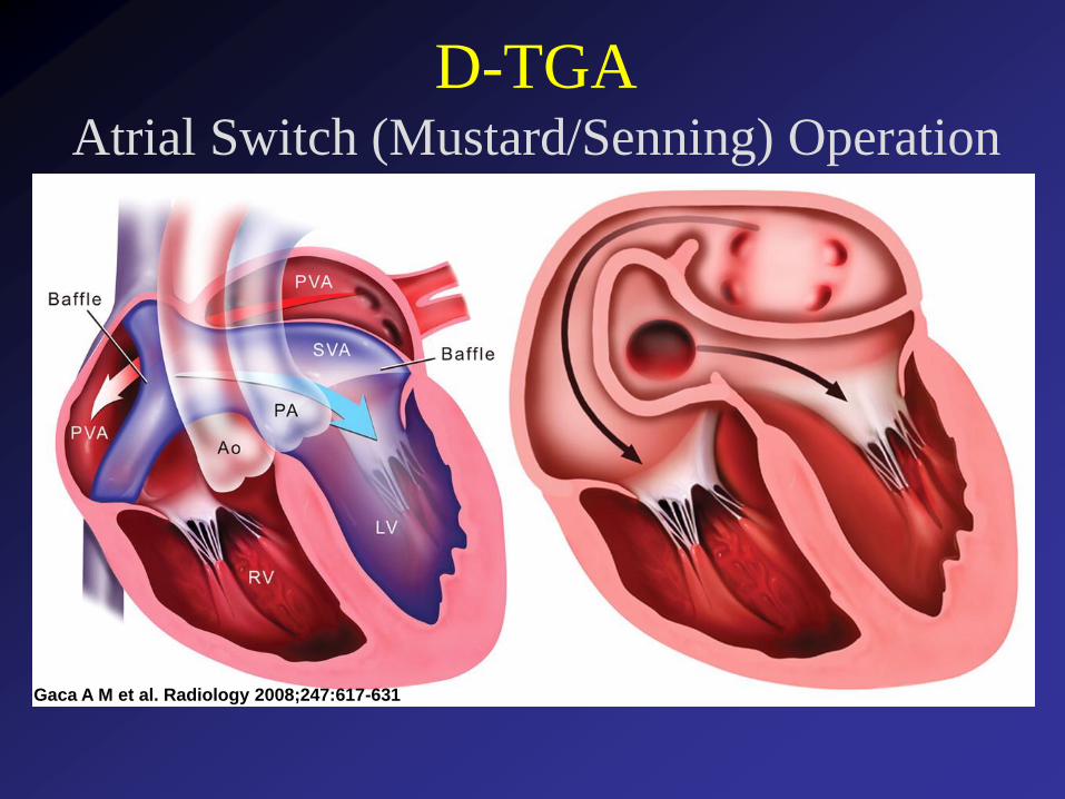
D-TGAAtrial Switch (Mustard/Senning) Operation
Gaca A M et al. Radiology 2008;247:617-631
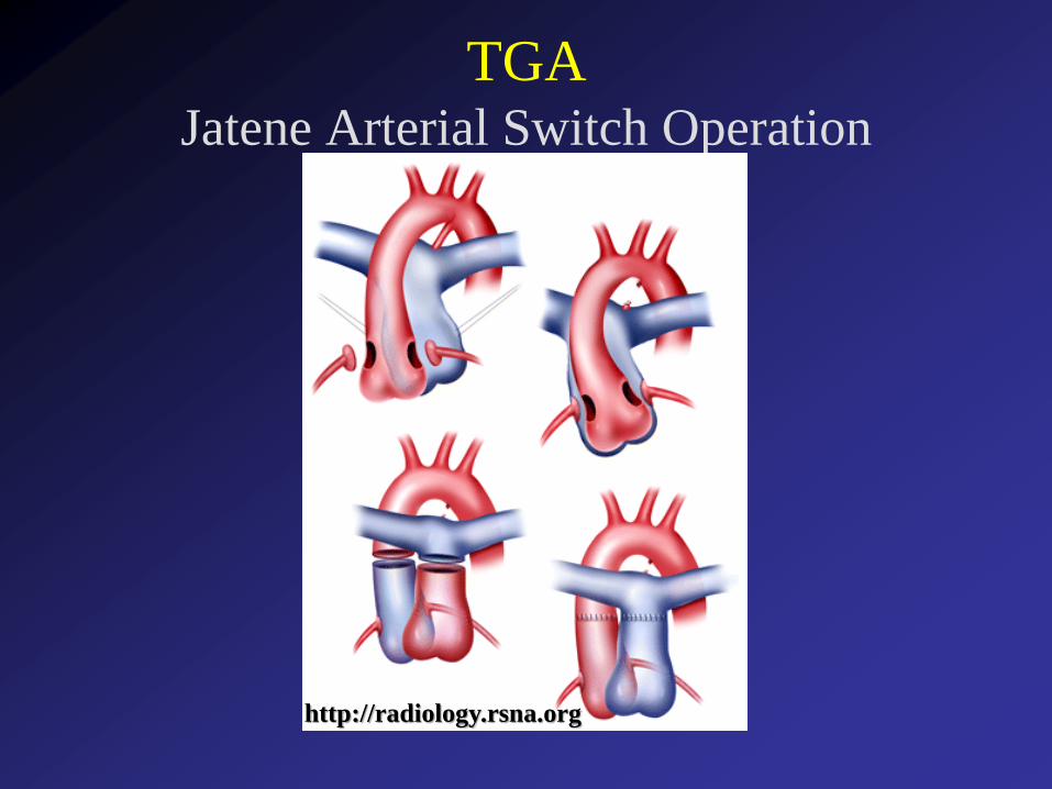
TGAJatene Arterial Switch Operation
http://radiology.rsna.org

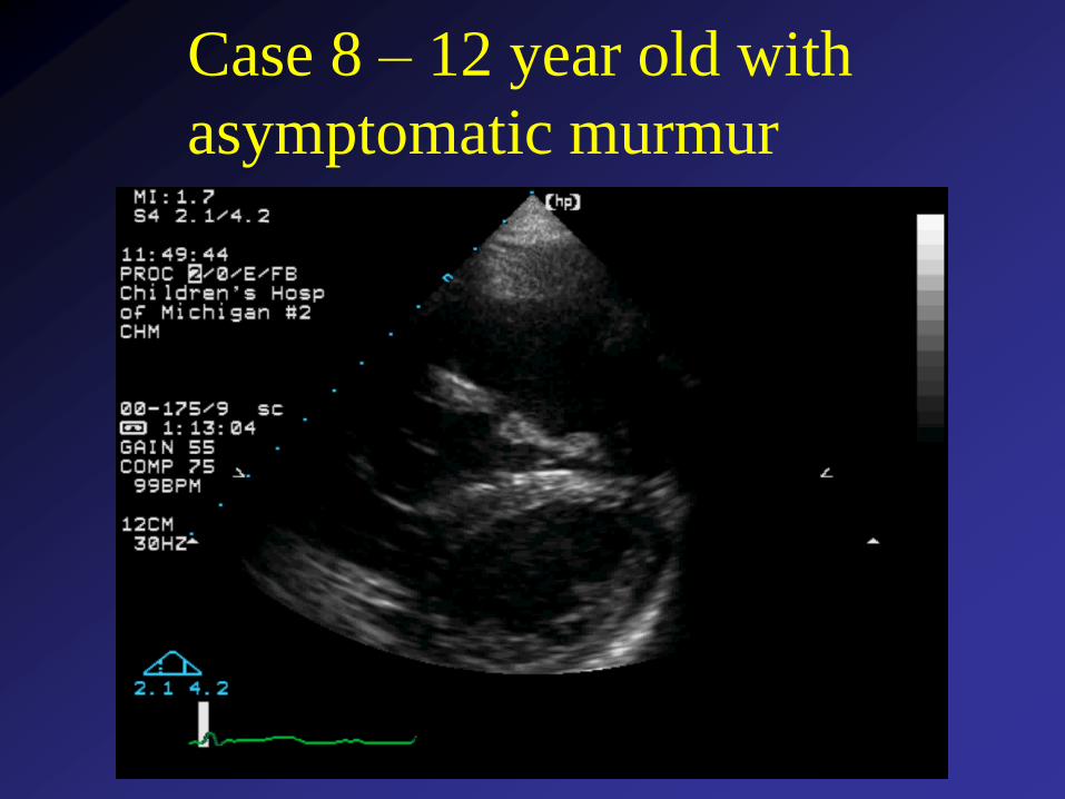
Case 8 – 12 year old with
asymptomatic murmur
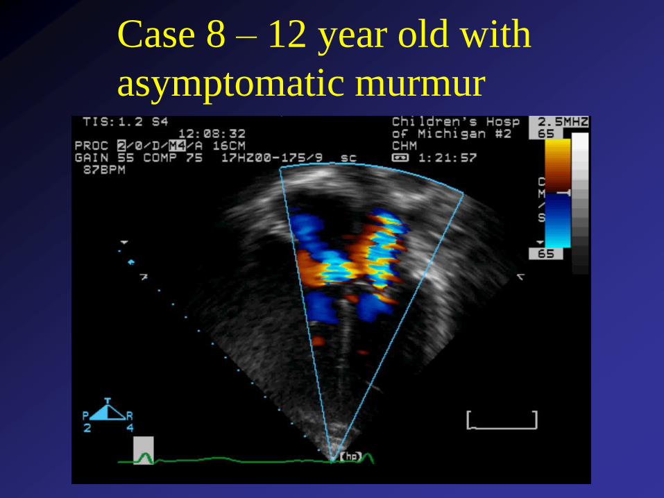
Case 8 – 12 year old with
asymptomatic murmur
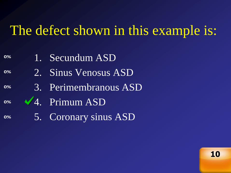
The defect shown in this example is:
0%
0%
0%
0%
0% 1. Secundum ASD
2. Sinus Venosus ASD
3. Perimembranous ASD
4. Primum ASD
5. Coronary sinus ASD
Countdown
10
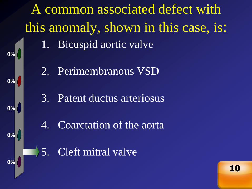
A common associated defect with
this anomaly, shown in this case, is:
0%
0%
0%
0%
0%
1. Bicuspid aortic valve
2. Perimembranous VSD
3. Patent ductus arteriosus
4. Coarctation of the aorta
5. Cleft mitral valve
Countdown
10
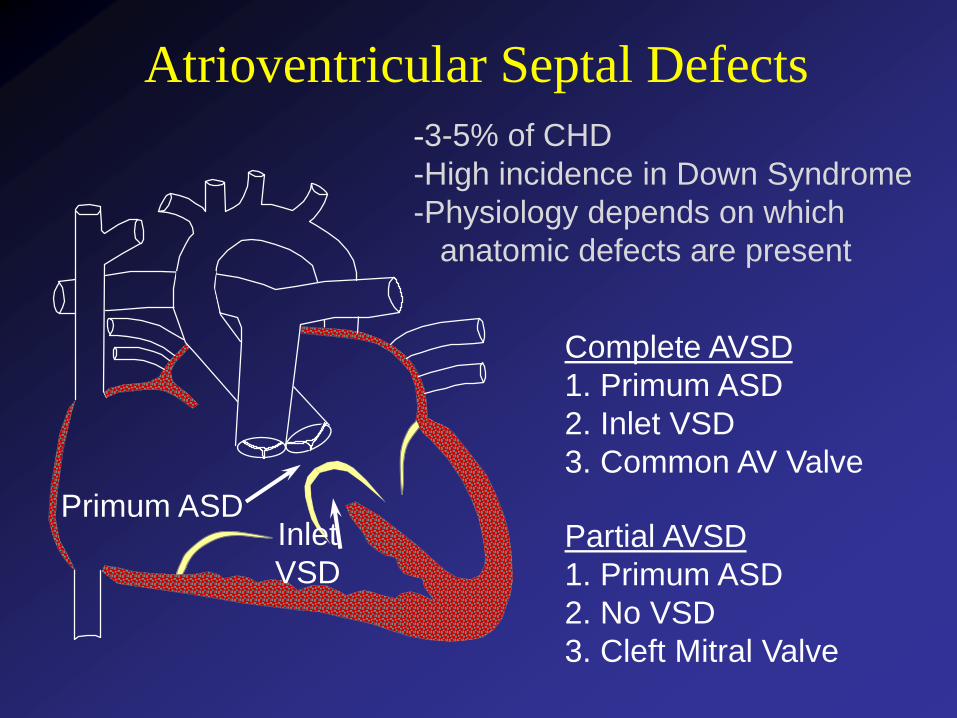
Atrioventricular Septal Defects
Primum ASD
Complete AVSD
1. Primum ASD
2. Inlet VSD
3. Common AV Valve
Partial AVSD
1. Primum ASD
2. No VSD
3. Cleft Mitral Valve
Inlet
VSD
-3-5% of CHD
-High incidence in Down Syndrome
-Physiology depends on which
anatomic defects are present
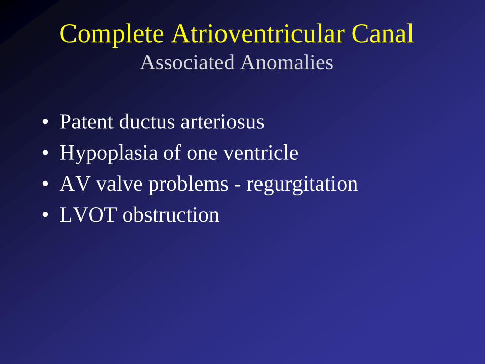
Complete Atrioventricular CanalAssociated Anomalies
• Patent ductus arteriosus
• Hypoplasia of one ventricle
• AV valve problems - regurgitation
• LVOT obstruction
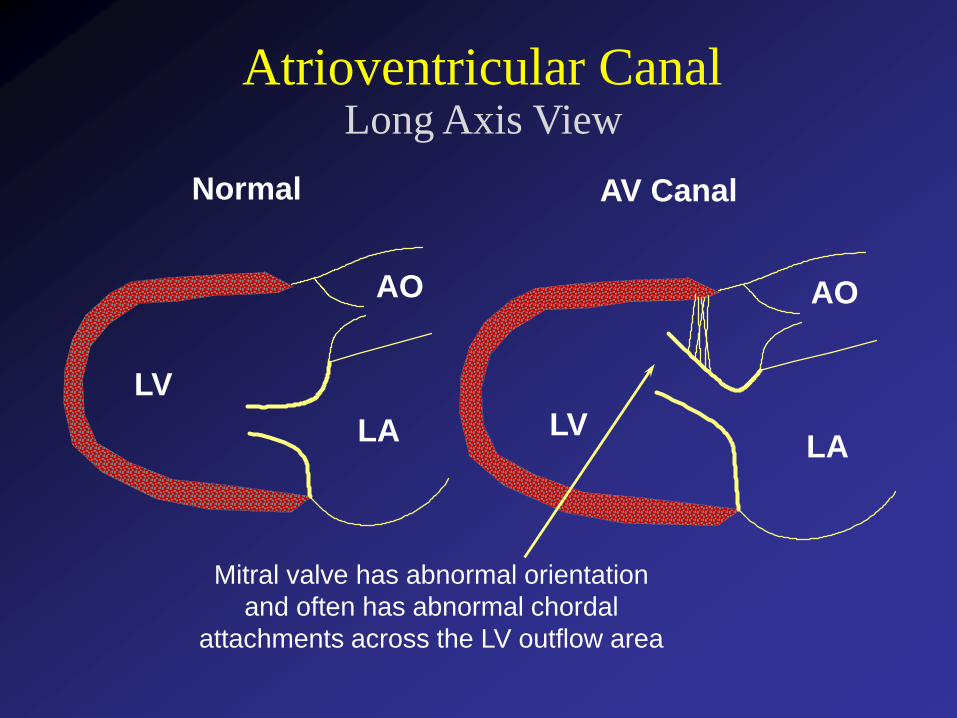
Atrioventricular CanalLong Axis View
LV
LA
AO
LVLA
AO
Normal AV Canal
Mitral valve has abnormal orientation
and often has abnormal chordal
attachments across the LV outflow area
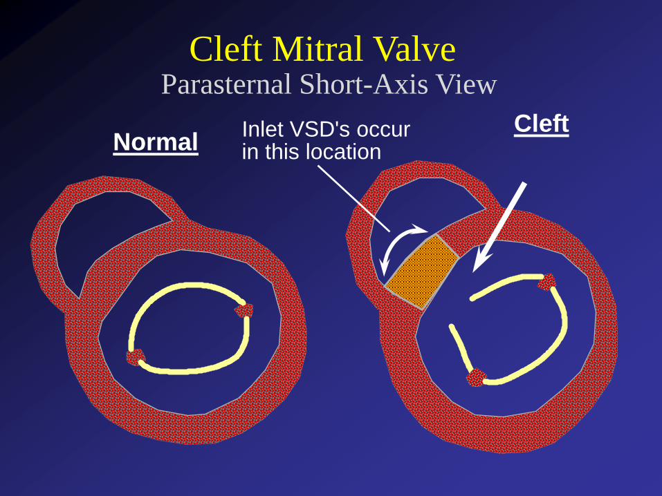
Cleft Mitral ValveParasternal Short-Axis View
NormalCleftInlet VSD's occur
in this location
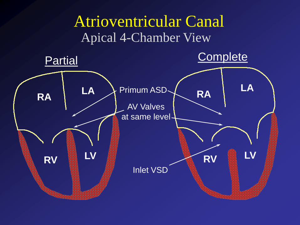
Apical 4-Chamber ViewAtrioventricular Canal
RA
RV
LA
LV
RA
RV
LA
LV
Primum ASD
Inlet VSD
Partial Complete
AV Valves
at same level
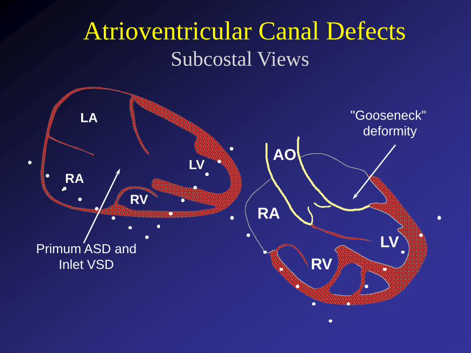
Atrioventricular Canal DefectsSubcostal Views
LA
RALV
RV
LV
RV
RA
AO
"Gooseneck"
deformity
Primum ASD and
Inlet VSD
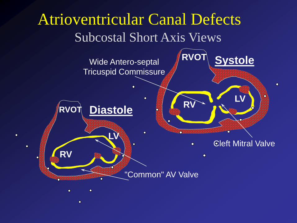
Subcostal Short Axis Views
Atrioventricular Canal Defects
Diastole
Systole
RV
LV
RVOTRV
LV
RVOT
Cleft Mitral Valve
Wide Antero-septal
Tricuspid Commissure
"Common" AV Valve
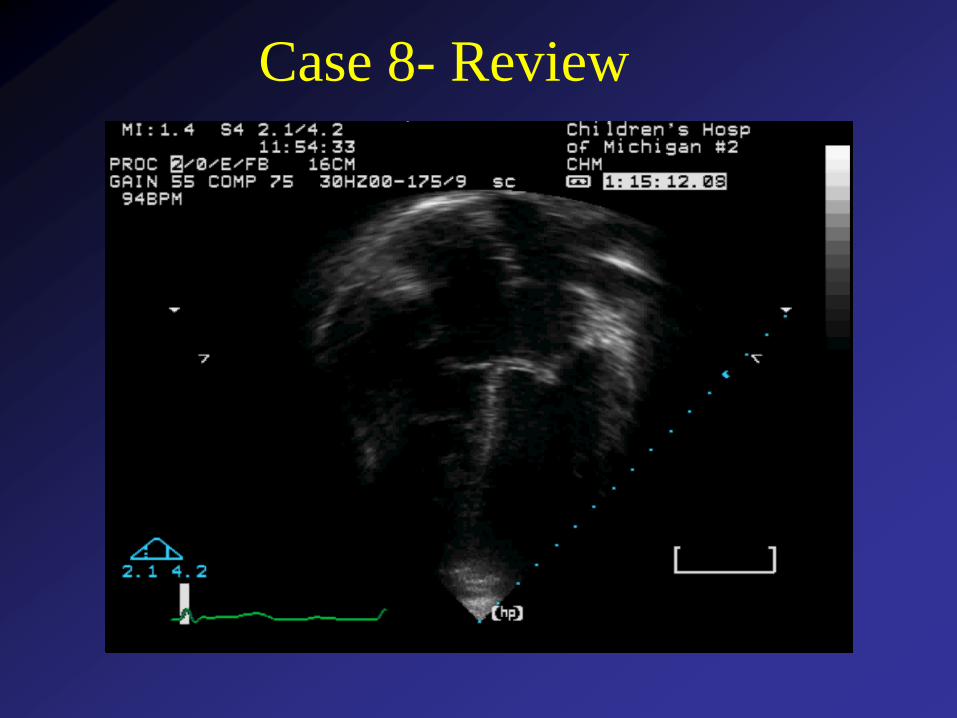
Case 8- Review

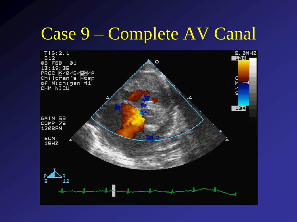
Case 9 – Complete AV Canal
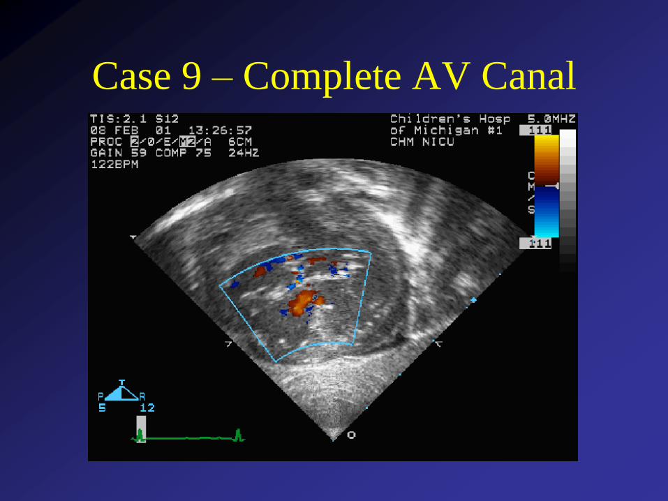
Case 9 – Complete AV Canal
AV Septal DefectsPhysiology
• Physiology dependent on which components
of AV septal defect are present
• If 1° ASD and no VSD - physiology similar to
isolated ASD (right sided volume overload)
• Complete AVSD - marked volume and
pressure overload (VSD shunt physiology)
• AV valve regurgitation may exacerbate
volume overload and symptoms of heart
failure
AV Septal DefectsSurgical Intervention
• Partial AVSD
Usually electively repaired age 2-4 years
Complicating features (AVV regurg.,
LVOTO) may necessitate earlier
intervention
• Complete AVSD
Usually repaired by 6 months of age
(earlier in trisomy 21) to prevent pulmonary
vascular obstructive disease

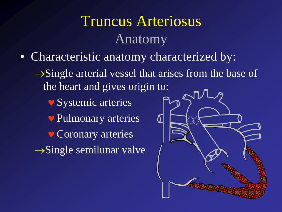
Truncus ArteriosusAnatomy
• Characteristic anatomy characterized by:
Single arterial vessel that arises from the base of
the heart and gives origin to:
Systemic arteries
Pulmonary arteries
Coronary arteries
Single semilunar valve
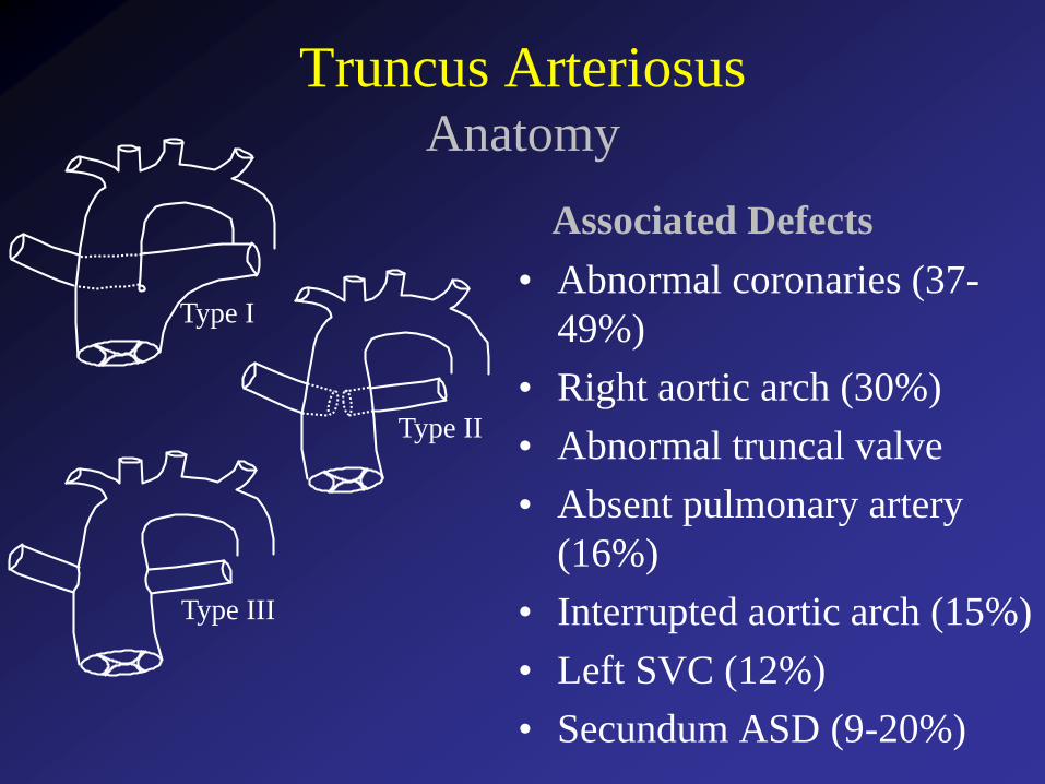
Type I
Truncus ArteriosusAnatomy
Associated Defects
• Abnormal coronaries (37-
49%)
• Right aortic arch (30%)
• Abnormal truncal valve
• Absent pulmonary artery
(16%)
• Interrupted aortic arch (15%)
• Left SVC (12%)
• Secundum ASD (9-20%)
Type II
Type III
Truncus ArteriosusClinical Aspects
• Patients usually present due to the presence
of a cardiac murmur
• Complete mixing of systemic and
pulmonary venous blood results in cyanosis
• Excessive pulmonary blood flow causes
sign and symptoms of congestive heart
failure
• The cyanosis is generally mild
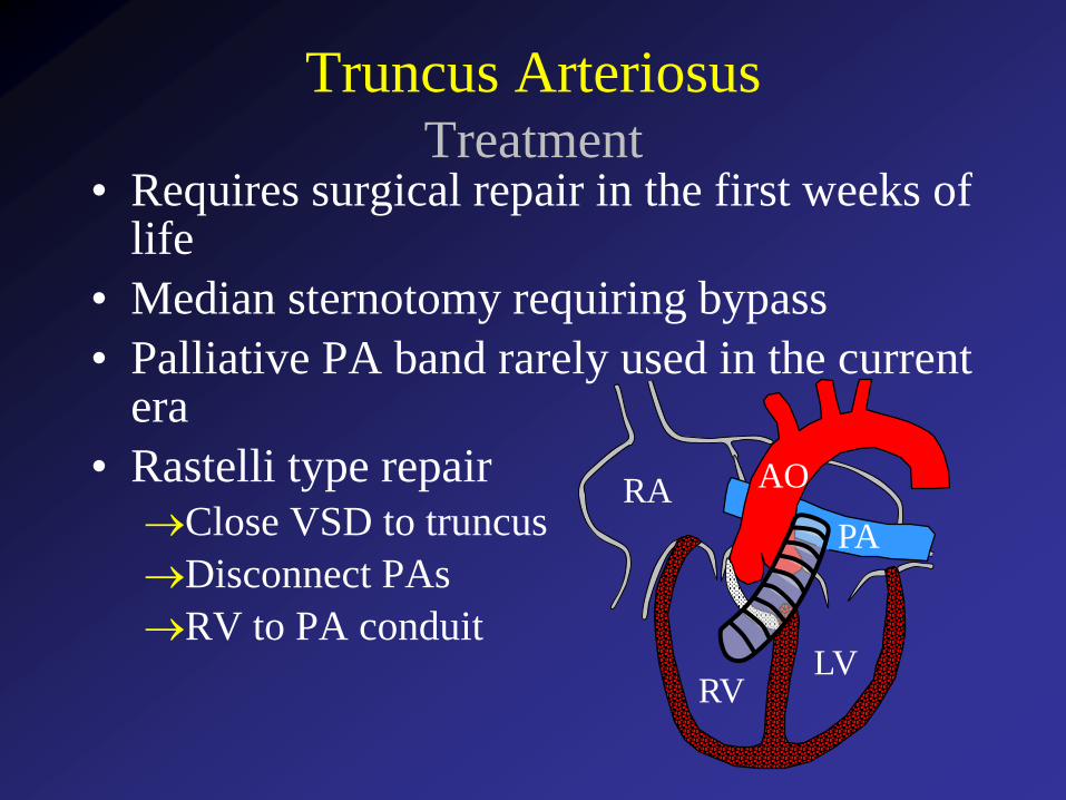
Truncus ArteriosusTreatment
• Requires surgical repair in the first weeks of life
• Median sternotomy requiring bypass
• Palliative PA band rarely used in the current era
• Rastelli type repairClose VSD to truncus
Disconnect PAs
RV to PA conduit
RVLV
RA AO
PA
Case 10Truncus Arteriosus
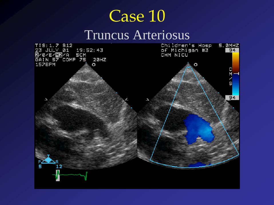
Case 10Truncus Arteriosus

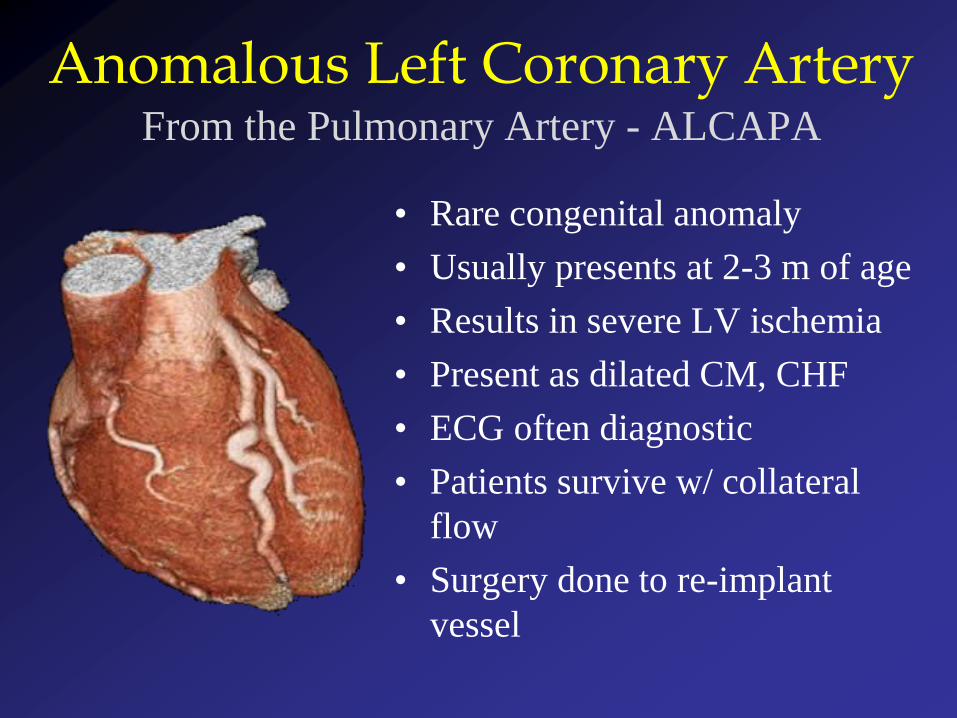
Anomalous Left Coronary ArteryFrom the Pulmonary Artery - ALCAPA
• Rare congenital anomaly
• Usually presents at 2-3 m of age
• Results in severe LV ischemia
• Present as dilated CM, CHF
• ECG often diagnostic
• Patients survive w/ collateral
flow
• Surgery done to re-implant
vessel
Anomalous Left Coronary ArteryEchocardiographic Clues
• Left ventricular dysfunction (usually severe)
• Mitral insufficiency – due to LV
dilation/dysfunction, papillary muscle infarction
• Endocardial fibroelastosis of LV and/or papillary
muscles
• Failure to identify proximal LCA from aorta
• Unusual flow into main pulmonary artery
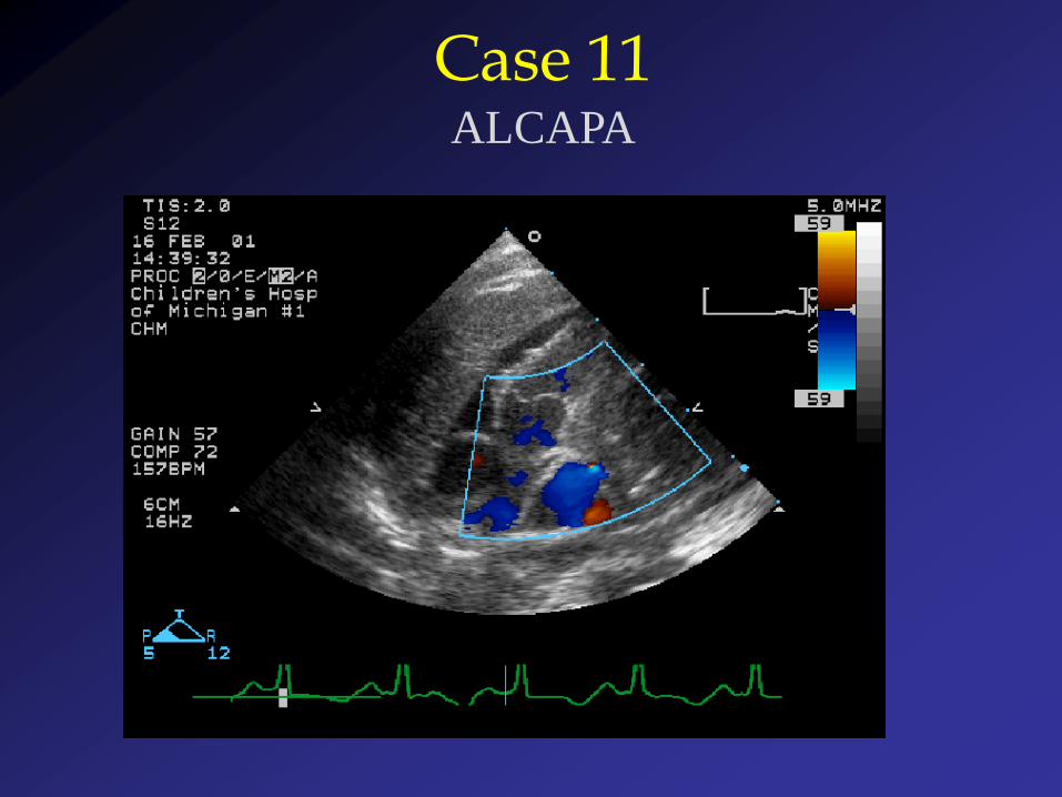
Case 11ALCAPA

RA
RV
LV
MPAAO
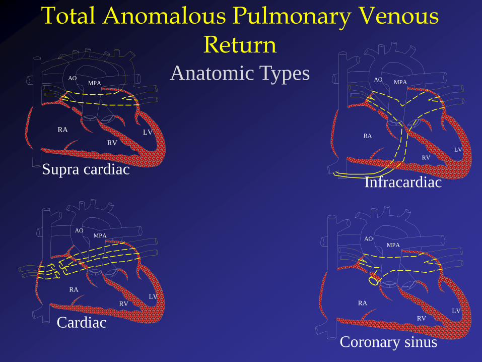
InfracardiacSupra cardiac
RA
RV
LV
MPAAO
Coronary sinus
RA
RV
LV
MPAAO
Cardiac
RA
RVLV
MPAAO
Total Anomalous Pulmonary Venous Return
Anatomic Types
Total Anomalous Pulmonary Venous Return
Echo Clues
• Enlarged right heart
• Right to left atrial shunting
• Unusual “membranes” in left atrium
• Abnormal flow in systemic venous system
• Obstruction may occur at different levels
Most common - infracardiac
• May be remarkably asymptomatic (in
absence of obstruction)
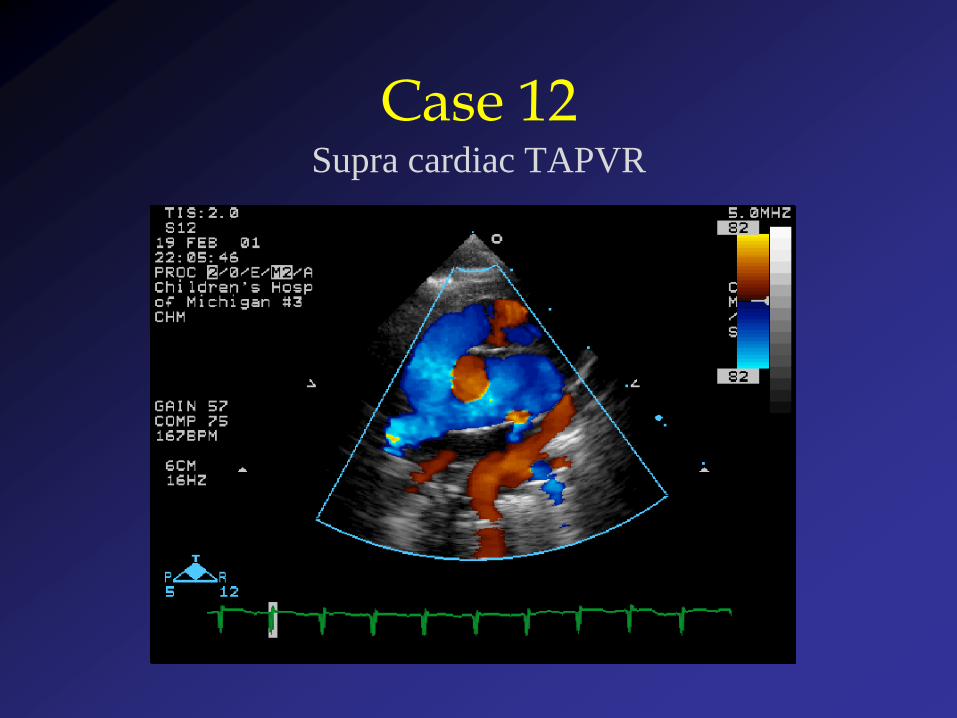
Case 12Supra cardiac TAPVR