Embed Size (px)
DESCRIPTION
Conduction System of the Heart 4. Faisal I. Mohammed, MD, PhD. Objectives. List the parts that comprise the conduction system Explain the mechanism of slow response action potential (pacemaker potential) Point out the regulation of the conduction system potential by Autonomic Nerves - PowerPoint PPT Presentation
Citation preview
1
Conduction System of the Heart4
Faisal I. Mohammed, MD, PhD
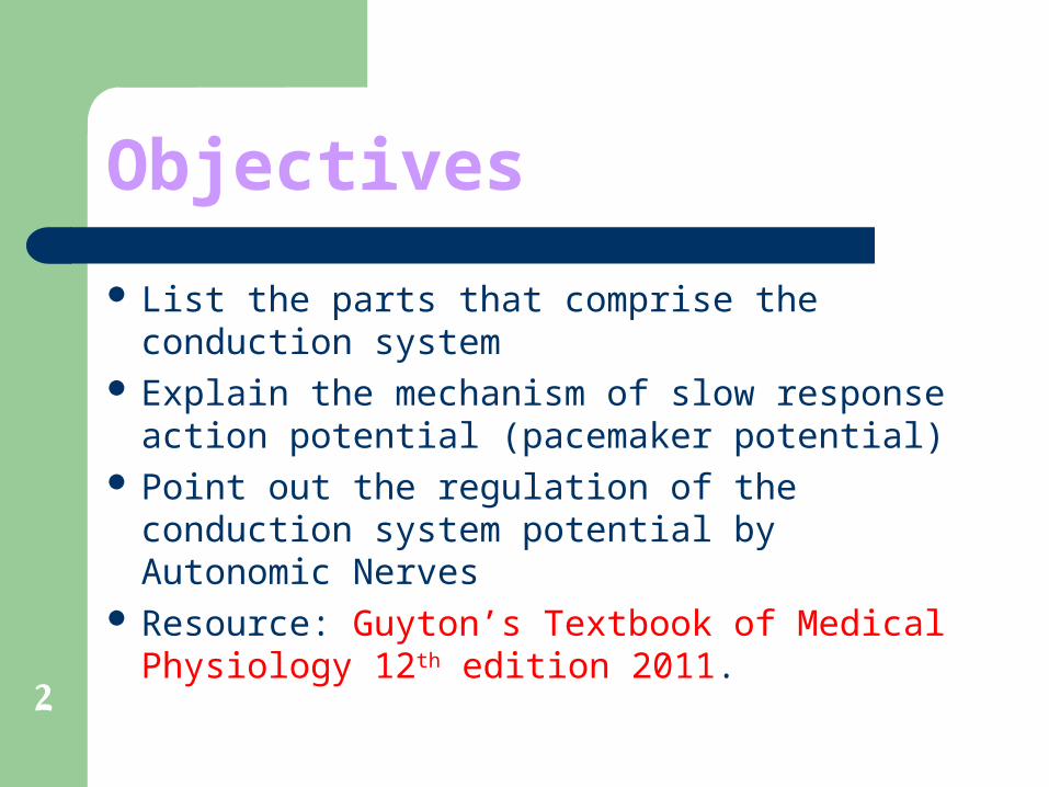
2
Objectives
List the parts that comprise the conduction system Explain the mechanism of slow response action
potential (pacemaker potential) Point out the regulation of the conduction system
potential by Autonomic Nerves Resource: Guyton’s Textbook of Medical
Physiology 12th edition 2011.
3
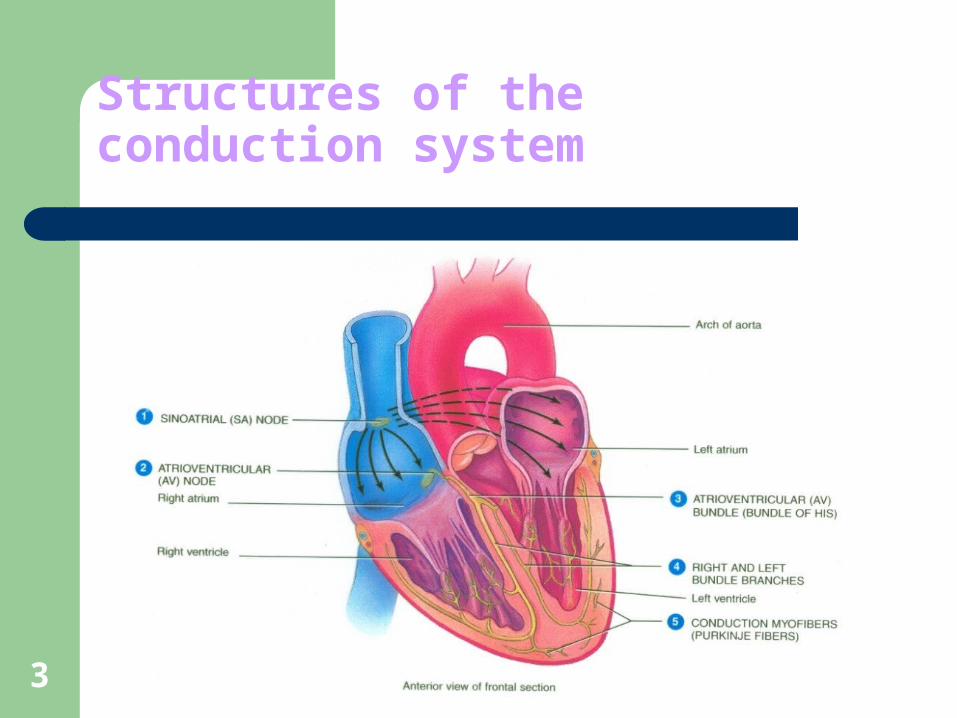
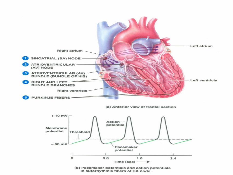
Structures of the conduction system
4
5
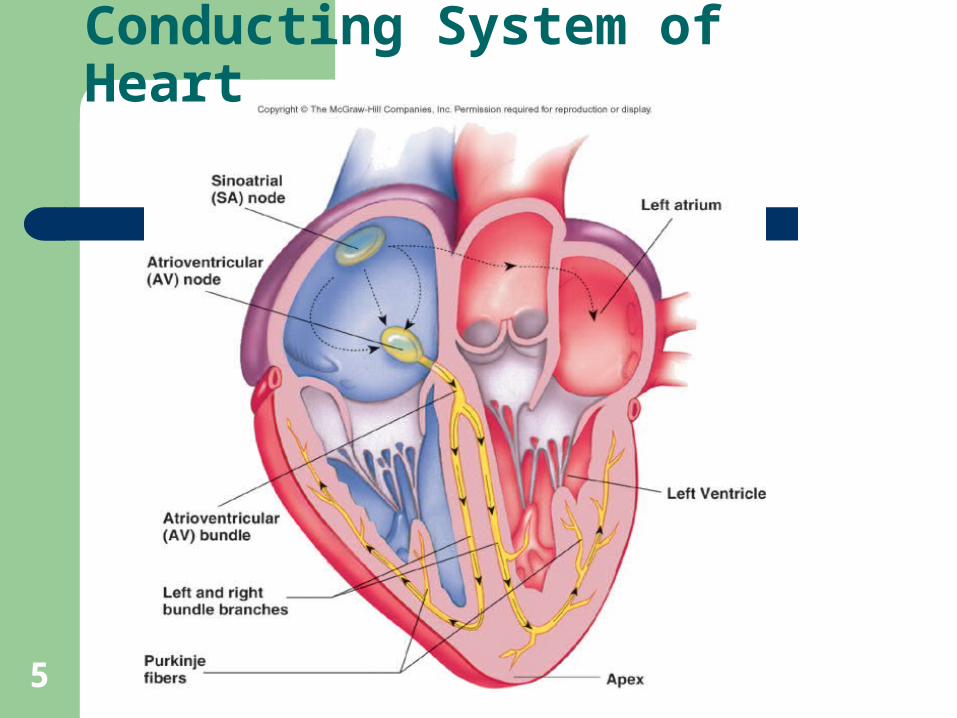
Conducting System of Heart
6
Heart Physiology: Sequence of Excitation
7
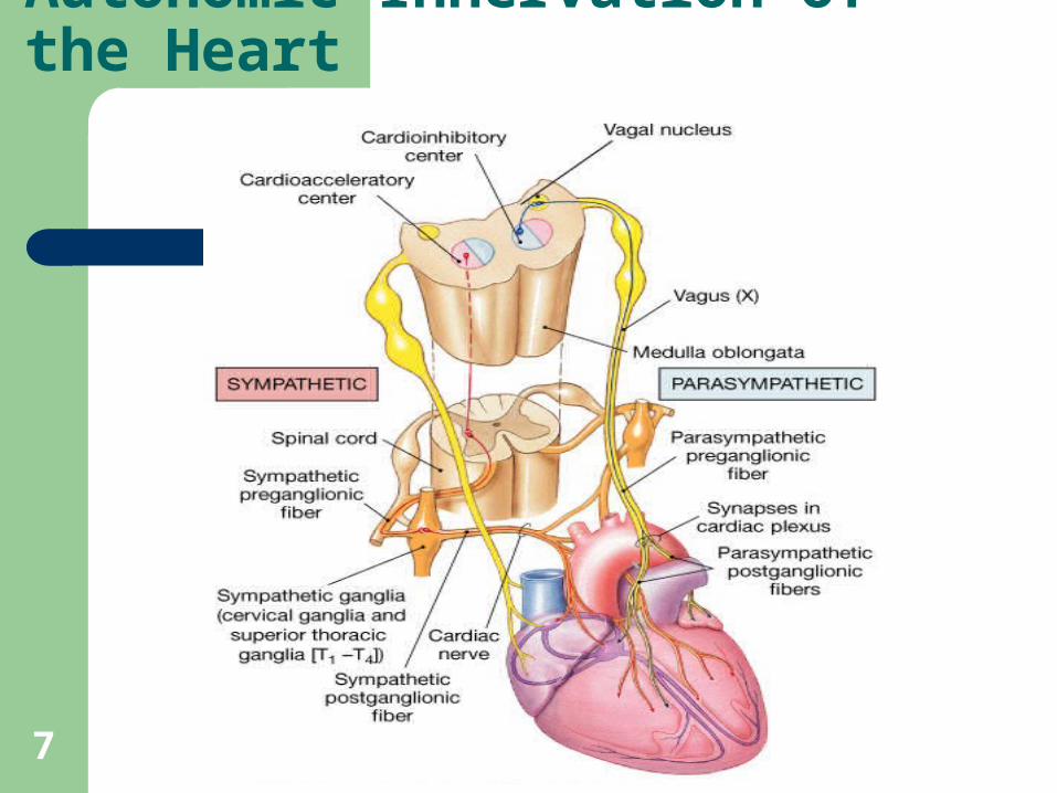
Autonomic Innervation of the Heart
8
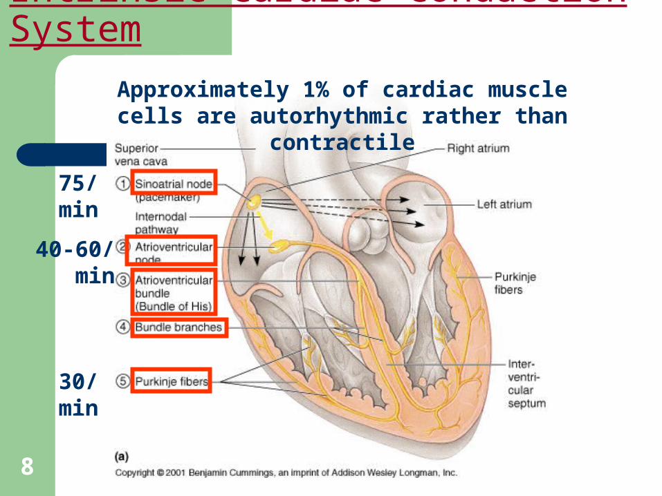
Intrinsic Cardiac Conduction System
Approximately 1% of cardiac muscle cells are autorhythmic rather than contractile
75/min
40-60/min
30/min
9
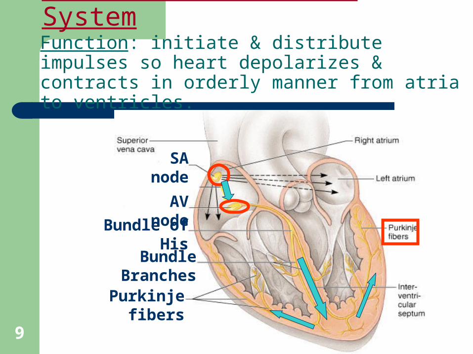
Intrinsic Conduction SystemFunction: initiate & distribute impulses so heart depolarizes & contracts in orderly manner from atria to ventricles.
SA node
AV node
Bundle of His
Bundle Branches
Purkinje fibers
10
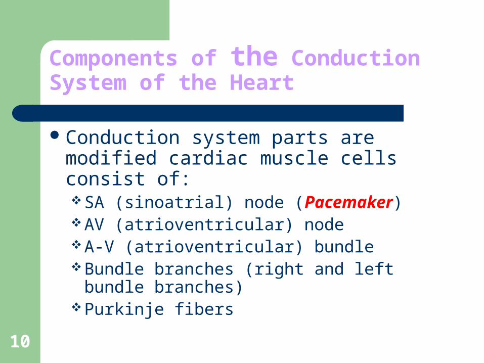
Components of the Conduction System of the Heart
Conduction system parts are modified cardiac muscle cells consist of:SA (sinoatrial) node (Pacemaker)AV (atrioventricular) nodeA-V (atrioventricular) bundleBundle branches (right and left bundle
branches)Purkinje fibers
11
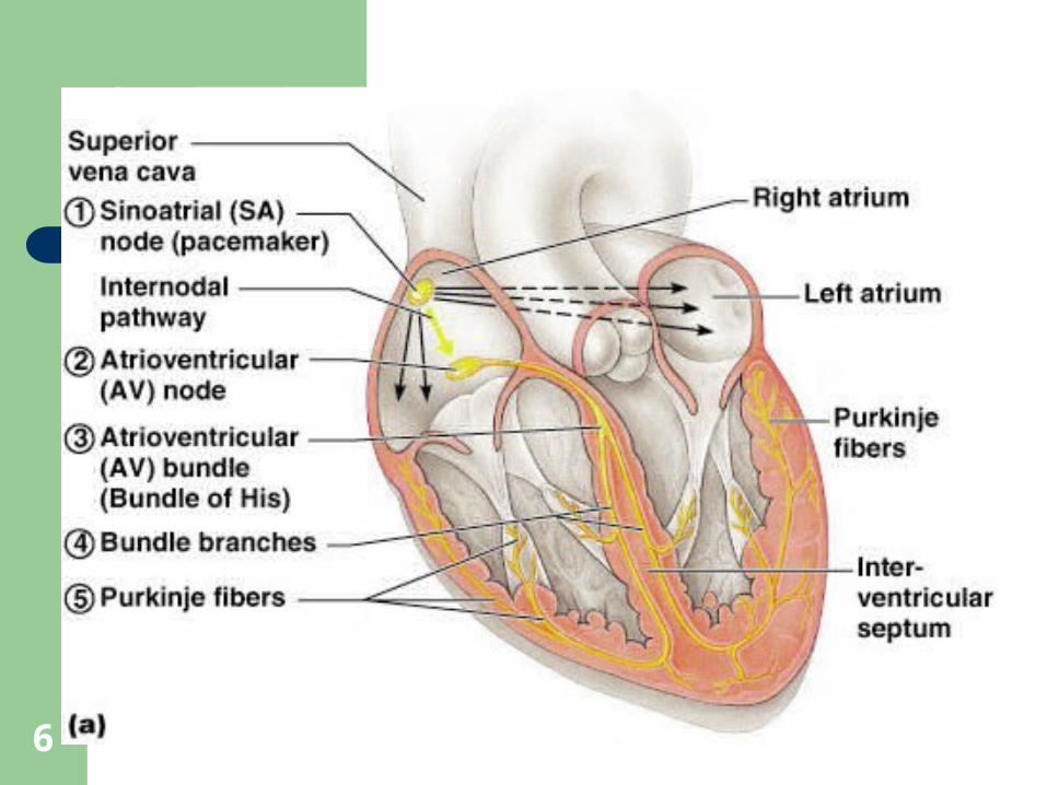
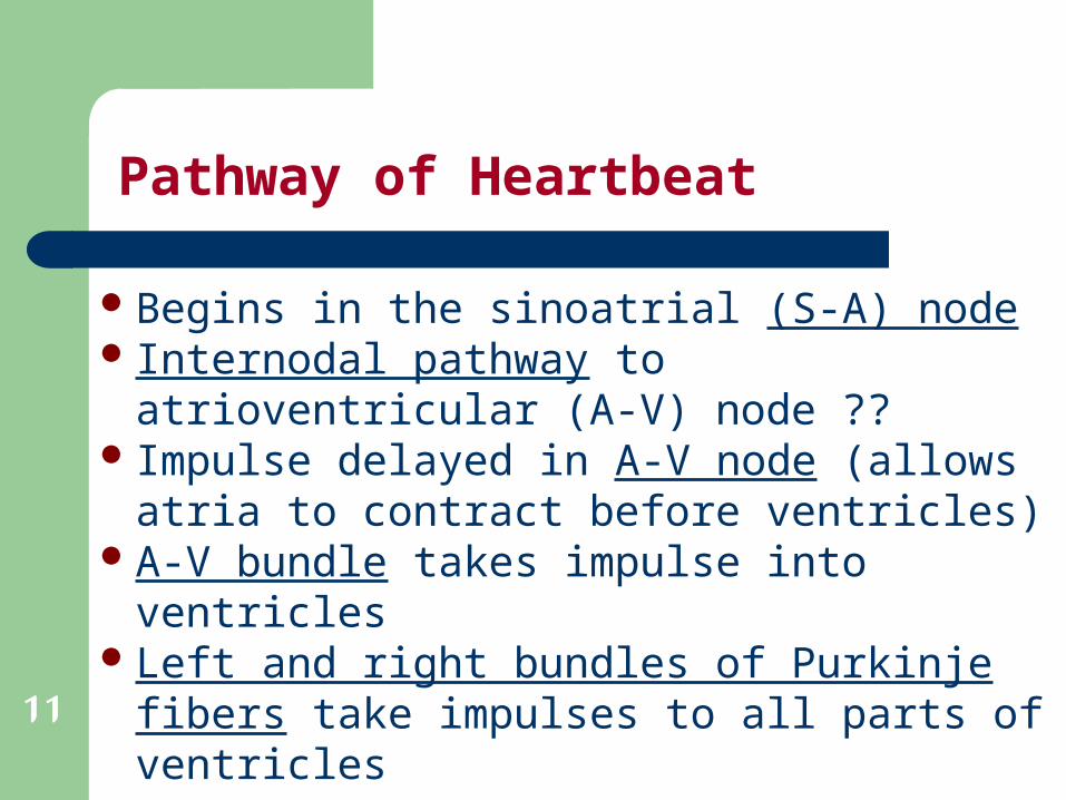
Pathway of Heartbeat
Begins in the sinoatrial (S-A) nodeInternodal pathway to atrioventricular (A-V)
node ?? Impulse delayed in A-V node (allows atria to
contract before ventricles)A-V bundle takes impulse into ventriclesLeft and right bundles of Purkinje fibers take
impulses to all parts of ventricles
12
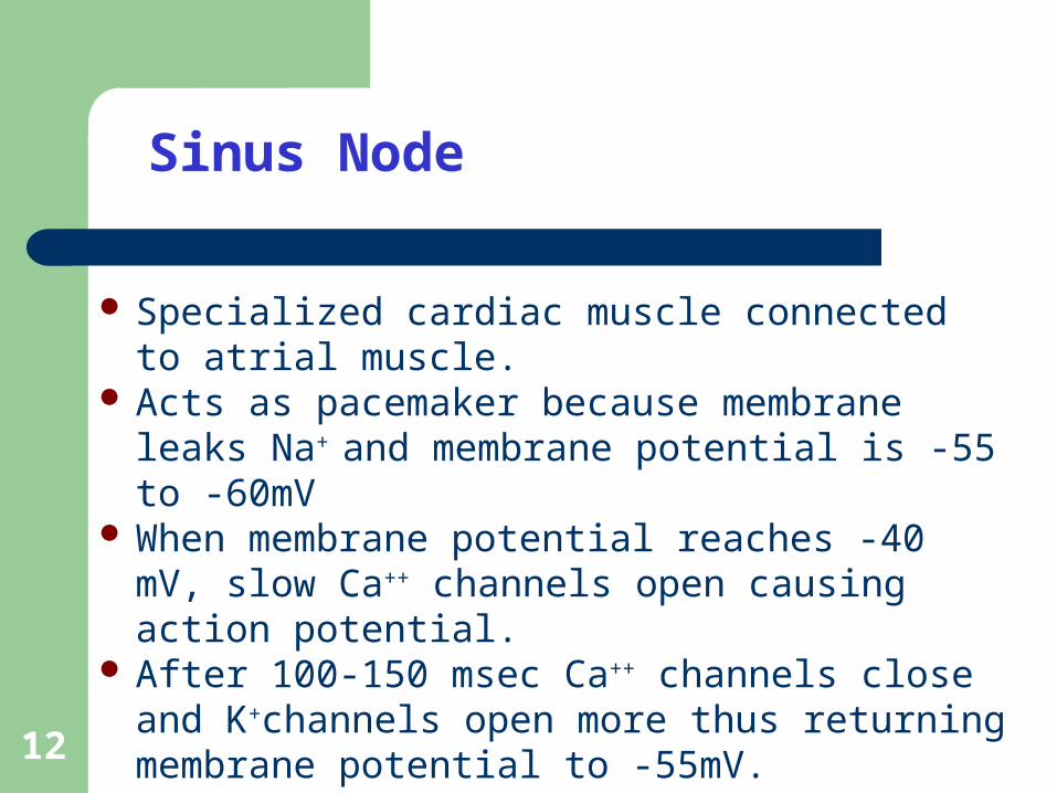
Sinus Node
Specialized cardiac muscle connected to atrial muscle.
Acts as pacemaker because membrane leaks Na+ and membrane potential is -55 to -60mV
When membrane potential reaches -40 mV, slow Ca++ channels open causing action potential.
After 100-150 msec Ca++ channels close and K+channels open more thus returning membrane potential to -55mV.
13
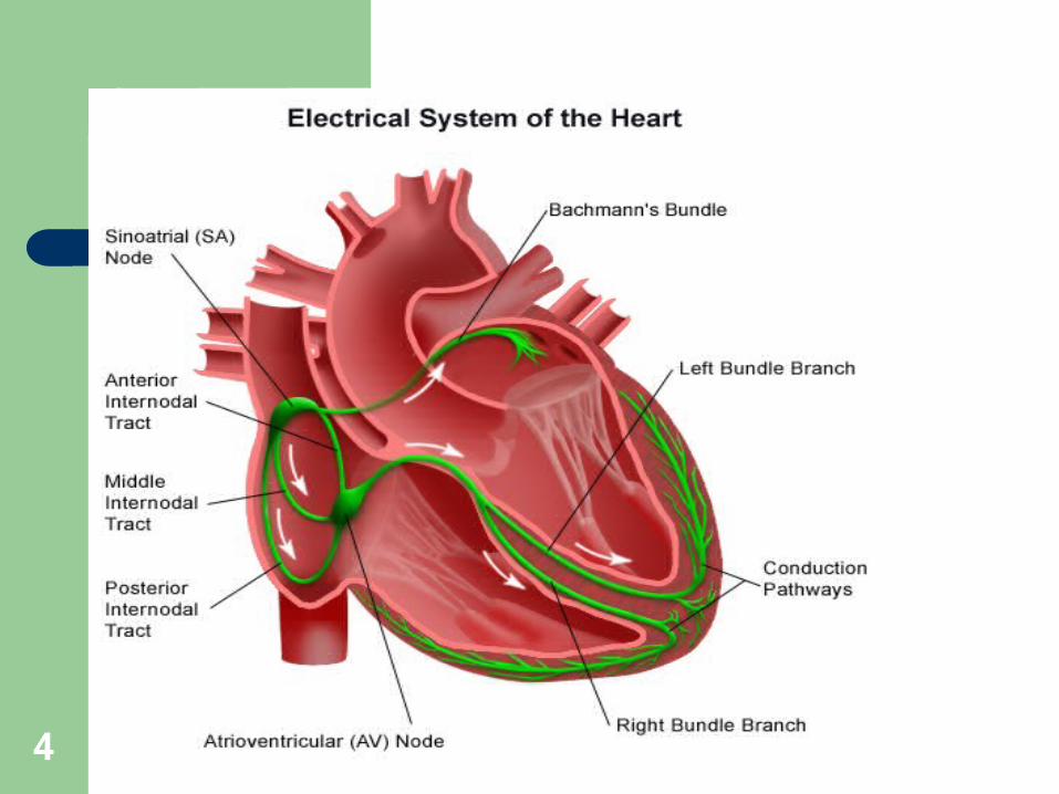
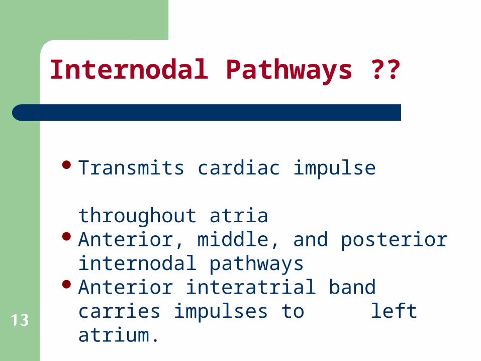
Internodal Pathways ??
Transmits cardiac impulse throughout atriaAnterior, middle, and posterior internodal
pathwaysAnterior interatrial band carries impulses to
left atrium.
14
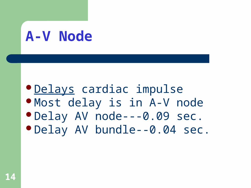
A-V Node
Delays cardiac impulseMost delay is in A-V nodeDelay AV node---0.09 sec.Delay AV bundle--0.04 sec.
15
Purkinje System
Fibers lead from A-V node through A-V bundle into Ventricles
Fast conduction; many gap junctions at intercalated disks
16
A-V Bundles
Normally one-way conduction through the bundles
Only conducting path between atria and ventricles is A-V node - A-V bundle
Divides into left and right bundlesTransmission time between A-V
bundles and last of ventricular fibers is 0.06 second (QRS time)
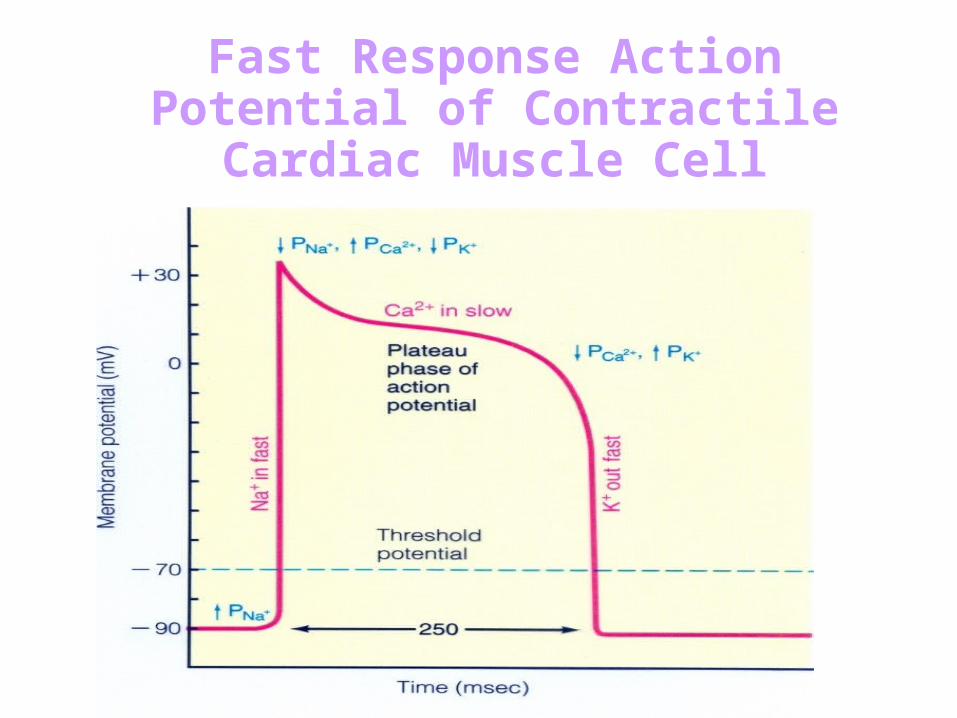
Fast Response Action Potential of Contractile Cardiac Muscle Cell
20
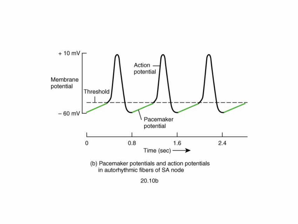
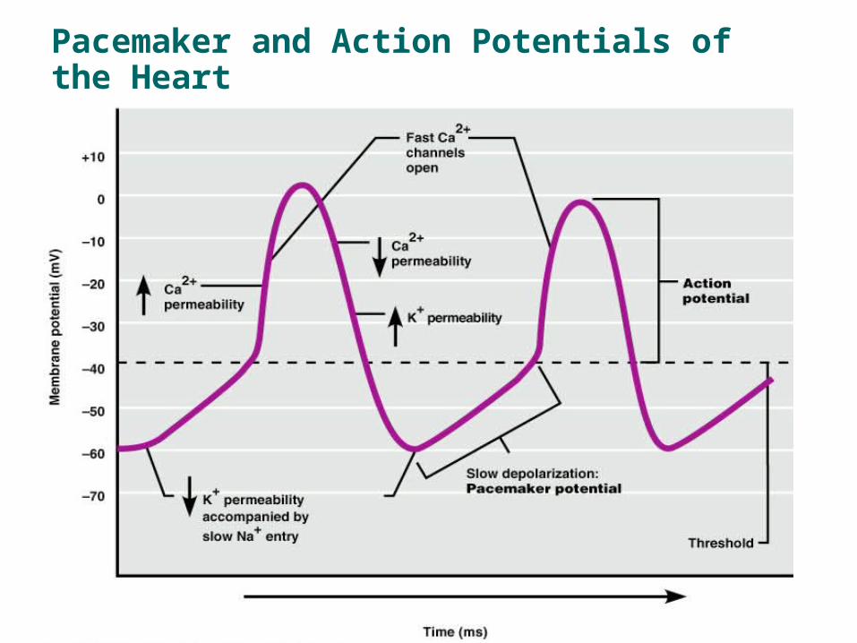
Pacemaker and Action Potentials of the Heart
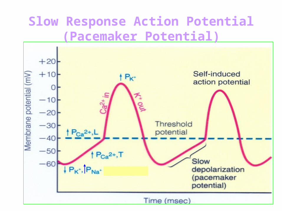
Slow Response Action Potential (Pacemaker Potential)
22
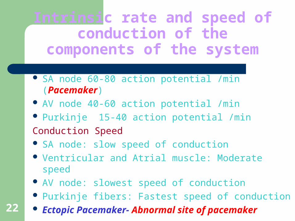
Intrinsic rate and speed of conduction of the components of the system
SA node 60-80 action potential /min (Pacemaker) AV node 40-60 action potential /min Purkinje 15-40 action potential /min
Conduction Speed SA node: slow speed of conduction Ventricular and Atrial muscle: Moderate speed AV node: slowest speed of conduction Purkinje fibers: Fastest speed of conduction Ectopic Pacemaker- Abnormal site of pacemaker
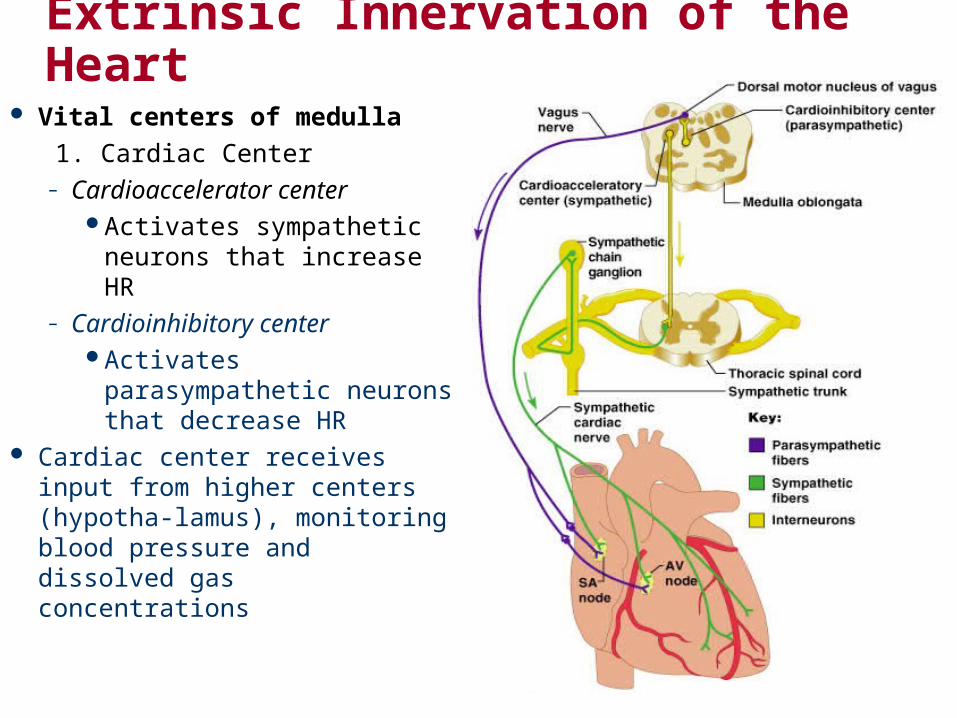
Extrinsic Innervation of the Heart
Vital centers of medulla
1. Cardiac Center– Cardioaccelerator center
Activates sympathetic neurons that increase HR
– Cardioinhibitory centerActivates parasympathetic
neurons that decrease HR Cardiac center receives input
from higher centers (hypotha-lamus), monitoring blood pressure and dissolved gas concentrations
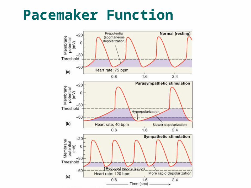
Pacemaker Function
Sympathetic – increases heart rate by Ca+2 & If channel (net Na+) flow
Parasympathetic – decreases rate by K+ efflux & Ca+2 influx
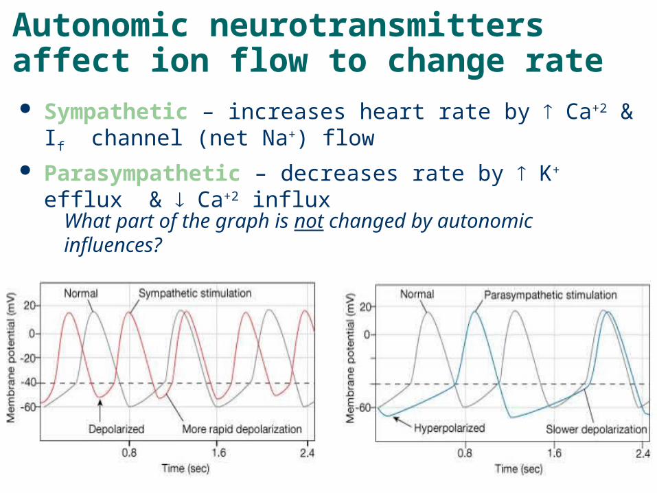
Autonomic neurotransmitters affect ion flow to change rate
What part of the graph is not changed by autonomic influences?
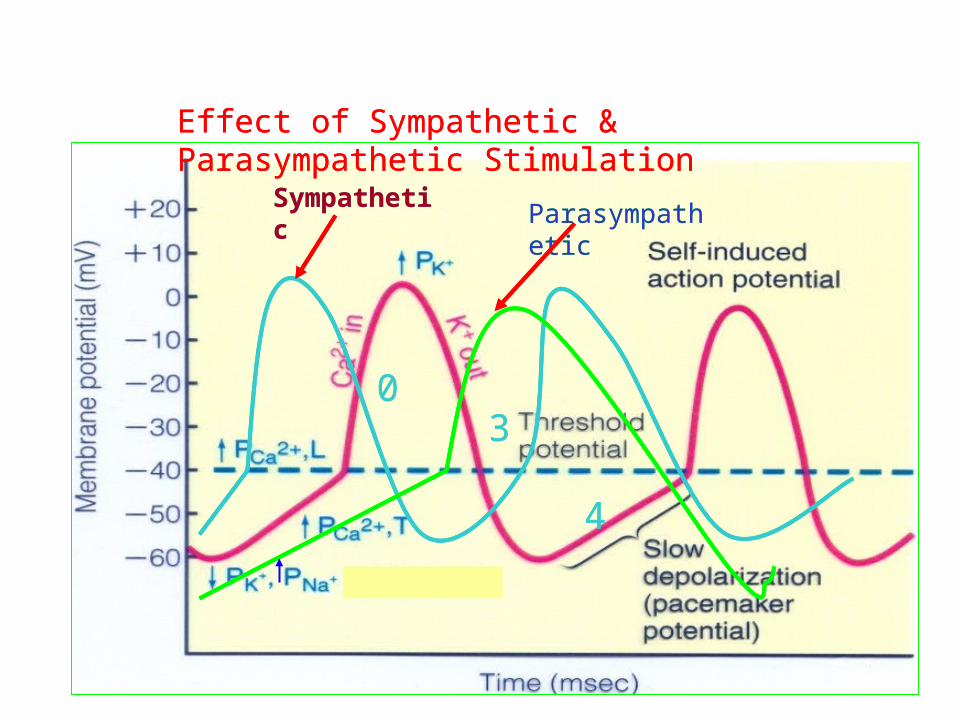
Effect of Sympathetic & Parasympathetic Stimulation
03
4
SympatheticParasympathetic
Effect of Sympathetic & Parasympathetic Stimulation
03
4
SympatheticParasympathetic
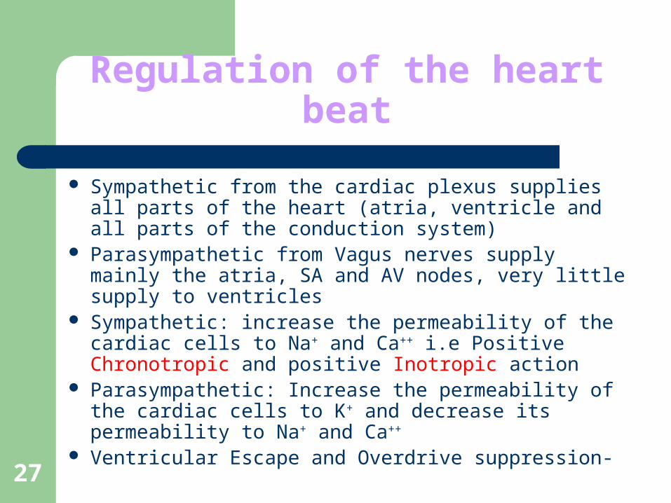
27
Regulation of the heart beat
Sympathetic from the cardiac plexus supplies all parts of the heart (atria, ventricle and all parts of the conduction system)
Parasympathetic from Vagus nerves supply mainly the atria, SA and AV nodes, very little supply to ventricles
Sympathetic: increase the permeability of the cardiac cells to Na+ and Ca++ i.e Positive Chronotropic and positive Inotropic action
Parasympathetic: Increase the permeability of the cardiac cells to K+ and decrease its permeability to Na+ and Ca++
Ventricular Escape and Overdrive suppression-
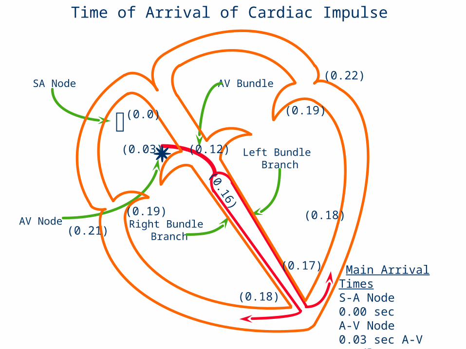
SA Node
AV Node
AV Bundle
Left Bundle Branch
Right Bundle Branch
(0.0)
(0.03) (0.12)
(0.19)
(0.21)
(0.22)
(0.19)
(0.18)
(0.17)
(0.18)
(0.16)
Time of Arrival of Cardiac Impulse
Main Arrival TimesS-A Node 0.00 secA-V Node 0.03 sec A-V Bundle 0.12 sec VentricularSeptum 0.16 sec
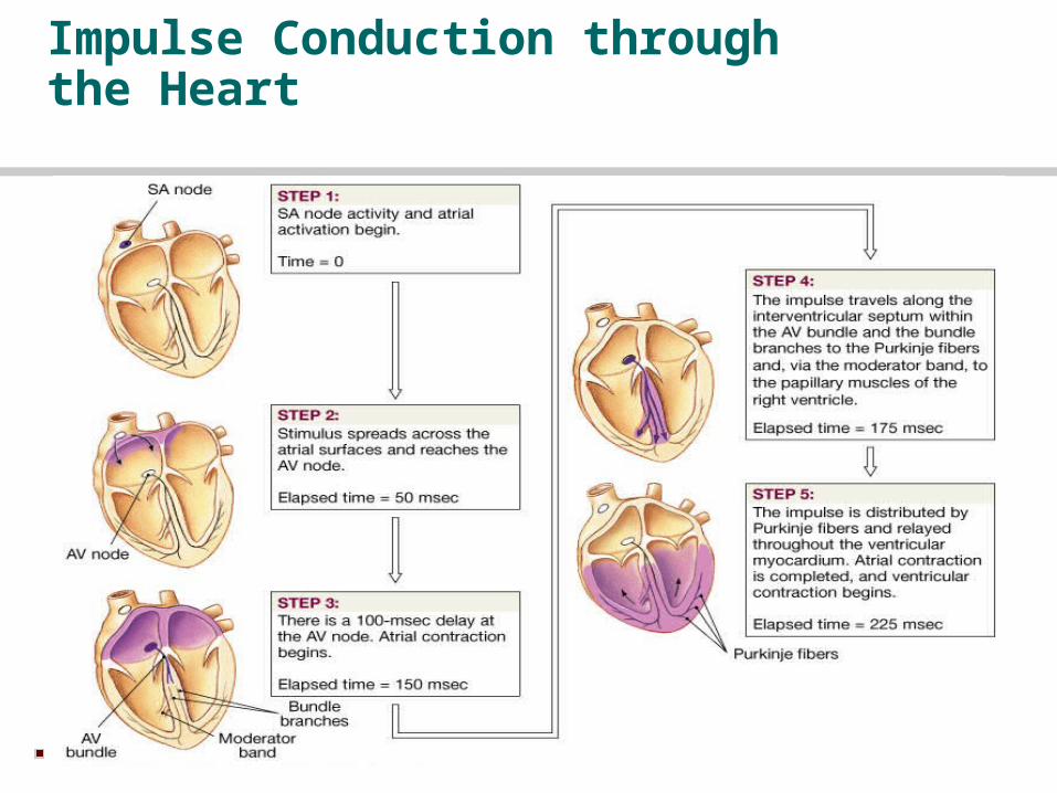
Impulse Conduction through the Heart
30
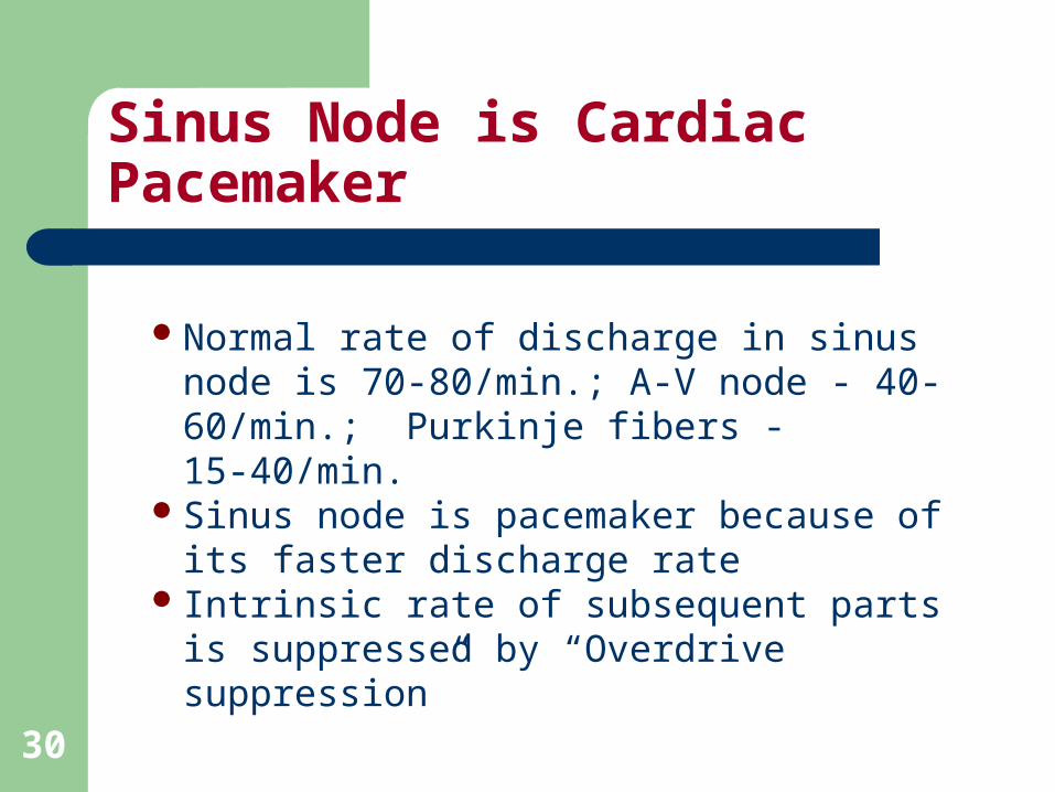
Sinus Node is Cardiac Pacemaker
Normal rate of discharge in sinus node is 70-80/min.; A-V node - 40-60/min.; Purkinje fibers - 15-40/min.
Sinus node is pacemaker because of its faster discharge rate
Intrinsic rate of subsequent parts is suppressed by “Overdrive suppression”
31
Ectopic Pacemaker
This is a portion of the heart with a more rapid discharge than the sinus node.
Also occurs when transmission from sinus node to A-V node is blocked (A-V block).
32
Ectopic Pacemaker (cont’d)
During sudden onset of A-V block, sinus node discharge does not get through, and next fastest area of discharge becomes pacemaker of heart beat (Purkinje system).
Delay in pickup of the heart beat is the “Stokes-Adams” syndrome. New pacemaker is in A-V node or penetrating part of A-V bundle.
33
Parasympathetic Effects on Heart Rate
Parasympathetic (vagal) nerves, which release acetylcholine at their endings, innervate S-A node and A-V junctional fibers proximal to A-V node.
Causes hyperpolarization because of increased K+ permeability in response to acetylcholine.
This causes decreased transmission of impulses maybe temporarily stopping heart rate.
Ventricular escape occurs.
34
Sympathetic Effects on Heart Rate
Releases norepinephrine at sympathetic ending
Causes increased sinus node discharge (Chronotropic effect)
Increases rate of conduction of impulse (Dromotropic effect)
Increases force of contraction in atria and ventricles (Inotropic effect)

Thank YouThank You