Embed Size (px)
Citation preview
Br Heart 1984; 52: 484-9
Conditions for effective Nd-YAG laser angioplasty
HERBERT J GESCHWIND, GEORGES BOUSSIGNAC, BERNARD TEISSEIRE,
NICOLE BENHAIEM, RENEE BITTOUN, DANIEL LAURENT
From the Deparnments of Clinical Investigation and Histopatology, University Hospital Henri-Mondor, Creteil,France
SUMMARY To establish the optimal conditions for recanalisation of obstructed arteries withoutdamage to vessel walls, a Nd-YAG laser coupled to a 0.2 mm diameter optic fibre was used on
obstructed human cadaver coronary and peripheral arteries and on popliteal arteries in amputatedlimbs. Vaporisation of atheromatous plaques was consistently obtained with an energy of 360-600 J
and a diluted blood perfusate (3 g/100 ml haemoglobin) at a rate of 20 mi/ The arterial wall was
protected from thermal injury by inserting the optic fibre into an inflated balloon catheter and bycooling the system with the perfusate. Since recanalisation of occluded arteries was consistentlyobtained without damage to the arterial wall or debris and thin and flexible optic fibres were easy to
guide in the arteries, perutaneous transluminal Nd-YAG laser angioplasty was used in obstructedfemoral and popliteal arteries in three patients.The first European trials in man showed the method to be feasible, effective, and harmless,
although further studies are required to improve penetration of the obstruction and increase thediameter of tunnel.
Lasers have been widely used in the treatment of dis-eases in several areas of medical practice. This techni-que has not, however, yet been applied in diseases ofthe heart and blood vessels. Experimental studieshave recently been undertaken to assess the efficacy oflaser radiation for removing obstructions in testtubes,' in human cadaver arteries,2 or in animalarteries.3 The obstructions were either thrombi oratheromatous plaques. The sources of laser that havebeen used comprised carbon dioxide, Nd-YAG(neodymium yttrium, aluminium, garnet), andmainly argon.4 These studies concluded thatatheromatous plaques and thrombi may berecanalised, but damage to the arterial wall oftenoccurred.
Since the optimum conditions for successfully per-forating obstructed arteries and avoiding injury to thearterial wall have not yet been determined, we carriedout experimental studies with a Nd-YAG lasercoupled to a fibreoptic system to establish the mainsuitable conditions required to relieve obstructions inarteries without perforating the arterial wall.5
Requests for reprints to Dr Herbert J Geschwind, CHU Henri Mon-dor, 51 avenue du Marechal de Lattre de Tassigny, 94010 Creteil,France.
Accepted for publication 2 August 1984
Material and methods
Experimental studies were performed on various mat-erial including: (a) 10 thrombi prepared from bloodsamples from normal human volunteers; the bloodwas permitted to clot and dry in test tubes of 5 mm indiameter, and the thrombi were cut to fixed lengths of5 mm; (b) 10 atheromatous plaques removed fromobstructed fresh human cadaver arteries and insertedinto test tubes; (c) 20 totally occluded fresh humancadaver coronary arteries cut to lengths of 5 mm; and(d) 10 totally occluded popliteal and tibial arterieslocated in freshly amputated limbs.A Nd-YAG laser with a wavelength of 1064 nm
(Robert et Carriere Biomedical) was coupled to afibreoptic system consisting of a 0-2 mm diametersilica fibre 3 mm long. One end was aligned with thefocused laser beam as it exited from the laser, and thefree end was used for laser emission. The power read-ing on the laser meter was 20-25 W and the actualpower at the fibretip 12 W. The main laser beam was
used with continuous emission, and duration ofexposure varied between 2 and 50 s. Peak emissionwas also used and the duration adjusted by an elec-tronic system to 0 2, 0-3, 0.5, and 0 7 s with automaticfiring in bursts at a rate of one emission every 5 s. The
484
on 25 April 2018 by guest. P
rotected by copyright.http://heart.bm
j.com/
Br H
eart J: first published as 10.1136/hrt.52.5.484 on 1 Novem
ber 1984. Dow
nloaded from
Conditions for effective Nd-YAG laser angioplasty
divergence at the tip of the optic fibre was 10°. Inorder accurately to locate the impact area from themain beam a low power red beam (having no effect ontissues) was emitted when desired by an He-Ne laser,thus showing the beam trajectory.The optic fibre was inserted into a balloon catheter
and both were advanced against the atheroscleroticplaque or the thrombus. The balloon was inflated inorder to maintain the coaxial position of the opticfibre. For laser treatment of coronary arteries, thedevice commonly used in coronary angioplasty proce-
dures was used. Both the optic fibre and the ballooncatheter were inserted into a guiding catheter so thatthe tip of the optic fibre was placed 3 mm beyond thetip of the balloon catheter. When test tubes were usedthe laser beam was directed through the open end.
During the procedure fluid was circulated in boththe guiding and balloon catheters using a TransomegaT4 pump. Experiments were achieved using: (a)blood (haemoglobin 15 g/dl); (b) saline; and (c) a mix-ture of blood and saline so that the haemoglobin con-centration was 3 g/dl. The rate of perfusion was
adjusted to 10, 20, and 50 m/min. Blood was filteredfrom the artery effluent through 8 and 3 ,um micro-pore filters.The effects of laser emission were evaluated by
gross inspection and microscopically; those in obs-tructed amputated limb arteries were assessed byrepeat angiography. In intact occluded coronaryartery segments the treatment was considered to besuccessful if the plaque was penetrated and a newlumen established. The diameter of holes was
measured using a stereoscopic microscope (WildHeerbrugg M8). Perforation was defined as a rupturethrough the entire thickness of the arterial wall.
After exposure to the laser, all tissue samples were
fixed in 10% formaldehyde, dehydrated, and embed-ded in paraffin. Tissue blocks were cut in sections of3 ,um thickness. Sections from each tissue were
stained with haematoxylin and eosin, Masson's tric-hrome, and orcein. Each section was evaluated bylight microscopy.The total energy delivered in joules was calculated
as the product of the amount of power (watts) and theduration of exposure (s).
Results
EFFECTS ON THROMBIRetraction of thrombi occurred in all cases, and a
reduction in size by 50-70% was obtained with 200 J.Totally obstructive thrombi were detached from thewalls of test tubes and arterial walls, and holes andperforations were created.
485
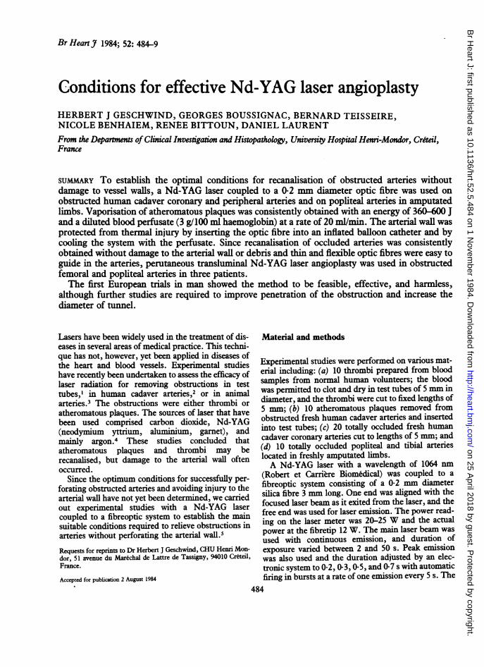
Fig. 1 Hole obtained with Nd-YAG laser emissiwn in anobstructed fresh human cadaver carotid artery viewed through asteeoscopic nmicroscope.
EFFECT ON ATHEROMATOUS PLAQUESThe effects were dependent on the colour, thickness,density of tissue,6 power intensity, and duration ofexposure to the laser as well as the composition andrate of the perfusion. With saline, no significanteffects were observed. With blood, thermal injuryoccurred at the entry of the plaque but no perforationresulted. By contrast, with diluted blood (3 g/100 mlhaemoglobin concentration) perfusion, perforation ofthe atheromatous plaque occurred (Fig. 1). The mostefficient perfusion rate was 20 ml/min with a perfusatetemperature of 22°C (Tables 1 and 2). Under these
Table 1 Results of laser treatment on atheromatous plaquesaccording to the perforate used. Values are mean (SD) (n=8)Perfusate Size of perforation
Length Entry Exit(mm) diameter diameter
(mm) (mm)
Saline (0.9 g/100 ml) 0.7(0.5)*** 0.4(0.3)*** 04**Diluted blood:Haemoglobin
concentration3 g/100 ml 4-8(0.2) 2-1(0-2) 1-2(0-3)
Haemoglobinconcentration10 g/lOO ml 3.8(0.4)*** 2-1(0 3) 0.3(0.4)***
Blood:Haemoglobin
concentration15 g/100 ml 2.2(0.5)*** 2-9(0-5) 0.3(0-3)***
***p<0-001.
on 25 April 2018 by guest. P
rotected by copyright.http://heart.bm
j.com/
Br H
eart J: first published as 10.1136/hrt.52.5.484 on 1 Novem
ber 1984. Dow
nloaded from
Geschwind, Boussignac, Teisseire, Benhaiem, Bittoun, Laurent
Table 2 Rault oflaser treatment on atheromatous plaquesaccording to temperature ofperfusate. Values aremean (SD) (n=8)
Sige of perforation Temperante40C 220C 370C
Length (mm) 4-6(0-4) 4-8(0.2) 4.8(0-2)Entry diameter (mm) 1-9(0-4) 2-1(0-2) 2-0(0-3)Exit diameter (mm) 1-0(0-4) 1-2(0-3) 1.2(0-2)
b
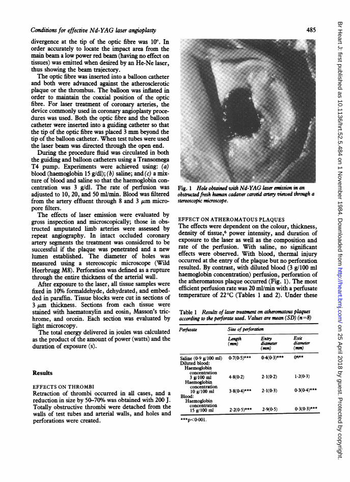
Fig. 2 An obstructed fresh human cadaver coronary artery (a)before laser treatme and (b) afterpenetaton with a laser beam(energy 3603'; en densiy 36 000 J/cm2).
experimental conditions the energy required to pene-trate a 5 mm long plaque was 12 W at the tip of theoptic fibre and the exposure duration 30 s, the energydensity being 36 000 J/cm2 (Fig. 2). Three firings of480 J each were necessary totally to penetrate plaques20 mm long. The diameter of the hole at the entry ofthe tunnel was 2 mm and 1 mm at the other end. Noeffect could be obtained when laser emission was usedin bursts. Five centimetres of occluded amputatedlimb arteries were cleared with nine successive firingsof 600 J each.
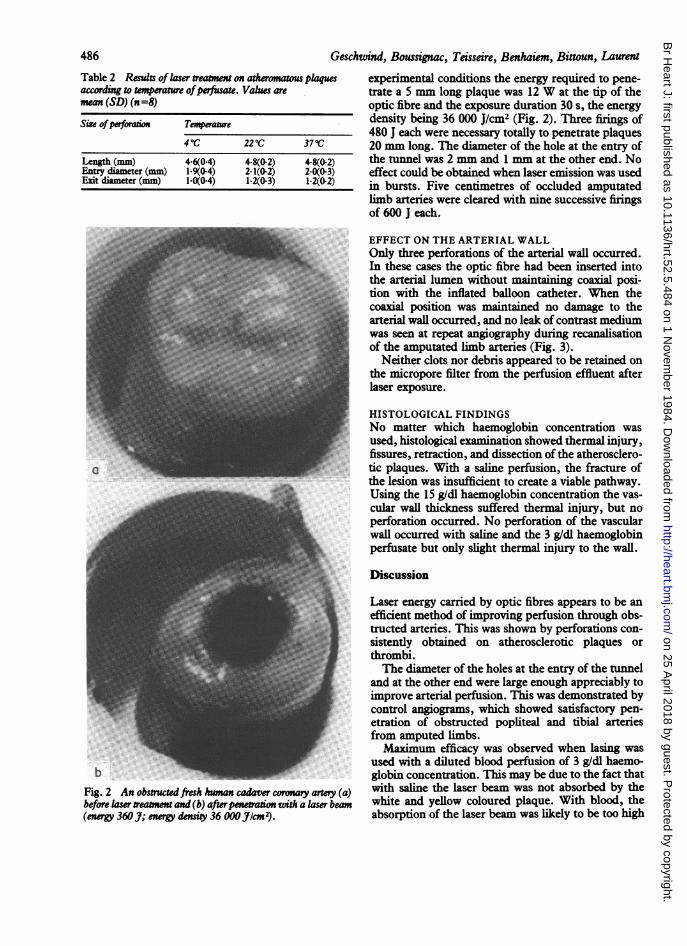
EFFECT ON THE ARTERIAL WALLOnly three perforations of the arterial wall occurred.In these cases the optic fibre had been inserted intothe arterial lumen without maintaining coaxial posi-tion with the inflated balloon catheter. When thecoaxial position was maintained no damage to thearterial wall occurred, and no leak of contrast mediumwas seen at repeat angiography during recanalisationof the amputated limb arteries (Fig. 3).
Neither clots nor debris appeared to be retained onthe micropore filter from the perfusion effluent afterlaser exposure.
HISTOLOGICAL FINDINGSNo matter which haemoglobin concentration wasused, histological examination showed thermal injury,fissures, retraction, and dissection of the atherosclero-tic plaques. With a saline perfusion, the fracture ofthe lesion was insufficient to create a viable pathway.Using the 15 g/dl haemoglobin concentration the vas-cular wall thickness suffered thermal injury, but noperforation occurred. No perforation of the vascularwall occurred with saline and the 3 g/dl haemoglobinperfusate but only slight thermal injury to the wall.
Discussion
Laser energy carried by optic fibres appears to be anefficient method of improving perfusion through obs-tructed arteries. This was shown by perforations con-sistently obtained on atherosclerotic plaques orthrombi.The diameter of the holes at the entry of the tunnel
and at the other end were large enough appreciably toimprove arterial perfusion. This was demonstrated bycontrol angiograms, which showed satisfactory pen-etration of obstructed popliteal and tibial arteriesfrom amputed limbs.Maximum efficacy was observed when lasing was
used with a diluted blood perfusion of 3 g/dl haemo-globin concentration. This may be due to the fact thatwith saline the laser beam was not absorbed by thewhite and yellow coloured plaque. With blood, theabsorption of the laser beam was likely to be too high
486
on 25 April 2018 by guest. P
rotected by copyright.http://heart.bm
j.com/
Br H
eart J: first published as 10.1136/hrt.52.5.484 on 1 Novem
ber 1984. Dow
nloaded from
Conditionsfor effective Nd-YAG laser angioplasty
b: C
Fig. 3 Angiograms of(a) an obstructed popliteal artery in afreshly amputated limb; (b) recanalisation ofpopliteal and tibial arteriesafter laser treatment; note retrograde opacification ofa previous obstructed bypass (arrow); (c) repermeation ofdistal vessels in the samelimb.
at the entry of the plaque, thus preventing the beamfrom penetrating into the atheroma. With dilutedblood, the absorption of the laser beam was sufficientto allow perforation to occur and the plaque to beentirely penetrated, since more residual energy wasavailable.The rate of perfusion also played a role in the
efficacy of vaporising the atheroma, since a high rateof perfusion prevented thermal injury. By contrast, alow rate of perfusion permitted blood to absorb thelaser radiation to too great an extent. Consequently,insufficient laser energy was available and thermalinjury to the plaque did not occur.An adequate rate of perfusion was required to pre-
vent the arterial wall from being injured by the laserbeam since the circulating fluid acts by cooling theartery. The arterial wall was protected by the coaxialposition of the tip of the optic fibre in the centre of thearterial lumen since the inflated balloon kept the fibretip at a distance from the arterial wall.
Laser treatment was effective only with continuousemission when the duration was long enough to createthermal injury to the obstruction. By contrast, whenbursts were used cooling occurred during the emis-sion intervals leading to a ineffective response.
The safety of the procedure was shown by the factthat no debris was retained on the micropore filters;thus no distal embolisation is likely to occur. Thisagrees with the findings of Choy et al who observedtotal vaporisation without debris then thrombi weresubjected to laser emission.3
Finally, since the optic fibre that was used in ourexperiments was very thin and flexible it could beinserted into all available balloon catheters includingthose commonly used for peripheral artery angi-oplasty and specifically those used for coronary angi-oplasty. The feasibility of guiding these devices inman was evident both in the femoral and iliac arteriesas well as in the coronary arteries.
CLINICAL STUDYSince these experimental studies consistently showedeffective recanalisation of totally occluded arterieswithout perforation of the arterial wall the methodwas recently applied in three patients. The protocolwas approved by the human research committee ofthis hospital and informed consent was obtained fromall patients. The first patient had a totally occludedfemoral artery, the second a totally occluded poplitealartery, and the third a femoral artery stenosis 5 mm
487
on 25 April 2018 by guest. P
rotected by copyright.http://heart.bm
j.com/
Br H
eart J: first published as 10.1136/hrt.52.5.484 on 1 Novem
ber 1984. Dow
nloaded from
Geschwind, Boussignac, Teisseire, Benhaiem, Bittoun, Laurent
b-
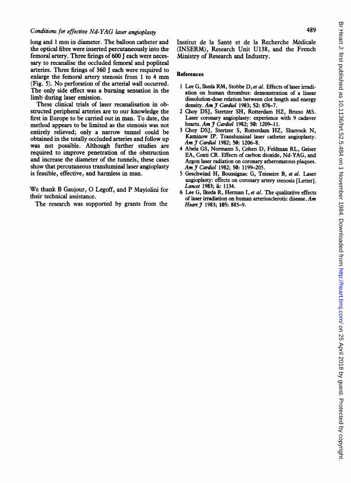
Fig. 5 Angiograms offemoral artery stenosis in man (a) beforeand (b) after enlargement of the stenosis by the laser.
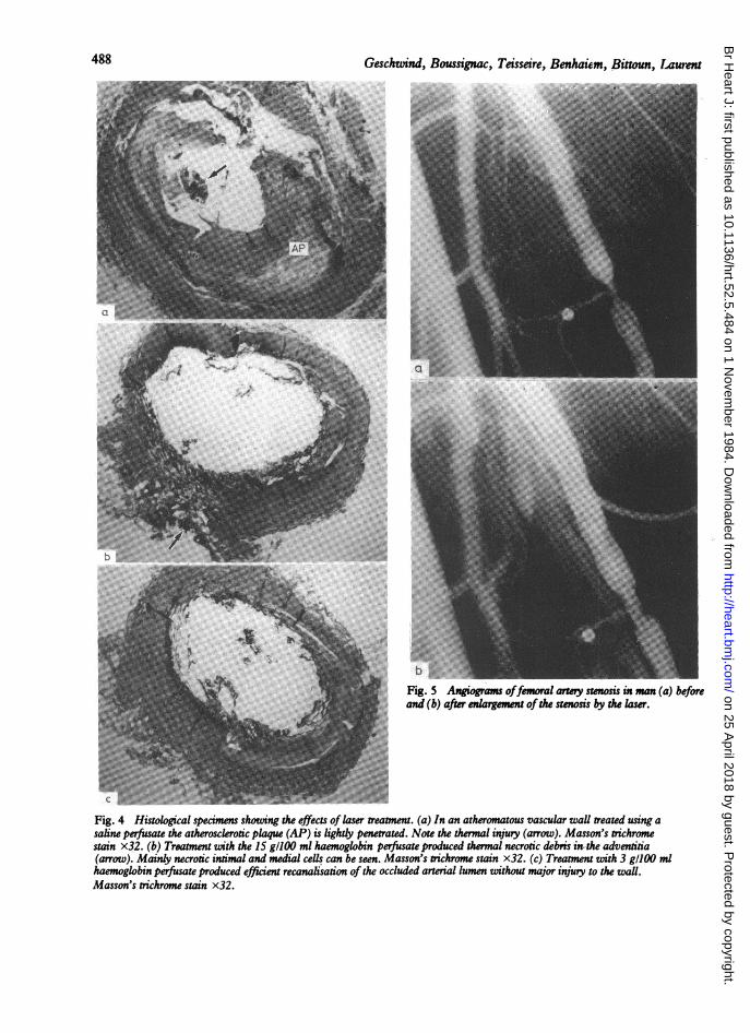
£Fig. 4 Histological specimens showting the effects of laser treatment. (a) In an atheromatous vascular wall treated using asaline perfusate the atherosclerotic plaque (AP) is lightly penetrated. Note the thermal injury (arrow). Masson's trichromestain x32. (b) Treatment with the 15 g/100 ml haemoglobin perfusate produced thermal necrotic debris in-the adventitia(arrow). Matnly necrotic intimal and medial cells can be seen. Masson's trichrome stain x32. (c) Treatment with 3 gl100 mlhaemoglobin perfusate produced efficient recanalisation of the occluded arterial lumen without major injury to the wall.Masson's trichrome stain x32.
488
on 25 April 2018 by guest. P
rotected by copyright.http://heart.bm
j.com/
Br H
eart J: first published as 10.1136/hrt.52.5.484 on 1 Novem
ber 1984. Dow
nloaded from
Conditions for effective Nd-YAG laser angioplasty
long and 1 mm in diameter. The balloon catheter andthe optical fibre were inserted percutaneously into thefemoral artery. Three firings of 600 J each were neces-sary to recanalise the occluded femoral and poplitealarteries. Three firings of 360 J each were required toenlarge the femoral artery stenosis from 1 to 4 mm(Fig. 5). No perforation of the arterial wall occurred.The only side effect was a burning sensation in thelimb during laser emission.These clinical trials of laser recanalisation in ob-
structed peripheral arteries are to our knowledge thefirst in Europe to be carried out in man. To date, themethod appears to be limited as the stenosis was notentirely relieved; only a narrow tunnel could beobtained in the totally occluded arteries and follow upwas not possible. Although further studies arerequired to improve penetration of the obstructionand increase the diameter of the tunnels, these casesshow that percutaneous transluminal laser angioplastyis feasible, effective, and harmless in man.
We thank B Gaujour, 0 Legoff, and P Mayiolini fortheir technical assistance.The research was supported by grants from the
489
Institut de la Sante et de la Recherche Medicale(INSERM), Research Unit U138, and the FrenchMinistry of Research and Industry.
References
1 Lee G, Ikeda RM, Stobbe D, et al. Effects of laser irradi-ation on human thrombus: demonstration of a lineardissolution-dose relation between clot length and energydensity. Am J Cardiol 1983; 52: 876-7.
2 Choy DSJ, Stertzer SH, Rotterdam HZ, Bruno MS.Laser coronary angioplasty: experience with 9 cadaverhearts. AmJ Cardiol 1982; 50: 1209-11.
3 Choy DSJ, Stertzer S, Rotterdam HZ, Sharrock N,Kaminow IP. Transluminal laser catheter angioplasty.Am J Cardiol 1982; 50: 1206-8.
4 Abela GS, Nornann S, Cohen D, Feldman RL, GeiserEA, Conti CR. Effects of carbon dioxide, Nd-YAG, andArgon laser radiation on coronary atheromatous plaques.Am J Cardiol 1982; 50: 1199-205.
5 Geschwind H, Boussignac G, Teisseire B, et al. Laserangioplasty: effects on coronary artery stenosis [Letter].Lancet 1983; ii: 1134.
6 Lee G, Ikeda R, Herman I, et al. The qualitative effectsof laser irradiation on human arteriosclerotic disease.AmHeartJa 1983; 105: 885-9.
on 25 April 2018 by guest. P
rotected by copyright.http://heart.bm
j.com/
Br H
eart J: first published as 10.1136/hrt.52.5.484 on 1 Novem
ber 1984. Dow
nloaded from


![[Shinobi] Bleach 484](https://img.pdfslide.us/doc/110x75/568bf1c41a28ab89339449cb/shinobi-bleach-484.jpg)

