Embed Size (px)
Citation preview
Computed Radiography in Pediatrics
Ka tsuh i ko Aok i
Computed rad iography (CR) has var ious advantages such as dynamic range. Diagnost ic accuracy w i t h CR using reduced radiat ion dose is considered compa- rable w i t h tha t of convent iona l f i lm-screen radiogra-
From the Depanment of Radiology, Shizuoka Children's Hospital, Shizuoka, Japan.
Address reprint requests to Katsuhiko Aoki, MD, Depart- ment of Radiotogy, Shizuoka Children's Hospital, 860 Urushi- yama, Shizuoka 420, Japan.
Copyright �9 1995 by W.B. Saunders Company 0897-1889/95/0801-102053.00/0
phy in d iagnosing apparent chest abnormal i t ies. In addi t ion to reducing the radiat ion dose, del ineat ion of the var ious structures in infants can be obta ined w i t h CR using postprocessing. Even in cases invo lv ing inadequate exposure, appropr ia te in format ion can be obta ined by postprocessing. For these reasons, the cumulat ive dose of radiat ion exposure wi l l be reduced. Thus, reduct ion of the radiat ion dose is the greatest advantage of using CR in pediatrics, Copyright �9 1995 by W.B. Saunders Company
KEY WORDS: computed radiography, pediatr ic radiol- ogy, reduced radiat ion exposure.
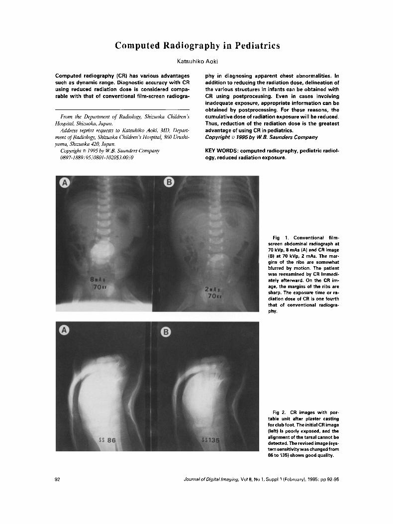
Fig 1, Convent ional f i lm- screen abdominal radiograph at 70 kVp, 8 mAs (A) and CR image (B) at 70 kVp, 2 mAs. The mar- gins of the ribs are somewhat blurred by motion. The patient was reexamined by CR immedi- ately afterward. On the CR im- age, the margins of the ribs are sharp. The exposure time of ra- diation dose of CR is one fourth that of conventional radiogra- phy.
Fig 2. CR images with por- table unit after plaster casting for club foot, The initial CR image (left) is poorly exposed, and the alignment of the tarsal cannot be detected. The revised image (sys- teta sensitivity was changed from 86 to 135) shows good quality.
92 JournalofDigitallmaging, Vol 8, No 1, Suppl 1 (February), 1995: pp 92-96
COMPL)TED RADtOGRAPHY iN PED)ATRICS 93
T HE COMPUTED RADIOGRAPHY sys- teta that was provided by Fuji Photo Film
Co Ltd (Tokyo, Japan) has been installed in Shizuoka Children's Hospital since November 1989. This new modality has great advantages in pediatric radiology.
REDUCTION OF THE RADIATION DOSE
The application of CR is berteficiat for chil- tiren, 1 wbo ate more sensitive to radiation hazards than adults. When a CR system is use& a reduction in radiation dose can be accom- plished as follows: (1) reduction of the radiation dose per exposure; (2) reduction of a total accumulated radiation dose for each patient during his life; or (3) reduction of the total dose of radiation to the population when mass screen- ings ate performed. Furthermore, dose reduc- tion is performed by shortening the exposure time (mAs). This may also prevent motion a~tifact and avoid repeat examinations (Fig l).
WlDE DYNAMIC RANGE
In pediatrics, x-ray examinations are fre- quently performed in the ward using portable unit. This is a case that CR is suitable because of wide dynamic range, one of the advantages of CR. Last year, 68.3% of all in-patient plain chest radiographs were taken with the portable unit in our hospital. The fitms obtained with a portable unit may be of poor qualRy as a result of inappropriate exposure, b~t ~he computer
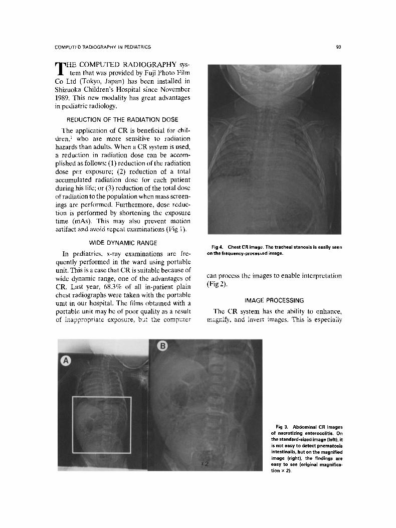
Fig 4. Chest CR image. The tracheal stenosis is easily seen on the frequency-pror image.
can process the images to enable interpretation (Fig 2).
IMAGE PROCESSING
The CR system has the ability to enhance, magnify, and invert images. This is especially
Fig 3. Abdominal CR images of necrotizing enterocolitis, On the standard-sized image (left), it is not easy to detect pnematosis intestinalis, but on the magnified image (right), the findings are easy to see (original magnifica- tion x 2).
94 KATSUHIKO AOKI
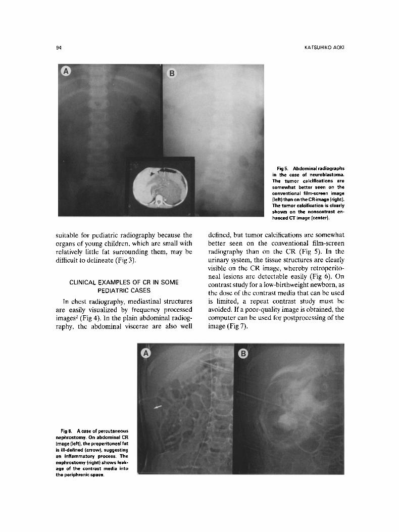
Fig5. Abdominalradiographs in the case of neuroblastoma. The tumor calcifications are somewhat better seen on the conventional film-screen image (left) than on the CR image (right). The tumor calcification is clearly shown on the noncontrast en- hanced CT image (center}.
suitable for pediatric radiography because the organs of young children, which are small with relatively little fat surrounding them, may be difficult to delineate (Fig 3).
CLINICAL EXAMPLES OF CR IN SOME PEDIATRIC CASES
In chest radiography, mediastinal structures are easily visualized by frequency processed images 2 (Fig 4). In the plain abdominal radiog- raphy, the abdominal viscerae are also well
defined, but tumor calcifications are somewhat better seen on the conventional film-screen radiography than on the CR (Fig 5). In the urinary system, the tissue structures are clearly visible on the CR image, whereby retroperito- neal lesions are detectabie easily (Fig 6). On contrast study fora low-birthweight newborn, as the dose of the contrast media that can be used is limited, a repeat contrast study must be avoided. Ir a poor-quality image is obtained, the computer can be used for postprocessing of the image (Fig 7).
Fig 6. A case of percutaneous nephrostomy. On abdominal CR image (left}, the properitoneal fat is Ul-defined (arrowl, suggesting ah inflammatory process. The nephrostomy (right) shows leak- age of the contrast media into the periphrenic space.
COMPUTED RADIOGRAPHY IN PEDIATRICS 95
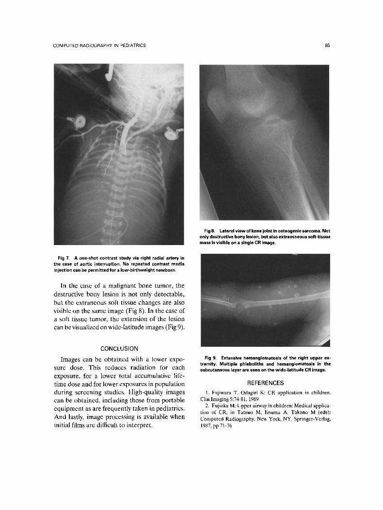
Fig 7. A one-shot contrast study via right radial artery in the case of aortic interruption. No repeated contrast media injection can be permitted for a Iow-birthweight newborn.
In the case of a malignant bone tumor, the destructive bony lesion is not only detectable, but the extraneous soft tissue changes are also visible on the same image (Fig 8). In the case of a soft tissue tumor, the extension of the lesion can be visualized on wide-latitude images (Fig 9).
Fig 8. Lateral view of knee joint in osteogenic sarcoma. Not only destructive bony lesion, but also extraosseous soft-tissue mass is visible on a single CR image.
CONCLUSlON
Images can be obtained with a lower expo- sure dose. This reduces radiation for each exposure, f o r a lower total accumulative life- time dose and for lower exposures in population during screening studies. High-quality images can be obtained, including those from portable equipment as are frequently taken in pediatrics. And lastly, image processing is available when initial films ate difficult to interpret.
Fig 9. Extensive hemangiomatosis of the right upper ex- tremity. Multiple phleboliths and hemangiomatosis in the subcutaneous layer are seen on the wide-latitude CR image.
REFERENCES
1. Fujiwara T, Odagiri K: CR application in children. Clin Imaging 5:74-81, 1989
2. Fujioka M: Upper airway in chiIdren: Medical applica- tion of CR, in Tateno M, Iinuma A, Takano M (eds): Computed Radiography. New York, NY, Springer-Verlag, 1987, pp 71-76