Embed Size (px)
Citation preview

1
Computational Study of the pKa Values of Potential Catalytic
Residues in the Active Site of Monoamine Oxidase B†
Rok Borštnar,a Matej Repič,a Shina Caroline Lynn Kamerlin,b Robert Vianello,a,c and
Janez Mavri a,d*
a Laboratory for Biocomputing and Bioinformatics, National Institute of Chemistry, Hajdrihova 19, SI–
1000 Ljubljana, Slovenia. E–mail: [email protected]
b Department of Cell and Molecular Biology, Uppsala University, Uppsala Biomedical Centre, Box 596,
SE–751 24 Uppsala, Sweden
c Quantum Organic Chemistry Group, Ruđer Bošković Institute, Bijenička cesta 54, HR–10000 Zagreb,
Croatia
d EN–FIST Centre of Excellence, Dunajska 156, SI–1000 Ljubljana, Slovenia
† This manuscript is dedicated to Professor Wilfred F. van Gunsteren on the occasion of his 65th
birthday.

2
ABSTRACT
Monoamine oxidase (MAO), which exists in two isozymic forms, MAO A and MAO B, is an important
flavoenzyme responsible for the metabolism of amine neurotransmitters such as dopamine, serotonin
and norepinephrine. Despite extensive research effort, neither the catalytic nor the inhibition
mechanisms of MAO have been completely understood. There has also been dispute with regard to
the protonation state of the substrate upon entering the active site, as well as the identity of residues
that are important for the initial deprotonation of irreversible acetylenic inhibitors, in accordance
with the recently proposed mechanism. Therefore, in order to investigate features essential for the
modes of action of MAO, we have calculated pKa values of three relevant tyrosine residues in the
MAO B active site, with and without dopamine bound as the substrate (as well as the pKa of the
dopamine itself in the active site). The calculated pKa values for Tyr188, Tyr398 and Tyr435 in the
complex are found to be shifted upwards to 13.0, 13.7 and 14.7, respectively, relative to 10.1 in
aqueous solution, ruling out the likelihood that they are viable proton acceptors. The altered tyrosine
pKa values could be rationalized as an interplay of two opposing effects: insertion of positively
charged bulky dopamine that lowers tyrosine pKa values, and subsequent removal of water molecules
from the active site that elevates tyrosine pKa values, in which the latter prevails. Additionally, the pKa
value of the bound dopamine (8.8) is practically unchanged compared to the corresponding value in
aqueous solution (8.9), as would be expected from a charged amine placed in a hydrophobic active
site consisting of aromatic moieties. We also observed potentially favorable cation–π interactions
between –NH3+ group on dopamine and aromatic moieties, which provide stabilizing effect to the
charged fragment. Thus, we offer here theoretical evidence that the amine is most likely to be
present in the active site in its protonated form, which is similar to the conclusion from experimental
studies of MAO A (Jones et al. J. Neural Trans. 2007, 114, 707–712). However, the free energy cost of

3
transferring the proton from the substrate to the bulk solvent is only 1.9 kcal mol–1, leaving open the
possibility that the amine enters the chemical step in its neutral form. In conjunction with additional
experimental and computational work, the data presented here should lead towards a deeper
understanding of mechanisms of the catalytic activity and irreversible inhibition of MAO B, which can
allow for the design of novel and improved MAO B inhibitors.
KEYWORDS
MAO B, flavoenzymes, enzyme catalysis, free energy calculations, dopamine degradation
4
INTRODUCTION
Flavoenzymes are enzymes that operate with either flavin mononucleotide (FMN) or flavin adenine
dinucleotide (FAD) cofactors. Prominent members of this family include the monoamine oxidases
(MAOs), which metabolize biogenic amines towards the corresponding imines. They are located in the
outer mitochondrial membranes of the brain, liver, intestinal, placental cells and platelets.1–3 In
MAOs, the FAD coenzyme is covalently bound to a cysteine through an 8α-‐thioether linkage.4–6 The
enzyme exists in two isozymic forms, MAO A and MAO B,7–9 which differ in substrate and inhibitor
specificities, as well as in their tissue distribution.1–3 MAOs have the role of regulating the
concentrations of neurotransmitters in living cells, and are a very promiscuous family of enzymes,
since they act on a number of diverse primary, secondary and tertiary alkyl and arylamines, although
their preference is for primary amines. MAO A is the more abundant isoform in humans, and is mainly
responsible for the oxidation of noradrenaline and serotonin. The imbalance in
noradrenaline/serotonin levels is known to cause depression-‐like symptoms and other mood
disorders.2 Hence, the selective inhibition of this isoform results in elevated noradrenaline and
serotonin concentrations, thus gradually improving the symptoms of depression. In contrast, MAO B
is responsible for the metabolism of histamine’s metabolite N–methylhistamine and dopamine.1 The
latter is an important neurotransmitter involved in the control of voluntary movement. It has been
established that insufficient dopaminergic stimulation of the basal ganglia is characteristic for
Parkinson’s disease.4 Hence, inhibition of MAO B is one of the strategies for the treatment of the
latter illness.10 Most MAO B inhibitors that are in clinical use nowadays are irreversible.10,11

5
Scheme 1. Atom numbering of the flavin moiety, without which MAO enzymes are catalytically
inactive. “R” denotes the ribityl adenosine diphosphate group, which is not shown here for clarity.
In our previous work, we studied the mechanism of the irreversible inhibition of MAO B by the
acetylenic inhibitors rasagiline and selegiline.12 In terms of the calculated barrier heights and the
overall exergonicity of the reaction, our study elucidated that the polar anionic mechanism is the
most probable, where the rate limiting step involves nucleophilic attack of the deprotonated inhibitor
onto the flavin. The chemical reaction takes place on the N5 atom of the flavin (Scheme 1), in
accordance with the available X-‐ray structures. 9,13,14 It followed that the latter reaction is preceded by
a facile enzymatic proton abstraction from the inhibitor’s terminal acetylene site. However, it has not
been possible to experimentally determine the identity of the relevant proton acceptor, which we
also did not determine in our computational study as it was performed on a model system involving
the flavin and inhibitors. Therefore, as a preliminary step towards a deeper understanding of the
chemical and the inhibition mechanisms, insight into the pKa values of potentially catalytically
relevant residues would be beneficial.
Three different potential catalytic mechanisms have been proposed to date: (1) a hydride mechanism,
(2) a radical mechanism and (3) a polar nucleophilic mechanism. In other words, it is assumed that the

6
catalytic rate-‐limiting step involves either the heterolytic H– abstraction in (1), or the homolytic H•
extraction in (2), or deprotonation of H+ in (3), all from the α–carbon atom of the substrate in the
vicinity of the amino group. A common feature of all three mechanisms is that the mentioned
activating stage is performed by N5 atom on the flavin and that dopamine enters the reaction in the
neutral form. Erdem et al.15 assumed that the hydride mechanism is unlikely to take place, because
hydride transfer is kinetically unfavorable.16 Using kinetic and structural analysis, and employing Taft
correlation to a series of benzylamine analogs, Miller and Edmondson17 provided strong experimental
evidence that proton transfer is an integral part of the rate limiting step, contrary to hydride anion
abstraction. This has led Edmondson and co-‐workers to propose the polar nucleophilic mechanism for
MAO enzymes, 17–24 although the latter has been disputed in the literature, mostly by Silverman,25–29
Ramsay,30–34 Scrutton35 and their co-‐workers, in favor of the radical mechanism. Finally, in a very
recent study Erdem and Büyükmenekşe36 investigated a biradical mechanism for MAO catalysis, but in
the same paper the authors declared it as improbable concluding that their results “present negative
evidence for the modelled biradical mechanism”. Nevertheless, it still remains a fact that, despite a
huge amount of research devoted to MAOs in the last couple of decades, there is still no consensus in
the literature about the exact mechanisms of the catalytic activity of MAO and its irreversible
inhibition.
Several important structural features of MAO B have been thoroughly emphasized when assessing
mechanisms of the catalysis/inhibition, but one is particularly relevant for the present work: the
hydrophobic nature of the MAO active site composed of aromatic moieties, that include tyrosines
(called the aromatic cage) and the FAD co-‐factor.37,38 It should be stressed that hydrophobicity of an
active site is not a black and white concept, it is difficult to define it, but on the other hand one can
7
relatively safely assume that it depends on the nature of the moieties comprising the active site. The
active site hydrophobicity was proposed to determine the protonation state of the substrate in MAO
active site, since MAO substrates are protonated in the cytoplasm, and are present as monocations
under physiological conditions. Edmondson and coworkers argued39 that because the free energy cost
associated with the transfer of a charged moiety into the hydrophobic active site is expected to be too
high, the substrate must enter the enzyme in its neutral form. However, experimental pH profiles for
kynuramine oxidation by MAO A and phenylethylamine degradation by MAO B would suggest that the
amine is most likely present in the active site in its protonated form, 40 though contradicting
arguments have been presented by Scrutton and co-‐workers,41 who, based on their pH dependent
measurements of kinetic isotope effects in MAO A, suggested that the active site is believed to be
organized for the activation of the neutral rather than charged form of the substrate. However, both
groups agree that the neutral form must enter the chemical step. The aromatic cage surrounding the
flavin co-‐factor also plays an important role in MAO enzymes. X-‐ray analysis revealed two tyrosyl
residues (Tyr398 and Tyr435 in human MAO B), constituting the aromatic cage, which both lie almost
perpendicular to flavin,7,39 suggesting a functional role in catalysis. It was proposed that they are
responsible for the orientation of a substrate towards the flavin, 37,38 but could also have direct
involvement in the proton transfer reactions.
Therefore, for all reasons stated, it is critical to know the pKa values of relevant residues and the
substrate within the MAO active site in order to progress in understanding catalytic and inhibition
mechanisms. However, these values are difficult to determine experimentally,42 and, similarly, while
experimental pH profiles can provide tremendous insight, it can be hard to conclusively determine the
identity of residues whose protonation state is being affected. Although there are many experimental

8
methods that enable determination of the overall titration curve of a protein, only a few
spectroscopic techniques posses sufficient resolution to allow for the determination of pKa values of
individual residues in a protein.43 For MAO enzymes, a lot of research efforts has been devoted by
Scrutton,41 Edmondson,44 Ramsay45 and their co-‐workers to experimentally measure pKa values, but
only data for several residues that are close to the surface of MAOs, and which are believed to form
the so-‐called “entrance” and “substrate” cavities7,39,46–48 were obtained. In addition, pKa calculations
continue to provide a significant challenge to computations.49–52 In the present work, we have
investigated pKa values of three tyrosine residues (Tyr188, Tyr 398 and Tyr 435) and the dopamine
molecule within MAO B active site. Both the free enzyme and the enzyme complexed with dopamine
were considered. We hope that the obtained acidity/basicity parameters will offer new insight into
features of MAO enzymes and help elucidating exact mechanisms of their activity and irreversible
inhibition.
COMPUTATIONAL METHODS
The starting point for our calculations was the high-‐resolution (1.6 Å) X-‐ray structure of MAO B in
complex with 2-‐(2-‐benzofuranyl)-‐2-‐imidazoline),13 which was obtained from the Protein Data Bank53
(accession code 2XFN). All ligands present in the crystal structure were removed and we manually
placed physiologically relevant dopamine monocation (Figure 1) in the active site, as it is a
characteristic substrate metabolized by MAO B.
9
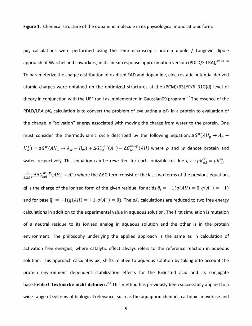
Figure 1. Chemical structure of the dopamine molecule in its physiological monocationic form.
pKa calculations were performed using the semi-‐macroscopic protein dipole / Langevin dipole
approach of Warshel and coworkers, in its linear response approximation version (PDLD/S-‐LRA),49,54–56
To parameterize the charge distribution of oxidized FAD and dopamine, electrostatic potential derived
atomic charges were obtained on the optimized structures at the (PCM)/B3LYP/6–31G(d) level of
theory in conjunction with the UFF radii as implemented in Gaussian09 program.57 The essence of the
PDLD/LRA pKa calculation is to convert the problem of evaluating a pKa in a protein to evaluation of
the change in “solvation” energy associated with moving the charge from water to the protein. One
must consider the thermodynamic cycle described by the following equation: ∆𝐺! 𝐴𝐻! → 𝐴!! +
𝐻!! = ∆𝐺! 𝐴𝐻! → 𝐴!! + 𝐻!! + ∆𝐺!!"!→! 𝐴! − ∆𝐺!"#
!→! 𝐴𝐻 where p and w denote protein and
water, respectively. This equation can be rewritten for each ionizable residue i, as: 𝑝𝐾!,!! = 𝑝𝐾!,!! −
!!!.!!"
∆∆𝐺!"#!→! 𝐴𝐻! → 𝐴!! where the ∆∆G term consist of the last two terms of the previous equation,
qi is the charge of the ionized form of the given residue, for acids 𝑞! = −1(𝑞 𝐴𝐻 = 0, 𝑞 𝐴! = −1)
and for base 𝑞! = +1(𝑞 𝐴𝐻 = +1, 𝑞 𝐴! = 0). The pKa calculations are reduced to two free energy
calculations in addition to the experimental value in aqueous solution. The first simulation is mutation
of a neutral residue to its ionized analog in aqueous solution and the other is in the protein
environment. The philosophy underlying the applied approach is the same as in calculation of
activation free energies, where catalytic effect always refers to the reference reaction in aqueous
solution. This approach calculates pKa shifts relative to aqueous solution by taking into account the
protein environment dependent stabilization effects for the Brønsted acid and its conjugate
base.Fehler! Textmarke nicht definiert.,54 This method has previously been successfully applied to a
wide range of systems of biological relevance, such as the aquaporin channel, carbonic anhydrase and
10
the bovine pancreatic trypsin inhibitor, to name a few examples.52,58–61
The protein studied here was first explicitly solvated using the surface constrained all atom solvent
(SCAAS) model,54 employing a water grid with a radius of 20 Å around the investigated residue. Long
range interactions were treated using the local reaction field (LRF) approach.62 The resulting system
was equilibrated by running a 50 ps molecular dynamics simulation using a 0.5 fs time step at 300 K.
After that, we evaluated pKa values using the PDLD/S-‐LRA approach, employing full atomic charges, by
averaging the corresponding values over the results obtained for 20 protein configuration windows,
connecting charged and uncharged states, each averaged over 25 ps of simulation with a 1 fs time
step, giving rise to a total simulation time of 500 ps for the entire thermodynamic perturbation.
Calculated pKa values are sensitive to the applied external dielectric constant during the simulations.
The choice of the correct dielectric constant to describe the protein interior is a very complicated
issue, which has been the subject of heated debates over the years. A variety of values were
suggested, ranging from ε = 2–80. For example, van Gunsteren and co-‐workers performed molecular
dynamics simulation using the GROMOS force field, and obtained a value of ε = 30 for the interior of
lysozyme.63 In our work we employed ε = 8–12 based on the discussion in reference 55. All PDLD/S-‐
LRA calculations were performed using the ENZYMIX force field and the MOLARIS simulation
package.54
RESULTS AND DISCUSSION
The results of pKa calculations of relevant residues in the MAO active site are shown in Table 1, and

11
the orientation of the relevant residues is illustrated in Fig. 2, as well as the corresponding pKas of the
tyrosine sidechain and dopamine in aqueous solution. Before we start analyzing the calculated results,
it is useful to bring about the fact that experimental aqueous solution pKa values of tyrosine (side
chain –OH deprotonation) and dopamine (aminoethyl –NH3+ deprotonation) assume 10.164 and 8.9,65
respectively. As a consequence, it follows that under physiological conditions tyrosine is a rather weak
acid and is mostly present in the neutral Tyr–OH form, and that dopamine assumes monocationic
form, being protonated at the free aminoethyl group.
Table 1. Calculated pKa values at different dielectric constants ε.a All values are averaged over 20 starting conformations, with the corresponding standard deviations shown in parentheses.
MAO B free enzyme MAO B in complex with protonated dopamine
pKw ε = 8 ε = 9 ε = 10 ε = 11 ε = 12 ε = 8 ε = 9 ε = 10 ε = 11 ε = 12
Tyr188 11.2
(0.019)
11.1
(0.018)
11.0
(0.020)
10.4
(0.022)
10.4
(0.020)
13.6
(0.030)
13.3
(0.022)
13.1
(0.018)
12.5
(0.024)
12.3
(0.019)
Tyr398 10.7
(0.020)
10.5
(0.023)
10.3
(0.020)
10.2
(0.020)
10.1
(0.018)
14.8
(0.019)
14.3
(0.019)
13.8
(0.016)
13.0
(0.020)
12.8
(0.016)
Tyr435 10.2
(0.018)
10.2
(0.016)
10.2
(0.019)
9.7
(0.040)
9.8
(0.017)
15.6
(0.022)
15.2
(0.019)
14.8
(0.019)
14.0
(0.020)
13.8
(0.017)
tyrosine 10.1
dopamine 8.7
(0.037)
8.7
(0.036)
8.7
(0.039)
8.9
(0.026)
8.9
(0.024) 8.9
a pKw denotes the corresponding experimental value in aqueous solution.
The apparent pKa value of each amino acid is influenced by the micro-‐environment provided by the
protein’s structure. The latter reflects inter-‐residue, residue-‐solvent and long-‐range electrostatic
interactions with other charged residues in the protein or salt ions in solution. In the free enzyme, the
pKas of the tyrosine sidechains considered in this work are within 0.8 pKa unit of the corresponding
value in aqueous solution (Table 1). The former assume 10.8, 10.4 and 10.0 for Tyr188, Tyr398 and

12
Tyr435, respectively, obtained as the average of the five calculated dielectric constant dependent
values (ε = 8–12). However, placing a protonated dopamine into the active site (Figure 2) causes an
upward pKa shift of up to 4.7 pKa units, as would be expected from placing a positively charged species
next to them into a hydrophobic active site, and in line with the suggestion of Edmondson and
coworkers.37,38 It turns out that in the complex, the hydroxy groups are made less acidic, which further
favors the neutral form relative to the aqueous solution. We can conclude that it is very unlikely that
these tyrosines could serve as proton acceptors either from the protonated substrate or particularly
during the initial deprotonation of irreversible acetylenic inhibitors. It turns out that, before the
protonated substrate enters the enzyme, its active site is not markedly hydrophobic resulting in
unchanged tyrosine pKa values. However, once dopamine monocation is positioned in the active site,
its steric requirements demand removal of nearby water molecules, which enhances the
hydrophobicity of the environment. As a consequence, the resulting tyrosine pKa values are controlled
by two opposing effects. Firstly, binding of the cation, which favors deprotonated form of tyrosine
sidechain, thus lowering its pKa values, and secondly the increased hydrophobic nature of the active
site, which works towards an increase in tyrosine pKa values. Our results demonstrate that the latter
effect prevails resulting in an overall upwards shift of tyrosine acidity constants.

13
Figure 2. Structure of the MAO B active site in complex with dopamine.
Visualization of the relevant crystal structures as well as the simulation trajectories reveals that all
three investigated tyrosine residues are found in front of the re side of flavin. Two of those (Tyr398
and Tyr435) form the so-‐called “aromatic cage” and the third one (Tyr188) is found between them,
just a bit further away from the flavin (Figure 2). Additionally, despite the hydrophobic nature of the
active site consisting of aromatic moieties, there appears to nevertheless still be a few water
molecules present, which are hydrogen bonded to the aforementioned tyrosines, and connecting the
tyrosine sidechains and the substrate with the N1 atom (Scheme 1) of the flavin. This is important,
because this location could serve as the potential proton accepting site, which together with N5
position forms two reactive centers on flavin. These are, for example, found both hydrogenated in the
reduced form of the flavin FADH2. This is in agreement with the recent study by North and co-‐
workers,66 who used Mulliken population analysis on several substituted flavins and showed that the
N1 atom bears more negative atomic charge and is more nucleophilic compared with the N5 atom,
14
which, on the other hand, shows electrophilic nature. Moreover, a large number of flavoenzymes have
the N1 atom of FAD interacting with positively charged residues.2 As a result, there is a possibility of
proton transfer from the dopamine to the bulk water, allowing for any of the suggested mechanisms
of catalysis and inhibition.
From Table 1, it can be seen that when dopamine is bound to MAO B it assumes a pKa value of 8.8
(Table 1), which would be expected from an amine bound in a hydrophobic active site consisting of
aromatic moieties. This is in accordance with experimental studies on MAO A by Ramsay and
coworkers.40 Scrutton and co-‐workers,41 however, considered several MAO A substrates and showed
that, upon binding to the active site, the corresponding amine pKa values were downshifted by as
much as two pKa units. Therefore, in order to verify our calculated pKa shift using the PDLD/S-‐LRA
approach, we also calculated the pKa shift of the dopamine using the free energy perturbation
adiabatic charging (FEP/AC) approach67 ,68 and the thermodynamic cycle outlined in Figure 3 of
reference 49 (see also references 69 and 70). That is, the charges of the dopamine were perturbed
from its charged to neutral form (with the proton being replaced by a dummy atom with no charge in
the neutral form) in 51 mapping frames of 50 ps length each (total simulation time 2.55 ns) in both
aqueous solution and the MAO B active site. This gave solvation free energy differences of –53.7 and –
54.1 kcal mol–1 in aqueous solution and in MAO B, respectively, which corresponds to a negligible
downward pKa shift of 0.3 kcal mol–1, using the relationship pKap = pKaw + ∆∆G/2.3RT (see for example
reference 70), where pKap and pKaw denote pKa values of dopamine in protein and in water, in the
same order. This is in good agreement with our PDLD/S-‐LRA calculations (Table 1), again suggesting
that the dopamine is present in the active site in its protonated form. From this value, it is also
possible to obtain the free energy cost for transferring a proton from the dopamine monocation to the
15
bulk using the relationship:71 ∆𝐺!"! = 2.303𝑅𝑇(𝑝𝐾!
!(𝑑𝑜𝑛𝑜𝑟)− 𝑝𝐻), which implies that, even if the
dopamine is present in its protonated form, it would be fairly easy to deprotonate it by proton transfer
to the bulk solvent, with a free energy cost of about 1.9 kcal mol–1 at physiological pH (7.4). It is also
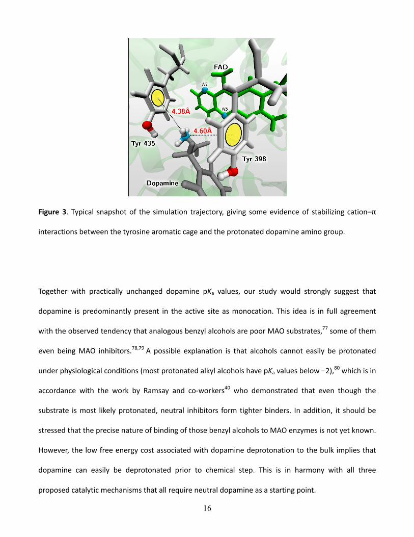
worth noting that analysis of the simulation trajectory reveals specific interactions of the protonated
amino group with the aromatic cage (Figure 3), where, in a typical snapshot, the observed distance
between the dopamine nitrogen atom and the centre of the phenyl ring on tyrosine residues assumes
values between 4–5 Å. This, in conjunction with the elevated tyrosine pKa values, suggests that these
residues might play an important role in stabilizing the protonated amine, either directly through
hydrogen bonding interactions with the relevant sidechains, or through cation–π interactions with the
aromatic cage. The former can be as strong as hydrogen bonding interactions,72 and are a well-‐
established pattern of molecular recognition in the systems of biological interest. 73 – 76 As an
illustration, the relevant experimentally determined gas-‐phase binding energy between protonated
methylamine MeNH3+ and benzene is as high as –18.8 kcal mol–1.72 Also, it was demonstrated that
cation–π interactions are crucial, for instance, in promoting binding of cationic agonists and
antagonists to the nicotinic acetylcholine receptor.73
16
Figure 3. Typical snapshot of the simulation trajectory, giving some evidence of stabilizing cation–π
interactions between the tyrosine aromatic cage and the protonated dopamine amino group.
Together with practically unchanged dopamine pKa values, our study would strongly suggest that
dopamine is predominantly present in the active site as monocation. This idea is in full agreement
with the observed tendency that analogous benzyl alcohols are poor MAO substrates,77 some of them
even being MAO inhibitors.78,79 A possible explanation is that alcohols cannot easily be protonated
under physiological conditions (most protonated alkyl alcohols have pKa values below –2),80 which is in
accordance with the work by Ramsay and co-‐workers40 who demonstrated that even though the
substrate is most likely protonated, neutral inhibitors form tighter binders. In addition, it should be
stressed that the precise nature of binding of those benzyl alcohols to MAO enzymes is not yet known.
However, the low free energy cost associated with dopamine deprotonation to the bulk implies that
dopamine can easily be deprotonated prior to chemical step. This is in harmony with all three
proposed catalytic mechanisms that all require neutral dopamine as a starting point.
17
CONCLUDING REMARKS
In this article we provide what is, to the best of our knowledge, the first systematic study of the pKa
values of titratable groups present in the active site of Monoamine oxidase B (MAO B). We have
considered here both the holoenzyme and the enzyme supplemented with the dopamine molecule to
form enzyme–substrate Michaelis complex, giving rise to the prechemical step of dopamine
degradation. Specifically, we have examined the pKas of the three tyrosine residues that constitute the
so-‐called aromatic cage,7,39 the pKa value of the dopamine itself, as well as the free energy cost of
potential deprotonation of the dopamine by bulk solvent. The calculations were performed using the
full dimensionality of the protein and extensive sampling.
It was demonstrated that, for the investigated tyrosine residues, their pKa values span the range
between 13.0–14.7 pKa units, which are increased from the corresponding water solution value of
10.1 providing strong support to the idea of the hydrophobic nature of the active site put forward by
Edmondson and co-‐workers,17 which could help in understanding the precise mechanism of the
catalytic step and the inhibition reaction of MAO enzymes. The altered tyrosine pKa values could be
rationalized as an interplay of two opposing effects: insertion of positively charged bulky dopamine
that lowers the tyrosine pKa values, and therewith associated removal of water molecules from the
active site that promotes hydrophobicity and elevates the tyrosine pKa values, in which the latter
prevails. Similarly, calculated pKa values for the dopamine suggest that the pKa of this species is
relatively unaffected by the change of environment, which would again be consistent with an amine
placed in a hydrophobic active site consisting of aromatic residues, and implying that the

18
corresponding deprotonation of the dopamine monocation by the bulk solvent would be fairly facile,
with a free energy cost of 1.9 kcal mol–1 at room temperature and physiological pH. It is worth noting
that, during the entire simulation time, few water molecules were present at the active site, and were
constantly exchanging with the bulk water molecules. This gives some additional evidence about the
nature of the active site, which is not conventionally water-‐free hydrophobic, but rather involves the
aromatic cage that is capable to form favorable quadrupolar interactions with dipolar (water) and
cationic (protonated substrate) species. Additional proof was provided by visualization of the
simulation trajectories showing a favorable orientation of the charged dopamine towards tyrosine
residues. We would like to emphasize at this stage that Edmondson and co-‐workers39 did raise a valid
point about the high free energy cost associated with transporting a protonated dopamine through a
membrane, so at this stage it is unclear precisely how the protonated dopamine could enter the MAO
B active site. However, this is an issue that is out of the scope of the present work, and our calculated
results tie in with experimental studies on MAO A,40 suggesting that the amine is present in the active
site in its protonated form, but that the relatively low cost of proton loss to the bulk is consistent with
all three proposed catalytic mechanisms that all require neutral dopamine to enter the chemical step.
Future studies will be directed towards elucidation of the exact catalytic and the inhibition
mechanisms of MAO B on the QM/MM level with proper thermal averaging and appropriate free
energy calculations.67 Quantization of the nuclear motion should yield values of the H/D kinetic
isotope effects that will discriminate between several possible mechanisms. In order to facilitate this,
the present results shed new light on features of MAO B active site and provide relevant constrains for
upcoming calculations. It remains a future challenge to apply the molecular dynamics methodology
for simulations at the constant pH81 and quantum dynamical treatment of proton dynamics82–84 to
19
MAO B active site, which we plan to address. Our ultimate goal will be to rationalize substrate and
inhibitor specificity and design novel reversible and irreversible inhibitors that are all potential drugs,
used in the clinical treatment of depression and certain neurological disorders like Parkinson disease.85
ACKNOWLEDGMENT
R.B., M.R. and J.M. would like to thank the Slovenian Research Agency for financial support in the
framework of the program group P1–0012 and within the corresponding research project contract No.
J1–2014. R.V. gratefully acknowledges the European Commission for an individual FP7 Marie Curie
Intra European Fellowship for Career Development; contract number PIEF–GA–2009–255038. S.C.L.K.
would like to thank the Swedish research council (VR, grant 2010-‐5026) for funding, as well as the Carl
Tryggers and Sven and Ebba Christina Hagberg foundations for generous stipends.

20
REFERENCES
(1) Joosten, V.; van Berkel, W. J. Curr. Opin. Chem. Biol. 2007, 11, 195–202.
(2) Fraaije, M. W.; Mattevi, A. Trends Biochem. Sci. 2000, 25, 126–132.
(3) Costa, E.; Green, A. R.; Koslow, S. H.; LeFevre, H. F.; Revuelta, A. V.; Wang, C. Pharmacol. Rev.
1972, 24, 167–190.
(4) Johnston, J. P. Biochem. Pharmacol. 1968, 17, 1285–1297.
(5) Wu, H. F.; Chen, K.; Shih, J. C. Mol. Pharmacol. 1993, 43, 888–893.
(6) Nandigama, R. K.; Edmondson, D. E. J. Biol. Chem. 2000, 275, 20527–20532.
(7) Binda, C.; Hubalek, F.; Li, M.; Edmondson, D. E.; Mattevi, A. FEBS Lett. 2004, 564, 225–228.
(8) Edmondson, D. E.; Mattevi, A.; Binda, C.; Li, M.; Hubalek, F. Curr. Med. Chem. 2004, 11, 1983–
1993.
(9) De Colibus, L.; Li, M.; Binda, C.; Lustig, A.; Edmondson, D. E.; Mattevi, A. Proc. Nat. Acad. Sci. USA
2005, 102, 12684–12689.
(10) Hefti, M. H.; Vervoort, J.; van Berkel, W. J. Eur. J. Biochem. 2003, 270, 4227–4242.
(11) Goodman, L. S.; Brunton, L. L.; Chabner, B.; Knollmann, B. C. Goodman & Gilman's
Pharmacological Basis of Therapeutics, McGraw-‐Hill, New York, 12th edn., 2011.
(12) Borštnar, R.; Repič, M.; Kržan, M.; Mavri, J.; Vianello, R. Eur. J. Org. Chem. 2011, 6419–6433.
(13) Bonivento, D.; Milczek, E. M.; McDonald, G. R.; Binda, C.; Holt, A.; Edmondson, D. E.; Mattevi, A. J.
Biol. Chem. 2010, 285, 36849–36856.
(14) Binda, C.; Hubálek, F.; Li, M.; Herzig, Y.; Sterling, J.; Edmondson, D. E.; Matevi, A. J. Med. Chem.
2005, 48, 8148–8154.
(15) Erdem, S. S.; Karahan, O.; Yildiz, I.; Yelekci, K. Org. Biomol. Chem. 2006, 4, 646–658.
21
(16) Binda, C.; Wang, J.; Li, M.; Hubálek, F.; Mattevi, A.; Edmondson, D. E. Biochemistry 2008, 47,
5616–5625.
(17) Miller, J. R.; Edmondson, D. E. Biochemistry 1999, 38, 13670–13683.
(18) Edmondson, D. E. Xenobiotica 1995, 25, 735–753.
(19) Edmondson, D. E.; Bhattacharrya, A. K.; Xu, J. Biochim. Biophys. Acta 2000, 1479, 52–58.
(20) Edmondson, D. E.; Bhattacharyya, A. K.; Walker, M. C. Biochemistry 1993, 32, 5196–5202.
(21) Husain, M.; Edmondson, D. E.; Singer, T. P. Biochemistry 1982, 21, 595–600.
(22) Nandigama, R. K.; Edmondson, D. E. Biochemistry 2000, 39, 15258–15265.
(23) Walker, M. C.; Edmondson, D. E. Biochemistry 1994, 33, 7088–7098.
(24) Upadhyay, A. K.; Wang, J.; Edmondson, D. E. Biochemistry 2008, 47, 526–536.
(25) Silverman, R. B.; Lu, X.; Zhou, J. J. P.; Swihart, A. J. Am. Chem. Soc. 1994, 116, 11590–11591.
(26) Silverman, R. B.; Lu, X. J. Am. Chem. Soc. 1994, 116, 4129–4130.
(27) Silverman, R. B. Acc. Chem. Res. 1995, 28, 335–342 and references cited therein.
(28) DeRose, V. J.; Woo, J. C.; Hawe, W. P.; Hoffman, B. M.; Silverman, R. B.; Yelekci, K. Biochemistry
1996, 35, 11085–11091.
(29) Silverman, R. B. Biochem. Soc. Trans. 1991, 19, 201–206.
(30) Ramsay, R. R.; Sablin, S. O.; Bachurin, S. O.; Singer, T. P. Biochemistry 1993, 32, 9025–9030.
(31) Rigby, S. E.; Hynson, R. M.; Ramsay, R. R.; Munro, A. W.; Scrutton, N. S. J. Biol. Chem. 2005, 280,
4627–4631.
(32) Sablin, S. O.; Ramsay, R. R. J. Biol. Chem. 1998, 273, 14074–14076.
(33) Ramsay, R. R. Biochemistry 1991, 30, 4624–4629.
(34) Kay, C. W.; El Mkami, H.; Molla, G.; Pollegioni, L.; Ramsay, R. R. J. Am. Chem. Soc. 2007, 129,
16091–16097.

22
(35) Scrutton, N. S. Nat. Prod. Rep. 2004, 21, 722–730.
(36) Erdem, S. S.; Buyukmenekse, B. J. Neural Transm. 2011, 118, 1021–1029.
(37) Li, M.; Binda, C.; Mattevi, A.; Edmondson, D. E. Biochemistry 2006, 45, 4775–4784.
(38) Akyuz, M. A.; Erdem, S. S.; Edmondson, D. E. J. Neural Transm. 2007, 114, 693–698.
(39) Binda, C.; Wang, J.; Pisani, L.; Caccia, C.; Carotti, A.; Salvati, P.; Edmondson, D. E.; Mattevi, A. J.
Med. Chem. 2007, 50, 5848–5852.
(40) Jones, T. Z. E.; Balsa, D.; Unzeta, M.; Ramsay, R. R. J. Neural Transm. 2007, 114, 707–712.
(41) Dunn, R. V.; Marshall, K. R.; Munro, A. W.; Scrutton, N. S. FEBS J. 2008, 275, 3850–3858.
(42) Grimsley, G. R.; Scholtz, J. M.; Pace, C. N. Protein Sci. 2009, 18, 247–251 and references cited
therein.
(43) Juffer, A. H. Biochem. Cell Biol. 1998, 76, 198–209.
(44) Milczek, E. M.; Binda, C.; Rovida, S.; Mattevi, A.; Edmondson, D. E. FEBS J. 2011, 278, 4860–4869.
(45) Ramsay, R. R. Vopr. Med. Khim. 1997, 43, 457–470.
(46) Ramsay, R. R.; Dunford, C.; Gillman, P. K. Br. J. Pharmacol. 2007, 152, 946–951.
(47) Ramsay, R. R.; Koerber, S. C.; Singer, T. P. Biochemistry 1987, 26, 3045–3050.
(48) Ramsay, R. R.; Sablin, S. O. Neurobiol. 1999, 7, 205–212.
(49) Kamerlin, S. C.; Haranczyk, M.; Warshel, A. J. Phys. Chem. B 2009, 113, 1253–1272 and references
cited therein.
(50) Vogel, H. J.; Juffer, A. H. Theor. Chem. Acc. 1999, 101, 159–162.
(51) Harris, T. K.; Turner, G. J. Life 2002, 53, 85–98.
(52) Kato, M.; Warshel, A. J. Phys. Chem. B 2006, 110, 11566–11570.
(53) Bernstein, F. C.; Koetzle, T. F.; Williams, G. J.; Meyer Jr., E. E.; Brice, M. D.; Rodgers, J. R.; Kennard,
O.; Shimanouchi, T.; Tasumi, M. J. Mol. Biol. 1977, 112, 535–542.
23
(54) Lee, F. S.; Chu, Z. T.; Warshel, A. J. Comp. Chem. 1993, 14, 161–185.
(55) Sham, Y. Y.; Chu, Z. T.; Tao, H.; Warshel, A. Proteins 2000, 39, 393–407.
(56) Kato, M.; Pisliakov, A. V.; Warshel, A. Proteins 2006, 64, 829–844.
(57) Gaussian 09, Revision A.1, Frisch, M. J.; Trucks, G. W.; Schlegel, H. B.; Scuseria, G. E.; Robb, M. A.;
Cheeseman, J. R.; Scalmani, G.; Barone, V.; Mennucci, B.; Petersson, G. A.; Nakatsuji, H.; Caricato, M.;
Li, X.; Hratchian, H. P.; Izmaylov, A. F.; Bloino, J.; Zheng, G.; Sonnenberg, J. L.; Hada, M.; Ehara, M.;
Toyota, K.; Fukuda, R.; Hasegawa, J.; Ishida, M.; Nakajima, T.; Honda, Y.; Kitao, O.; Nakai, H.; Vreven,
T.; Montgomery, Jr., J. A.; Peralta, J. E.; Ogliaro, F.; Bearpark, M.; Heyd, J. J.; Brothers, E.; Kudin, K. N.;
Staroverov, V. N.; Kobayashi, R.; Normand, J.; Raghavachari, K.; Rendell, A.; Burant, J. C.; Iyengar, S. S.;
Tomasi, J.; Cossi, M.; Rega, N.; Millam, N. J.; Klene, M.; Knox, J. E.; Cross, J. B.; Bakken, V.; Adamo, C.;
Jaramillo, J.; Gomperts, R.; Stratmann, R. E.; Yazyev, O.; Austin, A. J.; Cammi, R.; Pomelli, C.; Ochterski,
J. W.; Martin, R. L.; Morokuma, K.; Zakrzewski, V. G.; Voth, G. A.; Salvador, P.; Dannenberg, J. J.;
Dapprich, S.; Daniels, A. D.; Farkas, Ö.; Foresman, J. B.; Ortiz, J. V.; Cioslowski, J.; Fox, D. J. Gaussian,
Inc., Wallingford CT, 2009.
(58) Schutz, C. N.; Warshel, A. Proteins 2001, 44, 400–417.
(59) Braun–Sand, S.; Burykin, A.; Chu, Z. T.; Warshel, A. J. Phys. Chem. B 2005, 109, 583–592.
(60) Burykin, A.; Warshel, A. Biophys. J. 2003, 85, 3696–3706.
(61) Burykin, A.; Kato, M.; Warshel, A. Proteins 2003, 52, 412–426.
(62) Lee, F. S.; Warshel, A. J. Chem. Phys. 1992, 97, 3100–3107.
(63) Smith, P. E.; Brunne, R. M.; Mark, A. E.; van Gunsteren, W. F. J. Phys. Chem. 1993, 97, 2009–2014.
(64) Nespoulous, C.; Pernollet, J. C. Int. J. Pept. Protein. Res. 1994, 43, 154–159.
(65) Armstrong, J.; Barlow, R. B. Br. J. Pharmac. 1976, 57, 501–516.
(66) North, M. A.; Bhattacharyya, S.; Truhlar, D. G. J. Phys. Chem. B 2010, 114, 14907–14915.

24
(67) Warshel, A. Computer Modeling of Chemical Reactions in Enzymes and Solutions, Wiley–
Interscience, New York, 1997.
(68) Warshel, A.; Sussman, F.; King, G. Biochemistry 1986, 25, 8368–8372.
(69) Glennon, T. M.; Villa, J.; Warshel, A. Biochemistry 2000, 39, 9641–9651.
(70) Fothergill, M.; Goodman, M. F.; Petruska, J.; Warshel, A. J. Am. Chem. Soc. 1995, 117, 11619–
11627.
(71) Warshel, A. Biochemistry 1981, 20, 3167.
(72) Deakyne, C. A.; Meot-‐Ner (Mautner), M. J. Am. Chem. Soc. 1985, 107, 474–479.
(73) Ma, J. C.; Dougherty, D. A. Chem. Rev. 1997, 97, 1303–1324, and references cited therein.
(74) Dougherty, D. A. Science 1996, 271, 163–168.
(75) Gaberšček, Mavri, J. Chem. Phys. Lett. 1999, 308, 421–427.
(76) Mavri, J.; Koller, J.; Hadži, D. J. Mol. Struct. (Theochem) 1993, 283, 305–312.
(77) McEwen Jr., C. M.; Sasaki, G.; Lenz Jr., W. R. J. Biol. Chem. 1968, 243, 5217–5225.
(78) McEwen Jr., C. M. J. Biol. Chem. 1965, 240, 2011–2018.
(79) Severina, I. S. Eur. J. Biochem. 1973, 38, 239–246.
(80) Smith, M. B.; March, J. March’s Advanced Organic Chemistry: Reactions, Mechanisms and
Structure, 5th ed.; John Wiley & Sons: New York, 2001.
(81) Bürgi, R.; Kollman, P. A.; van Gunsteren, W. F. Proteins 2002, 47, 469–480.
(82) Mavri, J.; Berendsen, H. J. C.; van Gunsteren, W. F. J. Phys. Chem. 1993, 97, 13469–13476.
(83) Billeter, S. R.; van Gunsteren, W. F. Comp. Phys. Comm. 1997, 107, 61–91.
(84) Billeter, S. R.; van Gunsteren, W. F. J. Phys. Chem. A 1998, 102, 4669–4678.
(85) Westlund, K. N. in Monoamine oxidase inhibitors in neurological diseases, ed. A. N. Lieberman,
CRC Press, 1994, pp. 1–20.