Embed Size (px)
Citation preview
Computational Modeling of Closed-loop Peripheral Nerve Block Based on Halorhodopsin (NpHR)
BIOMEDE 599-003 Neural Engineering Prof. Cindy ChestekWinter 2016 Final Project Suseendrakumar Duraivel, Joseph Letner, Zhuohe LiuApr. 26, 2016

Objectives
❖ Introduction to optogenetics and neural inhibition solutions;
❖ Methods of computer finite element simulation
❖ Results of relations among modeling variables
❖ Discussion of findings and real world application
❖ Future work and conclusion
2

Introduction
3
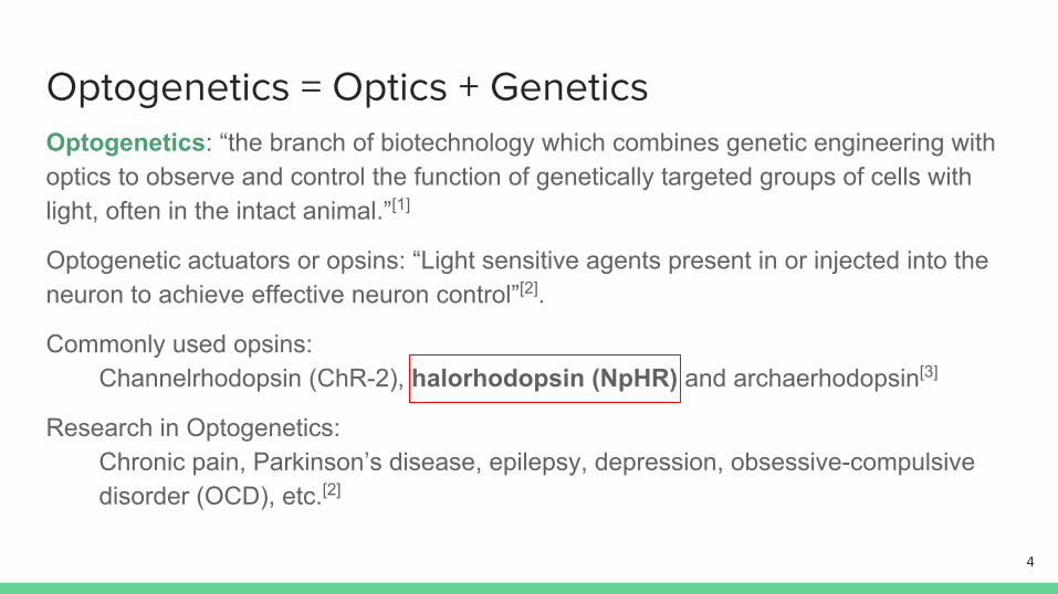
Optogenetics = Optics + GeneticsOptogenetics: “the branch of biotechnology which combines genetic engineering with optics to observe and control the function of genetically targeted groups of cells with light, often in the intact animal.”[1]
Optogenetic actuators or opsins: “Light sensitive agents present in or injected into the neuron to achieve effective neuron control”[2].
Commonly used opsins: Channelrhodopsin (ChR-2), halorhodopsin (NpHR) and archaerhodopsin[3]
Research in Optogenetics: Chronic pain, Parkinson’s disease, epilepsy, depression, obsessive-compulsive disorder (OCD), etc.[2]
4
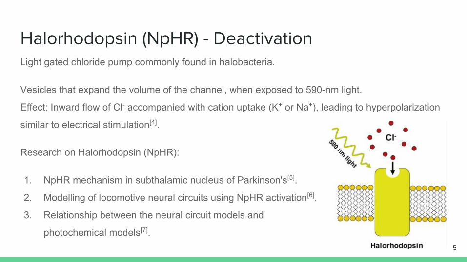
Halorhodopsin (NpHR) - DeactivationLight gated chloride pump commonly found in halobacteria.
Vesicles that expand the volume of the channel, when exposed to 590-nm light.
Effect: Inward flow of Cl- accompanied with cation uptake (K+ or Na+), leading to hyperpolarization
similar to electrical stimulation[4].
Research on Halorhodopsin (NpHR):
1. NpHR mechanism in subthalamic nucleus of Parkinson's[5].
2. Modelling of locomotive neural circuits using NpHR activation[6].
3. Relationship between the neural circuit models and
photochemical models[7].5
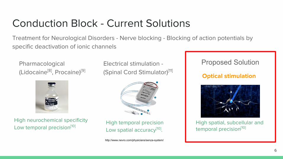
Conduction Block - Current SolutionsTreatment for Neurological Disorders - Nerve blocking - Blocking of action potentials by specific deactivation of ionic channels
http://www.nevro.com/physicians/senza-system/
High neurochemical specificity Low temporal precision[10]
High temporal precision Low spatial accuracy[10].
Optical stimulation
High spatial, subcellular and temporal precision[10]
6
Pharmacological (Lidocaine[8], Procaine){9]
Electrical stimulation - (Spinal Cord Stimulator)[11]
Proposed Solution
Project Goals
To computationally model a device, that is capable of actively detecting action potentials
propagating in a peripheral afferent neuron and blocking the propagation of the action potentials
downstream from the recording site by activating NpHR with optogenetic stimulation.
1. Establish a cable theory model adapted to neurons expressing NpHR based on the Hodgkin-
Huxley equations.
2. To build a closed-loop integrated system that applies the aforementioned cable theory model
to the inhibition of neuronal signal.
3. To optimize the system by adjusting the related parameters, to ensure effective inhibition
under various physiological conditions.
7

Methods
8
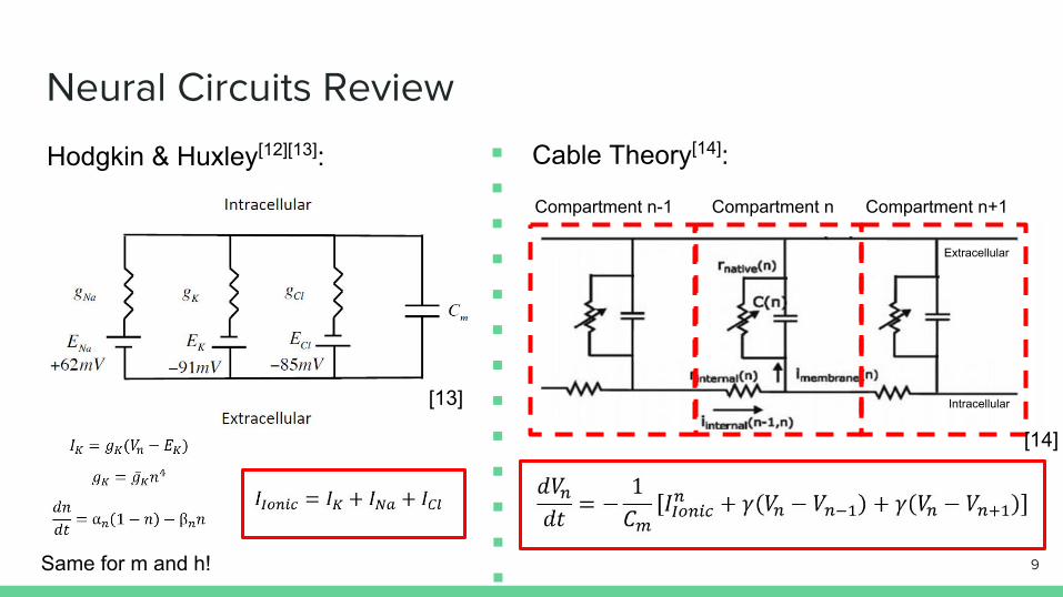
Neural Circuits Review
Compartment n-1 Compartment n Compartment n+1
Cable Theory[14]:Hodgkin & Huxley[12][13]:
Intracellular
Extracellular
Same for m and h! 9
[13]
[14]
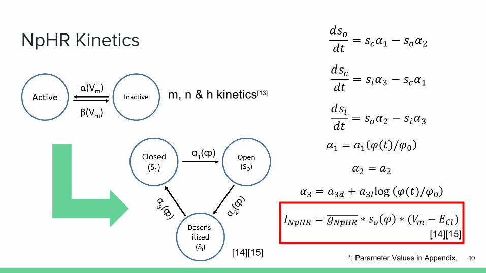
NpHR Kinetics
m, n & h kinetics[13]
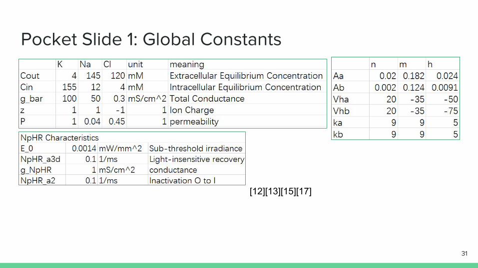
*: Parameter Values in Appendix.[14][15]
[14][15]
10
α1(ȹ)
α 2(ȹ
)α3 (ȹ
)
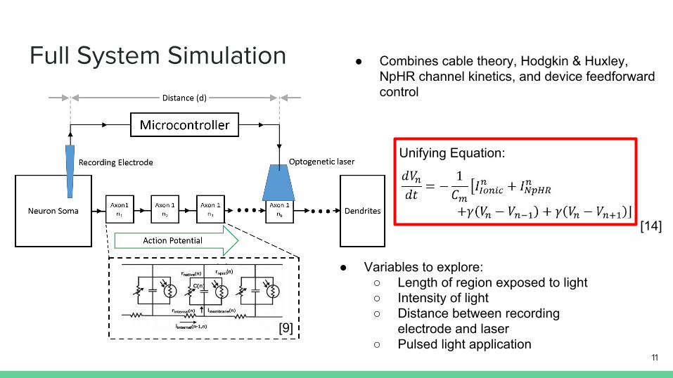
Full System Simulation
Unifying Equation:
● Combines cable theory, Hodgkin & Huxley, NpHR channel kinetics, and device feedforward control
● Variables to explore: ○ Length of region exposed to light○ Intensity of light○ Distance between recording
electrode and laser○ Pulsed light application
[9]
[14]
11
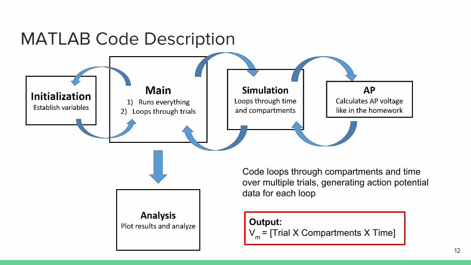
MATLAB Code Description
Output:Vm = [Trial X Compartments X Time]
Code loops through compartments and time over multiple trials, generating action potential data for each loop
12
Results
13
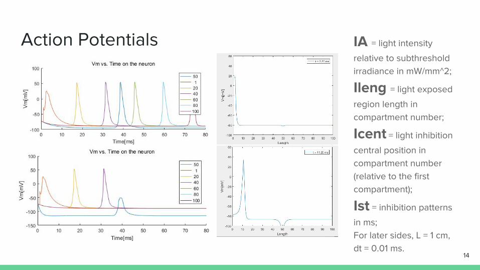
Action Potentials IA = light intensity
relative to subthreshold irradiance in mW/mm^2;
Ileng = light exposed
region length in compartment number;
Icent = light inhibition
central position in compartment number (relative to the first compartment);
Ist = inhibition patterns
in ms; For later sides, L = 1 cm, dt = 0.01 ms.
14
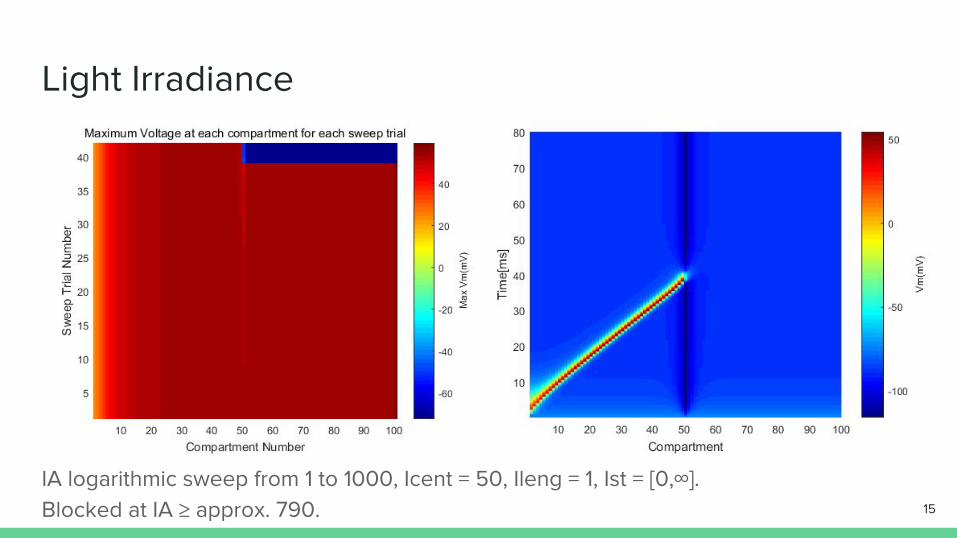
Light Irradiance
IA logarithmic sweep from 1 to 1000, Icent = 50, Ileng = 1, Ist = [0,∞]. Blocked at IA ≥ approx. 790. 15
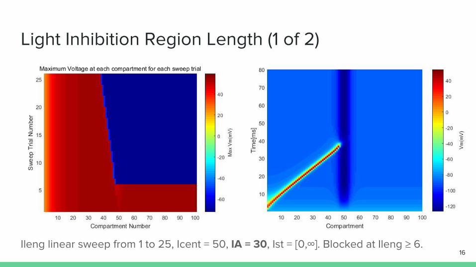
Light Inhibition Region Length (1 of 2)
Ileng linear sweep from 1 to 25, Icent = 50, IA = 30, Ist = [0,∞]. Blocked at Ileng ≥ 6. 16
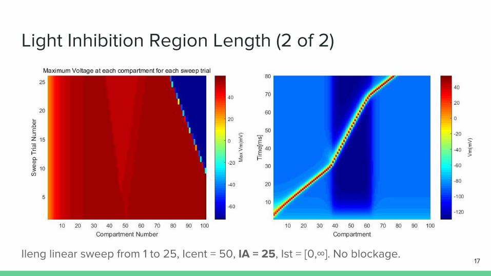
Light Inhibition Region Length (2 of 2)
Ileng linear sweep from 1 to 25, Icent = 50, IA = 25, Ist = [0,∞]. No blockage. 17
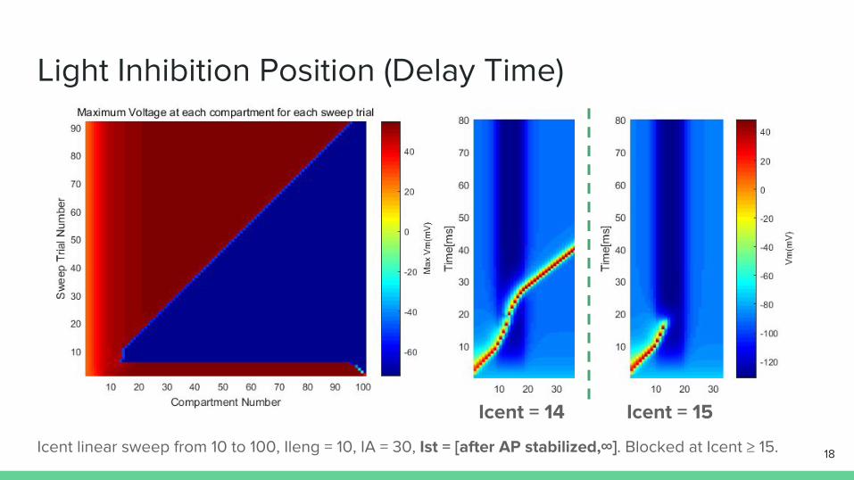
Light Inhibition Position (Delay Time)
Icent linear sweep from 10 to 100, Ileng = 10, IA = 30, Ist = [after AP stabilized,∞]. Blocked at Icent ≥ 15. 18
Icent = 14 Icent = 15
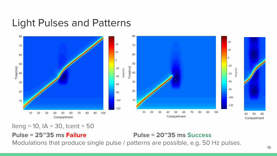
Light Pulses and Patterns
Ileng = 10, IA = 30, Icent = 50Pulse = 25~35 ms Failure Pulse = 20~35 ms Success
19Modulations that produce single pulse / patterns are possible, e.g. 50 Hz pulses.

Discussion
20
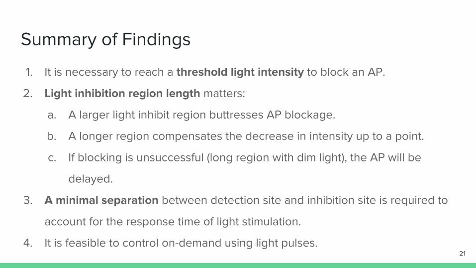
Summary of Findings
1. It is necessary to reach a threshold light intensity to block an AP.
2. Light inhibition region length matters:
a. A larger light inhibit region buttresses AP blockage.
b. A longer region compensates the decrease in intensity up to a point.
c. If blocking is unsuccessful (long region with dim light), the AP will be
delayed.
3. A minimal separation between detection site and inhibition site is required to
account for the response time of light stimulation.
4. It is feasible to control on-demand using light pulses. 21
Causal Explanations
22
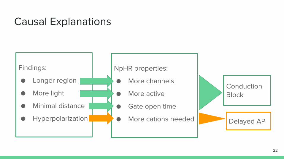
Findings:
● Longer region
● More light
● Minimal distance
● Hyperpolarization
NpHR properties:
● More channels
● More active
● Gate open time
● More cations needed
Conduction Block
Delayed AP
Connection to Real World Application
Loads of design requirements for an implantable device before marketing: Effective, low-power, small, cheap, etc. Tradeoffs: ➔ Closed loop: on-demand, but extra microcomputer processing and chip cost; ➔ Open loop: simple, but potentially battery-eating as sources always on. Restrictions: ➔ The actual genetic therapy; ➔ Device finite size; ➔ Light source limited output power; ➔ Accessibility of nerve for detection site and inhibition site. => Our nerve inhibition device is achievable in theory.
23

Future Work and Conclusion
24
Future Work - For Our Model
❖ There are compatibility issues of modeling parameters due to multiple
sources of neuron bioelectrical properties and model simplification.
1. Use specific parameters for specific afferent axons in humans
E.g. Sciatic nerve, amputation sites, etc.
2. Fix propagation speed of APs: only 0.1 m/s, and should be independent to
compartment number;
3. Include the influence of neuron diameter to inter-compartment
conductance (γ) and using better neuron geometry;
25
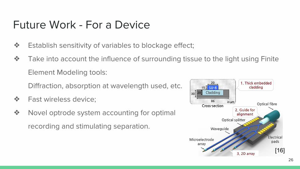
Future Work - For a Device
❖ Establish sensitivity of variables to blockage effect;
❖ Take into account the influence of surrounding tissue to the light using Finite
Element Modeling tools:
Diffraction, absorption at wavelength used, etc.
❖ Fast wireless device;
❖ Novel optrode system accounting for optimal
recording and stimulating separation.
26
[16]
Conclusion
● We have devised a computer model that simulates a pain blocking device
● Effectiveness relies on light intensity, separation of light source to electrode,
light application length, and pulse length
● NpHR demonstrates a robust mechanism for conduction block
● Future work relies on expanding of the simulation parameters and a better
physiological model
27

Thank you!Q & A
28
References[1] Miesenböck, G. (2009). The optogenetic catechism. Science, 326(5951), 395-399.[2] Zhang, F., Aravanis, A. M., Adamantidis, A., de Lecea, L., & Deisseroth, K. (2007). Circuit-breakers: optical technologies for probing neural signals and systems. Nat Rev Neurosci, 8(8), 577-581. doi:10.1038/nrn2192.[3] Boyden, E. S., Zhang, F., Bamberg, E., Nagel, G., & Deisseroth, K. (2005). Millisecond-timescale, genetically targeted optical control of neural activity. Nat Neurosci, 8(9), 1263-1268. doi:10.1038/nn1525.[4] B Schobert and J K Lanyi, Halorhodopsin is a light-driven chloride pump. J. Biol. Chem. 1982 257: 10306-.[5] Gradinaru, V., Mogri, M., Thompson, K. R., Henderson, J. M., & Deisseroth, K. (2009). Optical deconstruction of parkinsonian neural circuitry. Science, 324(5925), 354-359. doi:10.1126/science.1167093.[6] Inada, K., Kohsaka, H., Takasu, E., Matsunaga, T., & Nose, A. (2011). Optical dissection of neural circuits responsible for Drosophila larval locomotion with halorhodopsin. PLoS One, 6(12), e29019. doi:10.1371/journal.pone.0029019.[7] Nikolic, K., Jarvis, S., Grossman, N., & Schultz, S. (2013). Computational models of optogenetic tools for controlling neural circuits with light. Conf Proc IEEE Eng Med Biol Soc, 2013, 5934-5937. doi:10.1109/embc.2013.6610903.[8] Correa-Illanes, G., Roa, R., Pineros, J. L., & Calderon, W. (2012). Use of 5% lidocaine medicated plaster to treat localized neuropathic pain secondary to traumatic injury of peripheral nerves. Local Reg Anesth, 5, 47-53. doi:10.2147/lra.s31868[9] Franz, D. N., & Perry, R. S. (1974). Mechanisms for differential block among single myelinated and non-myelinated axons by procaine. J Physiol, 236(1), 193-210.[10] Dugue, G. P., Akemann, W., & Knopfel, T. (2012). A comprehensive concept of optogenetics. Prog Brain Res, 196, 1-28. doi:10.1016/b978-0-444-59426-6.00001-x.
29

References[11] Al-Kaisy, A., Van Buyten, J., Smet, I., Palmisani, S., Pang, D., & Smith, T. (2014). Sustained effectiveness of 10 kHz high-frequency spinal cord stimulation for patients with chronic, low back pain: 24-month results of a prospective multicenter study. Pain Medicine, 15(3), 347-354.[12] Hodgkin, A. L., & Huxley, A. F. (1952). A quantitative description of membrane current and its application to conduction and excitation in nerve.The Journal of physiology, 117(4), 500.[13] Chestek,C. A. Lecture 4 – Outline [PDF]. Retrieved from University of Michigan Canvas: https://ctools.umich.edu/gateway/[14] Grossman, N., Nikolic, K., Toumazou, C., & Degenaar, P. (2011). Modeling study of the light stimulation of a neuron cell with channelrhodopsin-2 mutants. IEEE Trans Biomed Eng, 58(6), 1742-1751. doi:10.1109/tbme.2011.2114883[15] Boyle, P. M., Karathanos, T. V., Entcheva, E., & Trayanova, N. A. (2015). Computational modeling of cardiac optogenetics: Methodology overview & review of findings from simulations. Comput Biol Med, 65, 200-208. doi:10.1016/j.compbiomed.2015.04.036[16] Son, Y., Lee, H. J., Kim, J., Shin, H., Choi, N., Lee, C. J., ... & Cho, I. J. (2015). In vivo optical modulation of neural signals using monolithically integrated two-dimensional neural probe arrays. Scientific reports, 5[17] Mainen, Z. F., Joerges, J., Huguenard, J. R., & Sejnowski, T. J. (1995). A model of spike initiation in neocortical pyramidal neurons. Neuron, 15(6), 1427-1439.
30
Pocket Slide 1: Global Constants
31
[12][13][15][17]
Pocket Slide 2: More Figures
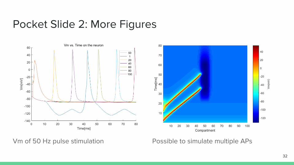
Vm of 50 Hz pulse stimulation Possible to simulate multiple APs
32