Embed Size (px)
Citation preview
Judith A. Spilker, Nanette Hock, Elaine Miller and Pamela H. MitchellDebbie Summers, Anne Leonard, Deidre Wentworth, Jeffrey L. Saver, Jo Simpson,
AssociationIschemic Stroke Patient : A Scientific Statement From the American Heart
Comprehensive Overview of Nursing and Interdisciplinary Care of the Acute
ISSN: 1524-4628 Copyright © 2009 American Heart Association. All rights reserved. Print ISSN: 0039-2499. OnlineStroke is published by the American Heart Association. 7272 Greenville Avenue, Dallas, TX 72514
doi: 10.1161/STROKEAHA.109.1923622009, 40:2911-2944: originally published online May 28, 2009Stroke
http://stroke.ahajournals.org/content/40/8/2911located on the World Wide Web at:
The online version of this article, along with updated information and services, is
http://stroke.ahajournals.org/ An erratum has been published regarding this article. Please see the attached page for:
http://www.lww.com/reprintsReprints: Information about reprints can be found online at [email protected]. E-mail:
Fax:Kluwer Health, 351 West Camden Street, Baltimore, MD 21202-2436. Phone: 410-528-4050. Permissions: Permissions & Rights Desk, Lippincott Williams & Wilkins, a division of Wolters http://stroke.ahajournals.org//subscriptions/Subscriptions: Information about subscribing to Stroke is online at
at AHA National Center on November 3, 2011http://stroke.ahajournals.org/Downloaded from

AHA Scientific Statement
Comprehensive Overview of Nursing and InterdisciplinaryCare of the Acute Ischemic Stroke Patient
A Scientific Statement From the American Heart Association
Debbie Summers, MSN, RN, FAHA, Chair; Anne Leonard, MPH, RN, FAHA, Co-Chair;Deidre Wentworth, MSN, RN; Jeffrey L. Saver, MD, FAHA; Jo Simpson, BSN, RN;
Judith A. Spilker, BSN, RN; Nanette Hock, MSN, RN, FAHA; Elaine Miller, DNS, RN, FAHA;Pamela H. Mitchell, PhD, RN, FAHA; on behalf of the
American Heart Association Council on Cardiovascular Nursing and the Stroke Council
Ischemic stroke represents 87% of all strokes.1 As world-wide initiatives move forward with stroke care, healthcare
providers and institutions will be called on to deliver the mostcurrent evidence-based care. The American Heart Associa-tion/American Stroke Association (AHA/ASA) charged apanel of healthcare professionals from several disciplineswith developing a practical, comprehensive overview of carefor the patient with acute ischemic stroke (AIS). This articlefocuses on educating nursing and allied healthcare profes-sionals about the roles and responsibilities of those who carefor patients with AIS.
Nurses play a pivotal role in all phases of care of the strokepatient. For the purposes of this article, the writing panel hasdefined 2 phases of stroke care: (1) The emergency orhyperacute care phase,2,3 which includes the prehospitalsetting and the emergency department (ED), and (2) the acutecare phase, which includes critical care units, intermediatecare units, stroke units, and general medical units.
Stroke is a complex disease that requires the efforts andskills of all members of the multidisciplinary team. Nursesare often responsible for the coordination of care throughoutthe continuum.4–9 Coordinated care of the AIS patient resultsin improved outcomes, decreased lengths of stay, and de-creased costs.10
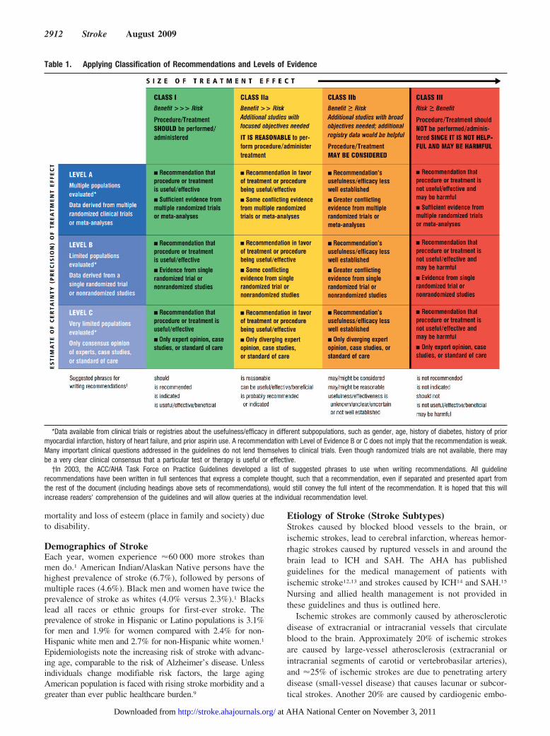
In developing this comprehensive overview, the writingpanel applied the rules of evidence and formulation ofstrength of evidence (recommendations) used by other AHA
writing groups11 (Table 1). We also cross-reference otherAHA guidelines as appropriate.
Overview of StrokeIt is important that nurses understand the burden of stroke asa public health issue in the United States. This will guidethem in developing appropriate skills to care for AIS patientsand to educate patients and families about secondary strokeprevention.
Epidemiology of StrokeThe AHA estimates that �780 000 strokes occur each year;600 000 of these are new strokes, and �180 000 are recurrentstrokes.1 Eighty-seven percent are ischemic strokes, 10% areintracranial hemorrhages (ICH), and 3% are subarachnoidhemorrhages (SAH). In 2007, the overall mortality rate fromstroke was 273 000, which makes stroke the third-leadingcause of death in the United States.1 Between 1979 and 2005,the annual number of hospital discharges with stroke as thediagnosis was �900 000.1 Direct and indirect costs associ-ated with stroke are estimated to be approximately $65.5billion.1 Direct costs are attributed to the initial hospitaliza-tion, skilled nursing care, physician and nursing care, medi-cations and durable medical equipment, home health care,and acute rehabilitation. Indirect costs include loss of prod-uctivity (loss of future earnings) due to morbidity and
The American Heart Association makes every effort to avoid any actual or potential conflicts of interest that may arise as a result of an outsiderelationship or a personal, professional, or business interest of a member of the writing panel. Specifically, all members of the writing group are requiredto complete and submit a Disclosure Questionnaire showing all such relationships that might be perceived as real or potential conflicts of interest.
This statement was approved by the American Heart Association Science Advisory and Coordinating Committee on February 12, 2009. A copy of thestatement is available at http://www.americanheart.org/presenter.jhtml?identifier�3003999 by selecting either the “topic list” link or the “chronologicallist” link (No. LS-2045). To purchase additional reprints, call 843-216-2533 or e-mail [email protected].
The American Heart Association requests that this document be cited as follows: Summers D, Leonard A, Wentworth D, Saver JL, Simpson J, SpilkerJA, Hock N, Miller E, Mitchell PH; on behalf of the American Heart Association Council on Cardiovascular Nursing and the Stroke Council.Comprehensive overview of nursing and interdisciplinary care of the acute ischemic stroke patient: a scientific statement from the American HeartAssociation. Stroke. 2009;40:2911–2944.
Expert peer review of AHA Scientific Statements is conducted at the AHA National Center. For more on AHA statements and guidelines development,visit http://www.americanheart.org/presenter.jhtml?identifier�3023366.
Permissions: Multiple copies, modification, alteration, enhancement, and/or distribution of this document are not permitted without the expresspermission of the American Heart Association. Instructions for obtaining permission are located at http://www.americanheart.org/presenter.jhtml?identifier�4431. A link to the “Permission Request Form” appears on the right side of the page.
(Stroke. 2009;40:2911-2944.)© 2009 American Heart Association, Inc.
Stroke is available at http://stroke.ahajournals.org DOI: 10.1161/STROKEAHA.109.192362
2911
;
at AHA National Center on November 3, 2011http://stroke.ahajournals.org/Downloaded from
mortality and loss of esteem (place in family and society) dueto disability.
Demographics of StrokeEach year, women experience �60 000 more strokes thanmen do.1 American Indian/Alaskan Native persons have thehighest prevalence of stroke (6.7%), followed by persons ofmultiple races (4.6%). Black men and women have twice theprevalence of stroke as whites (4.0% versus 2.3%).1 Blackslead all races or ethnic groups for first-ever stroke. Theprevalence of stroke in Hispanic or Latino populations is 3.1%for men and 1.9% for women compared with 2.4% for non-Hispanic white men and 2.7% for non-Hispanic white women.1
Epidemiologists note the increasing risk of stroke with advanc-ing age, comparable to the risk of Alzheimer’s disease. Unlessindividuals change modifiable risk factors, the large agingAmerican population is faced with rising stroke morbidity and agreater than ever public healthcare burden.9
Etiology of Stroke (Stroke Subtypes)Strokes caused by blocked blood vessels to the brain, orischemic strokes, lead to cerebral infarction, whereas hemor-rhagic strokes caused by ruptured vessels in and around thebrain lead to ICH and SAH. The AHA has publishedguidelines for the medical management of patients withischemic stroke12,13 and strokes caused by ICH14 and SAH.15
Nursing and allied health management is not provided inthese guidelines and thus is outlined here.
Ischemic strokes are commonly caused by atheroscleroticdisease of extracranial or intracranial vessels that circulateblood to the brain. Approximately 20% of ischemic strokesare caused by large-vessel atherosclerosis (extracranial orintracranial segments of carotid or vertebrobasilar arteries),and �25% of ischemic strokes are due to penetrating arterydisease (small-vessel disease) that causes lacunar or subcor-tical strokes. Another 20% are caused by cardiogenic embo-
Table 1. Applying Classification of Recommendations and Levels of Evidence
*Data available from clinical trials or registries about the usefulness/efficacy in different subpopulations, such as gender, age, history of diabetes, history of priormyocardial infarction, history of heart failure, and prior aspirin use. A recommendation with Level of Evidence B or C does not imply that the recommendation is weak.Many important clinical questions addressed in the guidelines do not lend themselves to clinical trials. Even though randomized trials are not available, there maybe a very clear clinical consensus that a particular test or therapy is useful or effective.
†In 2003, the ACC/AHA Task Force on Practice Guidelines developed a list of suggested phrases to use when writing recommendations. All guidelinerecommendations have been written in full sentences that express a complete thought, such that a recommendation, even if separated and presented apart fromthe rest of the document (including headings above sets of recommendations), would still convey the full intent of the recommendation. It is hoped that this willincrease readers’ comprehension of the guidelines and will allow queries at the individual recommendation level.
2912 Stroke August 2009
at AHA National Center on November 3, 2011http://stroke.ahajournals.org/Downloaded from
lism, most frequently from atrial fibrillation.16 Approximately30% of ischemic strokes are termed cryptogenic, for whichthe exact cause of stroke remains unknown.16
Hemorrhagic stroke is commonly caused by either primaryICH or SAH. Overall, ICH accounts for �10% of all strokesand SAH for �3%.1 Common causes and risk factors for ICHare hypertension (the number 1 cause), bleeding disorders,African-American ethnicity, aging, vascular malformations,excessive use/abuse of alcohol, and liver dysfunction.17–20
The primary cause of SAH is a ruptured cerebral aneurysm.
Crossing the Continuum of CarePhase 1 of stroke care, the emergency or hyperacute phase,encompasses the first 3 to 24 hours after onset of stroke. Thisphase generally incorporates the prehospital (activation ofemergency medical services [EMS]/9-1-1 and response) andED care protocols. The focus is on identifying stroke symp-toms and infarct location, assessing the patient for risk ofacute and long-term complications, and determining treat-ment options.
Phase 2 includes acute care, which encompasses the periodfrom 24 to 72 hours after onset of stroke. In this phase, thefocus is on clarifying the cause of stroke, preventing medicalcomplications, preparing the patient and family for discharge,and instituting long-term secondary prevention modalities.
The Emergency or Hyperacute Phase ofAIS Care
Optimal management of the AIS patient in the emergency orhyperacute phase of AIS care requires an accurate andsystematic evaluation that is coordinated and timely. Once apotential stroke is suspected, EMS personnel and nurses mustdetermine the time at which the patient was last known to bewell (last known well time). This time is the single mostimportant determinant of treatment options during the hyper-acute phase.
The Nurse’s RoleIn the prehospital setting, the leading healthcare team mem-ber is the emergency medical technician (EMT) or paramedic.Nurses may work as EMTs and paramedics, radio providersof online medical control to EMS personnel from basestations, and educators who teach EMS personnel aboutstroke and the care of stroke patients.
The key elements of prehospital care are stabilization ofthe airway, breathing, and circulation (the ABCs); identifica-tion of signs and symptoms of stroke; establishment orverification of the last known well time; provision of supple-mental oxygen to patients with hypoxemia; checking theblood glucose level; avoidance of the administration ofglucose-containing fluids (unless the patient is hypoglyce-mic); rapid initiation of transport (load and go); and deliveryof patients to receiving centers capable of rapidly caring foracute stroke.21 When recombinant tissue plasminogen activa-tor (rtPA) was approved as the first acute treatment for AIS,the paradigm of care of the stroke patient shifted, andemergency care of the stroke patient in the field emerged.22
The role of time in determining treatment eligibility andpatient outcome has generated a body of literature and
knowledge about appropriate care for AIS patients,22–35,37–45
from which healthcare providers have developed measures toquickly and easily identify and assess stroke patients.46,47
Textbooks and training courses for EMTs and paramedicsdiscuss stroke pathophysiology and identify stroke as amedical emergency. Understanding and recognizing specificstroke symptoms can be challenging.22–24,28,47–50 Evaluationof EMS practices has shown that stroke-specific knowl-edge has been deficient38 but that both overall knowledge28
and identification of stroke symptoms by EMTs andparamedics can be improved with additional stroke-specific education.19 –25,28,31,47–50
In many community and academic institutions, educationof EMS providers has become a function of the nurseeducator, a role that has expanded to the community. Themost widely available stroke teaching tool for this purpose21
is chapter 9 of the AHA stroke module.44,51 Educationalvideos and other tools are also available for the EMSaudience.52
Before beginning an EMS stroke education program, thenurse educator should verify local policies and regulationsgoverning acceptable practice for paramedics and EMTs inthat region or state. For example, in some communities, EMSproviders are not permitted to perform some recommendedpractices for acute stroke care, such as determining finger-stick glucose levels and starting intravenous lines. In othercommunities, higher standards that require specific assess-ment skills have been developed for EMS to enable them torespond more aggressively to AIS.10,27,47
As a part of their continuing education, EMS personnelmust also be provided with accurate information aboutacute stroke care and treatment capabilities in their com-munity.47 EMS units should know which hospitals areequipped to provide specific emergency stroke care, suchas those certified by the Joint Commission or their statehealth agencies.10,53–56
Continuing education of EMS personnel is challenging andrequires frequent updates. The nurse educator should keep inmind that a typical EMS provider cares for 4 to 10 strokepatients in a given year.44 As a result, field experience may belimited, and reinforcement of knowledge and practice incaring for acute stroke patients will be necessary. One studyconcluded that the knowledge gained from stroke trainingdecreased by �50% over 1 year57; therefore, educationalprograms about stroke might be repeated from 1 to severaltimes per year. In some states with mandated stroke systemsof care, EMS updates in stroke education will be required.21
The National Institutes of Health (NIH) proceedings Improv-ing the Chain of Recovery for Acute Stroke in Your Commu-nity is a useful resource for planning and organizing strokeeducational programs.47
Education Regarding Prehospital Assessment forAcute StrokeAlthough accurate identification of stroke symptoms is acritical success factor in early stroke treatment, the nurseeducator needs to include additional aspects of prehospitalstroke patient management.28,58,59 Recognition of strokesymptoms is an important factor in successful delivery of
Summers et al Nursing Care of the Acute Ischemic Stroke Patient 2913
at AHA National Center on November 3, 2011http://stroke.ahajournals.org/Downloaded from
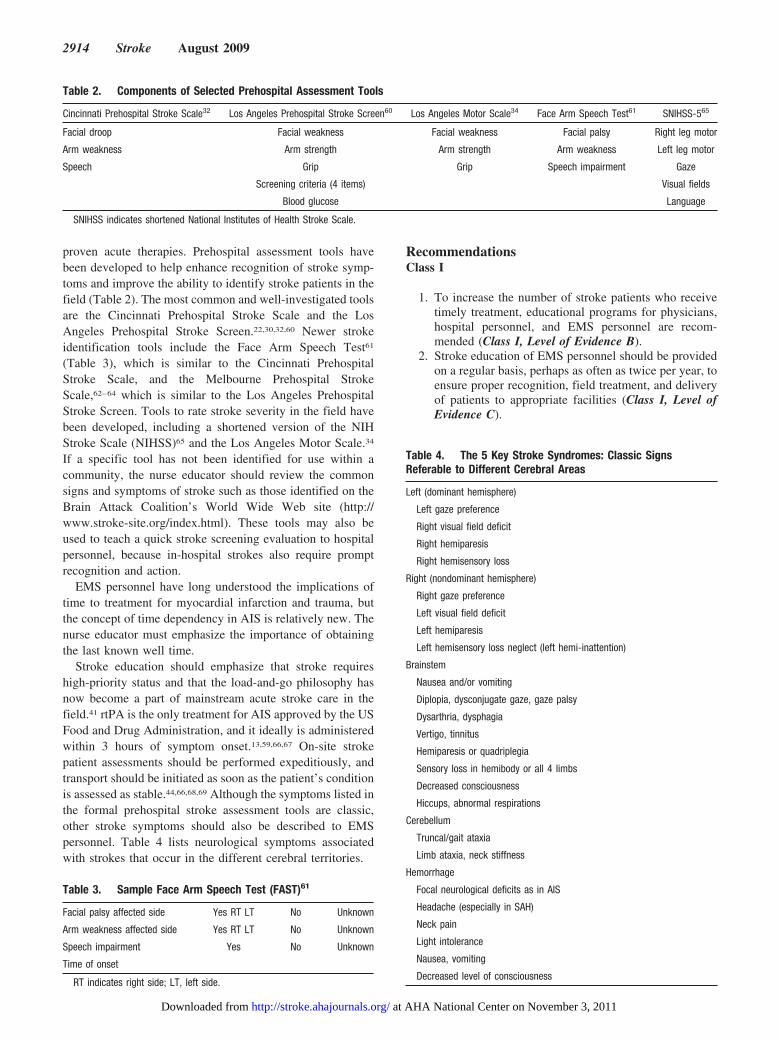
proven acute therapies. Prehospital assessment tools havebeen developed to help enhance recognition of stroke symp-toms and improve the ability to identify stroke patients in thefield (Table 2). The most common and well-investigated toolsare the Cincinnati Prehospital Stroke Scale and the LosAngeles Prehospital Stroke Screen.22,30,32,60 Newer strokeidentification tools include the Face Arm Speech Test61
(Table 3), which is similar to the Cincinnati PrehospitalStroke Scale, and the Melbourne Prehospital StrokeScale,62–64 which is similar to the Los Angeles PrehospitalStroke Screen. Tools to rate stroke severity in the field havebeen developed, including a shortened version of the NIHStroke Scale (NIHSS)65 and the Los Angeles Motor Scale.34
If a specific tool has not been identified for use within acommunity, the nurse educator should review the commonsigns and symptoms of stroke such as those identified on theBrain Attack Coalition’s World Wide Web site (http://www.stroke-site.org/index.html). These tools may also beused to teach a quick stroke screening evaluation to hospitalpersonnel, because in-hospital strokes also require promptrecognition and action.
EMS personnel have long understood the implications oftime to treatment for myocardial infarction and trauma, butthe concept of time dependency in AIS is relatively new. Thenurse educator must emphasize the importance of obtainingthe last known well time.
Stroke education should emphasize that stroke requireshigh-priority status and that the load-and-go philosophy hasnow become a part of mainstream acute stroke care in thefield.41 rtPA is the only treatment for AIS approved by the USFood and Drug Administration, and it ideally is administeredwithin 3 hours of symptom onset.13,59,66,67 On-site strokepatient assessments should be performed expeditiously, andtransport should be initiated as soon as the patient’s conditionis assessed as stable.44,66,68,69 Although the symptoms listed inthe formal prehospital stroke assessment tools are classic,other stroke symptoms should also be described to EMSpersonnel. Table 4 lists neurological symptoms associatedwith strokes that occur in the different cerebral territories.
RecommendationsClass I
1. To increase the number of stroke patients who receivetimely treatment, educational programs for physicians,hospital personnel, and EMS personnel are recom-mended (Class I, Level of Evidence B).
2. Stroke education of EMS personnel should be providedon a regular basis, perhaps as often as twice per year, toensure proper recognition, field treatment, and deliveryof patients to appropriate facilities (Class I, Level ofEvidence C).
Table 2. Components of Selected Prehospital Assessment Tools
Cincinnati Prehospital Stroke Scale32 Los Angeles Prehospital Stroke Screen60 Los Angeles Motor Scale34 Face Arm Speech Test61 SNIHSS-565
Facial droop Facial weakness Facial weakness Facial palsy Right leg motor
Arm weakness Arm strength Arm strength Arm weakness Left leg motor
Speech Grip Grip Speech impairment Gaze
Screening criteria (4 items) Visual fields
Blood glucose Language
SNIHSS indicates shortened National Institutes of Health Stroke Scale.
Table 3. Sample Face Arm Speech Test (FAST)61
Facial palsy affected side Yes RT LT No Unknown
Arm weakness affected side Yes RT LT No Unknown
Speech impairment Yes No Unknown
Time of onset
RT indicates right side; LT, left side.
Table 4. The 5 Key Stroke Syndromes: Classic SignsReferable to Different Cerebral Areas
Left (dominant hemisphere)
Left gaze preference
Right visual field deficit
Right hemiparesis
Right hemisensory loss
Right (nondominant hemisphere)
Right gaze preference
Left visual field deficit
Left hemiparesis
Left hemisensory loss neglect (left hemi-inattention)
Brainstem
Nausea and/or vomiting
Diplopia, dysconjugate gaze, gaze palsy
Dysarthria, dysphagia
Vertigo, tinnitus
Hemiparesis or quadriplegia
Sensory loss in hemibody or all 4 limbs
Decreased consciousness
Hiccups, abnormal respirations
Cerebellum
Truncal/gait ataxia
Limb ataxia, neck stiffness
Hemorrhage
Focal neurological deficits as in AIS
Headache (especially in SAH)
Neck pain
Light intolerance
Nausea, vomiting
Decreased level of consciousness
2914 Stroke August 2009
at AHA National Center on November 3, 2011http://stroke.ahajournals.org/Downloaded from
Education Priorities for Assessment andTreatment in the FieldNeurological assessment of the AIS patient should alwaysinclude the ABCs, vital signs, cardiac monitoring duringtransport, and baseline neurological assessment. Because thefield neurological examination will serve as a baseline forassessment of neurological improvement or worsening, theuse of a prehospital stroke scale is recommended.
EMS personnel on the scene should ask the patient’sfamily or bystanders when the patient was last known to benormal or without neurological deficits, ie, the last knownwell time. Documentation of this report of onset can behelpful in establishing an accurate time of stroke symptomonset.44,46,47,69 Ideally, standardized definitions should bedeveloped in EMS systems to define the specific onset dateand time. The date and time should be defined as the timewhen the stroke symptoms that brought the patient to thehospital first occurred. A specific time can be identifiedwithin a reasonable amount of certainty within �15 minutes.When possible, the information should be obtained directlyfrom the patient. If the patient is unable to give this informa-tion, EMS personnel should look to another reliable sourcefor this information.70 If the time of onset of stroke symptomsis not identifiable, a standard method of time parametersshould be used, such as morning (6:00 AM to 11:59 AM),afternoon (noon to 5:59 PM), evening (6:00 PM to 11:59 PM),and overnight (midnight to 5:59 AM).70 EMS providers mustemphasize to families the importance of traveling to thehospital with the patient, particularly if symptom onset iswithin the time frame for rtPA administration and thepatient’s language or decision-making capability is compro-mised. When family members cannot accompany the patient,EMS personnel should document the family’s contact infor-mation and provide it to the emergency physician.13
The current guidelines recommend the use of continuouscardiac monitoring during transport of a suspected strokepatient to determine the presence of cardiac arrhythmias.13 Ifthere is no standing field protocol for management of cardiacconditions, EMS personnel should contact the base station orreceiving institution if the electrocardiogram demonstratespossible acute myocardial ischemia or atrial fibrillation.Blood pressure should be monitored every 15 minutes, ormore often if severe hypertension (systolic blood pressure�200 mm Hg) or relative hypotension (systolic blood pres-sure �110 mm Hg) is observed during transport. Adminis-tration of antihypertensive drugs in the field is not recom-mended, because induced hypotension carries a possible riskof extending the area of cerebral infarct.47,58,68,71 Supplemen-tal oxygen should be given to hypoxic patients; in ambu-lances without oximetry capabilities, oxygen can be admin-istered at low levels, eg, 2 to 3 L/min. If pulse oximetry isavailable and the patient’s oxygen saturation is �92%, addi-tional oxygen is not needed.13 Transport with the head of the bedelevated �30° may help with oxygenation and may minimizethe possibility of aspiration.44,71,72 To decrease the risk ofaspiration, the patient should receive nothing by mouth (NPO).
Hypoglycemia, a common stroke mimic, can be identifiedquickly by measuring blood glucose during transport. Finger-stick tests can be performed if the emergency vehicle is
appropriately equipped and personnel are trained. Treatmentof severe hypoglycemia should be instituted promptly byEMS personnel. Intravenous access can be established in thefield, and non–glucose-containing intravenous fluids can bestarted if the patient is hypotensive. Establishment of intra-venous access should not delay transport.44,46,47,57,69,71
Finally, the nurse educator should emphasize the value ofearly notification of the receiving ED of the arrival of apotential acute stroke patient. Historic cardiac trials haveshown that prearrival notification of the ED enhances rapiddiagnostic workup, reducing time between symptom onsetand treatment.23,25,73
RecommendationsClass I
1. EMS personnel should be trained to administer a validatedprehospital stroke assessment, such as the CincinnatiPrehospital Stroke Scale or the Los Angeles PrehospitalStroke Screen (Class I, Level of Evidence B).
2. EMS personnel should be trained to determine the lastknown well time using standardized definitions tocollect the most accurate information (Class I, Level ofEvidence B).
3. EMS personnel should use the neurological/stroke as-sessment approach to gather basic physiological infor-mation about the patient and communicate the patient’scondition to the receiving hospital (Class I, Level ofEvidence B).
From the Field to the ED: Stroke Patient Triageand CareEmergency personnel initiate basic triage and care modalitiesin the field. Once the stroke patient arrives in the ED, patienttriage is usually a function of nursing staff. The EmergencyNurses Association and the American College of EmergencyPhysicians recommend a 5-level Emergency Severity Indexas a preferred system for triage in a busy ED.52 This indexputs all stroke patients in the level 2 or “needs immediateassessment” category, the same as for an unstable traumapatient or a critical care cardiac patient.46,52,69 The emergencynurse must be able to recognize neurological symptoms thatsuggest stroke and rapidly assess the initial time of symptomonset or the last known well time.44,46,57
The triage nurse should use specialized checklists, proto-cols, and other tools to identify stroke patients.10,44,47,57,74
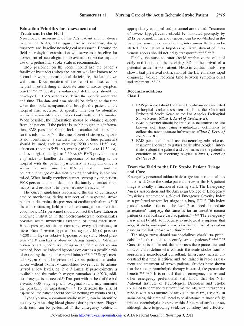
Once stroke is confirmed, the nurse uses these procedures andprotocols that define who contacts the acute stroke team orappropriate neurological consultant. Emergency nurses un-derstand that time is critical and are trained in rapid assess-ment and treatment of stroke patients. Studies have shownthat the sooner thrombolytic therapy is started, the greater thebenefit.23,44,46,75 It is critical that all emergency nurses andother emergency professional staff know that the NIH-National Institute of Neurological Disorders and Stroke(NINDS) benchmark treatment time for AIS with intravenousrtPA is within 60 minutes of arrival in the ED46 (Table 5). Insome cases, this time will need to be shortened to successfullyinitiate thrombolytic therapy within 3 hours of stroke onset,although there is growing evidence of safety and effective-
Summers et al Nursing Care of the Acute Ischemic Stroke Patient 2915
at AHA National Center on November 3, 2011http://stroke.ahajournals.org/Downloaded from
ness beyond the 3-hour window from stroke onset.76,77 TheAHA AIS Writing Committee has issued a Science Advisorystating that some eligible patients may be treated between the3- and 4.5-hour window after stroke. The recommendationcomes with several caveats and follows the inclusion criteriadescribed in the ECASS III results. The exceptions includepersons �80 years of age, those taking oral anticoagulantswith an international normalized ratio of �1.7, persons withan NIHSS score �25, and those with a history of stroke anddiabetes.77a
RecommendationsClass I
1. EDs should establish standard operating procedures andprotocols to triage stroke patients expeditiously (ClassI, Level of Evidence B).
2. Standard procedures and protocols should be estab-lished for benchmarking time to evaluate and treateligible stroke patients with rtPA expeditiously (Class I,Level of Evidence B).
3. Target treatment with rtPA should be within 1 hour ofthe patient’s arrival in the ED (Class I, Level ofEvidence A).
4. Eligible patients can be treated between the 3- to4.5-hour window when evaluated carefully for exclu-sions to treatment (Class I, Level of Evidence B).
Emergency Nursing Interventions in theEmergency/Hyperacute Phase of Stroke
The First 24 HoursStroke symptoms typically begin suddenly (but can evolveover minutes to hours) and are referable to the affected regionof the brain. Ischemic stroke symptoms are generally dividedinto those that affect the anterior and posterior cerebralcirculation (Table 4). To properly triage patients for AIStherapies such as rtPA, emergency nurses should be familiarwith both typical and unusual stroke presentations.
As in the prehospital phase, initial patient assessmentsmade by the emergency nurse are based on the principle ofassessing the ABCs, vital signs, and neurological assessment.The majority of AIS patients will present to the ED in ahemodynamically stable condition; however, ischemicstrokes involving the posterior circulation can require aggres-
sive airway management, especially if the patient has analtered level of consciousness.26,78
Circulatory collapse or cardiac arrest, although possible, isuncommon in isolated ischemic stroke.79 The occurrence ofeither may indicate other medical conditions such as acutemyocardial infarction, atrial fibrillation, or congestive heartfailure. Cardiac monitoring of all suspected stroke patients inthe ED helps identify these conditions.69
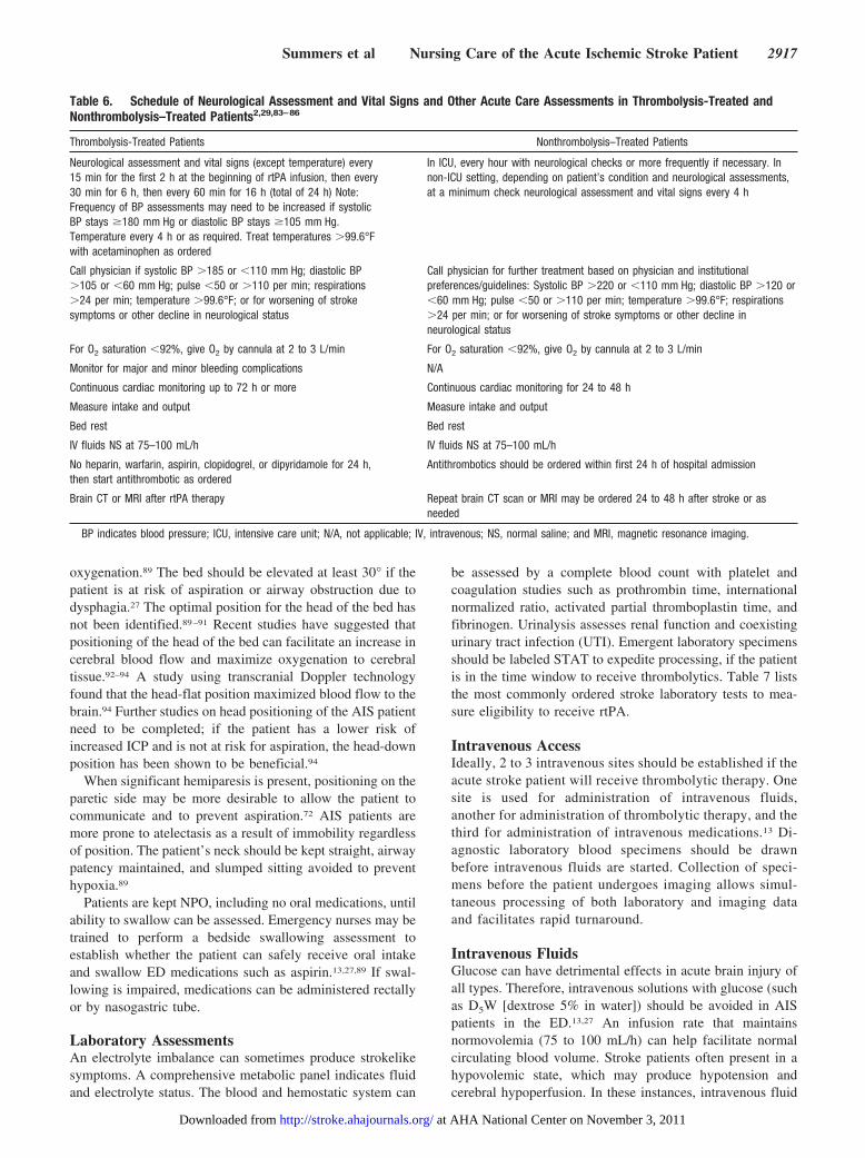
Initial ED documentation of the stroke patient begins withthe recording of all information included in the neurological/stroke assessment. Vital signs, including temperature, may bemeasured frequently as clinically indicated but not less thanevery 30 minutes while the patient is in the ED. Hyperthermiais associated with poor outcome in stroke patients13,80–82;therefore, it is important to consider treating any fever�99.6°F. During the 60-minute infusion of thrombolytictherapy, pulse and blood pressure should be checked at leastevery 15 minutes. Table 6 summarizes nursing care associ-ated with thrombolysis and nonthrombolysis treatment ofpatients with acute cerebral ischemia on the basis of theoriginal NIH-NINDS study protocol and AHA/ASAguidelines.2,29,83–86
Brain ImagingThe overriding objective of emergency evaluation of thestroke patient is to determine whether the stroke is anischemic infarction or ICH and to exclude a nonvascularlesion as the cause of symptoms. Additional objectives are tolocalize the lesion, determine its age and extent, and docu-ment its mechanism. Both computed tomography (CT) andmagnetic resonance imaging (MRI) are acceptable initialimaging modalities for acute evaluation. The most commonlyobtained study remains an immediate unenhanced (noncon-trast) head CT scan.13 The emergency nurse prepares thepatient for CT or MRI by explaining the test and may helptransport the patient to the scanner. The nurse should preno-tify the CT department that a patient with suspected acutestroke is in transport. This will allow technicians to reservethe scanner so that the patient can be imaged immediately onarrival. The CT scan should be completed in �25 minutes inpatients who are eligible for treatment with rtPA. The initialscan is one of the most important diagnostic tests in theemergency phase after stroke. Rapid acquisition and results ofimaging will define treatment.
Oxygenation, Positioning, and Oral IntakePatients with AIS are at risk of hypoxemia and oxygendesaturation. Maximization of oxygenation of all acute strokepatients has been examined in 1 quasi-randomized trial anddid not show clear findings of benefit from supplementaloxygen.87 There is general agreement, however, that hypoxicpatients will benefit from supplemental oxygen.13
Positioning of the head of the bed must be individualizedfor each patient. The traditional positioning at 25° to 30° isoften used for potentially increased intracranial pressure(ICP), at least until large lobar, ICH, space-occupying lesionsor other causes of increased ICP can be ruled out byimaging.88 Stroke patients with increased ICP and chronicrespiratory conditions may need head elevation for maximum
Table 5. NINDS Time Targets for Organized Triage of AcuteStroke Patients44: Key Evaluation Time Targets for the PotentialrtPA Candidate
Maximum intervals recommended by NINDS
Door-to–doctor first sees patient 10 min
Door-to–CT completed 25 min
Door-to–CT read 45 min
Door-to–thrombolytic therapy starts 60 min
Physician examination 15 min
Neurosurgical expertise available* 2 h
Admitted to monitored bed 3 h
CT indicates computed tomography.*On-site or by transfer to another facility.
2916 Stroke August 2009
at AHA National Center on November 3, 2011http://stroke.ahajournals.org/Downloaded from
oxygenation.89 The bed should be elevated at least 30° if thepatient is at risk of aspiration or airway obstruction due todysphagia.27 The optimal position for the head of the bed hasnot been identified.89–91 Recent studies have suggested thatpositioning of the head of the bed can facilitate an increase incerebral blood flow and maximize oxygenation to cerebraltissue.92–94 A study using transcranial Doppler technologyfound that the head-flat position maximized blood flow to thebrain.94 Further studies on head positioning of the AIS patientneed to be completed; if the patient has a lower risk ofincreased ICP and is not at risk for aspiration, the head-downposition has been shown to be beneficial.94
When significant hemiparesis is present, positioning on theparetic side may be more desirable to allow the patient tocommunicate and to prevent aspiration.72 AIS patients aremore prone to atelectasis as a result of immobility regardlessof position. The patient’s neck should be kept straight, airwaypatency maintained, and slumped sitting avoided to preventhypoxia.89
Patients are kept NPO, including no oral medications, untilability to swallow can be assessed. Emergency nurses may betrained to perform a bedside swallowing assessment toestablish whether the patient can safely receive oral intakeand swallow ED medications such as aspirin.13,27,89 If swal-lowing is impaired, medications can be administered rectallyor by nasogastric tube.
Laboratory AssessmentsAn electrolyte imbalance can sometimes produce strokelikesymptoms. A comprehensive metabolic panel indicates fluidand electrolyte status. The blood and hemostatic system can
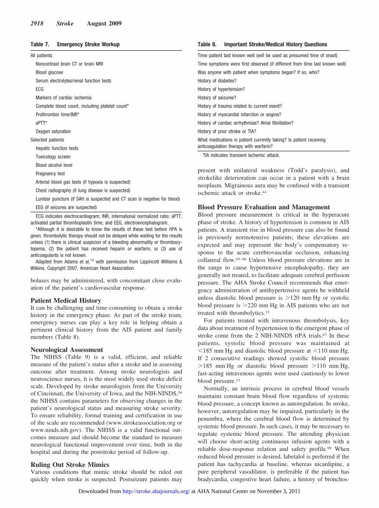
be assessed by a complete blood count with platelet andcoagulation studies such as prothrombin time, internationalnormalized ratio, activated partial thromboplastin time, andfibrinogen. Urinalysis assesses renal function and coexistingurinary tract infection (UTI). Emergent laboratory specimensshould be labeled STAT to expedite processing, if the patientis in the time window to receive thrombolytics. Table 7 liststhe most commonly ordered stroke laboratory tests to mea-sure eligibility to receive rtPA.
Intravenous AccessIdeally, 2 to 3 intravenous sites should be established if theacute stroke patient will receive thrombolytic therapy. Onesite is used for administration of intravenous fluids,another for administration of thrombolytic therapy, and thethird for administration of intravenous medications.13 Di-agnostic laboratory blood specimens should be drawnbefore intravenous fluids are started. Collection of speci-mens before the patient undergoes imaging allows simul-taneous processing of both laboratory and imaging dataand facilitates rapid turnaround.
Intravenous FluidsGlucose can have detrimental effects in acute brain injury ofall types. Therefore, intravenous solutions with glucose (suchas D5W [dextrose 5% in water]) should be avoided in AISpatients in the ED.13,27 An infusion rate that maintainsnormovolemia (75 to 100 mL/h) can help facilitate normalcirculating blood volume. Stroke patients often present in ahypovolemic state, which may produce hypotension andcerebral hypoperfusion. In these instances, intravenous fluid
Table 6. Schedule of Neurological Assessment and Vital Signs and Other Acute Care Assessments in Thrombolysis-Treated andNonthrombolysis–Treated Patients2,29,83–86
Thrombolysis-Treated Patients Nonthrombolysis–Treated Patients
Neurological assessment and vital signs (except temperature) every15 min for the first 2 h at the beginning of rtPA infusion, then every30 min for 6 h, then every 60 min for 16 h (total of 24 h) Note:Frequency of BP assessments may need to be increased if systolicBP stays �180 mm Hg or diastolic BP stays �105 mm Hg.Temperature every 4 h or as required. Treat temperatures �99.6°Fwith acetaminophen as ordered
In ICU, every hour with neurological checks or more frequently if necessary. Innon-ICU setting, depending on patient’s condition and neurological assessments,at a minimum check neurological assessment and vital signs every 4 h
Call physician if systolic BP �185 or �110 mm Hg; diastolic BP�105 or �60 mm Hg; pulse �50 or �110 per min; respirations�24 per min; temperature �99.6°F; or for worsening of strokesymptoms or other decline in neurological status
Call physician for further treatment based on physician and institutionalpreferences/guidelines: Systolic BP �220 or �110 mm Hg; diastolic BP �120 or�60 mm Hg; pulse �50 or �110 per min; temperature �99.6°F; respirations�24 per min; or for worsening of stroke symptoms or other decline inneurological status
For O2 saturation �92%, give O2 by cannula at 2 to 3 L/min For O2 saturation �92%, give O2 by cannula at 2 to 3 L/min
Monitor for major and minor bleeding complications N/A
Continuous cardiac monitoring up to 72 h or more Continuous cardiac monitoring for 24 to 48 h
Measure intake and output Measure intake and output
Bed rest Bed rest
IV fluids NS at 75–100 mL/h IV fluids NS at 75–100 mL/h
No heparin, warfarin, aspirin, clopidogrel, or dipyridamole for 24 h,then start antithrombotic as ordered
Antithrombotics should be ordered within first 24 h of hospital admission
Brain CT or MRI after rtPA therapy Repeat brain CT scan or MRI may be ordered 24 to 48 h after stroke or asneeded
BP indicates blood pressure; ICU, intensive care unit; N/A, not applicable; IV, intravenous; NS, normal saline; and MRI, magnetic resonance imaging.
Summers et al Nursing Care of the Acute Ischemic Stroke Patient 2917
at AHA National Center on November 3, 2011http://stroke.ahajournals.org/Downloaded from
boluses may be administered, with concomitant close evalu-ation of the patient’s cardiovascular response.
Patient Medical HistoryIt can be challenging and time-consuming to obtain a strokehistory in the emergency phase. As part of the stroke team,emergency nurses can play a key role in helping obtain apertinent clinical history from the AIS patient and familymembers (Table 8).
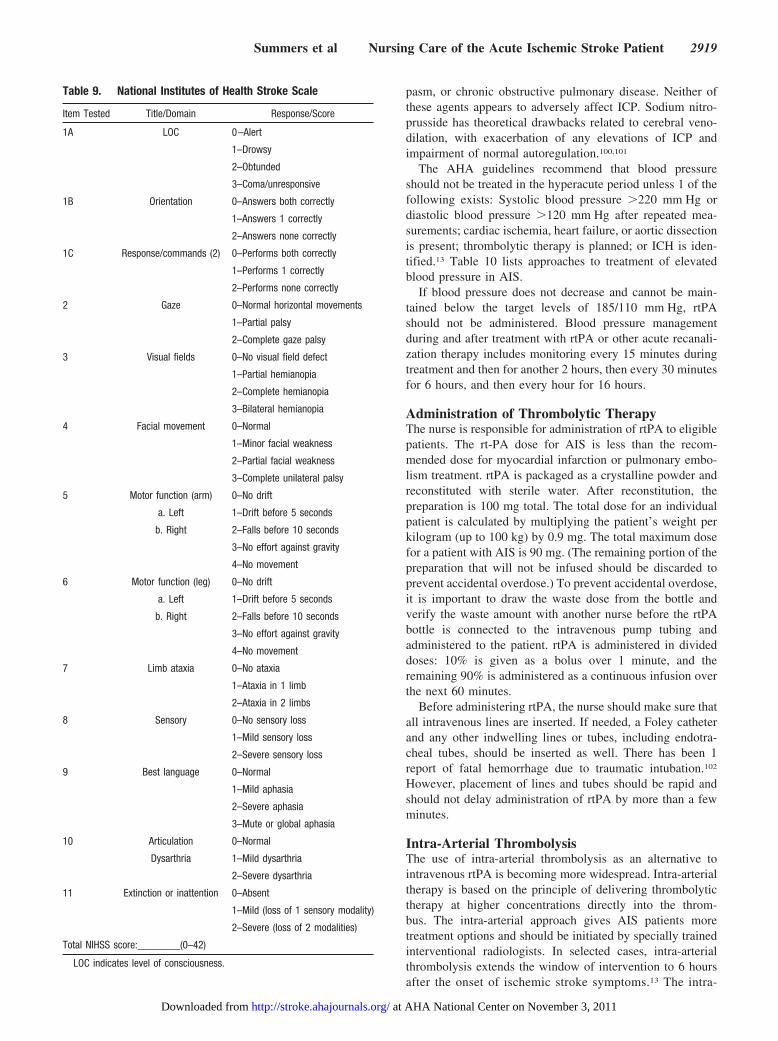
Neurological AssessmentThe NIHSS (Table 9) is a valid, efficient, and reliablemeasure of the patient’s status after a stroke and in assessingoutcome after treatment. Among stroke neurologists andneuroscience nurses, it is the most widely used stroke deficitscale. Developed by stroke neurologists from the Universityof Cincinnati, the University of Iowa, and the NIH-NINDS,26
the NIHSS contains parameters for observing changes in thepatient’s neurological status and measuring stroke severity.To ensure reliability, formal training and certification in useof the scale are recommended (www.strokeassociation.org orwww.ninds.nih.gov). The NIHSS is a valid functional out-comes measure and should become the standard to measureneurological functional improvement over time, both in thehospital and during the poststroke period of follow-up.
Ruling Out Stroke MimicsVarious conditions that mimic stroke should be ruled outquickly when stroke is suspected. Postseizure patients may
present with unilateral weakness (Todd’s paralysis), andstrokelike deterioration can occur in a patient with a brainneoplasm. Migrainous aura may be confused with a transientischemic attack or stroke.44
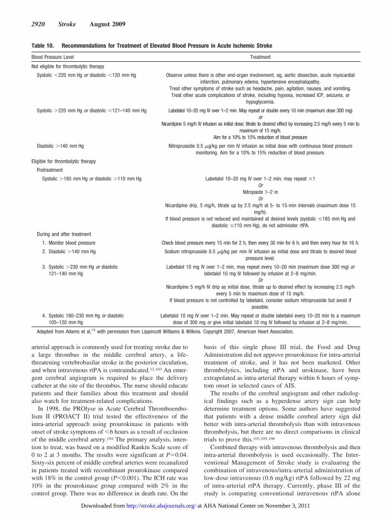
Blood Pressure Evaluation and ManagementBlood pressure measurement is critical in the hyperacutephase of stroke. A history of hypertension is common in AISpatients. A transient rise in blood pressure can also be foundin previously normotensive patients; these elevations areexpected and may represent the body’s compensatory re-sponse to the acute cerebrovascular occlusion, enhancingcollateral flow.95–98 Unless blood pressure elevations are inthe range to cause hypertensive encephalopathy, they aregenerally not treated, to facilitate adequate cerebral perfusionpressure. The AHA Stroke Council recommends that emer-gency administration of antihypertensive agents be withheldunless diastolic blood pressure is �120 mm Hg or systolicblood pressure is �220 mm Hg in AIS patients who are nottreated with thrombolytics.13
For patients treated with intravenous thrombolysis, keydata about treatment of hypertension in the emergent phase ofstroke come from the 2 NIH-NINDS rtPA trials.67 In thesepatients, systolic blood pressure was maintained at�185 mm Hg and diastolic blood pressure at �110 mm Hg.If 2 consecutive readings showed systolic blood pressure�185 mm Hg or diastolic blood pressure �110 mm Hg,fast-acting intravenous agents were used cautiously to lowerblood pressure.13
Normally, an intrinsic process in cerebral blood vesselsmaintains constant brain blood flow regardless of systemicblood pressure, a concept known as autoregulation. In stroke,however, autoregulation may be impaired, particularly in thepenumbra, where the cerebral blood flow is determined bysystemic blood pressure. In such cases, it may be necessary toregulate systemic blood pressure. The attending physicianwill choose short-acting continuous infusion agents with areliable dose-response relation and safety profile.99 Whenreduced blood pressure is desired, labetalol is preferred if thepatient has tachycardia at baseline, whereas nicardipine, apure peripheral vasodilator, is preferable if the patient hasbradycardia, congestive heart failure, a history of bronchos-
Table 8. Important Stroke/Medical History Questions
Time patient last known well (will be used as presumed time of onset)
Time symptoms were first observed (if different from time last known well)
Was anyone with patient when symptoms began? If so, who?
History of diabetes?
History of hypertension?
History of seizures?
History of trauma related to current event?
History of myocardial infarction or angina?
History of cardiac arrhythmias? Atrial fibrillation?
History of prior stroke or TIA?
What medications is patient currently taking? Is patient receivinganticoagulation therapy with warfarin?
TIA indicates transient ischemic attack.
Table 7. Emergency Stroke Workup
All patients
Noncontrast brain CT or brain MRI
Blood glucose
Serum electrolytes/renal function tests
ECG
Markers of cardiac ischemia
Complete blood count, including platelet count*
Prothrombin time/INR*
aPTT*
Oxygen saturation
Selected patients
Hepatic function tests
Toxicology screen
Blood alcohol level
Pregnancy test
Arterial blood gas tests (if hypoxia is suspected)
Chest radiography (if lung disease is suspected)
Lumbar puncture (if SAH is suspected and CT scan is negative for blood)
EEG (if seizures are suspected)
ECG indicates electrocardiogram; INR, international normalized ratio; aPTT,activated partial thromboplastin time; and EEG, electroencephalogram.
*Although it is desirable to know the results of these test before rtPA isgiven, thrombolytic therapy should not be delayed while waiting for the resultsunless (1) there is clinical suspicion of a bleeding abnormality or thrombocy-topenia, (2) the patient has received heparin or warfarin, or (3) use ofanticoagulants is not known.
Adapted from Adams et al,13 with permission from Lippincott Williams &Wilkins. Copyright 2007, American Heart Association.
2918 Stroke August 2009
at AHA National Center on November 3, 2011http://stroke.ahajournals.org/Downloaded from
pasm, or chronic obstructive pulmonary disease. Neither ofthese agents appears to adversely affect ICP. Sodium nitro-prusside has theoretical drawbacks related to cerebral veno-dilation, with exacerbation of any elevations of ICP andimpairment of normal autoregulation.100,101
The AHA guidelines recommend that blood pressureshould not be treated in the hyperacute period unless 1 of thefollowing exists: Systolic blood pressure �220 mm Hg ordiastolic blood pressure �120 mm Hg after repeated mea-surements; cardiac ischemia, heart failure, or aortic dissectionis present; thrombolytic therapy is planned; or ICH is iden-tified.13 Table 10 lists approaches to treatment of elevatedblood pressure in AIS.
If blood pressure does not decrease and cannot be main-tained below the target levels of 185/110 mm Hg, rtPAshould not be administered. Blood pressure managementduring and after treatment with rtPA or other acute recanali-zation therapy includes monitoring every 15 minutes duringtreatment and then for another 2 hours, then every 30 minutesfor 6 hours, and then every hour for 16 hours.
Administration of Thrombolytic TherapyThe nurse is responsible for administration of rtPA to eligiblepatients. The rt-PA dose for AIS is less than the recom-mended dose for myocardial infarction or pulmonary embo-lism treatment. rtPA is packaged as a crystalline powder andreconstituted with sterile water. After reconstitution, thepreparation is 100 mg total. The total dose for an individualpatient is calculated by multiplying the patient’s weight perkilogram (up to 100 kg) by 0.9 mg. The total maximum dosefor a patient with AIS is 90 mg. (The remaining portion of thepreparation that will not be infused should be discarded toprevent accidental overdose.) To prevent accidental overdose,it is important to draw the waste dose from the bottle andverify the waste amount with another nurse before the rtPAbottle is connected to the intravenous pump tubing andadministered to the patient. rtPA is administered in divideddoses: 10% is given as a bolus over 1 minute, and theremaining 90% is administered as a continuous infusion overthe next 60 minutes.
Before administering rtPA, the nurse should make sure thatall intravenous lines are inserted. If needed, a Foley catheterand any other indwelling lines or tubes, including endotra-cheal tubes, should be inserted as well. There has been 1report of fatal hemorrhage due to traumatic intubation.102
However, placement of lines and tubes should be rapid andshould not delay administration of rtPA by more than a fewminutes.
Intra-Arterial ThrombolysisThe use of intra-arterial thrombolysis as an alternative tointravenous rtPA is becoming more widespread. Intra-arterialtherapy is based on the principle of delivering thrombolytictherapy at higher concentrations directly into the throm-bus. The intra-arterial approach gives AIS patients moretreatment options and should be initiated by specially trainedinterventional radiologists. In selected cases, intra-arterialthrombolysis extends the window of intervention to 6 hoursafter the onset of ischemic stroke symptoms.13 The intra-
Table 9. National Institutes of Health Stroke Scale
Item Tested Title/Domain Response/Score
1A LOC 0–Alert
1–Drowsy
2–Obtunded
3–Coma/unresponsive
1B Orientation 0–Answers both correctly
1–Answers 1 correctly
2–Answers none correctly
1C Response/commands (2) 0–Performs both correctly
1–Performs 1 correctly
2–Performs none correctly
2 Gaze 0–Normal horizontal movements
1–Partial palsy
2–Complete gaze palsy
3 Visual fields 0–No visual field defect
1–Partial hemianopia
2–Complete hemianopia
3–Bilateral hemianopia
4 Facial movement 0–Normal
1–Minor facial weakness
2–Partial facial weakness
3–Complete unilateral palsy
5 Motor function (arm) 0–No drift
a. Left 1–Drift before 5 seconds
b. Right 2–Falls before 10 seconds
3–No effort against gravity
4–No movement
6 Motor function (leg) 0–No drift
a. Left 1–Drift before 5 seconds
b. Right 2–Falls before 10 seconds
3–No effort against gravity
4–No movement
7 Limb ataxia 0–No ataxia
1–Ataxia in 1 limb
2–Ataxia in 2 limbs
8 Sensory 0–No sensory loss
1–Mild sensory loss
2–Severe sensory loss
9 Best language 0–Normal
1–Mild aphasia
2–Severe aphasia
3–Mute or global aphasia
10 Articulation 0–Normal
Dysarthria 1–Mild dysarthria
2–Severe dysarthria
11 Extinction or inattention 0–Absent
1–Mild (loss of 1 sensory modality)
2–Severe (loss of 2 modalities)
Total NIHSS score:________(0–42)
LOC indicates level of consciousness.
Summers et al Nursing Care of the Acute Ischemic Stroke Patient 2919
at AHA National Center on November 3, 2011http://stroke.ahajournals.org/Downloaded from
arterial approach is commonly used for treating stroke due toa large thrombus in the middle cerebral artery, a life-threatening vertebrobasilar stroke in the posterior circulation,and when intravenous rtPA is contraindicated.12,103 An emer-gent cerebral angiogram is required to place the deliverycatheter at the site of the thrombus. The nurse should educatepatients and their families about this treatment and shouldalso watch for treatment-related complications.
In 1998, the PROlyse in Acute Cerebral Thromboembo-lism II (PROACT II) trial tested the effectiveness of theintra-arterial approach using prourokinase in patients withonset of stroke symptoms of �6 hours as a result of occlusionof the middle cerebral artery.104 The primary analysis, inten-tion to treat, was based on a modified Rankin Scale score of0 to 2 at 3 months. The results were significant at P�0.04.Sixty-six percent of middle cerebral arteries were recanalizedin patients treated with recombinant prourokinase comparedwith 18% in the control group (P�0.001). The ICH rate was10% in the prourokinase group compared with 2% in thecontrol group. There was no difference in death rate. On the
basis of this single phase III trial, the Food and DrugAdministration did not approve prourokinase for intra-arterialtreatment of stroke, and it has not been marketed. Otherthrombolytics, including rtPA and urokinase, have beenextrapolated as intra-arterial therapy within 6 hours of symp-tom onset in selected cases of AIS.
The results of the cerebral angiogram and other radiolog-ical findings such as a hyperdense artery sign can helpdetermine treatment options. Some authors have suggestedthat patients with a dense middle cerebral artery sign didbetter with intra-arterial thrombolysis than with intravenousthrombolysis, but there are no direct comparisons in clinicaltrials to prove this.103,105,106
Combined therapy with intravenous thrombolysis and thenintra-arterial thrombolysis is used occasionally. The Inter-ventional Management of Stroke study is evaluating thecombination of intravenous/intra-arterial administration oflow-dose intravenous (0.6 mg/kg) rtPA followed by 22 mgof intra-arterial rtPA therapy. Currently, phase III of thestudy is comparing conventional intravenous rtPA alone
Table 10. Recommendations for Treatment of Elevated Blood Pressure in Acute Ischemic Stroke
Blood Pressure Level Treatment
Not eligible for thrombolytic therapy
Systolic �220 mm Hg or diastolic �120 mm Hg Observe unless there is other end-organ involvement, eg, aortic dissection, acute myocardialinfarction, pulmonary edema, hypertensive encephalopathy.
Treat other symptoms of stroke such as headache, pain, agitation, nausea, and vomiting.Treat other acute complications of stroke, including hypoxia, increased ICP, seizures, or
hypoglycemia.
Systolic �220 mm Hg or diastolic �121–140 mm Hg Labetalol 10–20 mg IV over 1–2 min. May repeat or double every 10 min (maximum dose 300 mg)or
Nicardipine 5 mg/h IV infusion as initial dose; titrate to desired effect by increasing 2.5 mg/h every 5 min tomaximum of 15 mg/h.
Aim for a 10% to 15% reduction of blood pressure
Diastolic �140 mm Hg Nitroprusside 0.5 �g/kg per min IV infusion as initial dose with continuous blood pressuremonitoring. Aim for a 10% to 15% reduction of blood pressure.
Eligible for thrombolytic therapy
Pretreatment
Systolic �185 mm Hg or diastolic �110 mm Hg Labetalol 10–20 mg IV over 1–2 min; may repeat �1Or
Nitropaste 1–2 inOr
Nicardipine drip, 5 mg/h, titrate up by 2.5 mg/h at 5- to 15-min intervals (maximum dose 15mg/h).
If blood pressure is not reduced and maintained at desired levels (systolic �185 mm Hg anddiastolic �110 mm Hg), do not administer rtPA.
During and after treatment
1. Monitor blood pressure Check blood pressure every 15 min for 2 h, then every 30 min for 6 h, and then every hour for 16 h.
2. Diastolic �140 mm Hg Sodium nitroprusside 0.5 �g/kg per min IV infusion as initial dose and titrate to desired bloodpressure level.
3. Systolic �230 mm Hg or diastolic121–140 mm Hg
Labetalol 10 mg IV over 1–2 min, may repeat every 10–20 min (maximum dose 300 mg) orlabetalol 10 mg IV followed by infusion at 2–8 mg/min.
OrNicardipine 5 mg/h IV drip as initial dose, titrate up to desired effect by increasing 2.5 mg/h
every 5 min to maximum dose of 15 mg/h.If blood pressure is not controlled by labetalol, consider sodium nitroprusside but avoid if
possible.
4. Systolic 180–230 mm Hg or diastolic105–120 mm Hg
Labetalol 10 mg IV over 1–2 min. May repeat or double labetalol every 10–20 min to a maximumdose of 300 mg or give initial labetalol 10 mg IV followed by infusion at 2–8 mg/min.
Adapted from Adams et al,13 with permission from Lippincott Williams & Wilkins. Copyright 2007, American Heart Association.
2920 Stroke August 2009
at AHA National Center on November 3, 2011http://stroke.ahajournals.org/Downloaded from
with rtPA plus endovascular interventions.103,107 Furtherstudies will provide more data on the efficacy of intra-ar-terial thrombolysis.
Mechanical DevicesThe Merci Retriever (Concentric Medical, Mountain View,Calif) was the first retrieval device approved for clot retrac-tion in AIS patients who were not candidates for rtPA or whohad failed intravenous therapy.108–110 The Penumbra System(Penumbra Inc, Alameda, Calif) was the second retrievaldevice approved to remove blood clots in patients withAIS.111 These devices have been used in combination withintravenous or intra-arterial therapy. Noser et al109 havesuggested that aggressive mechanical clot disruption mayhelp increase recanalization rates compared with intra-arterialthrombolysis. This information is not yet supported byclinical trials.
Several other approaches to recanalization with devicecatheters are available. The EKOS catheter (EKOS Corp,Bothell, Wash) used in the Interventional Management ofStroke phase I and II trials delivered intra-arterial rtPA withconcurrent intra-arterial low-energy ultrasound. Use of trans-cranial Doppler ultrasound to enhance the thrombolytic ac-tivity of intravenous rtPA is being evaluated in phase IIclinical trials.112 These device catheters have been evaluatedin safety and technical efficacy trials. Randomized, controlledclinical trials of both the Merci and EKOS devices must becompleted to evaluate their clinical efficacy.107,113
RecommendationsClass I
1. Emergency personnel should be highly trained in strokecare (Class I, Level of Evidence B).
2. Frequent neurological/stroke assessments should bedone (Class I, Level of Evidence C); these should bedone more frequently for patients receiving rtPA.
3. Supplemental oxygen should be given to patients withan oxygen saturation of �92% and a decreased level ofconsciousness (Class I, Level of Evidence C). There islittle evidence that supplemental oxygen should beprovided routinely.
4. The stroke patient’s head should be positioned inneutral alignment with the body, and the head of the bedshould be elevated 25° to 30° to help the patient handleoral secretions, especially if dysphagia is present (ClassI, Level of Evidence C).
5. Stroke patients in the ED should be kept NPO (notgiven anything orally) until ability to swallow is as-sessed (Class I, Level of Evidence B).
6. Intravenous access should be obtained in at least 2 sites,with 1 site for administration of rtPA and 1 site fordelivery of intravenous fluids or other medications if thepatient is a candidate for rtPA (Class I, Level ofEvidence C).
7. Only nondextrose, normotonic intravenous fluids suchas normal saline should be used in the AIS patient(Class I, Level of Evidence C).
8. Intravenous rtPA should be administered without delayand should not be excluded in an eligible patient (ClassI, Level of Evidence A).
9. See Table 11 for additional medical recommendations.
Phase 2: Acute CareDuring the acute care phase, nursing care should focus oncontinued stabilization of the stroke patient through frequentevaluation of neurological status, blood pressure manage-ment, and prevention of complications. Medical managementfocuses on establishing the cause or etiology of AIS, preven-tion of treatment-related complications, and evaluation ofsecondary prevention strategies. There is considerable evi-dence that dedicated stroke teams, units, and coordinated careimprove clinical outcomes in the acute care phase.5,7,57,114–121
Clinical Pathways and Stroke Order SetsSpecific order sets (standing orders) that address issues suchas control of blood glucose, parameters to treat fever, andconsultations with other multidisciplinary team membersshould be developed. The Brain Attack Coalition recom-mends succinct, organized stroke care in its recommendationsfor the development of primary stroke centers.10
Specific Care in the Acute Phase: ImmediateMedical and Nursing ManagementA key element of the management of patients with acutestroke is to prevent deterioration and medical complications,
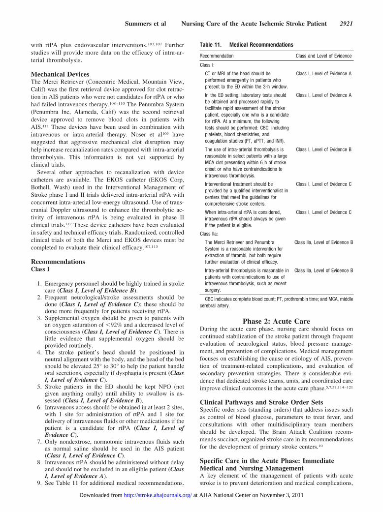
Table 11. Medical Recommendations
Recommendation Class and Level of Evidence
Class I:
CT or MRI of the head should beperformed emergently in patients whopresent to the ED within the 3-h window.
Class I, Level of Evidence A
In the ED setting, laboratory tests shouldbe obtained and processed rapidly tofacilitate rapid assessment of the strokepatient, especially one who is a candidatefor rtPA. At a minimum, the followingtests should be performed: CBC, includingplatelets, blood chemistries, andcoagulation studies (PT, aPTT, and INR).
Class I, Level of Evidence A
The use of intra-arterial thrombolysis isreasonable in select patients with a largeMCA clot presenting within 6 h of strokeonset or who have contraindications tointravenous thrombolysis.
Class I, Level of Evidence B
Interventional treatment should beprovided by a qualified interventionalist incenters that meet the guidelines forcomprehensive stroke centers.
Class I, Level of Evidence C
When intra-arterial rtPA is considered,intravenous rtPA should always be givenif the patient is eligible.
Class I, Level of Evidence C
Class IIa:
The Merci Retriever and PenumbraSystem is a reasonable intervention forextraction of thrombi, but both requirefurther evaluation of clinical efficacy.
Class IIa, Level of Evidence B
Intra-arterial thrombolysis is reasonable inpatients with contraindications to use ofintravenous thrombolysis, such as recentsurgery.
Class IIa, Level of Evidence B
CBC indicates complete blood count; PT, prothrombin time; and MCA, middlecerebral artery.
Summers et al Nursing Care of the Acute Ischemic Stroke Patient 2921
at AHA National Center on November 3, 2011http://stroke.ahajournals.org/Downloaded from
such as respiratory problems associated with smoking orpneumonia, hypertension, hyperglycemia, dehydration, mal-nourishment, fever, coronary artery disease, cerebral edema,infection, and thromboembolism (deep vein thrombosis[DVT] or pulmonary embolism). These all worsen overallpatient outcome.13,82,122–131
To provide high-quality care, nurses must coordinate theactivities of the multidisciplinary team. Clinical pathways orphysician standing orders can guide the team in managingstroke patients and are useful for coordinating diagnostic testsand appropriate therapies and care issues. Clinical pathwaysimprove coordination of acute stroke care and dischargeplanning, decrease hospital costs, decrease readmission rates,reduce length of hospital stay, and enhance usefulness ofoutcome measurement and quality improvement.114,132,133
Examples of professional resources including clinical path-ways, guidelines, and standing orders can be found on theAHA Web site (http://www.americanheart.org/presenter.jhtml?identifier�3047992) and the Brain Attack CoalitionWeb site (http://www.stroke-site.org).
Intensive ManagementExperts estimate up to 30% of all stroke patients willdeteriorate in the first 24 hours.13,129 This statistic supportsthe need for intensive monitoring by nurses specificallytrained in acute stroke care. Patients who receivethrombolytic therapy should also be monitored closely for atleast 24 hours after treatment. Care may be provided in adesignated intensive care unit or a stroke unit with continuouscardiac telemetry. In either area, nurses are trained in the careof patients after thrombolysis, are aware of bleeding compli-cations, are trained in the use of appropriate neurologicalassessment tools, and are adept at recognizing the signs ofincreasing ICP often related to large stroke lesions.86 Thenurse-patient ratio is 1:2 for the first 24 hours; then, if thepatient’s condition is stable, the ratio is 1:4 asappropriate.53,134
Bleeding assessment after administration of rtPA is theresponsibility of the clinical nurse, who monitors the patientfor major and minor bleeding complications in the first 24 to36 hours after administration of rtPA.84,135 ICH is the majorbleeding complication associated with thrombolytic thera-py.83,135 In the NINDS trials, 6.4% of treated patients hadsymptomatic ICH, which is defined as “any CT-documentedhemorrhage that was temporally related to deterioration in thepatient’s clinical condition in the judgment of the clinicalinvestigator” within 36 hours of treatment.135 Studies haveshown that there is a natural rate of hemorrhagic transforma-tion in ischemic stroke, and some studies suggest thatpetechial hemorrhages are frequently found in almost allcerebral infarcts.135–138 The use of thrombolytics increases therisk of serious hemorrhagic transformation. The nurse mustidentify which patients are at higher risk of ICH. One studyhas shown that only 3% of patients with an NIHSS score of�10 who were treated with rtPA had symptomatic ICHcompared with 17% of those with an NIHSS score �20.135
Other studies have shown that hemorrhagic transformationsare more frequent when there has been a deviation from thenational guidelines treatment protocol.139–142 Age �80 years
was determined to be an independent factor in developmentof hemorrhage after administration of rtPA.75,143 Hemor-rhagic transformation should be suspected if there is a changein level of consciousness, elevation of blood pressure, dete-rioration in motor examination, onset of new headache, ornausea and vomiting. If hemorrhage is suspected, the rtPAinfusion should be discontinued immediately.84 Managementof ICH includes immediate physician notification and attain-ment of rapid brain imaging and laboratory work, includingprothrombin time/international normalized ratio, activatedpartial thromboplastin time, fibrinogen level, complete bloodcount with platelets, and, if not already done, type andcross-match. The nursing staff must be prepared to administer6 to 8 U of cryoprecipitate containing factor VIII and 6 to 8U of platelets.83,84 The physician will decide on further actionin collaboration with other team members, such as theconsulting neurosurgeon. Facilities that treat patients withthrombolytics should have a hemorrhage algorithm (Table12) and clinical guidelines to expedite assessment and man-agement of a new ICH. Other major bleeding complicationsobserved after thrombolytic therapy are retroperitoneal, gen-itourinary, and gastrointestinal hemorrhages. Minor bleedingcomplications are common, such as oozing from gums andvenipuncture sites, as well as hematuria and hemoptysis.84
Assessment of the patient’s skin may identify hematomas orareas of ecchymosis or purpura. If the patient has antecubitalvenous access, automatic blood pressure cuffs should be usedwith caution to prevent formation of a hematoma in thepatient’s arm. The cuff site should be checked frequently,rotated, and repositioned every 2 hours. If petechiae arenoticed under the automatic blood pressure cuff, use of thecuff should be discontinued. To prevent trauma during oralcare, soft sponges should be used instead of toothbrushes inthe first 24 hours. Invasive procedures such as arterialpunctures or insertion of catheters or nasogastric tubes shouldalso be avoided in the first 24 hours after treatment.84
Neurological Stroke Assessment, Includingthe NIHSSIntensive monitoring of stroke patients includes frequentmonitoring of neurological assessment, including blood pres-sure, heart rate, and respirations. In patients treated withthrombolysis, blood pressure should be assessed at 15-minuteintervals for 2 hours, every 30 minutes for the next 6 hours,and then once per hour until 24 hours after initiation ofthrombolytic therapy. A complete bedside NIHSS assessmentmay be performed on admission to the intensive care unit,and an abbreviated version can be performed with morefrequent assessments (Table 9).86 A complete NIHSS scaleshould be done if there is evidence of neurological declineor an increase in the abbreviated score.65,84
The NIHSS provides valuable prognostic information andhas been correlated with infarct volume.13,86,144 Patients withan NIHSS score of �10 have a much more favorable outcomeat 1 year than patients with an NIHSS score of �20.144 Thenurse can use the NIHSS to identify patients who are at higherrisk for ICH after thrombolytic treatment. In the NINDS rtPAtrial, patients with an NIHSS score of �22 had a 17% risk ofICH, whereas those with an NIHSS score of �10 had only a
2922 Stroke August 2009
at AHA National Center on November 3, 2011http://stroke.ahajournals.org/Downloaded from
3% risk of ICH.13 The NIHSS may be useful for working withfamilies on discharge planning needs.145,146 In 1 study ofischemic and hemorrhagic stroke patients, a 24-hour NIHSSscore of �5 increased by nearly 5-fold the likelihood ofdischarge to home rather than inpatient rehabilitation or askilled nursing facility.145,146
Ongoing Blood Pressure ManagementBlood pressure is a critical vital sign in the AIS patient. It isnot uncommon to see variations in blood pressure after AIS.96
Blood pressure is elevated in �40% to 80% of all AISpatients,96 especially in the first 24 to 48 hours after stroke,and will fall 10 to 14 days after the acute phase.147 Elevatedblood pressure may increase cerebral perfusion in the ische-mic zone, where autoregulation is lost and perfusion ispressure dependent.148 The current guidelines recommendmaintaining blood pressure at �180/105 mm Hg for 24 hoursin patients who have received thrombolytic therapy.13 It isrecommended that antihypertensive treatment be initiated fornonthrombolytic candidates only if systolic blood pressure is�220 mm Hg or diastolic pressure is �110 mm Hg.13
Blood pressure should be monitored and assessed contin-ually for causative factors of rises. Elevated blood pressure
may be due to a physiological response to hypoxia, increasingICP, hemorrhagic transformation, full bladder, pain, nausea, aloud environment, or preexisting hypertension.13,149,150 TheASA blood pressure guidelines may be included in standingphysician orders to expedite treatment of elevated bloodpressure in the phase that immediately follows thrombolytictherapy.13 At present, the optimum blood pressure in theimmediate poststroke period is unclear and controversial, andfurther scientific evidence is needed.151,152 There is evidence,however, that rapid lowering of blood pressure may induceworsening of neurological symptoms by inducing loweredperfusion pressures to the area of ischemia.149,153,154
Arterial hypotension is rare in the AIS patient but may beassociated with volume depletion or decreased cardiac outputrelated to arrhythmias or myocardial ischemia. Patients withhypotension require evaluation with advanced neurologicalnursing assessment and telemetry monitoring. Treatmentconsists of volume replacement with normal saline andcorrection of arrhythmias.
Temperature ManagementFever appears to exacerbate the ischemic injury to neuronsand is associated with increased morbidity and mortality,
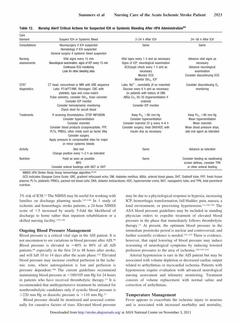
Table 12. Nursing Alert! Critical Actions for Suspected ICH or Systemic Bleeding After rtPA Administration84
CareElement Suspect ICH or Systemic Bleed 2–24 h After ICH 24–36 h After ICH
Consultations Neurosurgery if ICH suspectedHematology if ICH suspected
General surgery if systemic bleed suspected
Same Same
Nursingassessments
Vital signs every 15 minNeurological examination, signs of ICP every 15 min
Continuous ECG monitoringLook for other bleeding sites
Vital signs every 1 h and as necessarySigns of ICP, neurological examination
GCS/pupil check every 1 h and asnecessary
Monitor ECGMonitor SVO2, ICP
Advance vital signs asnecessary
Advance neurologicalexamination
Consider discontinuing ECG
STATdiagnostics
CT head, noncontrast or MRI with GRE sequenceLabs: PT/aPTT/INR, fibrinogen, CBC with
platelets, type and cross-matchPulse oximetry, consider SVO2, brain oximeter
Consider ICP monitorConsider hemodynamic monitoring
Check stool for occult blood
Labs: Na2�, osmolality (if on mannitol)Glucose every 6 h and as necessary
(in patients with history of DM)ABGs CO2 30–35 (hyperventilation if
ordered)Consider ICP monitor
Consider discontinuing O2
monitoring
Treatments If receiving thrombolytics, STOP INFUSIONConsider hyperventilation
Consider mannitolConsider blood products (cryoprecipitate, FFP,PLTs, PRBCs, other meds such as factor VIIa)
Consider surgeryApply pressure to compressible sites for major
or minor systemic bleeds
Keep PO2 �90 mm HgConsider hyperventilation
Consider mannitol 25 g every 4–6 hConsider surgery; treat DKA/HOC with
insulin drip as necessary
Keep PO2 �90 mm HgWean hyperventilation
Wean mannitolWean blood pressure drips,add oral agent as tolerated
Activity Bed restChange position every 1–2 h as tolerated
Same Advance as tolerated
Nutrition Feed as soon as possibleNPO
Consider enteral feedings with NGT or DHT
Same Consider feeding as swallowingscreen defines, consider TPN
or other enteral feeding
NINDS rtPA Stroke Study Group hemorrhage algorithm.83,84
GCS indicates Glasgow Coma Scale; GRE, gradient refocused echo; DM, diabetes mellitus; ABGs, arterial blood gases; DHT, Dobhoff tube; FFP, fresh frozenplasma; PLTs, platelets; PRBCs, packed red blood cells; DKA, diabetic ketoacidosis; HOC, hyperosmolar coma; NGT, nasogastric tube; and TPN, total parenteralnutrition.
Summers et al Nursing Care of the Acute Ischemic Stroke Patient 2923
at AHA National Center on November 3, 2011http://stroke.ahajournals.org/Downloaded from
particularly in acute stroke.155 Data from additional meta-analyses found a correlation between temperature elevationand cerebral infarct volume.80 Even an increase of 1°F is apredictor of poorer patient outcome and is an independentfactor in short- and long-term mortality rates.82,129,156,157 Therationale for this additional injury may be related to increasedmetabolic demands and free radical production. Immediatetreatment of the source of the fever will reduce its dura-tion.82,157 One approach to maintaining normothermia is toimmediately begin acetaminophen at 99.6°F.158,159 Even morerapid induction of cooling can be achieved by additionallytreating temperature elevation with indwelling catheter tem-perature control systems or surface cooling systems.158,159
Continuous Cardiac MonitoringCardiac monitoring is recommended for all ischemic strokepatients.13,160,161 Studies suggest that insular lesions can leadto cardiac arrhythmias and sudden cardiac death.13 Arrhyth-mias such as ventricular ectopy, tachycardia, and heart blockshave been associated with AIS.162 Right hemispheric infarctshave been associated with a higher incidence of arrhythmias,possibly due to sympathetic and parasympathetic nervoussystem dysfunction.163 Atrial fibrillation, often paroxysmal, iscommonly first detected only after it has caused cardioem-bolic stroke. If cardiac output is compromised, arrhythmiasmay further aggravate an already compromised cerebralblood flow. If not completed as part of the initial ED workup,a 12-lead ECG can be completed on admission. Many strokepatients have underlying cardiac problems and are at risk foran acute myocardial infarction during the acute stages ofstroke. Patients may also need a cardiac evaluation by acardiologist during the acute stages of stroke. If telemetry isunavailable, a Holter monitor can be used to check forarrhythmias.13
Assessment of OxygenationMonitoring of oxygen saturation will reduce the risk ofneurological deterioration related to hypoxemia. Supplemen-tal oxygen at 2 to 4 L/min is recommended for an oxygensaturation of �92%.13,164 Many factors compromise adequateoxygenation, for example, decreased level of consciousness,aspiration, and atelectasis. Vigilant assessment of the pa-tient’s lung sounds and ability to swallow will keep the nurseaware of pending threats to adequate oxygenation.13,135,165 Ifan oxygen saturation of 92% cannot be maintained, arterialblood gases and a chest radiograph are recommended. In theabsence of hypoxemia, supplemental oxygen is notrecommended.87
AngioedemaLarge prospective cohort studies have shown that orolingualangioedema occurs in a small proportion (1% to 2%) ofpatients with AIS or acute myocardial ischemia treated withrtPA. This was more commonly seen in patients with frontalcortex and insular ischemia, in patients who had receivedalteplase and were concurrently taking angiotensin-converting enzyme (ACE) inhibitors.102 In most cases, symp-toms were mild and transient. The pharmacological insert that
accompanies rtPA addresses this and recommends that pa-tients be monitored during infusion and for several hoursafterward for signs of allergic reaction that exhibits asorolingual angioedema (http://www.gene.com/gene/products/information/cardiovascular/activase/insert.jsp). Treatment in-cludes immediate discontinuation of rtPA and administrationof antihistamines, intravenous corticosteroids, or epinephrine.It is important for emergency and intensive care unit nurses toevaluate patients closely for throat or mouth edema and lookfor any difficulty in breathing due to angioedema.
Blood Glucose Monitoring: HyperglycemiaHyperglycemia in critically ill patients has long been associ-ated with complications. Infarct expansion, hemorrhagic con-version, and poor clinical outcomes have been reported in theAIS population.124 Even the benefit of recanalization after useof thrombolytics may be reduced.166–168 Increased bloodglucose provides additional substrate for anaerobic metabo-lism, which promotes lactic acidosis and free radical produc-tion. Elevated serum glucose is common in the acute phase ofstroke and may be related to uncontrolled or undetecteddiabetes mellitus or stress-induced hyperglycemia associatedwith cortisol and norepinephrine release at the time ofinsult.167,169–174 In 1 study, elevated glucose was present intwo thirds of AIS patients.175 Treatment with insulin confersa protective effect in critically ill patients.164,165 The ASA2007 “Guidelines for the Early Management of Adults WithIschemic Stroke” recommend the use of rapid-acting insulinfor a blood glucose level �140 mg/dL.13
Several studies have shown that elevated blood glucose isan independent factor in poor functional outcomes, increasedinfarct size, increased length of stay (7 versus 6 days),increased mortality at 30 days, and increased cost ($6611versus $5262).176–178 Not only did patients have poorer out-comes associated with hyperglycemia, but in an analysis of theNINDS rtPA trial, it was found that the risk of hemorrhagictransformation increased by 75% per 100 mg/dL of bloodglucose.167,177–180 Poorer outcomes were also seen in patientswith a glucose level �140 mg/dL after administration of rtPA.There is speculation that elevated glucose levels may preventearly reperfusion and eliminate the benefit of rtPA.166–168
Several nonrandomized studies have suggested improvedoutcome if there is an acute reversal of hyperglycemia.167,181–184
The Glucose Insulin in Stroke Trial (GIST), a multicenter,randomized trial, recruited 933 patients and evaluated the useof variable-dose glucose and potassium insulin (GKI) versussaline infusions in stroke patients with glucose levels of 6.0 to17.0 mmol/L (�300 mg/dL and defined as mild to moderatehyperglycemia).185 The purpose of the GKI infusion was tomaintain glucose at 4 to 7 mmol/L (euglycemic); there was noglucose intervention in the control group after stroke. Mostpatients were entered in the study within 24 hours after onsetof stroke symptoms. The primary outcome was death at 90days. The study was stopped early owing to low recruitment.Based on the intention-to-treat data, there was no significantreduction in morbidity or mortality at 90 days in the inter-vention group (GKI versus control: odds ratio 1.14, 95%confidence interval 0.86 to 1.52, P�0.37). Plasma glucose
2924 Stroke August 2009
at AHA National Center on November 3, 2011http://stroke.ahajournals.org/Downloaded from
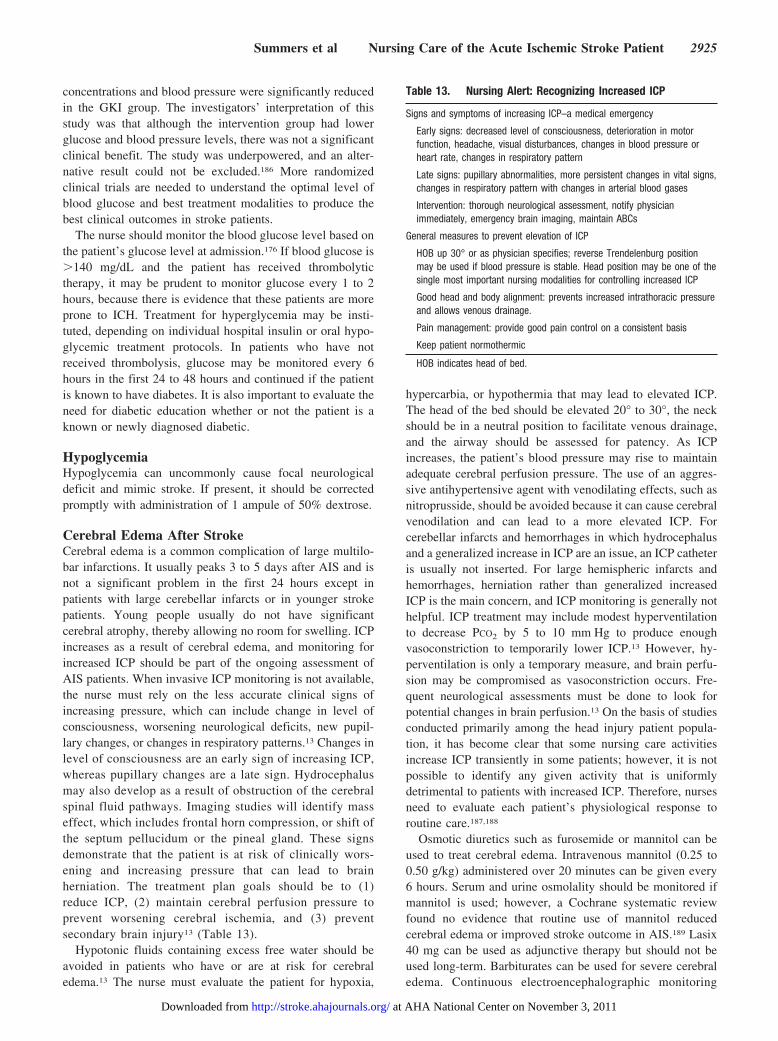
concentrations and blood pressure were significantly reducedin the GKI group. The investigators’ interpretation of thisstudy was that although the intervention group had lowerglucose and blood pressure levels, there was not a significantclinical benefit. The study was underpowered, and an alter-native result could not be excluded.186 More randomizedclinical trials are needed to understand the optimal level ofblood glucose and best treatment modalities to produce thebest clinical outcomes in stroke patients.
The nurse should monitor the blood glucose level based onthe patient’s glucose level at admission.176 If blood glucose is�140 mg/dL and the patient has received thrombolytictherapy, it may be prudent to monitor glucose every 1 to 2hours, because there is evidence that these patients are moreprone to ICH. Treatment for hyperglycemia may be insti-tuted, depending on individual hospital insulin or oral hypo-glycemic treatment protocols. In patients who have notreceived thrombolysis, glucose may be monitored every 6hours in the first 24 to 48 hours and continued if the patientis known to have diabetes. It is also important to evaluate theneed for diabetic education whether or not the patient is aknown or newly diagnosed diabetic.
HypoglycemiaHypoglycemia can uncommonly cause focal neurologicaldeficit and mimic stroke. If present, it should be correctedpromptly with administration of 1 ampule of 50% dextrose.
Cerebral Edema After StrokeCerebral edema is a common complication of large multilo-bar infarctions. It usually peaks 3 to 5 days after AIS and isnot a significant problem in the first 24 hours except inpatients with large cerebellar infarcts or in younger strokepatients. Young people usually do not have significantcerebral atrophy, thereby allowing no room for swelling. ICPincreases as a result of cerebral edema, and monitoring forincreased ICP should be part of the ongoing assessment ofAIS patients. When invasive ICP monitoring is not available,the nurse must rely on the less accurate clinical signs ofincreasing pressure, which can include change in level ofconsciousness, worsening neurological deficits, new pupil-lary changes, or changes in respiratory patterns.13 Changes inlevel of consciousness are an early sign of increasing ICP,whereas pupillary changes are a late sign. Hydrocephalusmay also develop as a result of obstruction of the cerebralspinal fluid pathways. Imaging studies will identify masseffect, which includes frontal horn compression, or shift ofthe septum pellucidum or the pineal gland. These signsdemonstrate that the patient is at risk of clinically wors-ening and increasing pressure that can lead to brainherniation. The treatment plan goals should be to (1)reduce ICP, (2) maintain cerebral perfusion pressure toprevent worsening cerebral ischemia, and (3) preventsecondary brain injury13 (Table 13).
Hypotonic fluids containing excess free water should beavoided in patients who have or are at risk for cerebraledema.13 The nurse must evaluate the patient for hypoxia,
hypercarbia, or hypothermia that may lead to elevated ICP.The head of the bed should be elevated 20° to 30°, the neckshould be in a neutral position to facilitate venous drainage,and the airway should be assessed for patency. As ICPincreases, the patient’s blood pressure may rise to maintainadequate cerebral perfusion pressure. The use of an aggres-sive antihypertensive agent with venodilating effects, such asnitroprusside, should be avoided because it can cause cerebralvenodilation and can lead to a more elevated ICP. Forcerebellar infarcts and hemorrhages in which hydrocephalusand a generalized increase in ICP are an issue, an ICP catheteris usually not inserted. For large hemispheric infarcts andhemorrhages, herniation rather than generalized increasedICP is the main concern, and ICP monitoring is generally nothelpful. ICP treatment may include modest hyperventilationto decrease PCO2 by 5 to 10 mm Hg to produce enoughvasoconstriction to temporarily lower ICP.13 However, hy-perventilation is only a temporary measure, and brain perfu-sion may be compromised as vasoconstriction occurs. Fre-quent neurological assessments must be done to look forpotential changes in brain perfusion.13 On the basis of studiesconducted primarily among the head injury patient popula-tion, it has become clear that some nursing care activitiesincrease ICP transiently in some patients; however, it is notpossible to identify any given activity that is uniformlydetrimental to patients with increased ICP. Therefore, nursesneed to evaluate each patient’s physiological response toroutine care.187,188
Osmotic diuretics such as furosemide or mannitol can beused to treat cerebral edema. Intravenous mannitol (0.25 to0.50 g/kg) administered over 20 minutes can be given every6 hours. Serum and urine osmolality should be monitored ifmannitol is used; however, a Cochrane systematic reviewfound no evidence that routine use of mannitol reducedcerebral edema or improved stroke outcome in AIS.189 Lasix40 mg can be used as adjunctive therapy but should not beused long-term. Barbiturates can be used for severe cerebraledema. Continuous electroencephalographic monitoring
Table 13. Nursing Alert: Recognizing Increased ICP
Signs and symptoms of increasing ICP–a medical emergency
Early signs: decreased level of consciousness, deterioration in motorfunction, headache, visual disturbances, changes in blood pressure orheart rate, changes in respiratory pattern
Late signs: pupillary abnormalities, more persistent changes in vital signs,changes in respiratory pattern with changes in arterial blood gases
Intervention: thorough neurological assessment, notify physicianimmediately, emergency brain imaging, maintain ABCs
General measures to prevent elevation of ICP
HOB up 30° or as physician specifies; reverse Trendelenburg positionmay be used if blood pressure is stable. Head position may be one of thesingle most important nursing modalities for controlling increased ICP
Good head and body alignment: prevents increased intrathoracic pressureand allows venous drainage.
Pain management: provide good pain control on a consistent basis
Keep patient normothermic
HOB indicates head of bed.
Summers et al Nursing Care of the Acute Ischemic Stroke Patient 2925
at AHA National Center on November 3, 2011http://stroke.ahajournals.org/Downloaded from
should be performed if barbiturates are administered. Hypo-thermia can also be used to treat elevated ICP.13 Unfortu-nately, all of these modalities are short-lived and palliative atbest. The recommendations for reducing cerebral edema needfurther study with regard to AIS. Currently, there is noclinical evidence that the measures discussed reduce cerebraledema or improve outcome in patients with ischemic brainswelling.13,189
If hydrocephalus is present, fluid drainage through anintraventricular catheter can rapidly reduce ICP. Surgicaldecompression (hemicraniectomy) is the most definitive andinvasive treatment of massive cerebral edema. Large cerebel-lar infarctions and hemorrhages that cause direct cerebellarcompression of the brain stem are best treated with surgicaldecompression.190–192 Surgical evacuation may be done inpatients with large hemispheric infarcts, but survivors havesevere residual neurological deficits.193–200 Subsequent to a2002 Cochrane review that found some evidence for benefitfrom surgical decompression,201 larger nonrandomized pro-spective trials have shown a reduction in the mortality rate ofpatients who have hemicraniectomy of large lesions involv-ing the middle cerebral artery territory.193–200,202 A recentpooled analysis of 3 randomized, controlled clinical trials(DEcompressive Craniectomy In MALignant middle cerebralartery infarcts [DECIMAL], DEcompressive Surgery for theTreatment of malignant INfarction of the middle cerebralarterY [DESTINY], and Hemicraniectomy After Middlecerebral artery infarction with Life-threatening Edema Trial[HAMLET]) in patients with malignant middle cerebralinfarct showed that decompressive surgery performed within48 hours of stroke onset reduced mortality and yielded morefavorable outcomes.123,203
SeizuresSeizures are a possible complication of large corticalstrokes and can be potentially life-threatening if notcontrolled. They can occur at the time of AIS, during thefirst few days after the event, or several months later. Nostudy has specifically tested the usefulness of anticonvul-sant medications in preventing or controlling seizures afterstroke. Drugs proven to be of value in preventing seizuresfrom other causes, however, are recommended for patientswho have had more than 1 seizure after stroke.204 Routineprophylactic administration of anticonvulsant drugs tostroke survivors who have not had seizures should beavoided.13 A study of 3552 SAH patients in 21 countriesfound that anticonvulsant drugs were commonly used butwere associated with more in-hospital complications andpoorer outcomes.205 No similar studies were found regard-ing AIS.
If an anticonvulsant drug is required, the nurse musteducate the patient and family about the seizure condition,pharmacological management, the medication regimen fortreatment of seizures, and the side effects and precautions.The patient must be told to never adjust or take additionalmedications without consulting the physician. Patients maynot have another seizure, but they and their families should beeducated that the risk is ongoing.
RecommendationsClass I
1. Stroke neurological assessments should be performedevery 4 hours after the hyperacute phase of stroke,and then frequency should be based on the patient’sstability and other comorbid conditions (Class I,Level of Evidence B).
2. Temperatures �99.6°F should be managed aggressively(Class I, Level of Evidence C).
3. Continuous cardiac monitoring of the stroke patientshould be provided for at least 24 to 48 hours afterstroke to detect potential cardiac problems (Class I,Level of Evidence B).
4. Careful, frequent monitoring and assessment for wors-ening of neurological deficits or bleeding should beperformed for up to 24 hours after thrombolytic therapy(Class I, Level of Evidence B).
5. Hyperglycemia should be treated in patients with aserum glucose concentration �140 mg/dL (Class I,Level of Evidence C).
6. Management of arterial hypertension in the acute phaseshould be approached with caution because of the lackof data available to guide management (Class I, Levelof Evidence C).
7. Oxygenation should be evaluated with an oxygen satu-ration monitor (Class I, Level of Evidence C).
8. To prevent aspiration pneumonia, the patient’s lungsshould be auscultated, and the patient should be evalu-ated for signs of respiratory compromise and dysphagia(Class I, Level of Evidence C). Nurses should reportseizure activity, and treatment should begin immedi-ately (Class I, Level of Evidence B). Prophylactictreatment of seizures should not be given.
Class IIa
1. It is reasonable to use clinical pathways, protocols, orpreprinted stroke order sets to organize care of thestroke patient (Class IIa, Level of Evidence B).
Diagnostic Testing During the Acute Phase
Overview of Neuroimaging in StrokeDiagnostic imaging techniques include CT, MRI, magneticresonance angiography, CT angiography, conventional an-giography, and carotid and transcranial ultrasound of thecervical and cephalic arteries.13 Imaging of extracranial andintracranial cerebral blood vessels may be essential forunderstanding the mechanism of AIS. The presence of ahigh-grade stenosis, occlusion, dissection, or vascular anom-aly indicates the events that led to the stroke. The nurse willneed to educate the patient and family regarding each test andwhat to expect.
Computed TomographyImaging in the hyperacute phase was discussed above. Arepeat CT or MRI brain scan should be done 24 hours afterthe initial stroke event in patients treated with thrombolysis orin any situation in which the patient is clinically deterioratingto assess hemorrhagic transformation and infarct progression.The CT scan is still considered the gold standard in AIS
2926 Stroke August 2009
at AHA National Center on November 3, 2011http://stroke.ahajournals.org/Downloaded from
treatment, although recent technology has led to more sophis-ticated multimodal approaches in stroke imaging with CTevaluation. CT perfusion and CT angiography provide a mapof cerebral blood volume, cerebral blood flow, and meantransit time. These studies identify the ischemic core andpneumbra regions that can guide the decision for furtherinterventional treatment.
Magnetic Resonance ImagingMRI demonstrates evidence of ischemic injury to the brainearlier than CT for all ischemic stroke subtypes. Magneticresonance angiography is a useful noninvasive procedure forevaluating extracranial and intracranial vessels. Newer mul-timodal techniques (eg, diffusion-weighted imaging andperfusion-weighted imaging) have further increased the sen-sitivity of MRI. The advantage of diffusion-weighted imagingis early identification of the ischemic area within minutesof onset of acute stroke. Perfusion-weighted imagingindicates the hemodynamic status of cerebral blood flow.A diffusion-perfusion mismatch shows an ischemic pen-umbra change in perfusion without a diffusion abnormal-ity. This is an opportunity to identify irreversible versusreversible cerebral tissue. If there is no mismatch, the hopeof salvageable tissue is less. Treatment modalities and timeto treatment may change as more is learned about theischemic penumbra.13,206 –208
UltrasonographyCarotid duplex scanning is the standard ultrasound testinitially used to screen for cervical internal carotid stenosis.Demonstration of stenosis �60% is highly accurate; how-ever, differentiation between severe (95% to 99%) stenosisand 100% occlusion is not completely reliable.
Cerebral AngiographyCerebral angiography is the best tool to accurately evaluatethe surface characteristics of a stenosed artery and is consid-ered the “gold standard” for measuring the degree of stenosisof a cervical or cephalic artery. After the procedure, the nursewill perform frequent measurement of vital signs, neurolog-ical assessment, femoral and pedal pulse checks, and groinchecks. The radiologist should be notified if a groin hema-toma or change in velocity of the pedal pulse develops.Postprocedure activity is restricted according to the closuredevice used and the patient’s status.
Transesophageal and Transthoracic EchocardiographyAll patients with ischemic stroke or transient ischemic attackshould undergo a comprehensive assessment of cardiovascu-lar risk to identify those with the highest likelihood ofmorbidity and mortality due to unrecognized coronary heartdisease or the presence of a cardioembolic source ofstroke.13,209,210 Transthoracic echocardiography is excellentfor identifying ventricular sources, such as a dyskineticventricular wall segment, whereas transesophageal echocar-diography excels at identifying atrial and aortic sources, suchas patent foramen ovale or aortic arch atherosclerosis. Atransesophageal echocardiogram is sensitive for detectingapical thrombi and atrial septal defects or patent foramenovale.
Transthoracic echocardiography is the less invasive of the2 procedures and is the most commonly ordered initial test toevaluate for a cardioembolic source of stroke. During atransesophageal echocardiogram, the nurse must monitor thepatient closely and position the patient to decrease risk ofaspiration if nausea or vomiting occurs.210,211
Chest RadiographA chest radiograph is no longer routinely recommendedduring the ED evaluation of stroke unless an underlyingpulmonary or cardiac problem is suspected. Obtaining a chestradiograph may take valuable time that is needed to admin-ister rtPA within the 3-hour window. Therefore, a chestradiograph is not a high priority in the ED but is done duringthe acute phase of hospitalization.
RecommendationClass I
1. All nurses should be familiar with the basic neuroim-aging testing for stroke patients so that they can educateand prepare patients and families (Class I, Level ofEvidence C).
General Supportive Care of Stroke: Rememberingthe BasicsMedical and nursing management both focus on theprevention of subacute complications of stroke, includingmalnutrition, aspiration, pneumonia, UTI, bowel or blad-der dysfunction, DVT, pulmonary embolism, contractures,joint abnormalities, and skin breakdown.130 Depression isalso common after stroke.212 Even in specialized stroke units,up to 63% of patients experience 1 or more complicationsafter acute stroke. The most common complications during thefirst week in a Norwegian stroke unit were pain, fever, progress-ing stroke, and UTI; immobilization-related complications, suchas pressure ulcers and clinical signs of DVT and pulmonaryembolism, had a very low incidence in this modern strokeunit.213 However, many stroke patients are not cared for inspecialized units, so it behooves nurses to continue to be vigilantfor the following areas of care related to prevention or rapiddetection of complications.5,7,57,114–121,132,214–223
InfectionPneumonia and UTI are frequently seen in the acute phaseafter stroke. Fever or a change in level of consciousnessshould give the nurse a high index of suspicion for infection.Stroke patients frequently present to the hospital with acompromised chest radiograph or a UTI.
Pneumonia is a serious complication occurring in the first48 to 72 hours after AIS and accounts for approximately 15%to 25% of deaths associated with stroke.13,224 Stroke-associated pneumonia increases length of stay, mortality, andhospital costs.224 The most common cause of pneumonia isaspiration due to dysphagia.225 Immobility and atelectasis canalso lead to development of pneumonia. The patient’s airwayand oxygenation must be monitored closely; some patientsmay require endotracheal intubation and mechanical ventila-tion. Early mobility and good pulmonary care can helpprevent pneumonia.224 Preventive measures in intubated pa-
Summers et al Nursing Care of the Acute Ischemic Stroke Patient 2927
at AHA National Center on November 3, 2011http://stroke.ahajournals.org/Downloaded from
tients include ventilation in a semirecumbent position, posi-tioning of the airway, suctioning, early mobility, and short-ened use of intubation, if feasible.226 In a randomized trial,early prophylaxis with levofloxacin was beneficial inpreventing systemic infections.227 Nursing managementincludes prompt recognition and reporting of fever; thesource of the fever should be sought and treatment begunimmediately. Early management of nausea and vomitingcan help prevent aspiration pneumonia; use of antiemeticmedications is warranted in this situation. Suctioning ofthe airway should be done carefully if increased ICP ispresent.228
UTIs are common, occurring in approximately 15% to 60%of stroke patients, and independently predict poor out-come.122,130,229–231 The use of an indwelling catheter andchanges in sphincter control increase the risk of UTI. Indwell-ing catheters should be avoided if possible but are oftenrequired in the acute phase of stroke. The catheter should beremoved as soon as the patient is medically and neurologi-cally stable. Intermittent catheterization may lessen the risk ofinfection. External catheters, incontinence pants, and inter-mittent catheterization are alternatives to an indwelling cath-eter. The patient should be assessed for UTI if there is achange in level of consciousness and no known reason forneurological deterioration. A urinalysis and urine cultureshould be obtained if UTI is suspected.13,229,232,233
Bowel and Bladder CareConstipation is the most common bowel problem. The nurseshould assess the patient’s premorbid bowel eliminationpattern, bowel sounds, and abdominal distention, if present.The patient should also be evaluated for hydration andimpaction. The nurse must request medications if needed ordevelop a bowel program, which can integrate the use of stoolsofteners, laxatives, and enemas to prevent constipation earlyafter stroke.
The most common urinary complication is incontinence,which occurs 30% to 60% of the time in the early recoveryperiod.234 An infarct in the frontal lobe or the pons can leadto incontinence. Voiding problems include neurogenic blad-der; hyperreflexia with urge incontinence, urgency, andfrequency; and urinary retention with or without overflowincontinence. After the indwelling catheter is removed, inter-mittent catheterization may be necessary to retrain the blad-der. Intermittent catheterization should occur every 4 to 6hours to prevent filling of the bladder beyond 500 mL and tostimulate normal physiological filling and emptying. Inter-mittent catheterization is recommended if postvoid residualurine volume is �100 mL.235
Urinary incontinence can increase the incidence of derma-titis, skin breakdown, UTIs, and perineal thrush. Urinaryincontinence also interferes with rehabilitation and is themajor factor in patients being discharged to nursing homes.236
Voiding strategies should be incorporated into the daily planof care. The nurse must initiate a bladder-training program todecrease the number of incontinent episodes. The patientshould be offered a commode, bedpan, or urinal every 2 hoursduring waking hours and every 4 hours at night. Neurologicaldeficits may complicate the task of going to the bathroom.
High fluid intake during the day and decreased fluid intake inthe evening should be encouraged.
Mobility and the Musculoskeletal SystemStroke patients may be initially kept on bed rest but should bemobilized when they are hemodynamically stable. Earlymobilization reduces risk of atelectasis, pneumonia, DVT,and pulmonary embolism. Complications from immobilityaccount for up to 51% of deaths in the first 30 days afterischemic stroke.237,238 Immobility can also lead to contrac-tures, orthopedic complications, atrophy, and nerve pressurepalsies. The nurse should monitor the first transfer from bedto an upright position, because some patients may haveneurological worsening during movement. Joints on theparalyzed side must be positioned higher than joints proximalto it. The nurse must assess for deformities that may be foundon the affected side (eg, shoulder adduction). Subluxation ofthe affected shoulder is common, and special care should betaken to avoid pulling on the affected arm and shoulder whenrepositioning or moving the patient. Nursing interventions,including range-of-motion and positioning techniques, canprevent joint contractures and atrophy.237,239
Pulmonary Embolism and DVTPulmonary embolism occurs more commonly than is sus-pected clinically and accounts for a substantial number ofdeaths after AIS. Wijdicks240 found a sudden death rate of50% in patients with pulmonary embolism that occurredbetween 3 and 120 days after the initial stroke event. In thatstudy, prophylaxis for DVT was noted in only 4 of 30patients. Pulmonary embolism was associated with DVTdiagnosed clinically or at autopsy in 11 patients and in theparalyzed leg. More recently, Indredavik and colleagues213
found pulmonary embolism in fewer than 2.5% of patientsduring the first week in a specialized stroke unit. DVT andpulmonary emboli were more likely to occur in the first 3months after stroke, with an incidence of 2.5% and 1.2%,respectively.
Both ischemic and hemorrhagic stroke patients are at riskfor developing DVT, especially as a result of paralysis orimpaired mobility. Prevention of DVT is one of the coreperformance measures for primary stroke center certification,and initiation of prevention has become a quality indicator inseveral populations.241,242 Safe ambulation should be startedas soon after stroke as possible. Pneumatic compressiondevices and compression stockings can be used to preventpulmonary embolism.243,244
Until recently, anticoagulants such as low-molecular-weight heparinoids and unfractionated heparin were alsoused.245,246 The results of the Prevention of VTE after AcuteIschemic stroke with LMWH enoxaparin (PREVAIL) Trialshowed that a 40-mg injection of enoxaparin once daily wasmore effective than 5000 IU of unfractionated heparin twicea day for prevention of DVT in AIS patients.247 Strokepatients taking anticoagulants should be assessed for bleedingdaily.
FallsFalls are a common cause of injury in stroke patients, with hipfractures the most prevalent injury.248 Hip fractures in the first
2928 Stroke August 2009
at AHA National Center on November 3, 2011http://stroke.ahajournals.org/Downloaded from
7 days after stroke are associated with a poor prognosis andhave been recognized as a consequence of hemiplegia sincethe 1950s.249 Most fractures occur on the paretic side.250
Patients with right hemispheric infarcts that cause neglect orinattention have the highest fall risk.
Minimization of fall risk is a global responsibility. Nursesmust implement fall-prevention programs and educate otherstaff and family members about risks and fall precautions.These may include identifying patients at risk, use of alarmsystems, use of special equipment (eg, enclosure beds), andplacing call buttons and the patient’s belongings near thepatient to prevent the patient from reaching for something andrisking a fall. Voiding times should be scheduled to preventfalls that occur when a patient tries to go to the bathroom. Insome cases, it may be necessary to have a sitter stay with thepatient to ensure the patient’s safety.249
Skin CareStroke patients are at risk for skin breakdown because of lossof sensation and impaired circulation, older age, decreasedlevel of consciousness, and inability to move themselvesbecause of paralysis.239 Related complications such as incon-tinence can accelerate skin breakdown. Major pressure areasare the heels, sacrum, and lateral malleoli. Patients should beexamined for skin breakdown when repositioned and aftersitting. Special care should be taken when moving patients toavoid excessive friction or pressure.239 Patients should not beleft in 1 position for �2 hours. The skin must be kept cleanand dry, and special mattresses should be used where indi-cated. The Braden Scale is commonly used to predict the riskof the development of pressure ulcers (decubitus). Nursingpersonnel can use this predictive model to design the care ofstroke patients with immobility and those who are at risk forthe development of skin breakdown.251–253
Detection of Dysphagia and Prevention of AspirationAspiration is frequently a result of dysphagia. In 2001,Galvan254 found that approximately half of all aspirations dueto dysphagia are “silent” and go unrecognized until there is apulmonary manifestation or complication. Order sets shouldinclude a swallow assessment before oral intake, performedby a nurse; evaluation by a speech language pathologist; andinstitution of NPO status with intravenous normal saline at 75to 100 mL/h until evaluation by the speech language pathol-ogist. Swallow assessment requires an evidence-based tool.225
The Massey Bedside Swallowing Screen has been successfullyevaluated for interrater reliability and predictive validity.255
Optimally, swallow assessment is performed soon after thepatient’s arrival in the ED. Until then, the patient shouldremain on NPO status, which means no ice chips, no oralmedications, no water, and no exceptions.256 The nurseshould assess swallowing by direct observation, looking forthe presence of choking, coughing, a wet voice, a delay ininitiating swallow, uncoordinated chewing or swallowing,extended time eating or drinking, pocketing of food, and lossof food from the mouth. When oral intake is authorized, thenurse should follow the speech language pathologist’s rec-ommendations,257 which include improving the patient’sability to concentrate while eating with minimal distractions.These assessments and interventions may make the difference
between improved recovery and increased morbidity or evenmortality.256
Nutritional CompromiseFifty percent of patients with severe strokes were reported tobe malnourished at 2 to 3 weeks after the stroke.258 Malnu-trition was associated with higher complications and poorerfunctional outcomes.259,260 To avoid nutritional compromise,nutritional intervention should occur no later than 3 to 4 daysafter diagnosis of dysphagia.5 The Ontario Heart and StrokeAssociation suggests that early gastrostomy should be con-sidered5 if it is anticipated that dysphagia will continuebeyond 6 weeks; however, dysphagia resolves in at least 87%of stroke patients.257
A nutritional assessment of a patient can be performed bytaking a diet history, with simple assessments such as bodymass index or more complex anthropometric measures suchas midarm circumference or triceps skin-fold thickness. In thestroke patient, it can be difficult to obtain a diet historybecause of communication problems, and anthropometricmeasures may not be accurate owing to paralysis of the arm.
When compromise of the patient’s nutritional health issuspected, serum albumin levels may be checked; this is oftencited as an index for long-term maintenance but may not bereflective of nutritional status.261,262 Serum albumin falls inacute illness owing to increased catabolism. No specific toolhas been developed or tested in acute stroke patients toevaluate nutritional assessments.263
A dietitian can provide a more accurate assessment ofnutritional health and nutrient needs. Without adequate nu-trition, there is a risk of weight loss, impaired immunesystem, increased weakness, increased length of stay, andmortality. Nutritional assessment should be performed on thestroke unit at the time of admission and throughout thehospital stay. The simplest but most valuable thing the nursecan do to monitor nutrition is to monitor the patient’s weightand weight change over time and monitor the patient’s dietaryintake.263–265
Stroke may compromise the patient’s ability to self-feed,which can impact self-esteem. A pleasant environment thatencompasses patience and encouragement fosters hope in thestroke patient. Use of seasonings, the serving of foods atappropriate temperatures, and augmentation of food presen-tation may enhance appeal and compensate for the patient’sloss of taste or smell. An interdisciplinary approach totreating dysphagia will result in early detection and earlyintervention to minimize the impact on the patient’s life,family, and healthcare costs.
RecommendationsMedical complications after stroke are common. The basicsof care become critical care in the stroke patient. An over-riding concept is patient and family education at all steps ofthe acute care process.
Class I
1. Infections, such as pneumonia and UTI, should beidentified and treated immediately with antibiotics(Class I, Level of Evidence B).
Summers et al Nursing Care of the Acute Ischemic Stroke Patient 2929
at AHA National Center on November 3, 2011http://stroke.ahajournals.org/Downloaded from
2. Early bowel and bladder care should be instituted toprevent complications such as constipation and urinaryretention or infection (Class I, Level of Evidence A).Use of indwelling catheters should be avoided if possi-ble because of the risk of UTI (Class I, Level ofEvidence A).
3. Early implementation of anticoagulant therapy or phys-ical compression modalities should be considered for allstroke patients who cannot ambulate at 2 days and whoare at risk for DVT or pulmonary embolus (Class I,Level of Evidence A). Early mobility should always beattempted if safe for the patient (Class I, Level ofEvidence B).
4. Fall precautions should be initiated, and the strokepatient should be told not to ambulate without assis-tance (Class I, Level of Evidence B).
5. Frequent turning should be instituted in bedriddenpatients to prevent skin breakdown (Class I, Level ofEvidence A). Use of the Braden Scale in nursingpractice can assist in the prediction of stroke patientsat high risk of developing pressure ulcers (Class I,Level of Evidence A). Range-of-motion exercisesshould start in the early phase of acute stroke careonce risk has been assessed (Class I, Level ofEvidence C).
6. A swallow screen should be performed in the first 24hours after stroke, preferably by the speech languagepathologist (Class I, Level of Evidence B). Nursesshould be familiar with bedside swallow assessment if aformal evaluation cannot be done within the specifiedperiod. Stroke patients should be kept NPO until thescreen has been performed (Class I, Level of EvidenceB). Further studies of dysphagia in the setting of acutestroke should be performed.
7. Patients who cannot swallow should have a nasogastrictube placed, or if severity warrants, a percutaneousendoscopic gastrostomy tube should be placed (Class I,Level of Evidence B). Assessment of proper hydrationis included in this recommendation.
Class IIa
1. If an indwelling catheter is required, excellent pericareand prevention of infection modalities should be insti-tuted to prevent complications (Class IIa, Level ofEvidence C).
2. The stroke patient can be fed either by intravenousinfusion or through nasogastric or percutaneous en-doscopic gastrostomy tubes (Class IIa, Level ofEvidence B).
Class IIb
1. Nurses may provide passive range-of-motion exercisesbetween physical therapy visits to help patients main-tain joint mobility and prevent complications of immo-bility (Class IIb, Level of Evidence C).
The Nurse’s Role in Secondary Preventionof StrokeNurses historically have played an important role in patientand family education, both at the bedside and in the outpatientsetting. Stroke education includes explaining stroke, discuss-ing risk factors (with emphasis placed on those risk factors
that are modifiable266), and describing secondary preventionmeasures, including compliance with prescribed medications.The AHA guidelines for secondary prevention of stroke arebeing revised. Pertinent recommendations from the 2006guidelines are provided in Table 14.267 Nurses play a signif-icant role in education and lifestyle modification strategies,and therefore, they must stay current with changing standardsand how they translate into practice. Suggestions for reducingblood pressure include adopting the DASH (Dietary Ap-proaches to Stop Hypertension) diet, engaging in regularaerobic activity, and moderating alcohol intake.268–273 Nursesshould be familiar with smoking cessation programs andmaterials and should include this information in the dischargeplan of care.274 Guidelines for smoking cessation for both theclinician and the consumer are available from the Office ofthe Surgeon General (www.surgeongeneral.gov/tobacco/default.htm) and the Agency for Health Care Policy andResearch.
For the stroke patient with atrial fibrillation, the nurseshould explain that use of warfarin (to maintain an interna-tional normalized ratio of 2 to 3) is recommended forprevention of stroke unless an absolute contraindication toanticoagulation exists. The nurse should also discuss thepotential side effect of bleeding, interactions between warfa-rin and other drugs or certain foods, and the importance ofcompliance. The patient should be informed that weeklytesting is required when warfarin is begun, then monthlytesting when the international normalized ratio is stable.Specific educational aids are found in Table 15.
Nurses can educate stroke patients about secondary pre-vention modalities. Patient education is a standard perfor-mance measure in primary stroke center certification (seehttp://www.jointcommission.org/CertificationPrograms/PrimaryStrokeCenters/stroke_measure_set.htm or http://www.jointcommission.org/CertificationPrograms/PrimaryStrokeCenters/guide_table_contents.htm). Patients should be taught therationale for administration of an antiplatelet drug and itspotential side effects. They should also be taught that anti-platelet therapy is lifelong, not short term.
Discharge PlanningThe discharge destination of stroke patients requires compre-hensive planning.275 The NINDS suggests that 30% of strokesurvivors will recover almost completely or with minorimpairments.67,276 Another 40% will require subacute care,and 10% will require care in a skilled nursing facility. Theremaining 15% will die soon after stroke onset. Of allsurvivors, �14% will experience recurrent stroke within 1year of the primary event. Eventually, 68% to 74% of strokesurvivors will require the care of family members in thehome.277 Detailed planning for postacute stroke care willoptimize outcomes and reduce risk and may control financialburden.
The average length of hospital stay for a stroke patient in2006 was 4.9 days, which may not be enough time to learn thetrue impact of a stroke.278 Order sets that include stroke teamreferrals will lead to initiation of early rehabilitation inter-vention. Such referrals include physiotherapy, occupationaltherapy, speech therapy, nutritional assessment, psychology,
2930 Stroke August 2009
at AHA National Center on November 3, 2011http://stroke.ahajournals.org/Downloaded from
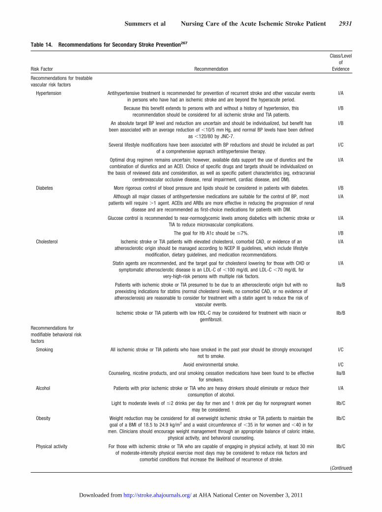
Table 14. Recommendations for Secondary Stroke Prevention267
Risk Factor Recommendation
Class/Levelof
Evidence
Recommendations for treatablevascular risk factors
Hypertension Antihypertensive treatment is recommended for prevention of recurrent stroke and other vascular eventsin persons who have had an ischemic stroke and are beyond the hyperacute period.
I/A
Because this benefit extends to persons with and without a history of hypertension, thisrecommendation should be considered for all ischemic stroke and TIA patients.
I/B
An absolute target BP level and reduction are uncertain and should be individualized, but benefit hasbeen associated with an average reduction of �10/5 mm Hg, and normal BP levels have been defined
as �120/80 by JNC-7.
I/B
Several lifestyle modifications have been associated with BP reductions and should be included as partof a comprehensive approach antihypertensive therapy.
I/C
Optimal drug regimen remains uncertain; however, available data support the use of diuretics and thecombination of diuretics and an ACEI. Choice of specific drugs and targets should be individualized on
the basis of reviewed data and consideration, as well as specific patient characteristics (eg, extracranialcerebrovascular occlusive disease, renal impairment, cardiac disease, and DM).
I/A
Diabetes More rigorous control of blood pressure and lipids should be considered in patients with diabetes. I/B
Although all major classes of antihypertensive medications are suitable for the control of BP, mostpatients will require �1 agent. ACEIs and ARBs are more effective in reducing the progression of renal
disease and are recommended as first-choice medications for patients with DM.
I/A
Glucose control is recommended to near-normoglycemic levels among diabetics with ischemic stroke orTIA to reduce microvascular complications.
I/A
The goal for Hb A1c should be �7%. I/B
Cholesterol Ischemic stroke or TIA patients with elevated cholesterol, comorbid CAD, or evidence of anatherosclerotic origin should be managed according to NCEP III guidelines, which include lifestyle
modification, dietary guidelines, and medication recommendations.
I/A
Statin agents are recommended, and the target goal for cholesterol lowering for those with CHD orsymptomatic atherosclerotic disease is an LDL-C of �100 mg/dL and LDL-C �70 mg/dL for
very-high-risk persons with multiple risk factors.
I/A
Patients with ischemic stroke or TIA presumed to be due to an atherosclerotic origin but with nopreexisting indications for statins (normal cholesterol levels, no comorbid CAD, or no evidence ofatherosclerosis) are reasonable to consider for treatment with a statin agent to reduce the risk of
vascular events.
IIa/B
Ischemic stroke or TIA patients with low HDL-C may be considered for treatment with niacin orgemfibrozil.
IIb/B
Recommendations formodifiable behavioral riskfactors
Smoking All ischemic stroke or TIA patients who have smoked in the past year should be strongly encouragednot to smoke.
I/C
Avoid environmental smoke. I/C
Counseling, nicotine products, and oral smoking cessation medications have been found to be effectivefor smokers.
IIa/B
Alcohol Patients with prior ischemic stroke or TIA who are heavy drinkers should eliminate or reduce theirconsumption of alcohol.
I/A
Light to moderate levels of �2 drinks per day for men and 1 drink per day for nonpregnant womenmay be considered.
IIb/C
Obesity Weight reduction may be considered for all overweight ischemic stroke or TIA patients to maintain thegoal of a BMI of 18.5 to 24.9 kg/m2 and a waist circumference of �35 in for women and �40 in for
men. Clinicians should encourage weight management through an appropriate balance of caloric intake,physical activity, and behavioral counseling.
IIb/C
Physical activity For those with ischemic stroke or TIA who are capable of engaging in physical activity, at least 30 minof moderate-intensity physical exercise most days may be considered to reduce risk factors and
comorbid conditions that increase the likelihood of recurrence of stroke.
IIb/C
(Continued)
Summers et al Nursing Care of the Acute Ischemic Stroke Patient 2931
at AHA National Center on November 3, 2011http://stroke.ahajournals.org/Downloaded from
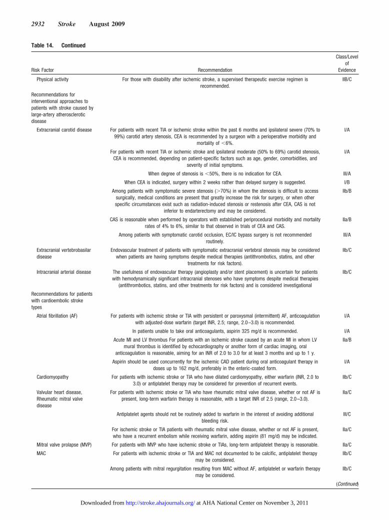
Table 14. Continued
Risk Factor Recommendation
Class/Levelof
Evidence
Physical activity For those with disability after ischemic stroke, a supervised therapeutic exercise regimen isrecommended.
IIB/C
Recommendations forinterventional approaches topatients with stroke caused bylarge-artery atheroscleroticdisease
Extracranial carotid disease For patients with recent TIA or ischemic stroke within the past 6 months and ipsilateral severe (70% to99%) carotid artery stenosis, CEA is recommended by a surgeon with a perioperative morbidity and
mortality of �6%.
I/A
For patients with recent TIA or ischemic stroke and ipsilateral moderate (50% to 69%) carotid stenosis,CEA is recommended, depending on patient-specific factors such as age, gender, comorbidities, and
severity of initial symptoms.
I/A
When degree of stenosis is �50%, there is no indication for CEA. III/A
When CEA is indicated, surgery within 2 weeks rather than delayed surgery is suggested. I/B
Among patients with symptomatic severe stenosis (�70%) in whom the stenosis is difficult to accesssurgically, medical conditions are present that greatly increase the risk for surgery, or when otherspecific circumstances exist such as radiation-induced stenosis or restenosis after CEA, CAS is not
inferior to endarterectomy and may be considered.
IIb/B
CAS is reasonable when performed by operators with established periprocedural morbidity and mortalityrates of 4% to 6%, similar to that observed in trials of CEA and CAS.
IIa/B
Among patients with symptomatic carotid occlusion, EC/IC bypass surgery is not recommendedroutinely.
III/A
Extracranial vertebrobasilardisease
Endovascular treatment of patients with symptomatic extracranial vertebral stenosis may be consideredwhen patients are having symptoms despite medical therapies (antithrombotics, statins, and other
treatments for risk factors).
IIb/C
Intracranial arterial disease The usefulness of endovascular therapy (angioplasty and/or stent placement) is uncertain for patientswith hemodynamically significant intracranial stenoses who have symptoms despite medical therapies
(antithrombotics, statins, and other treatments for risk factors) and is considered investigational
IIb/C
Recommendations for patientswith cardioembolic stroketypes
Atrial fibrillation (AF) For patients with ischemic stroke or TIA with persistent or paroxysmal (intermittent) AF, anticoagulationwith adjusted-dose warfarin (target INR, 2.5; range, 2.0–3.0) is recommended.
I/A
In patients unable to take oral anticoagulants, aspirin 325 mg/d is recommended. I/A
Acute MI and LV thrombus For patients with an ischemic stroke caused by an acute MI in whom LVmural thrombus is identified by echocardiography or another form of cardiac imaging, oral
anticoagulation is reasonable, aiming for an INR of 2.0 to 3.0 for at least 3 months and up to 1 y.
IIa/B
Aspirin should be used concurrently for the ischemic CAD patient during oral anticoagulant therapy indoses up to 162 mg/d, preferably in the enteric-coated form.
I/A
Cardiomyopathy For patients with ischemic stroke or TIA who have dilated cardiomyopathy, either warfarin (INR, 2.0 to3.0) or antiplatelet therapy may be considered for prevention of recurrent events.
IIb/C
Valvular heart disease,Rheumatic mitral valvedisease
For patients with ischemic stroke or TIA who have rheumatic mitral valve disease, whether or not AF ispresent, long-term warfarin therapy is reasonable, with a target INR of 2.5 (range, 2.0–3.0).
IIa/C
Antiplatelet agents should not be routinely added to warfarin in the interest of avoiding additionalbleeding risk.
III/C
For ischemic stroke or TIA patients with rheumatic mitral valve disease, whether or not AF is present,who have a recurrent embolism while receiving warfarin, adding aspirin (81 mg/d) may be indicated.
IIa/C
Mitral valve prolapse (MVP) For patients with MVP who have ischemic stroke or TIAs, long-term antiplatelet therapy is reasonable. IIa/C
MAC For patients with ischemic stroke or TIA and MAC not documented to be calcific, antiplatelet therapymay be considered.
IIb/C
Among patients with mitral regurgitation resulting from MAC without AF, antiplatelet or warfarin therapymay be considered.
IIb/C
(Continued)
2932 Stroke August 2009
at AHA National Center on November 3, 2011http://stroke.ahajournals.org/Downloaded from
and social services. Once assessed by each discipline, thepatient’s discharge planning process may begin.
Neurological findings identified during the acute phase andthroughout recovery will direct medical management, reha-bilitation, and the nursing plan of care. Deficits includealtered level of consciousness, confusion, behavioral distur-bances, cognitive deficits in higher functions such as memoryand ability to learn, motor deficits, disturbance in balance andcoordination, somatosensory deficits, disorders of vision,unilateral neglect, speech and language deficits, swallowingdisorder (dysphagia), and affective disorder. The rehabilita-tion medicine team (physiatrist, and speech, physical, andoccupational therapists) will evaluate the patient during theacute hospitalization and develop a rehabilitation plan fordischarge that will address the neurological deficits andappropriate rehabilitation needs. In addition, the rehabilita-tion team will give recommendations on the next level of careplacement. The nurse caring for the patient must communicatethe discharge plan to the patient and family and be the liaisonbetween the patient/family and rehabilitation team and dischargeplanners. Table 16 identifies key strategies or components thatwill help nurses to facilitate successful discharge planning andtransition to the next level of care.266,279,280
The goal of discharge planning is to ensure a safe transitionbetween the acute care facility, rehabilitation, outpatientsettings, primary care physician, and community while main-taining a continuity of care that will optimize rehabilitationpotential and ensure proper secondary prevention as appro-priate. Discharge planners (eg, social workers, case manag-ers) will integrate the concerns and expectations of theinterdisciplinary team with those of the patient, family, andassociated support system(s). Meeting the discharge needs forless obvious deficits (eg, memory problems) may pose asgreat a challenge as addressing needs related to overt hemi-paresis or aphasia. Teaching patients, families, and supportsystems how to identify and deal with the nuances ofstroke will facilitate community reintegration and optimizeoutcomes. A study from a Cochrane review281 found thatrecovering stroke patients placed on a structured care
pathway demonstrated significant improvement in func-tional ability and quality of life. Another study found thatimproved patient outcomes were highly dependent on supportsystems, including family support and an organized systemsapproach to meeting the discharge needs of the patient andfamily.282
What if the stroke victim is too disabled to participate inor benefit from rehabilitation? Hospital staff plays acrucial role in discharge decisions.283 Continued careoptions are skilled nursing care to assess for futurerehabilitation potential, skilled nursing care alone, pallia-tive care, or hospice care.
In a 2003 report on public health and aging, the Centers forDisease Control and Prevention noted that among the Medi-care population, postacute care was one of the fastest-growing categories, and stroke had the highest number ofpost–acute care beneficiaries.284 Additional findings suggestthat after adjustment for stroke severity, organized homehealth care for this population resulted in overall improvedfunctional outcomes and increased cost-effectiveness com-pared with other discharge facility options.284 Reported costsof nursing home care approached $15.2 billion comparedwith a home healthcare cost of $3.8 billion.285 These 2resources should alert discharge planners to the potential forand challenges of an increasing trend to home discharge.
Although outcomes for home care of stroke survivors arepositive, home care may be associated with negative healthoutcomes for caregivers.277 Therefore, appropriate referrals tocommunity resources, physician follow-up, support groups,governmental agencies, free services, faith-based communi-ties, and research opportunities must be incorporated intodischarge planning. However detailed it may be, dischargeplanning may not address all family questions or preparefamily members for future issues. It is, however, the backdropagainst which healing continues.
The Advanced Practice Nurse’s Role in AcuteStroke CareThe role of the nurse-practitioner or clinical nurse specialist isvital to the care of AIS patients throughout the continuum.
Table 14. Continued
Risk Factor Recommendation
Class/Levelof
Evidence
Aortic valve disease For patients with ischemic stroke or TIA and aortic valve disease who do not have AF, antiplatelettherapy may be considered.
IIb/C
Prosthetic heart valves For patients with ischemic stroke or TIA who have modern mechanical prosthetic heart valves, oralanticoagulants are recommended, with an INR target of 3.0 (range, 2.5–3.5).
I/B
For patients with mechanical prosthetic heart valves who have an ischemic stroke or systemicembolism despite adequate therapy with oral anticoagulants, aspirin 75 to 100 mg/d, in addition to oral
anticoagulants, and maintenance of the INR at a target of 3.0 (range, 2.5–3.5) is reasonable.
IIa/B
For patients with ischemic stroke or TIA who have bioprosthetic heart valves with no other source ofthromboembolism, anticoagulation with warfarin (INR, 2.0–3.0) may be considered.
IIb/C
JNC-7 indicates Seventh Report of the Joint National Committee on Prevention, Detection, Evaluation, and Treatment of High Blood Pressure; ACEI,angiotensin-converting enzyme inhibitor; ARBs, angiotensin receptor blockers; Hb, hemoglobin; CAD, coronary artery disease; NCEP, National Cholesterol EducationPanel; CHD, coronary heart disease; LDL-C, low-density lipoprotein cholesterol; BMI, body mass index; CEA, carotid endarterectomy; CAS, carotid angioplasty andstenting; EC/IC, extracranial/intracranial; AF, atrial fibrillation; MI, myocardial infarction; LV, left ventricular; MVP, mitral valve prolapse; and MAC, mitral annularcalcification.
Summers et al Nursing Care of the Acute Ischemic Stroke Patient 2933
at AHA National Center on November 3, 2011http://stroke.ahajournals.org/Downloaded from
These roles will be referred to as advanced practice nurses(APNs) for the purpose of the present report.
The APN plays an essential role in planning and leading ateam to develop clinical tools, organizing team members anddepartments to expedite activities in the care of the AISpatient, and monitoring outcomes and initiating quality initi-atives to improve care. The Brain Attack Coalition recom-mends that a primary stroke center have at least 1 APN toimplement and coordinate program activities.53
One study showed that financial outcomes were signifi-cantly improved when APNs functioned as outcome manag-ers and collaborated with the multidisciplinary team to carefor AIS patients.286 Patients had shorter lengths of stay, lowerrates of UTI and skin breakdown, and less time untilmobilization. The hospital had 2306 fewer patient-days and atotal cost savings of $2 467 328.
Stroke programs have shown that APNs have been key inimproving recognition and management of AIS pa-tients.287–290 The use of APNs in the Calgary StrokeProgram enhanced patient care and patient satisfactionwith continuity of care throughout hospitalization. Im-provements in the care process were also noted: CT imagetime was reduced from 60 to 30 minutes, door-to-needletime in administration of rtPA decreased from 90 to 60minutes, and consultation was faster.
The APN can integrate education, research, manage-ment, leadership, and consultation into the clinical role inmaking decisions about clinical management, in diagnosticreasoning, and in developing therapeutic interventions forthe care of AIS patients. APNs are assuming leadershiproles in developing programs such as dysphagia teams,
Table 15. Stroke Educational Programs
ASA/AHA (www.strokeassociation.org and www.americanheart.org)
Stroke: Patient Education Tool Kit
Power to End Stroke
African American Power to End Stroke
Power to End Stroke—Family Reunion Toolkit
Stroke Connection magazine
How Stroke Affects Behavior: Our Guide to Physical and EmotionalChanges
Living With Atrial Fibrillation: Our Guide to Managing a Key Stroke RiskFactor
Living With Disability After Stroke
Sex After Stroke: Our Guide to Intimacy After Stroke
Stroke: Are You at Risk? Our Guide to Stroke Risk Factors
Understanding Stroke: Our Guide to Explaining Stroke and How to ReduceYour Risk
Caring for Someone With Aphasia
High Blood Pressure and Stroke
Warning Signs of Stroke: Our Easy-Reading Guide to Emergency Action
Being a Stroke Family Caregiver
Smoking and Your Risk of Stroke
Just Move: Our Guide to Physical Activity
Diabetes, Heart Disease, and Stroke
NINDS (www.ninds.nih.gov)
What You Need to Know About Stroke
Stroke Risk Factors and Symptoms
Brain Basics: Preventing Stroke
Neurological Diagnostic Tests and Procedures
Questions and Answers About Stroke
Questions and Answers About Carotid Endarterectomy
National Stroke Association (www.stroke.org)
Stroke Smart magazine
Stroke Fact Sheet
African Americans and Stroke Brochure
Cholesterol Brochure
Explaining Stroke Brochure
Intracranial Atherosclerosis Brochure
Recurrent Stroke Prevention Brochure
Reducing Risk and Recognizing Symptoms Brochure
Transient Ischemic Attack Brochure
Stroke Rapid Response EMS/Prehospital Education
Hip Hop Stroke–Brainiac Kids Stroke Education
Brain Attack Coalition (www.stroke-site.org)
Table 16. Effective Nursing Strategies for SuccessfulDischarge Planning266,279,280
● Involve patient’s family/caregiver in assessment of postdischarge needsand decision making and treatment planning.
● Family and team meeting to discuss patient progress, rehabilitation goals,discharge needs or discharge issues, explanation of next level of care,providing care and support associated with these deficits and discussways to cope with stress associated with these impairments (eg,cognitive loss, urinary incontinence).
● Encourage the patient’s family/caregiver to participate in the rehabilitationsessions and to be trained to assist the patient with functional activities.
● Predischarge needs assessment of home before discharge performed byan occupational therapist to evaluate home environment and determinesafety, need for home modification(s), and equipment needs.
● Consider availability of support services and the desires of the patient’sfamily/caregiver.
● Caregiver training (multidisciplinary education about communicationstrategies if the patient is aphasic, positioning and handling, transfers,shoulder care, how to promote independence, according to individualpatient’s strengths and limitations).
● Postdischarge follow-up (ensure plans are made for medical care,secondary prevention, rehabilitation, social support, home care, andnursing if needed, caregiver support and education).
● Provide education for patient’s family/caregivers on stroke pathology,stroke prevention, stroke signs and symptoms, actions to take, follow-upappointments/therapy, treatment plan, and community resources and howto access. Education should be provided in interactive and written formatand documented in the medical record to facilitate communication ofeducation completed to other healthcare providers.
● Liaison with community providers, linkage with appropriate resources.
● Regular review of individual patient and caregiver psychosocial andsupport needs.
● Provision of information on discharge plans and postdischargemanagement to primary care physicians and community services.
● If the patient is aphasic, the nurse and rehabilitation team should assistin establishing a communication pattern with the patient’sfamily/caregiver before discharge.
2934 Stroke August 2009
at AHA National Center on November 3, 2011http://stroke.ahajournals.org/Downloaded from
incontinence teams, and anticoagulation clinics and areactively involved in discharge, palliative care, and second-ary prevention clinics.291–299
The Nurse’s Role in Stroke Education inthe CommunityA landmark study evaluating the knowledge of patientspresenting to the ED was first conducted in 1997 and repeatedin 1998 and 2003.300 The first study showed that 40% ofpatients admitted did not know the signs, symptoms, or riskfactors of stroke. A need for further public education toincrease awareness of stroke warning signs was identified,and all healthcare professionals were encouraged to becomeinvolved in community education.300 In 1998, a population-based interview was conducted in the greater Cincinnati,Ohio, area to establish the public’s knowledge of risk factorsand warning signs of acute stroke.301 Fifty-seven percentcorrectly named only 1 of the 5 warning signs of strokedefined by the NINDS, and 68% could identify only 1stroke risk factor. Only 57% of patients with hypertensionsaid that they were at risk for stroke. Sixty percent ofpatients �75 years of age correctly identified 1 warningsign of stroke compared with only 47% of patients �75years of age.301 As a result of organized communityeducation in the greater Cincinnati region, communityawareness of stroke warning signs significantly improvedover time, but knowledge of risk of stroke based onindividual risk factors did not improve.302
Studies have shown that some groups have less knowledgeabout stroke, delay seeking medical attention, and do notphone 9-1-1. These groups include the elderly, women, thosewith lower levels of education, and racial/ethnic populationssuch as blacks and Latinos. Healthcare professionals musteducate the public about stroke warning signs and risk factorsand actions to take when symptoms occur.303,304
The effect of stroke screening on knowledge and behav-ioral changes has been studied.305 One screening used theNational Stroke Association guidelines. Participants wereevaluated before and after screening and 3 months after theevent. Knowledge increased from 59% to 94% immediatelyafter the event but at 3 months was only 77%. Only 27% ofpatients attending the screening had implemented an inter-vention to decrease stroke risk.305 Similar studies showed that�50% of those attending a stroke screening made at least 1behavioral change to reduce stroke risk and that communityeducation improved the participant’s knowledge of strokewarning signs and symptoms.306–308
The National Stroke Association states that “80% ofstrokes can be prevented” (http://www.stroke.org/site/PageServer?pagename�PREVENT). This should compelnurses to educate the public about primary and secondarystroke prevention, including stroke awareness and stroke riskfactors, and to call 9-1-1 if warning signs of stroke occur. Theacronym FAST (Face, Arm, Speech, Time) is being used as away to educate the public. “The Beauty Shop Project”educated beauticians on the FAST acronym and asked themto educate their clients about acute stroke signs by empha-sizing the FAST acronym. After the project, 51% of partici-pants could identify at least 3 stroke warning signs compared
with 41% at the beginning of the project. Ninety-threepercent recognized the need to call 9-1-1 compared with85% before the project began (http://www.webmd.com/stroke/news/20070207/hip-hop-to-stroke-awareness).
The Massachusetts Health Promotion Clearinghouse Cataloghas a 3-minute video on stroke awareness titled Stroke HeroesAct FAST. Three scenarios educate viewers about recognizingand responding to the signs of stroke using the FAST model(http://www.maclearinghouse.com/CatalogHDSP.htm).
In 2004, the National Stroke Association created “Hip HopStroke,” a stroke education program for school-aged children.The interactive program is presented for 1 hour a day for 3days. Hip-hop music and the character “Brainiac” teachchildren about lifestyle changes, warning signs of stroke, andthe importance of calling 9-1-1 (http://www.stroke.org/site/PageNavigator/HipHopStroke).
Nurses have many avenues through which to educate thepublic: Churches, schools, and community and professionalorganizations. The AHA/ASA and National Stroke Associa-tion have educational information on their Web sites, as wellas materials for stroke education (Table 15). Stroke educationcan be done through blood pressure, lipid, and glucosescreenings; health education forums; and smoking cessation,exercise, and nutritional programs.
The Nurse’s Role in Certification of PrimaryStroke CentersCertification of stroke centers has gained national recognitionsince publication of the Brain Attack Coalition recommenda-tions in June 200010 amid concern that stroke centers should
Table 17. Specific Roles of Nursing in the Joint CommissionPrimary Stroke Center Certification
Bring team together toward common goal.
Organize team to accomplish stroke center certification.
Educate in-house nursing personnel, medical personnel, and ancillarypersonnel on stroke-related issues and philosophy of the Joint Commissionstroke center certification.
Understand the Joint Commission requirements and understand deficitshospital may have in achieving goal.
Develop preprinted or standing order sets and critical pathways as requiredby the Joint Commission.
Develop stroke care procedures for strategic in-hospital units that care forstroke patients.
Appoint quality-assurance nurse to collect data on execution of performancemeasures.
Understand whether hospital is capable of responding to recommendationsof Brain Attack Coalition.
Understand hospital’s medical record coding to ensure it is appropriatelycapturing ICD-10 and DRGs for proper billing for optimum reimbursementpotential.
Recognize that nursing personnel play an important role in the applicationprocess for Joint Commission certification in pulling together requireddocuments for the site visit.
Ensure that hospital personnel are prepared for the Joint Commissionreviewer.
Monitor performance measures established by the Joint Commission.
ICD indicates International Classification of Diseases; DRGs, Diagnosis-Related Groups.
Summers et al Nursing Care of the Acute Ischemic Stroke Patient 2935
at AHA National Center on November 3, 2011http://stroke.ahajournals.org/Downloaded from
require certification by a rigorous examining body. The JointCommission, with the support of the AHA and ASA, developeda certification program using the ideas proposed in the BrainAttack Coalition paper as the template for the model (http://www.jointcommission.org/CertificationPrograms/PrimaryS-trokeCenters/). In 2004, the certification program was rolled out.More than 200 hospitals were certified by the end of 2006.5
Nurses will play an important role in obtaining andmaintaining stroke center certification (Table 17), just as theydo in the Joint Commission’s general hospital accreditation.Nurses will ensure that hospital personnel are properlytrained and knowledgeable about all aspects of stroke careand will interact with the multidisciplinary team through-out the care continuum. Nurses ensure that a patient’sneeds are met by all the disciplines involved in care and inmany cases help integrate this care so that patients aredischarged having received optimum care through bestpractices. Nurses will work to ensure that all aspects of thiscomplicated disease are managed in a way that willproduce positive outcomes (Table 18).
The AHA/ASA patient education toolkit is available toassist nurses and other healthcare professionals in providingpatient and family education about stroke. The toolkit can beordered online through the AHA/ASA Web site (www.strokeas-sociation.org) or by calling the AHA/ASA directly (1–888-4-STROKE). Information about Joint Commission primarystroke center certification can be found at the Joint CommissionWeb site (www.jointcommission. org/CertificationPrograms/PrimaryStrokeCenters/).
The Nurse’s Role in Quality Improvement InitiativesNurses historically have been at the forefront of inpatienthospital quality improvement programs. This role will con-
tinue to expand as nurses institute quality improvementprograms for secondary prevention such as Get With theGuidelines Stroke, a patient management tool, or the Cover-dale registry, both of which are used to collect data forprimary stroke center certification.309,310 Get With theGuidelines Stroke offers evidence-based, best-practice in-formation that can be compared with other hospitals usingthis quality improvement tool. Information derived fromthis data collection program can also assist in patient andfamily education.
In summary, the present scientific statement describes nurs-ing’s vital role in the first 2 phases of stroke, the emergency orhyperacute phase and the acute phase. Using strength of evi-dence, the recommendations on the organization and integrationof care are designed to improve outcomes, decrease lengths ofstay, decrease costs, and decrease event recurrence.10
Table 18. Performance Measures From the Joint CommissionPrimary Stroke Center Certification Program
Stroke-1 Deep vein thrombosis prophylaxis
Stroke-2 Discharged on antithrombotic therapy
Stroke-3 Patients with atrial fibrillation receiving anticoagulation therapy
Stroke-4 Thrombolytic therapy administered
Stroke-5 Antithrombotic therapy by end of hospital day two
Stroke-6 Discharged on statin medication
Stroke-7 Dysphagia screening
Stroke-8 Stroke education
Stroke-9 Smoking cessation/advice/counseling
Stroke-10 Assessed for rehabilitation
DisclosuresWriting Group Disclosures
Writing GroupMember Employment Research Grant
Other ResearchSupport
Speakers’Bureau/Honoraria
OwnershipInterest
Consultant/Advisory Board Other
DebbieSummers
Saint Luke’s Brainand Stroke Institute
None None Concentric Medical*;Genentech*
None ConcentricMedical*;
National StrokeAssociation*
None
Anne Leonard University of TexasHealth ScienceCenter at San
Antonio
None None None None None AHA ScienceConsultant†
DeidreWentworth
Catholic HealthcareWest, MercyNeurological
Institute of NorthernCalifornia
None None Genentech, Inc*; NationalStroke Association*
None National StrokeAssociation*;PhotoThera,
Inc†
None
Jeffrey L.Saver
UCLA MedicalCenter
None ConcentricMedical*
BMS/Sanofi*; BoehringerIngelheim*
None AGA Medical*;Alsius Corp*;
Biosite*;BMS/Sanofi*;BoehringerIngelheim*;Co-Axia*;
Johnson &Johnson*
None
(Continued)
2936 Stroke August 2009
at AHA National Center on November 3, 2011http://stroke.ahajournals.org/Downloaded from
References1. Rosamond W, Flegal K, Furie K, Go A, Greenlund K, Haase N,
Hailpern SM, Ho M, Howard V, Kissela B, Kittner S, Lloyd-Jones D,McDermott M, Meigs J, Moy C, Nichol G, O’Donnell C, Roger V,Sorlie P, Steinberger J, Thom T, Wilson M, Hong Y. Heart disease andstroke statistics: 2008 update: a report from the American Heart Asso-ciation Statistics Committee and Stroke Statistics Subcommittee. Cir-culation. 2008;117:e25–e146.
2. Donnarumma R, Kongable G, Barch C, Braimah J, Bratina P, Daley S,Rapp K, Sailor S, Spilker J. Overview: hyperacute rt-PA stroke treatment:the NINDS rt-PA Stroke Study Group. J Neurosci Nurs. 1997;29:351–355.
3. Bader MK, Palmer S. What’s the “hyper” in hyperacute stroke?Strategies to improve outcomes in ischemic stroke patients presentingwithin 6 hours. AACN Adv Crit Care. 2006;17:194–214.
4. Birbeck GL, Zingmond DS, Cui X, Vickrey BG. Multispecialty strokeservices in California hospitals are associated with reduced mortality.Neurology. 2006;66:1527–1532.
5. Stroke Unit Trialists’ Collaboration. Organised inpatient (stroke unit)care for stroke. Cochrane Database Syst Rev. 2007;No. 4:CD000197.
6. Long AF, Kneafsey R, Ryan J, Berry J. The role of the nurse withinthe multi-professional rehabilitation team. J Adv Nurs. 2002;37:70 –78.
7. Jahnke HK, Zadrozny D, Garrity T, Hopkins S, Frey JL, Christopher M.Stroke teams and acute stroke pathways: one emergency department’stwo-year experience. J Emerg Nurs. 2003;29:133–139.
8. Gillum RF. New considerations in analyzing stroke and heart diseasemortality trends: the Year 2000 Age Standard and the InternationalStatistical Classification of Diseases and Related Health Problems, 10thRevision. Stroke. 2002;33:1717–1721.
9. Seshadri S, Beiser A, Kelly-Hayes M, Kase CS, Au R, Kannel WB,Wolf PA. The lifetime risk of stroke: estimates from the FraminghamStudy. Stroke. 2006;37:345–350.
10. Alberts MJ, Hademenos G, Latchaw RE, Jagoda A, Marler JR, MaybergMR, Starke RD, Todd HW, Viste KM, Girgus M, Shephard T, Emr M,Shwayder P, Walker MD. Recommendations for the establishment of
Writing Group Disclosures, Continued
Writing GroupMember Employment Research Grant
Other ResearchSupport
Speakers’Bureau/Honoraria
OwnershipInterest
Consultant/Advisory Board Other
Jo Simpson Morton Plant MeaseHealth Care
None None None None None None
Judith A. Spilker University ofCincinnati
None None Genentech*; NationalStroke Association*
None Co-Axia*;National Stroke
Association*
None
Nanette Hock Kaiser PermanenteNorthern California
None None None None None None
Elaine Miller University ofCincinnati College
of Nursing
None None None None None None
Pamela H.Mitchell
University ofWashington
Principal Investigatorof NIH research
grant: Psychosocial/Behavioral
Intervention in PSD,R01 NR07755,
4/1/02–3/31/08†
None None None None None
This table represents the relationships of writing group members that may be perceived as actual or reasonably perceived conflicts of interest as reported on the DisclosureQuestionnaire, which all members of the writing group are required to complete and submit. A relationship is considered to be �significant� if (1) the person receives $10 000or more during any 12-month period, or 5% or more of the person’s gross income; or (2) the person owns 5% or more of the voting stock or share of the entity, or owns$10 000 or more of the fair market value of the entity. A relationship is considered to be �modest� if it is less than �significant� under the preceding definition.
*Modest.†Significant.
Reviewer Disclosures
Reviewer EmploymentResearch
GrantOther Research
SupportSpeakers’
Bureau/HonorariaExpert
WitnessOwnership
InterestConsultant/Advisory
Board Other
Harold Adams University ofIowa
None None None None None None None
Linda Baas University ofCincinnati
None None None None None None None
Rosemarie B.King
NorthwesternUniversity
NIH† None None None None None 3 Rehabilitation researchsites, Stroke CaregiverStudy—admit stroke
survivors†
This table represents the relationships of reviewers that may be perceived as actual or reasonably perceived conflicts of interest as reported on the DisclosureQuestionnaire, which all reviewers are required to complete and submit. A relationship is considered to be �significant� if (1) the person receives $10 000 or moreduring any 12-month period, or 5% or more of the person’s gross income; or (2) the person owns 5% or more of the voting stock or share of the entity, or owns$10 000 or more of the fair market value of the entity. A relationship is considered to be �modest� if it is less than �significant� under the preceding definition.
*Modest.†Significant.
Summers et al Nursing Care of the Acute Ischemic Stroke Patient 2937
at AHA National Center on November 3, 2011http://stroke.ahajournals.org/Downloaded from
primary stroke centers: Brain Attack Coalition. JAMA. 2000;283:3102–3109.
11. Sackett DL. Rules of evidence and clinical recommendations on the useof antithrombotic agents. Chest. 1989;95(suppl):2S–4S.
12. Adams HP Jr, Adams RJ, Brott T, del Zoppo GJ, Furlan A, GoldsteinLB, Grubb RL, Higashida R, Kidwell C, Kwiatkowski TG, Marler JR,Hademenos GJ; Stroke Council of the American Stroke Association.Guidelines for the early management of patients with ischemic stroke: ascientific statement from the Stroke Council of the American StrokeAssociation. Stroke. 2003;34:1056–1083.
13. Adams HP Jr, del Zoppo G, Alberts MJ, Bhatt DL, Brass L, Furlan A,Grubb RL, Higashida RT, Jauch EC, Kidwell C, Lyden PD, Mor-genstern LB, Qureshi AI, Rosenwasser RH, Scott PA, Wijdicks EF.Guidelines for the early management of adults with ischemic stroke: aguideline from the American Heart Association/American Stroke Asso-ciation Stroke Council, Clinical Cardiology Council, CardiovascularRadiology and Intervention Council, and the Atherosclerotic PeripheralVascular Disease and Quality of Care Outcomes in Research Interdis-ciplinary Working Groups: the American Academy of Neurologyaffirms the value of this guideline as an educational tool for neurologists[published corrections appear in Stroke. 2007;38:e38 and 2007;38:e96].Stroke. 2007;38:1655–1711.
14. Broderick J, Connolly S, Feldmann E, Hanley D, Kase C, Krieger D,Mayberg M, Morgenstern L, Ogilvy CS, Vespa P, Zuccarello M.Guidelines for the management of spontaneous intracerebral hemor-rhage in adults: 2007 update: a guideline from the American HeartAssociation/American Stroke Association Stroke Council, High BloodPressure Research Council, and the Quality of Care and Outcomes inResearch Interdisciplinary Working Group [republished in Circulation.2007;116:e391–e413]. Stroke. 2007;38:2001–2023.
15. Mayberg MR, Batjer HH, Dacey R, Diringer M, Haley EC, Heros RC,Sternau LL, Torner J, Adams HP Jr, Feinberg W. Guidelines for themanagement of aneurysmal subarachnoid hemorrhage: a statement forhealthcare professionals from a special writing group of the StrokeCouncil, American Heart Association. Stroke. 1994;25:2315–2328.
16. Albers GW, Amarenco P, Easton JD, Sacco RL, Teal P. Antithromboticand thrombolytic therapy for ischemic stroke: the Seventh ACCP Con-ference on Antithrombotic and Thrombolytic Therapy. Chest. 2004;126(suppl):483S–512S.
17. Sturgeon JD, Folsom AR, Longstreth WT Jr, Shahar E, Rosamond WD,Cushman M. Hemostatic and inflammatory risk factors for intracerebralhemorrhage in a pooled cohort. Stroke. 2008;39:2268–2273.
18. Sturgeon JD, Folsom AR, Longstreth WT Jr, Shahar E, Rosamond WD,Cushman M. Risk factors for intracerebral hemorrhage in a pooledprospective study. Stroke. 2007;38:2718–2725.
19. Okada Y, Yamaguchi T, Minematsu K, Miyashita T, Sawada T,Sadoshima S, Fujishima M, Omae T. Hemorrhagic transformation incerebral embolism. Stroke. 1989;20:598–603.
20. Sahni R, Weinberger J. Management of intracerebral hemorrhage. VascHealth Risk Manag. 2007;3:701–709.
21. Acker JE III, Pancioli AM, Crocco TJ, Eckstein MK, Jauch EC,Larrabee H, Meltzer NM, Mergendahl WC, Munn JW, Prentiss SM,Sand C, Saver JL, Eigel B, Gilpin BR, Schoeberl M, Solis P, Bailey JR,Horton KB, Stranne SK. Implementation strategies for emergencymedical services within stroke systems of care: a policy statement fromthe American Heart Association/American Stroke Association ExpertPanel on Emergency Medical Services Systems and the Stroke Council.Stroke. 2007;38:3097–3115.
22. Kothari RU, Pancioli A, Liu T, Brott T, Broderick J. Cincinnati Pre-hospital Stroke Scale: reproducibility and validity. Ann Emerg Med.1999;33:373–378.
23. Barsan WG, Brott TG, Broderick JP, Haley EC, Levy DE, Marler JR.Time of hospital presentation in patients with acute stroke. Arch InternMed. 1993;153:2558–2561.
24. Marler JR. Early stroke diagnosis saves time. Ann Emerg Med. 1999;33:450–451.
25. Bratina P, Greenberg L, Pasteur W, Grotta JC. Current emergencydepartment management of stroke in Houston, Texas. Stroke. 1995;26:409–414.
26. Brott T, Reed RL. Intensive care for acute stroke in the communityhospital setting: the first 24 hours. Stroke. 1989;20:694–697.
27. Carrozzella J, Jauch EC. Emergency stroke management: a new era.Nurs Clin North Am. 2002;37:35–57, vi.
28. Crocco TJ, Moreno R, Jauch EC, Racine AN, Pio BJ, Liu T, Kothari RU.Teaching ACLS stroke objectives to prehospital providers: a case-basedapproach. Prehosp Emerg Care. 2003;7:229–234.
29. Daley S, Braimah J, Sailor S, Kongable GL, Barch C, Rapp K, BratinaP, Spilker J, Donnarumma R. Education to improve stroke awarenessand emergent response: the NINDS rt-PA Stroke Study Group.J Neurosci Nurs. 1997;29:393–396.
30. Kidwell CS, Starkman S, Eckstein M, Weems K, Saver JL. Identifyingstroke in the field: prospective validation of the Los Angeles prehospitalstroke screen (LAPSS). Stroke. 2000;31:71–76.
31. Kothari R, Barsan W, Brott T, Broderick J, Ashbrock S. Frequency andaccuracy of prehospital diagnosis of acute stroke. Stroke. 1995;26:937–941.
32. Kothari R, Hall K, Brott T, Broderick J. Early stroke recognition:developing an out-of-hospital NIH Stroke Scale. Acad Emerg Med.1997;4:986–990.
33. Lacy CR, Suh DC, Bueno M, Kostis JB. Delay in presentation andevaluation for acute stroke: Stroke Time Registry for OutcomesKnowledge and Epidemiology (S.T.R.O.K.E.). Stroke. 2001;32:63–69.
34. Llanes JN, Kidwell CS, Starkman S, Leary MC, Eckstein M, Saver JL.The Los Angeles Motor Scale (LAMS): a new measure to characterizestroke severity in the field. Prehosp Emerg Care. 2004;8:46–50.
35. Morris DL, Rosamond W, Madden K, Schultz C, Hamilton S. Pre-hospital and emergency department delays after acute stroke: theGenentech Stroke Presentation Survey. Stroke. 2000;31:2585–2590.
36. Deleted in proof.37. Morris DL, Rosamond WD, Hinn AR, Gorton RA. Time delays in
accessing stroke care in the emergency department. Acad Emerg Med.1999;6:218–223.
38. Porteous GH, Corry MD, Smith WS. Emergency medical services dis-patcher identification of stroke and transient ischemic attack. PrehospEmerg Care. 1999;3:211–216.
39. Smith WS, Corry MD, Fazackerley J, Isaacs SM. Improved paramedicsensitivity in identifying stroke victims in the prehospital setting.Prehosp Emerg Care. 1999;3:207–210.
40. Emergency Nurses Association. Position statements: ENA boardapproves statement on joint ENA/ACEP five-level triage task force.Available at: http://www.ahrq.gov/research/esi/esi2.htm. Accessed July8, 2007.
41. Williams JE, Rosamond WD, Morris DL. Stroke symptom attributionand time to emergency department arrival: the delay in accessing strokehealthcare study. Acad Emerg Med. 2000;7:93–96.
42. Williams LS, Bruno A, Rouch D, Marriott DJ. Stroke patients’knowledge of stroke: influence on time to presentation. Stroke. 1997;28:912–915.
43. Zweifler RM, York D, U TT, Mendizabal JE, Rothrock JF. Accuracy ofparamedic diagnosis of stroke. J Stroke Cerebrovasc Dis. 1998;7:446–448.
44. Field JM, ed. Advanced Cardiovascular Life Support. Dallas, Tex:American Heart Association; 2006:103–117.
45. Sahni R. Acute stroke: implications for prehospital care: National Asso-ciation of EMS Physicians Standards and Clinical Practice Committee.Prehosp Emerg Care. 2000;4:270–272.
46. Marler JR, Jones PW, Emr M, eds. Proceedings of a National Sym-posium on Rapid Identification and Treatment of Acute Stroke.Bethesda, Md: National Institute of Neurological Disorders and Stroke;1997. NIH publication No. 97–4239.
47. National Institutes of Health. Improving the Chain of Recovery for AcuteStroke in Your Community. Bethesda, Md: National Institute of Neuro-logical Disorders and Stroke; 2003. NIH publication No. 03–5348.
48. Pepe PE, Zachariah BS, Sayre MR, Floccare D. Ensuring the chain ofrecovery for stroke in your community. Acad Emerg Med. 1998;5:352–358.
49. Wojner AW, Morgenstern L, Alexandrov AV, Rodriguez D, Persse D,Grotta JC. Paramedic and emergency department care of stroke: baselinedata from a citywide performance improvement study. Am J Crit Care.2003;12:411–417.
50. Wojner-Alexandrov AW, Alexandrov AV, Rodriguez D, Persse D,Grotta JC. Houston paramedic and emergency stroke treatment andoutcomes study (HoPSTO). Stroke. 2005;36:1512–1518.
51. American Heart Association. Acute stroke case. In: Advanced Cardio-vascular Life Support Provider Manual. Dallas, Tex: American HeartAssociation; 2006:103–117.
52. Gilboy N, Tanabe P, Travers D, Rosenau A, Eitel D. Emergency SeverityIndex, Version 4: Implementation Handbook. Rockville, Md: Agency for
2938 Stroke August 2009
at AHA National Center on November 3, 2011http://stroke.ahajournals.org/Downloaded from
Healthcare Research and Quality; 2005. AHRQ publication No.05–0046-2.
53. Alberts MJ, Latchaw RE, Selman WR, Shephard T, Hadley MN, BrassLM, Koroshetz W, Marler JR, Booss J, Zorowitz RD, Croft JB, MagnisE, Mulligan D, Jagoda A, O’Connor R, Cawley CM, Connors JJ,Rose-DeRenzy JA, Emr M, Warren M, Walker MD; Brain AttackCoalition. Recommendations for comprehensive stroke centers: a con-sensus statement from the Brain Attack Coalition. Stroke. 2005;36:1597–1616.
54. Florida Stroke Act. 2004–325.55. Division for Heart Disease and Stroke Prevention. State program: Mas-
sachusetts basic implementation. Available at: http://www.cdc.gov/dhdsp/state_program/ma.htm. Accessed May 4, 2009.
56. Gropen TI, Gagliano PJ, Blake CA, Sacco RL, Kwiatkowski T,Richmond NJ, Leifer D, Libman R, Azhar S, Daley MB; NYSDOHStroke Center Designation Project Workgroup. Quality improvement inacute stroke: the New York State Stroke Center Designation Project.Neurology. 2006;67:88–93.
57. Schwamm LH, Pancioli A, Acker JE III, Goldstein LB, Zorowitz RD,Shephard TJ, Moyer P, Gorman M, Johnston SC, Duncan PW, GorelickP, Frank J, Stranne SK, Smith R, Federspiel W, Horton KB, Magnis E,Adams RJ. Recommendations for the establishment of stroke systems ofcare: recommendations from the American Stroke Association’s TaskForce on the Development of Stroke Systems. Circulation. 2005;111:1078–1091.
58. Suyama J, Crocco T. Prehospital care of the stroke patient. Emerg MedClin North Am. 2002;20:537–552.
59. Crocco TJ, Kothari RU, Sayre MR, Liu T. A nationwide prehospitalstroke survey. Prehosp Emerg Care. 1999;3:201–206.
60. Kidwell CS, Saver JL, Schubert GB, Eckstein M, Starkman S. Designand retrospective analysis of the Los Angeles Prehospital Stroke Screen(LAPSS). Prehosp Emerg Care. 1998;2:267–273.
61. Harbison J, Hossain O, Jenkinson D, Davis J, Louw SJ, Ford GA.Diagnostic accuracy of stroke referrals from primary care, emergencyroom physicians, and ambulance staff using the face arm speech test.Stroke. 2003;34:71–76.
62. Bray JE, Martin J, Cooper G, Barger B, Bernard S, Bladin C. Aninterventional study to improve paramedic diagnosis of stroke. PrehospEmerg Care. 2005;9:297–302.
63. Bray JE, Martin J, Cooper G, Barger B, Bernard S, Bladin C. Paramedicidentification of stroke: community validation of the Melbourneambulance stroke screen. Cerebrovasc Dis. 2005;20:28–33.
64. Mosley I, Nicol M, Donnan G, Patrick I, Dewey H. Stroke symptomsand the decision to call for an ambulance. Stroke. 2007;38:361–366.
65. Tirschwell DL, Longstreth WT Jr, Becker KJ, Gammans RE Sr, Sab-ounjian LA, Hamilton S, Morgenstern LB. Shortening the NIH StrokeScale for use in the prehospital setting. Stroke. 2002;33:2801–2806.
66. Marler JR. Introduction. In: Proceedings of a National Symposium onRapid Identification and Treatment of Acute Stroke. Bethesda, Md:National Institute of Neurological Disorders and Stroke; 1997. NIHpublication No. 97–4239.
67. National Institute of Neurological Disorders and Stroke rt-PA StrokeStudy Group. Tissue plasminogen activator for acute ischemic stroke.N Engl J Med. 1995;333:1581–1587.
68. Sayre MR. Damage control: past, present, and future of prehospitalstroke management. Emerg Med Clin North Am. 2002;20:877–886.
69. Thurman RJ, Jauch EC. Acute ischemic stroke: emergent evaluation andmanagement. Emerg Med Clin North Am. 2002;20:609–630, vi.
70. Rosamond WD, Reeves MJ, Johnson A, Evenson KR; Paul CoverdellNational Acute Stroke Registry Prototype Investigators. Documentationof stroke onset time: challenges and recommendations. Am J Prev Med.2006;31(suppl 2):S230–234.
71. Boatright JR. New urgency for rapid transport of patients with stroke toappropriate hospitals. J Emerg Nurs. 2003;29:344–346.
72. Brainin M, Funk G, Dachenhausen A, Huber G, Matz K, Eckhardt R.Stroke emergency: evidence favours laying the patient on the pareticside. Wien Med Wochenschr. 2004;154:568–570.
73. Kereiakes DJ, Weaver WD, Anderson JL, Feldman T, Gibler B,Aufderheide T, Williams DO, Martin LH, Anderson LC, Martin JS,McKendall G, Sherrid M, Greenberg H, Teichman SL. Time delays inthe diagnosis and treatment of acute myocardial infarction: a tale ofeight cities: report from the Pre-hospital Study Group and the CincinnatiHeart Project. Am Heart J. 1990;120:773–780.
74. Hacke W, Kaste M, Skyhoj Olsen T, Bogousslavsky J, Orgogozo JM.Acute treatment of ischemic stroke: European Stroke Initiative (EUSI).Cerebrovasc Dis. 2000;10(suppl 3):22–33.
75. Hacke W, Donnan G, Fieschi C, Kaste M, von Kummer R, Broderick JP,Brott T, Frankel M, Grotta JC, Haley EC Jr, Kwiatkowski T, Levine SR,Lewandowski C, Lu M, Lyden P, Marler JR, Patel S, Tilley BC, AlbersG, Bluhmki E, Wilhelm M, Hamilton S; ATLANTIS Trials Investi-gators; ECASS Trials Investigators; NINDS rt-PA Study Group Inves-tigators. Association of outcome with early stroke treatment: pooledanalysis of ATLANTIS, ECASS, and NINDS rt-PA Stroke Trials.Lancet. 2004;368:768–774.
76. Hacke W, Kaste M, Bluhmki E, Brozman M, Davalos A, Guidetti D,Larrue V, Lees KR, Medeghri Z, Machnig T, Schneider D, von KummerR, Wahlgren N, Toni D; ECASS Investigators. Thrombolysis withalteplase 3 to 4.5 hours after acute ischemic stroke. N Engl J Med.2008;359:1317–1329.
77. Wahlgren N, Ahmed N, Davalos A, Hacke W, Millan M, Muir K, RoineRO, Toni D, Lees KR; SITS Investigators. Thrombolysis with alteplase3–4.5 h after acute ischaemic stroke (SITS-ISTR): an observationalstudy. Lancet. 2008;372:1303–1309.
77a.del Zoppo GJ, Saver JL, Jauch EC, Adams HP Jr; on behalf of the AmericanHeart Association Stroke Council. Expansion of the time window fortreatment of acute ischemic stroke with intravenous tissue plasminogenactivator: a science advisory from the American Heart Association/American Stroke Association. Stroke. May 28, 2009. doi:10.1161/STROKEAHA.109.192535. Available at: http://stroke.ahajournals.org/cgi/reprint/STROKEAHA.109.192535.
78. Brott T, Bogousslavsky J. Treatment of acute ischemic stroke. N EnglJ Med. 2000;343:710–722.
79. Jauch E, Kissella B, Stettler B. Acute stroke management. UpdatedApril 9, 2007. Available at: http://emedicine.medscape.com/article/1159752-overview. Accessed November 26, 2008.
80. Reith J, Jørgensen HS, Pedersen PM, Nakayama H, Raaschou HO,Jeppesen LL, Olsen TS. Body temperature in acute stroke: relation tostroke severity, infarct size, mortality, and outcome. Lancet. 1996;347:422–425.
81. Ginsberg MD, Busto R. Combating hyperthermia in acute stroke: asignificant clinical concern. Stroke. 1998;29:529–534.
82. Wang Y, Lim LL, Levi C, Heller RF, Fisher J. Influence of admissionbody temperature on stroke mortality. Stroke. 2000;31:404–409.
83. Barch C, Spilker J, Bratina P, Rapp K, Daley S, Donnarumma R, SailorS, Braimah J, Kongable G; NINDS rt-PA Stroke Study Group. Nursingmanagement of acute complications following rt-PA in acute ischemicstroke. J Neurosci Nurs. 1997;29:367–372.
84. Braimah J, Kongable G, Rapp K, Daley S, Bratina P, Sailor S, Barch C,Donnarumma R, Spilker J; NINDS rt-PA Stroke Study Group. Nursingcare of acute stroke patients after receiving rt-PA therapy. J NeurosciNurs. 1997;29:373–383.
85. Bratina P, Rapp K, Barch C, Kongable G, Donnarumma R, Spilker J,Daley S, Braimah J, Sailor S; NINDS rt-PA Stroke Study Group.Pathophysiology and mechanisms of acute ischemic stroke. J NeurosciNurs. 1997;29:356–360.
86. Spilker J, Kongable G, Barch C, Braimah J, Brattina P, Daley S,Donnarumma R, Rapp K, Sailor S; NINDS rt-PA Stroke Study Group.Using the NIH Stroke Scale to assess stroke patients. J Neurosci Nurs.1997;29:384–392.
87. Ronning OM, Guldvog B. Should stroke victims routinely receive sup-plemental oxygen? A quasi-randomized controlled trial. Stroke. 1999;30:2033–2037.
88. Mitchell PH. Intracranial hypertension: influence of nursing careactivities. Nurs Clin North Am. 1986;21:563–576.
89. Rowat AM, Wardlaw JM, Dennis MS, Warlow CP. Patient positioninginfluences oxygen saturation in the acute phase of stroke. CerebrovascDis. 2001;12:66–72.
90. Pang JA, Yeung VT, Zhang YG. Do postural changes affect gasexchange in acute hemiplegia? Br J Clin Pract. 1988;42:501–502.
91. Tyson SF, Nightingale P. The effects of position on oxygen saturation inacute stroke: a systematic review. Clin Rehabil. 2004;18:863–871.
92. Wojner AW, El-Mitwalli A, Alexandrov AV. Effect of head positioningon intracranial blood flow velocities in acute ischemic stroke: a pilotstudy. Crit Care Nurs Q. 2002;24:57–66.
93. Schwarz S, Georgiadis D, Aschoff A, Schwab S. Effects of bodyposition on intracranial pressure and cerebral perfusion in patients withlarge hemispheric stroke. Stroke. 2002;33:497–501.
Summers et al Nursing Care of the Acute Ischemic Stroke Patient 2939
at AHA National Center on November 3, 2011http://stroke.ahajournals.org/Downloaded from
94. Wojner-Alexander AW, Garami Z, Chernyshev OY, Alexandrov AV.Heads down: flat positioning improves blood flow velocity in acuteischemic stroke. Neurology. 2005;64:1354–1357.
95. Phillips AM, Jardine DL, Parkin PJ, Hughes T, Ikram H. Brain stemstroke causing baroreflex failure and paroxysmal hypertension. Stroke.2000;31:1997–2001.
96. Semplicini A, Maresca A, Boscolo G, Sartori M, Rocchi R, Giantin V,Forte PL, Pessina AC. Hypertension in acute ischemic stroke: a com-pensatory mechanism or an additional damaging factor? Arch InternMed. 2003;163:211–216.
97. Fischberg GM, Lozano E, Rajamani K, Ameriso S, Fisher MJ. Strokeprecipitated by moderate blood pressure reduction. J Emerg Med. 2000;19:339–346.
98. Treib J, Haass A, Stoll M, Grauer M. Monitoring and management ofantihypertensive therapy induced deterioration in acute ischemic stroke.Am J Hypertens. 1996;9:513–514.
99. Rose JC, Mayer SA. Optimizing blood pressure in neurological emer-gencies [published correction appears in Neurocrit Care. 2006;4:98;dosage error in text]. Neurocrit Care. 2004;1:287–289.
100. Cottrell JE, Patel K, Turndorf H, Ransohoff J. Intracranial pressurechanges induced by sodium nitroprusside in patients with intracranialmass lesions. J Neurosurg. 1978;48:329–331.
101. Weiss MH, Spence J, Apuzzo ML, Heiden JS, McComb JG, Kurze T.Influence of nitroprusside on cerebral pressure autoregulation.Neurosurgery. 1979;4:56–59.
102. Hill MD, Lye T, Moss H, Barber PA, Demchuk AM, Newcommon NJ,Green TL, Kenney C, Cole-Haskayne A, Buchan AM. Hemi-orolingualangioedema and ACE inhibition after alteplase treatment of stroke.Neurology. 2003;60:1525–1527.
103. Agarwal P, Kumar S, Hariharan S, Eshkar N, Verro P, Cohen B, Sen S.Hyperdense middle cerebral artery sign: can it be used to select intra-arterial versus intravenous thrombolysis in acute ischemic stroke? Cere-brovasc Dis. 2004;17:182–190.
104. Furlan A, Higashida R, Wechsler L, Gent M, Rowley H, Kase C, PessinM, Ahuja A, Callahan F, Clark WM, Silver F, Rivera F. Intra-arterialprourokinase for acute ischemic stroke: the PROACT II study: a ran-domized controlled trial: Prolyse in Acute Cerebral Thromboembolism.JAMA. 1999;282:2003–2011.
105. Qureshi AI. Endovascular treatment of cerebrovascular diseases andintracranial neoplasms. Lancet. 2004;363:804–813.
106. Mohammad Y, Xavier AR, Christoforidis G, Bourekas E, Slivka A.Qureshi grading scheme for angiographic occlusions strongly correlateswith the initial severity and in-hospital outcome of acute ischemicstroke. J Neuroimaging. 2004;14:235–241.
107. IMS Study Investigators. Combined intravenous and intra-arterial recan-alization for acute ischemic stroke: the Interventional Management ofStroke Study. Stroke. 2004;35:904–911.
108. Smith WS, Sung G, Starkman S, Saver JL, Kidwell CS, Gobin YP,Lutsep HL, Nesbit GM, Grobelny T, Rymer MM, Silverman IE,Higashida RT, Budzik RF, Marks MP; MERCI Trial Investigators.Safety and efficacy of mechanical embolectomy in acute ischemicstroke: results of the MERCI trial. Stroke. 2005;36:1432–1438.
109. Noser EA, Shaltoni HM, Hall CE, Alexandrov AV, Garami Z,Cacayorin ED, Song JK, Grotta JC, Campbell MS III. Aggressivemechanical clot disruption: a safe adjunct to thrombolytic therapy inacute stroke? Stroke. 2005;36:292–296.
110. Flint AC, Duckwiler GR, Budzik RF, Liebeskind DS, Smith WS;MERCI and Multi MERCI Writing Committee. Mechanical throm-bectomy of intracranial internal carotid occlusion: pooled results of theMERCI and Multi MERCI Part I trials. Stroke. 2007;38:1274–1280.
111. Bose A, Henkes H, Alfke K, Reith W, Mayer TE, Berlis A, Branca V,Sit SP; Penumbra Phase 1 Stroke Trial Investigators. The PenumbraSystem: a mechanical device for the treatment of acute stroke due tothromboembolism. AJNR Am J Neuroradiol. 2008;29:1409–1413.
112. Alexandrov AV, Molina CA, Grotta JC, Garami Z, Ford SR,Alvarez-Sabin J, Montaner J, Saqqur M, Demchuk AM, Moye LA, HillMD, Wojner AW; CLOTBUST Investigators. Ultrasound-enhancedsystemic thrombolysis for acute ischemic stroke. N Engl J Med. 2004;351:2170–2178.
113. Ernst R, Pancioli A, Tomsick T, Kissela B, Woo D, Kanter D, Jauch E,Carrozzella J, Spilker J, Broderick J. Combined intravenous and intra-arterial recombinant tissue plasminogen activator in acute ischemicstroke. Stroke. 2000;31:2552–2557.
114. Rymer MM, Summers D, Soper P. Development of clinical pathways forstroke management: an example from Saint Luke’s Hospital, KansasCity. Clin Geriatr Med. 1999;15:741–764.
115. Bonnono C, Criddle LM, Lutsep H, Stevens P, Kearns K, Norton R.Emergi-paths and stroke teams: an emergency department approach toacute ischemic stroke. J Neurosci Nurs. 2000;32:298–305.
116. Furåker C, Hellstrom-Muhli U, Walldal E. Quality of care in relation toa critical pathway from the staff’s perspective. J Nurs Manag. 2004;12:309–316.
117. Gibbon B, Watkins C, Barer D, Waters K, Davies S, Lightbody L,Leathley M. Can staff attitudes to team working in stroke care beimproved? J Adv Nurs. 2002;40:105–111.
118. Kwan J, Hand P, Dennis M, Sandercock P. Effects of introducing anintegrated care pathway in an acute stroke unit. Age Ageing. 2004;33:362–367.
119. Panella M, Marchisio S, Di Stanislao F. Reducing clinical variationswith clinical pathways: do pathways work? Int J Qual Health Care.2003;15:509–521.
120. Sulch D, Melbourn A, Perez I, Kalra L. Integrated care pathways andquality of life on a stroke rehabilitation unit. Stroke. 2002;33:1600–1604.
121. Wilkinson G, Parcell M, MacDonald A. Cerebrovascular accidentclinical pathway. J Qual Clin Pract. 2000;20:109–112.
122. Roth EJ, Lovell L, Harvey RL, Heinemann AW, Semik P, Diaz S.Incidence of and risk factors for medical complications during strokerehabilitation. Stroke. 2001;32:523–529.
123. Katzan IL, Cebul RD, Husak SH, Dawson NV, Baker DW. The effect ofpneumonia on mortality among patients hospitalized for acute stroke.Neurology. 2003;60:620–625.
124. Bae HJ, Yoon DS, Lee J, Kim BK, Koo JS, Kwon O, Park JM.In-hospital medical complications and long-term mortality after ische-mic stroke. Stroke. 2005;36:2441–2445.
125. Heuschmann PU, Kolominsky-Rabas PL, Misselwitz B, Hermanek P,Leffmann C, Janzen RW, Rother J, Buecker-Nott HJ, Berger K. Pre-dictors of in-hospital mortality and attributable risks of death afterischemic stroke: the German Stroke Registers Study Group. Arch InternMed. 2004;164:1761–1768.
126. Baird TA, Parsons MW, Phanh T, Butcher KS, Desmond PM, TressBM, Colman PG, Chambers BR, Davis SM. Persistent poststroke hyper-glycemia is independently associated with infarct expansion and worseclinical outcome. Stroke. 2003;34:2208–2214.
127. Castillo J, Davalos A, Noya M. Aggravation of acute ischemic stroke byhyperthermia is related to an excitotoxic mechanism. Cerebrovasc Dis.1999;9:22–27.
128. Castillo J, Davalos A, Marrugat J, Noya M. Timing for fever-relatedbrain damage in acute ischemic stroke. Stroke. 1998;29:2455–2460.
129. Castillo J. Deteriorating stroke: diagnostic criteria, predictors, mech-anisms and treatment. Cerebrovasc Dis. 1999;9(suppl 3):1–8.
130. Langhorne P, Stott DJ, Robertson L, MacDonald J, Jones L, McAlpineC, Dick F, Taylor GS, Murray G. Medical complications after stroke: amulticenter study. Stroke. 2000;31:1223–1229.
131. Rocco A, Pasquini M, Cecconi E, Sirimarco G, Ricciardi MC, VicenziniE, Altieri M, Di Piero V, Lenzi GL. Monitoring after the acute stage ofstroke: a prospective study. Stroke. 2007;38:1225–1228.
132. Brown MJ. Stroke management: beginnings. Outcomes Manag NursPract. 2000;4:34–38.
133. Wolff AM, Taylor SA, McCabe JF. Using checklists and reminders inclinical pathways to improve hospital inpatient care. Med J Aust. 2004;181:428–431.
134. Furlan AJ. Overview: hospital care of acute stroke. In: Proceedings of aNational Symposium on Rapid Identification and Treatment of AcuteStroke. Bethesda, Md: National Institute of Neurological Disorders andStroke; 1997. NIH publication No. 97–4239.
135. The NINDS t-PA Stroke Study Group. Intracerebral hemorrhage afterintravenous t-PA therapy for ischemic stroke. Stroke. 1997;28:2109–2118.
136. Alexandrov AV, Black SE, Ehrlich LE, Caldwell CB, Norris JW. Pre-dictors of hemorrhagic transformation occurring spontaneously and onanticoagulants in patients with acute ischemic stroke. Stroke. 1997;28:1198–1202.
137. Toni D, Fiorelli M, Bastianello S, Sacchetti ML, Sette G, Argentino C,Montinaro E, Bozzao L. Hemorrhagic transformation of brain infarct:predictability in the first 5 hours from stroke onset and influence onclinical outcome. Neurology. 1996;46:341–345.
2940 Stroke August 2009
at AHA National Center on November 3, 2011http://stroke.ahajournals.org/Downloaded from
138. Motto C, Ciccone A, Aritzu E, Boccardi E, De Grandi C, Piana A,Candelise L. Hemorrhage after an acute ischemic stroke: MAST-I Col-laborative Group. Stroke. 1999;30:761–764.
139. Lopez-Yunez AM, Bruno A, Williams LS, Yilmaz E, Zurru C, Biller J.Protocol violations in community-based rTPA stroke treatment are asso-ciated with symptomatic intracerebral hemorrhage. Stroke. 2001;32:12–16.
140. Grotta JC, Burgin WS, El-Mitwalli A, Long M, Campbell M,Morgenstern LB, Malkoff M, Alexandrov AV. Intravenous tissue-typeplasminogen activator therapy for ischemic stroke: Houston experience1996 to 2000. Arch Neurol. 2001;58:2009–2013.
141. Katzan IL, Furlan AJ, Lloyd LE, Frank JI, Harper DL, Hinchey JA,Hammel JP, Qu A, Sila CA. Use of tissue-type plasminogen activator foracute ischemic stroke: the Cleveland area experience. JAMA. 2000;283:1151–1158.
142. Albers GW, Bates VE, Clark WM, Bell R, Verro P, Hamilton SA.Intravenous tissue-type plasminogen activator for treatment of acutestroke: the Standard Treatment with Alteplase to Reverse Stroke(STARS) study. JAMA. 2000;283:1145–1150.
143. Simon JE, Sandler DL, Pexman JH, Hill MD, Buchan AM; CalgaryStroke Programme. Is intravenous recombinant tissue plasminogen ac-tivator (rt-PA) safe for use in patients over 80 years old with acuteischaemic stroke? - The Calgary experience. Age Ageing. 2004;33:143–149.
144. Kwiatkowski TG, Libman RB, Frankel M, Tilley BC, Morgenstern LB,Lu M, Broderick JP, Lewandowski CA, Marler JR, Levine SR, Brott T.Effects of tissue plasminogen activator for acute ischemic stroke at oneyear: National Institute of Neurological Disorders and Stroke Recom-binant Tissue Plasminogen Activator Stroke Study Group. N EnglJ Med. 1999;340:1781–1787.
145. Schlegel D, Kolb SJ, Luciano JM, Tovar JM, Cucchiara BL, LiebeskindDS, Kasner SE. Utility of the NIH Stroke Scale as a predictor of hospitaldisposition. Stroke. 2003;34:134–137.
146. Saposnik G, Webster F, O’Callaghan C, Hachinski V. Optimizing dis-charge planning: clinical predictors of longer stay after recombinanttissue plasminogen activator for acute stroke. Stroke. 2005;36:147–150.
147. Semplicini A, Calo L. Administering antihypertensive drugs after acuteischemic stroke: timing is everything. CMAJ. 2005;172:625–626.
148. Marchal G, Young AR, Baron JC. Early postischemic hyperperfusion:pathophysiologic insights from positron emission tomography. J CerebBlood Flow Metab. 1999;19:467–482.
149. Johnston KC, Mayer SA. Blood pressure reduction in ischemic stroke: atwo-edged sword? Neurology. 2003;61:1030–1031.
150. Phillips SJ. Pathophysiology and management of hypertension in acuteischemic stroke. Hypertension. 1994;23:131–136.
151. Potter J, Robinson T, Ford G, James M, Jenkins D, Mistri A, Bulpitt C,Drummond A, Jagger C, Knight J, Markus H, Beevers G, Dewey M,Lees K, Moore A, Paul S; CHIRPS Trial Group. CHHIPS (ControllingHypertension and Hypotension Immediately Post-Stroke) Pilot Trial:rationale and design. J Hypertens. 2005;23:649–655.
152. Chalmers J. The management of blood pressure in acute stroke. LancetNeurol. 2003;2:593.
153. Goldstein LB. Blood pressure management in patients with acute ische-mic stroke [Published correction appears in Hypertension. 2004;43:e35].Hypertension. 2004;43:137–141.
154. Vemmos KN, Tsivgoulis G, Spengos K, Synetos A, Manios E, Vassi-lopoulou S, Zis V, Zakopoulos N. Blood pressure course in acuteischaemic stroke in relation to stroke subtype. Blood Press Monit.2004;9:107–114.
155. Hajat C, Hajat S, Sharma P. Effects of poststroke pyrexia on strokeoutcome: a meta-analysis of studies in patients. Stroke. 2000;31:410–414.
156. Kilpatrick MM, Lowry DW, Firlik AD, Yonas H, Marion DW. Hyper-thermia in the neurosurgical intensive care unit. Neurosurgery. 2000;47:850–855.
157. Kammersgaard LP, Jørgensen HS, Rungby JA, Reith J, Nakayama H,Weber UJ, Houth J, Olsen TS. Admission body temperature predictslong-term mortality after acute stroke: the Copenhagen Stroke Study.Stroke. 2002;33:1759–1762.
158. Knoll T, Wimmer ML, Gumpinger F, Haberl RL. The low normo-thermia concept: maintaining a core body temperature between 36 and37 degrees C in acute stroke unit patients. J Neurosurg Anesthesiol.2002;14:304–308.
159. Singh V. Critical care assessment and management of acute ischemicstroke. J Vasc Interv Radiol. 2004;15(pt 2):S21–S27.
160. Cavallini A, Micieli G, Marcheselli S, Quaglini S. Role of monitoring inmanagement of acute ischemic stroke patients. Stroke. 2003;34:2599–2603.
161. Sulter G, Elting JW, Langedijk M, Maurits NM, De Keyser J. Admittingacute ischemic stroke patients to a stroke care monitoring unit versus aconventional stroke unit: a randomized pilot study. Stroke. 2003;34:101–104.
162. Christensen H, Boysen G, Christensen AF, Johannesen HH. Insularlesions, ECG abnormalities, and outcome in acute stroke. J NeurolNeurosurg Psychiatry. 2005;76:269–271.
163. Korpelainen JT, Sotaniemi KA, Makikallio A, Huikuri HV, Myllyla VV.Dynamic behavior of heart rate in ischemic stroke. Stroke. 1999;30:1008–1013.
164. Díez-Tejedor E, Fuentes B. Acute care in stroke: the importance of earlyintervention to achieve better brain protection. Cerebrovasc Dis. 2004;17(suppl 1):130–137.
165. Perry L. Screening swallowing function of patients with acute stroke:part two: detailed evaluation of the tool used by nurses. J Clin Nurs.2001;10:474–481.
166. Yong M, Kaste M. Dynamic of hyperglycemia as a predictor of strokeoutcome in the ECASS-II trial. Stroke. 2008;39:2749–2755.
167. Lindsberg PJ, Roine RO. Hyperglycemia in acute stroke. Stroke. 2004;35:363–364.
168. Leigh R, Zaidat OO, Suri MF, Lynch G, Sundararajan S, Sunshine JL,Tarr R, Selman W, Landis DM, Suarez JI. Predictors of hyperacuteclinical worsening in ischemic stroke patients receiving thrombolytictherapy. Stroke. 2004;35:1903–1907.
169. Van den Berghe G, Schoonheydt K, Becx P, Bruyninckx F, WoutersPJ. Insulin therapy protects the central and peripheral nervous system ofintensive care patients. Neurology. 2005;64:1348–1353.
170. Miranda-Ruiz R, Castanon-Gonzalez JA. Hyperglycemia in critically illpatients: clinical implications for treatment [in Spanish]. Cir Cir. 2004;72:517–524.
171. Nazzaro P, Ciancio L, Vulpis V, Triggiani R, Schirosi G, Pirrelli A.Stress-induced hemodynamic responses are associated with insulinresistance in mild hypertensives. Am J Hypertens. 2002;15(pt1):865–871.
172. Alvarez-Sabín J, Molina CA, Montaner J, Arenillas JF, Huertas R, RiboM, Codina A, Quintana M. Effects of admission hyperglycemia onstroke outcome in reperfused tissue plasminogen activator–treatedpatients. Stroke. 2003;34:1235–1241.
173. Aronson D, Rayfield EJ. How hyperglycemia promotes atherosclerosis:molecular mechanisms. Cardiovasc Diabetol. 2002;1:1.
174. Melamed E. Reactive hyperglycaemia in patients with acute stroke.J Neurol Sci. 1976;29:267–275.
175. Juvela S, Siironen J, Kuhmonen J. Hyperglycemia, excess weight, andhistory of hypertension as risk factors for poor outcome and cerebralinfarction after aneurysmal subarachnoid hemorrhage. J Neurosurg.2005;102:998–1003.
176. Bruno A, Levine SR, Frankel MR, Brott TG, Lin Y, Tilley BC, LydenPD, Broderick JP, Kwiatkowski TG, Fineberg SE; NINDS rt-PA StrokeStudy Group. Admission glucose level and clinical outcomes in theNINDS rt-PA Stroke Trial. Neurology. 2002;59:669–674.
177. Els T, Klisch J, Orszagh M, Hetzel A, Schulte-Monting J, SchumacherM, Lucking CH. Hyperglycemia in patients with focal cerebral ischemiaafter intravenous thrombolysis: influence on clinical outcome and infarctsize. Cerebrovasc Dis. 2002;13:89–94.
178. Williams LS, Rotich J, Qi R, Fineberg N, Espay A, Bruno A, FinebergSE, Tierney WR. Effects of admission hyperglycemia on mortality andcosts in acute ischemic stroke. Neurology. 2002;59:67–71.
179. Lindsberg PJ, Soinne L, Roine RO, Salonen O, Tatlisumak T, Kallela M,Happola O, Tiainen M, Haapaniemi E, Kuisma M, Kaste M.Community-based thrombolytic therapy of acute ischemic stroke inHelsinki. Stroke. 2003;34:1443–1449.
180. Bruno A, Biller J, Adams HP Jr, Clarke WR, Woolson RF, Williams LS,Hansen MD. Acute blood glucose level and outcome from ischemicstroke: Trial of ORG 10172 in Acute Stroke Treatment (TOAST) Inves-tigators. Neurology. 1999;52:280–284.
181. Klijn CJ, Hankey GJ. Management of acute ischaemic stroke: newguidelines from the American Stroke Association and European StrokeInitiative. Lancet Neurol. 2003;2:698–701.
182. Gray CS, Hildreth AJ, Alberti GK, O’Connell JE; GIST Collaboration.Poststroke hyperglycemia: natural history and immediate management[published correction appears in Stroke. 2004;35:1229]. Stroke. 2004;35:122–126.
Summers et al Nursing Care of the Acute Ischemic Stroke Patient 2941
at AHA National Center on November 3, 2011http://stroke.ahajournals.org/Downloaded from
183. van den Berghe G, Wouters P, Weekers F, Verwaest C, Bruyninckx F,Schetz M, Vlasselaers D, Ferdinande P, Lauwers P, Bouillon R.Intensive insulin therapy in the critically ill patients. N Engl J Med.2001;345:1359–1367.
184. Malmberg K; DIGAMI (Diabetes Mellitus, Insulin Glucose Infusion inAcute Myocardial Infarction) Study Group. Prospective randomisedstudy of intensive insulin treatment on long term survival after acutemyocardial infarction in patients with diabetes mellitus. BMJ. 1997;314:1512–1515.
185. Scott JF, Robinson GM, French JM, O’Connell JE, Alberti KG, GrayCS. Glucose potassium insulin infusions in the treatment of acute strokepatients with mild to moderate hyperglycemia: the Glucose Insulin inStroke Trial (GIST). Stroke. 1999;30:793–799.
186. Gray CS, Hildreth AJ, Sandercock PA, O’Connell JE, Johnston DE,Cartlidge NE, Bamford JM, James OF, Alberti KG; GIST TrialistsCollaboration. Glucose-potassium-insulin infusions in the managementof post-stroke hyperglycaemia: the UK Glucose Insulin in Stroke Trial(GIST-UK). Lancet Neurol. 2007;6:397–406.
187. Chudley S. The effect of nursing activities on intracranial pressure. Br JNurs. 1994;3:454–459.
188. Price AM, Collins TJ, Gallagher A. Nursing care of the acute headinjury: a review of the evidence. Nurs Crit Care. 2003;8:126–133.
189. Bereczki D, Liu M, do Prado GF, Fekete I. Mannitol for acute stroke.Cochrane Database Syst Rev. 2001;No. 1:CD001153.
190. Chen HJ, Lee TC, Wei CP. Treatment of cerebellar infarction by decom-pressive suboccipital craniectomy. Stroke. 1992;23:957–961.
191. Koh MS, Goh KY, Tung MY, Chan C. Is decompressive craniectomyfor acute cerebral infarction of any benefit? Surg Neurol. 2000;53:225–230.
192. Tulyapronchote R, Malkoff MD, Selhorst JB, Gomez CR. Treatment ofcerebellar infarction by decompression suboccipital craniectomy.Stroke. 1993;24:478–480.
193. Lanzino DJ, Lanzino G. Decompressive craniectomy for space-occupying supratentorial infarction: rationale, indications, and outcome.Neurosurg Focus. 2000;8:e3.
194. Jaeger M, Soehle M, Meixensberger J. Improvement of brain tissueoxygen and intracranial pressure during and after surgical decom-pression for diffuse brain oedema and space occupying infarction. ActaNeurochir Suppl. 2005;95:117–118.
195. Malm J, Bergenheim AT, Enblad P, Hårdemark HG, Koskinen LO,Naredi S, Nordstrom CH, Norrving B, Uhlin J, Lindgren A. TheSwedish Malignant Middle cerebral artery Infarction Study: long-termresults from a prospective study of hemicraniectomy combined withstandardized neurointensive care. Acta Neurol Scand. 2006;113:25–30.
196. Schwab S, Hacke W. Surgical decompression of patients with largemiddle cerebral artery infarcts is effective. Stroke. 2003;34:2304–2305.
197. Fraser JF, Hartl R. Decompressive craniectomy as a therapeutic optionin the treatment of hemispheric stroke. Curr Atheroscler Rep. 2005;7:296–304.
198. Curry WT Jr, Sethi MK, Ogilvy CS, Carter BS. Factors associated withoutcome after hemicraniectomy for large middle cerebral artery territoryinfarction. Neurosurgery. 2005;56:681–692.
199. Hanley DF, Hacke W. Critical care and emergency medicine neurology.Stroke. 2004;35:365–366.
200. Gupta R, Connolly ES, Mayer S, Elkind MS. Hemicraniectomy formassive middle cerebral artery territory infarction: a systematic review.Stroke. 2004;35:539–543.
201. Morley NC, Berge E, Cruz-Flores S, Whittle IR. Surgical decom-pression for cerebral oedema in acute ischaemic stroke. CochraneDatabase Syst Rev. 2002;No. 3:CD003435.
202. Foerch C, Lang JM, Krause J, Raabe A, Sitzer M, Seifert V, SteinmetzH, Kessler KR. Functional impairment, disability, and quality of lifeoutcome after decompressive hemicraniectomy in malignant middlecerebral artery infarction. J Neurosurg. 2004;101:248–254.
203. Vahedi K, Hofmeijer J, Juettler E, Vicaut E, George B, Algra A,Amelink GJ, Schmiedeck P, Schwab S, Rothwell PM, Bousser MG, vander Worp HB, Hacke W; DECIMAL, DESTINY, and HAMLET Inves-tigators. Early decompressive surgery in malignant infarction of themiddle cerebral artery: a pooled analysis of three randomised controlledtrials. Lancet Neurol. 2007;6:215–222.
204. De Reuck J, De Groote L, Van Maele G. Single seizure and epilepsy inpatients with a cerebral territorial infarct. J Neurol Sci. 2008;271:127–130.
205. Rosengart AJ, Huo JD, Tolentino J, Novakovic RL, Frank JI, GoldenbergFD, Macdonald RL. Outcome in patients with subarachnoid hemorrhagetreated with antiepileptic drugs. J Neurosurg. 2007;107:253–260.
206. Schramm P, Schellinger PD, Klotz E, Kallenberg K, Fiebach JB,Kulkens S, Heiland S, Knauth M, Sartor K. Comparison of perfusioncomputed tomography and computed tomography angiography sourceimages with perfusion-weighted imaging and diffusion-weightedimaging in patients with acute stroke of less than 6 hours’ duration.Stroke. 2004;35:1652–1658.
207. Lee LJ, Kidwell CS, Alger J, Starkman S, Saver JL. Impact on strokesubtype diagnosis of early diffusion-weighted magnetic resonanceimaging and magnetic resonance angiography. Stroke. 2000;31:1081–1089.
208. Kidwell CS, Alger JR, Saver JL. Beyond mismatch: evolving paradigmsin imaging the ischemic penumbra with multimodal magnetic resonanceimaging. Stroke. 2003;34:2729–2735.
209. Wong TY, Klein R, Sharrett AR, Couper DJ, Klein BE, Liao DP,Hubbard LD, Mosley TH; ARIC Investigators, Atherosclerosis Risk InCommunities Study. Cerebral white matter lesions, retinopathy, andincident clinical stroke. JAMA. 2002;288:67–74.
210. Society of Gastroenterology Nurses and Associates. SGNA guidelinesfor nursing care of the patient receiving sedation and analgesia in thegastrointestinal endoscopy setting. Gastroenterol Nurs. 2000;23:125–129.
211. Goodwin SA. Pharmacologic management of patients undergoing con-scious sedation. Clin Nurse Spec. 2001;15:269–271.
212. Williams LS, Jones WJ, Shen J, Robinson RL, Weinberger M, KroenkeK. Prevalence and impact of depression and pain in neurology outpa-tients. J Neurol Neurosurg Psychiatry. 2003;74:1587–1589.
213. Indredavik B, Rohweder G, Naalsund E, Lydersen S. Medical compli-cations in a comprehensive stroke unit and an early supported dischargeservice. Stroke. 2008;39:414–420.
214. Pearson SD, Goulart-Fisher D, Lee TH. Critical pathways as a strategyfor improving care: problems and potential. Ann Intern Med. 1995;123:941–948.
215. Summers D, Soper PA. Implementation and evaluation of stroke clinicalpathways and the impact on cost of stroke care. J Cardiovasc Nurs.1998;13:69–87.
216. Wentworth DA, Atkinson RP. Implementation of an acute strokeprogram decreases hospitalization costs and length of stay. Stroke.1996;27:1040–1043.
217. Langhorne P. Organisation of acute stroke care. Br Med Bull. 2000;56:436–443.
218. Rudd AG, Hoffman A, Irwin P, Pearson M, Lowe D; IntercollegiateWorking Party for Stroke. Stroke units: research and reality: results fromthe National Sentinel Audit of Stroke. Qual Saf Health Care. 2005;14:7–12.
219. Rudd AG, Hoffman A, Irwin P, Lowe D, Pearson MG. Stroke unit careand outcome: results from the 2001 National Sentinel Audit of Stroke(England, Wales, and Northern Ireland). Stroke. 2005;36:103–106.
220. Launois R, Giroud M, Megnigbeto AC, Le Lay K, Presente G, MahagneMH, Durand I, Gaudin AF. Estimating the cost-effectiveness of strokeunits in France compared with conventional care. Stroke. 2004;35:770–775.
221. Krespi Y, Gurol ME, Coban O, Tuncay R, Bahar S. Stroke unit versusneurology ward: a before and after study. J Neurol. 2003;250:1363–1369.
222. Steiner MM, Brainin M. The quality of acute stroke units on anation-wide level: the Austrian Stroke Registry for acute stroke units.Eur J Neurol. 2003;10:353–360.
223. Brown MM. Brain attack: a new approach to stroke. Clin Med. 2002;2:60–65.
224. Hilker R, Poetter C, Findeisen N, Sobesky J, Jacobs A, Neveling M,Heiss WD. Nosocomial pneumonia after acute stroke: implications forneurological intensive care medicine. Stroke. 2003;34:975–981.
225. Hinchey JA, Shephard T, Furie K, Smith D, Wang D, Tonn S; StrokePractice Improvement Network Investigators. Formal dysphagiascreening protocols prevent pneumonia. Stroke. 2005;36:1972–1976.
226. Upadya A, Thorevska N, Sena KN, Manthous C, Amoateng-AdjepongY. Predictors and consequences of pneumonia in critically ill patientswith stroke. J Crit Care. 2004;19:16–22.
227. Chamorro A, Horcajada JP, Obach V, Vargas M, Revilla M, Torres F,Cervera A, Planas AM, Mensa J. The Early Systemic Prophylaxis ofInfection After Stroke study: a randomized clinical trial. Stroke. 2005;36:1495–1500.
2942 Stroke August 2009
at AHA National Center on November 3, 2011http://stroke.ahajournals.org/Downloaded from
228. Celik SA, Kanan N. A current conflict: use of isotonic sodium chloridesolution on endotracheal suctioning in critically ill patients. DCCN -Dimensions of Critical Care Nursing. 2006;25:11–14.
229. Aslanyan S, Weir CJ, Diener HC, Kaste M, Lees KR; GAIN Interna-tional Steering Committee and Investigators. Pneumonia and urinarytract infection after acute ischaemic stroke: a tertiary analysis of theGAIN International trial. Eur J Neurol. 2004;11:49–53.
230. Kong KH, Young S. Incidence and outcome of poststroke urinaryretention: a prospective study. Arch Phys Med Rehabil. 2000;81:1464–1467.
231. McLean DE. Medical complications experienced by a cohort of strokesurvivors during inpatient, tertiary-level stroke rehabilitation. Arch PhysMed Rehabil. 2004;85:466–469.
232. Field TS, Green TL, Roy K, Pedersen J, Hill MD. Trends in hospitaladmission for stroke in Calgary. Can J Neurol Sci. 2004;31:387–393.
233. Ween JE, Alexander MP, D’Esposito M, Roberts M. Incontinence afterstroke in a rehabilitation setting: outcome associations and predictivefactors. Neurology. 1996;47:659–663.
234. Gelber DA, Good DC, Laven LJ, Verhulst SJ. Causes of urinary incon-tinence after acute hemispheric stroke. Stroke. 1993;24:378–382.
235. Chan H. Bladder management in acute care of stroke patients: a qualityimprovement project. J Neurosci Nurs. 1997;29:187–190.
236. Patel M, Coshall C, Rudd AG, Wolfe CD. Natural history and effects on2-year outcomes of urinary incontinence after stroke. Stroke. 2001;32:122–127.
237. Bernhardt J, Dewey H, Thrift A, Donnan G. Inactive and alone: physicalactivity within the first 14 days of acute stroke unit care. Stroke.2004;35:1005–1009.
238. Bamford J, Dennis M, Sandercock P, Burn J, Warlow C. The frequency,causes and timing of death within 30 days of a first stroke: theOxfordshire Community Stroke Project. J Neurol Neurosurg Psychiatry.1990;53:824–829.
239. Brandstater ME, Shutter LA. Rehabilitation interventions during acutecare of stroke patients. Top Stroke Rehabil. 2002;9:48–56.
240. Wijdicks EF, Scott JP. Pulmonary embolism associated with acutestroke. Mayo Clin Proc. 1997;72:297–300.
241. Jacobs BS, Baker PL, Roychoudhury C, Mehta RH, Levine SR.Improved quality of stroke care for hospitalized Medicare beneficiariesin Michigan. Stroke. 2005;36:1227–1231.
242. Roychoudhury C, Jacobs BS, Baker PL, Schultz D, Mehta RH, LevineSR. Acute ischemic stroke in hospitalized Medicare patients: evaluationand treatment. Stroke. 2004;35:e22–e23.
243. Black PM, Crowell RM, Abbott WM. External pneumatic calf com-pression reduces deep venous thrombosis in patients with rupturedintracranial aneurysms. Neurosurgery. 1986;18:25–28.
244. Kamran SI, Downey D, Ruff RL. Pneumatic sequential compressionreduces the risk of deep vein thrombosis in stroke patients. Neurology.1998;50:1683–1688.
245. Gould MK, Dembitzer AD, Doyle RL, Hastie TJ, Garber AM. Low-molecular-weight heparins compared with unfractionated heparin fortreatment of acute deep venous thrombosis: a meta-analysis of ran-domized, controlled trials. Ann Intern Med. 1999;130:800–809.
246. Geerts WH, Pineo GF, Heit JA, Bergqvist D, Lassen MR, Colwell CW,Ray JG. Prevention of venous thromboembolism: the Seventh ACCPConference on Antithrombotic and Thrombolytic Therapy. Chest. 2004;126(suppl):338S–400S.
247. Sherman DG, Albers GW, Bladin C, Fieschi C, Gabbai AA, Kase CS,O’Riordan W, Pineo GF; PREVAIL Investigators. The efficacy andsafety of enoxaparin versus unfractionated heparin for the prevention ofvenous thromboembolism after acute ischaemic stroke (PREVAILStudy): an open-label randomised comparison. Lancet. 2007;369:1347–1355.
248. Jørgensen L, Engstad T, Jacobsen BK. Higher incidence of falls inlong-term stroke survivors than in population controls: depressivesymptoms predict falls after stroke. Stroke. 2002;33:542–547.
249. Kelly-Hayes M. Stroke outcome measures. J Cardiovasc Nurs. 2004;19:301–307.
250. Poole KE, Reeve J, Warburton EA. Falls, fractures, and osteoporosisafter stroke: time to think about protection? Stroke. 2002;33:1432–1436.
251. Braden BJ, Bergstrom N. Clinical utility of the Braden scale for pre-dicting pressure sore risk. Decubitus. 1989;2:44–46, 50–51.
252. Bergstrom N, Demuth PJ, Braden BJ. A clinical trial of the Braden Scalefor predicting pressure sore risk. Nurs Clin North Am. 1987;22:417–428.
253. Comfort EH. Reducing pressure ulcer incidence through Braden Scalerisk assessment and support surface use. Adv Skin Wound Care. 2008;21:330–334.
254. Galvan TJ. Dysphagia: going down and staying down. Am J Nurs.2001;101:37–42, quiz 43.
255. Massey R, Jedlicka D. The Massey Bedside Swallowing Screen.J Neurosci Nurs. 2002;34:252–253, 257–260.
256. Scottish Intercollegiate Guidelines Network (SIGN). Management ofPatients with Stroke: Identification and Management of Dysphagia: ANational Clinical Guideline. Edinburgh, United Kingdom: ScottishIntercollegiate Guidelines Network; 2004. No. 78.
257. Paik NJ, Han TR, Park JW, Lee EK, Park MS, Hwang IK. Categori-zation of dysphagia diets with the line spread test. Arch Phys MedRehabil. 2004;85:857–861.
258. Finestone HM, Greene-Finestone LS, Wilson ES, Teasell RW. Malnu-trition in stroke patients on the rehabilitation service and at follow-up:prevalence and predictors. Arch Phys Med Rehabil. 1995;76:310–316.
259. Aptaker RL, Roth EJ, Reichhardt G, Duerden ME, Levy CE. Serumalbumin level as a predictor of geriatric stroke rehabilitation outcome.Arch Phys Med Rehabil. 1994;75:80–84.
260. Finestone HM, Greene-Finestone LS, Wilson ES, Teasell RW. Pro-longed length of stay and reduced functional improvement rate in mal-nourished stroke rehabilitation patients. Arch Phys Med Rehabil. 1996;77:340–345.
261. Hickey JV. The Clinical Practice of Neurological and NeurosurgicalNursing. Philadelphia, Pa: Lippincott, Williams & Wilkins; 2003.
262. Mead GE, Donaldson L, North P, Dennis MS. An informal assessmentof nutritional status in acute stroke for use in an international multicentretrial of feeding regimens. Int J Clin Pract. 1998;52:316–318.
263. Nightingale JM, Walsh N, Bullock ME, Wicks AC. Three simplemethods of detecting malnutrition on medical wards. J R Soc Med.1996;89:144–148.
264. Frisancho AR. New standards of weight and body composition by framesize and height for assessment of nutritional status of adults and theelderly. Am J Clin Nutr. 1984;40:808–819.
265. Ha L, Iversen PO, Hauge T. Nutrition for elderly acute stroke patients[in Norwegian]. Tidsskr Nor Laegeforen. 2008;128:1946–1950.
266. Smith J, Forster A, House A, Knapp P, Wright J, Young J. Informationprovision for stroke patients and their caregivers. Cochrane DatabaseSyst Rev. 2008; No. 2: CD001919.
267. Sacco RL, Adams R, Albers G, Alberts MJ, Benavente O, Furie K,Goldstein LB, Gorelick P, Halperin J, Harbaugh R, Johnston SC, KatzanI, Kelly-Hayes M, Kenton EJ, Marks M, Schwamm LH, Tomsick T.Guidelines for prevention of stroke in patients with ischemic stroke ortransient ischemic attack: a statement for healthcare professionals fromthe American Heart Association/American Stroke Association Councilon Stroke: co-sponsored by the Council on Cardiovascular Radiologyand Intervention: the American Academy of Neurology affirms thevalue of this guideline. Stroke. 2006;37:577–617.
268. Sacks FM, Svetkey LP, Vollmer WM, Appel LJ, Bray GA, Harsha D,Obarzanek E, Conlin PR, Miller ER III, Simons-Morton DG, Karanja N,Lin PH; DASH-Sodium Collaborative Research Group. Effects on bloodpressure of reduced dietary sodium and the Dietary Approaches to StopHypertension (DASH) diet. N Engl J Med. 2001;344:3–10.
269. Vollmer WM, Sacks FM, Ard J, Appel LJ, Bray GA, Simons-MortonDG, Conlin PR, Svetkey LP, Erlinger TP, Moore TJ, Karanja N; DASH-Sodium Collaborative Research Group. Effects of diet and sodiumintake on blood pressure: subgroup analysis of the DASH-sodium trial.Ann Intern Med. 2001;135:1019–1028.
270. Chobanian AV, Hill M. National Heart, Lung, and Blood InstituteWorkshop on Sodium and Blood Pressure: a critical review of currentscientific evidence. Hypertension. 2000;35:858–863.
271. Kelley GA, Kelley KS. Progressive resistance exercise and resting bloodpressure: a meta-analysis of randomized controlled trials. Hypertension.2000;35:838–843.
272. Whelton SP, Chin A, Xin X, He J. Effect of aerobic exercise on bloodpressure: a meta-analysis of randomized, controlled trials. Ann InternMed. 2002;136:493–503.
273. Xin X, He J, Frontini MG, Ogden LG, Motsamai OI, Whelton PK.Effects of alcohol reduction on blood pressure: a meta-analysis ofrandomized controlled trials. Hypertension. 2001;38:1112–1117.
274. Litt J, Ling MY, McAvoy B. How to help your patients quit:practice-based strategies for smoking cessation. Asia Pacific FamilyMedicine. 2003;2:175–179.
Summers et al Nursing Care of the Acute Ischemic Stroke Patient 2943
at AHA National Center on November 3, 2011http://stroke.ahajournals.org/Downloaded from
275. Early Supported Discharge Trialists. Services for reducing duration ofhospital care for acute stroke patients. Cochrane Database Syst Rev.2005;No. 2:CD000443.
276. National Institute of Neurological Disorders Stroke rt-PA Stroke StudyGroup. Recombinant tissue plasminogen activator for minor strokes: theNational Institute of Neurological Disorders and Stroke rt-PA StrokeStudy experience. Ann Emerg Med. 2005;46:243–252.
277. Bakas T, Austin JK, Jessup SL, Williams LS, Oberst MT. Time anddifficulty of tasks provided by family caregivers of stroke survivors.J Neurosci Nurs. 2004;36:95–106.
278. DeFrances CJ, Lucas CA, Buie VC, Golosinskiy A. 2006 NationalHospital Discharge Survey. National Health Statistics Reports, No. 5.Hyattsville, Md: National Center for Health Statistics; 2008:12. Table 4.
279. Lindsay P, Bayley M, McDonald A, Graham ID, Warner G, Phillips S.Toward a more effective approach to stroke: Canadian Best PracticeRecommendations for Stroke Care [published correction appears inCMAJ. 2008;179:56]. CMAJ. 2008;178:1418–1425.
280. VA/DoD Clinical Practice Guideline Working Group. Management ofStroke Rehabilitation. Washington, DC: VHA, Department of VeteransAffairs and Health Affairs, Department of Defense; February 2003.Office of Quality and Performance publication No. 10Q CPG/STR-03.
281. Shepperd S, Parkes J, McClaren J, Phillips C. Discharge planning fromhospital to home. Cochrane Database Syst Rev. 2004;No. 1:CD000313.
282. Clark PA, Drain M, Gesell SB, Mylod DM, Kaldenberg DO, HamiltonJ. Patient perceptions of quality in discharge instruction. Patient EducCouns. 2005;59:56–68.
283. Gresham GE, Duncan PW, Stason WB, et al. Post-Stroke Rehabilitation.Clinical Practice Guideline, No. 16. Rockville, Md: US Department ofHealth and Human Services, Public Health Service, Agency for HealthCare Policy and Research; 1995. AHCPR publication No. 95–0062.
284. Centers for Disease Control and Prevention (CDC). Public health andaging: hospitalizations for stroke among adults aged �/�65 years:United States, 2000. MMWR Morb Mortal Wkly Rep. 2003;52:586–589.
285. Rosamond W, Flegal K, Friday G, Furie K, Go A, Greenlund K, HaaseN, Ho M, Howard V, Kissela B, Kittner S, Lloyd-Jones D, McDermottM, Meigs J, Moy C, Nichol G, O’Donnell CJ, Roger V, Rumsfeld J,Sorlie P, Steinberger J, Thom T, Wasserthiel-Smoller S, Hong Y;American Heart Association Statistics Committee and Stroke StatisticsSubcommittee. Heart disease and stroke statistics: 2007 update: a reportfrom the American Heart Association Statistics Committee and StrokeStatistics Subcommittee [published correction appears in Circulation.2007;115:e172]. Circulation. 2007;115:e69–e171.
286. Russell D, VorderBruegge M, Burns SM. Effect of an outcomes-managed approach to care of neuroscience patients by acute care nursepractitioners. Am J Crit Care. 2002;11:353–362.
287. Green T, Newcommon N. Advancing nursing practice: the role of thenurse practitioner in an acute stroke program. J Neurosci Nurs. 2006;38(suppl):328–330.
288. Minchin A, Wensley M. The medical nurse practitioner’s role in earlystroke recognition. Nurs Times. 2003;99:33–35.
289. Bahouth MN, LaMonte MP. Acute ischemic stroke: evaluation andmanagement strategies. Topics in Advanced Practice Nursing eJournal.2005;5:1–11.
290. Fitzpatrick M, Birns J. Thrombolysis for acute ischaemic stroke and therole of the nurse. Br J Nurs. 2004;13:1170–1174.
291. Lees L, Sharpe L, Edwards A. Nurse-led dysphagia screening in acutestroke patients. Nurs Stand. 2006;21:35–42.
292. Shaughnessy M, Michael KM, Normandt P. When stroke strikes: min-imizing impact and maximizing recovery. Adv Nurse Pract. 2006;14:26–33; quiz 34.
293. Neal-Boylan L. The rehabilitation nurse specialist in home care. HomeHealthc Nurse. 2006;24:457–458.
294. Gordon C, Weller C. A continence pathway for acute stroke care. NursTimes. 2006;102:57–58.
295. Werner H. The benefits of the dysphagia clinical nurse specialist role.J Neurosci Nurs. 2005;37:212–215.
296. Allen K, Hazelett S, Jarjoura D, Wright K, Clough L, Weinhardt J.Improving stroke outcomes: implementation of a postdischarge caremanagement model. J Clin Outcomes Manag. 2004;11:707–717.
297. Hamlin SK, Brown TR. Comparing weight reduction and medications intreating hypertension: a systematic literature review. Internet J Adv NursPract. 1999;3(2). Available at: http://www.ispub.com/journal/the_internet_journal_of_advanced_nursing_practice/volume_9_number_2_8/article/comparing_weight_reduction_and_medications_in_treating_mild_hypertension_a_systematic_literature_review.html.Accessed May 5, 2009.
298. Frank RR. Caring for terminally ill patients: one hospital’s teamapproach. J Critical Illness. 1999;14:51–55.
299. McBride KL, White CL, Sourial R, Mayo N. Postdischarge nursinginterventions for stroke survivors and their families. J Adv Nurs. 2004;47:192–200.
300. Kothari R, Sauerbeck L, Jauch E, Broderick J, Brott T, Khoury J, Liu T.Patients’ awareness of stroke signs, symptoms, and risk factors. Stroke.1997;28:1871–1875.
301. Pancioli AM, Broderick J, Kothari R, Brott T, Tuchfarber A, Miller R,Khoury J, Jauch E. Public perception of stroke warning signs andknowledge of potential risk factors. JAMA. 1998;279:1288–1292.
302. Schneider AT, Pancioli AM, Khoury JC, Rademacher E, Tuchfarber A,Miller R, Woo D, Kissela B, Broderick JP. Trends in communityknowledge of the warning signs and risk factors for stroke. JAMA.2003;289:343–346.
303. Nicol MB, Thrift AG. Knowledge of risk factors and warning signs ofstroke. Vasc Health Risk Manag. 2005;1:137–147.
304. Zerwic J, Hwang SY, Tucco L. Interpretation of symptoms and delay inseeking treatment by patients who have had a stroke: exploratory study.Heart Lung. 2007;36:25–34.
305. DeLemos CD, Atkinson RP, Croopnick SL, Wentworth DA, Akins PT.How effective are “community” stroke screening programs at improvingstroke knowledge and prevention practices? Results of a 3-monthfollow-up study. Stroke. 2003;34:e247–e249.
306. Willoughby DF, Sanders L, Privette A. The impact of a stroke screeningprogram. Public Health Nurs. 2001;18:418–423.
307. Becker K, Fruin M, Gooding T, Tirschwell D, Love P, Mankowski T.Community-based education improves stroke knowledge. CerebrovascDis. 2001;11:34–43.
308. Ferris A, Robertson RM, Fabunmi R, Mosca L. American Heart Asso-ciation and American Stroke Association national survey of stroke riskawareness among women. Circulation. 2005;111:1321–1326.
309. LaBresh KA, Ellrodt AG, Gliklich R, Liljestrand J, Peto R. Get with theguidelines for cardiovascular secondary prevention: pilot results. ArchIntern Med. 2004;164:203–209.
310. Schwamm L, Reeves MJ, Frankel M. Designing a sustainable nationalregistry for stroke quality improvement. Am J Prev Med. 2006;31(suppl2):S251–S257.
KEY WORDS: AHA Scientific Statements � stroke, ischemic � strokemanagement � emergency service, hospital � tissue plasminogen activator� nursing care
2944 Stroke August 2009
at AHA National Center on November 3, 2011http://stroke.ahajournals.org/Downloaded from
Correction
In the article by Summers et al, “Comprehensive Overview of Nursing and Interdisciplinary Careof the Acute Ischemic Stroke Patient: A Scientific Statement From the American HeartAssociation,” which published ahead of print on May 28, 2009, and appeared in the August 2009issue of the journal,1 a correction was needed.
On page 2920, in Table 10, the entry in the first column for “Eligible for thrombolytic therapy,Pretreatment, Systolic �185 mm Hg or diastolic �110 mm Hg,” the titration dose for nicardipinedrip in the corresponding second column was incorrectly listed. Therefore, “…titrate up by 0.25mg/h at…has been changed to, “…titrate up by 2.5 mg/h at…”
This correction has been made to the current online version of the article, which is available athttp://stroke.ahajournals.org/cgi/content/full/40/8/2911.
1[Correction for Vol 40, Number 8, August 2009. Pages 2911–2944.](Stroke. 2010;41:e563.)© 2010 American Heart Association, Inc.
Stroke is available at http://stroke.ahajournals.org DOI: 10.1161/STR.0b013e3181efa9de
e563 at AHA National Center on November 3, 2011http://stroke.ahajournals.org/Downloaded from