Embed Size (px)
Citation preview

Archives • 2014 • vol.1 •137-147
Composition and anti-inflammatory activity of extracts from three Paeonia species
Rita Patrizia Aquino1, Antonietta Santoro1, Lucia Prota1, Teresa Mencherini1, Elio Esposito2, Matilde Valeria Ursini2, Patrizia Picerno1, Stefania Nori1, Francesca Sansone1*, Paola Russo1
1Department of Pharmacy, University of Salerno, Via Giovanni Paolo II, 84084, Fisciano, Salerno, Italy 2Institute of Genetics and Biophysics ‘Adriano Buzzati-Traverso’ (CNR), 80131 Naples, Italy.
Abstract
Ethnopharmacological relevance: The roots of Paeonia species are widely used in Traditional ChineseMedicine for various diseases and, mainly, for their anti-inflammatory activity. Aim of the study: Thisresearch aimed to investigate the composition of extracts from three peonies, the herbaceous species,P.lactiflora, and the tree peonies of the section Moutan, P. rockii, and P. ostii,and their property to inhibitinflammation in bronchial epithelial and Human Embryonic Kidney (HEK 293/T) cells. Materials andmethods: Paeonia apolar extracts were obtained by maceration from dried roots of three species usingchloroform as extraction solvent. Composition and concentrations of the chemotaxonomic markers,benzoic acid and monoterpene derivatives, were established by a HPLC-DAD method. Total polyphenolscontent was determined by the Folin-Ciocalteau colorimetric assay. Anti-inflammatory activity wasstudied in bronchial epithelial cells affected by Cystic Fibrosis (CF), CuFi1, and the normal counterpartNuLi1. Cell proliferation and viability were evaluated by BrdU incorporation and MTT assays. The ability ofthe extracts to modulate cytokines (IL-6) and chemokines (IL-8 and RANTES) secretion was tested byELISA specific immunoassays. Moreover, the anti-inflammatory activity was confirmed in HEK 293/T cellstransfected with a NF- κB reporter plasmid determining NF- κB activity by luciferase assay. Results: Thetotal polyphenol content, expressed as benzoic acid equivalents, ranged from 105.2-110.0 (P.rockii andP.ostii) to 347.0 (P. lactiflora) μg/mg. HPLC analysis indicated that the amount of benzoic acid andmonoterpenes, paeoniflorigenone and benzoylpaeoniflorin, was almost superimposable (10.4%, 15.1%,and 6.5% w/w) in P. rockii and P. ostii, whereas P. lactiflora was characterized by a very high concentrationof benzoic acid (34.5% w/w). All Paeonia extracts at sub-toxic concentrations strongly reduced RANTESproduction in unstimulated as well as TNFα-stimulated CuFi1 cells. Moreover, P. lactiflora also reduced IL-8 secretion. In NuLi1 cells Paeonia extracts determined only a reduction of RANTES, even though with alesser extent. This interesting effect on chemokine secretion seems to be correlated to a direct inhibitionof NF- κB activity, as revealed in HEK 293/T cells. Conclusion: Among the three peonies, P. lactiflora,possess anti-inflammatory activity due to inhibition of chemokines (IL-8 and RANTES) release,particularly in CF cells. Results confirmed the traditional anti-inflammatory use of peony and suggested arelevant potential application in the treatment of Cystic Fibrosis.
Keywords: P. rockii, P. ostii, P. lactiflora extracts; Cystic Fibrosis; Inflammation; Chemokines
April 30, 2014
_______________________________________
http://pharmacologyonline.silae.it
ISSN: 1827-8620
IntroductionThe genus Paeonia (Paeoniaceae family)
comprises ca. 30 species divided into two groups:the herbaceous kinds, such as P. lactiflora, and thetree Peony, such as P. rockii and P. ostii, belongingto the section Moutan, distributed in Asia andMediterranean region (Zhao et al., 2008). Thedried root of P. lactiflora (Radix Paeoniae) is one ofthe most well-known traditional medicinal herb inChina, Korea, and Japan for more than 1200 years,used for various biological activities (He et al.,2010; Li et al., 2012; Wu et al., 2010). The anti-inflammatory property of P. lactiflora have widelybeen reported (He and Dai, 2011; Jiang et al., 2011;Ou, 2008; Wu et al., 2010); extracts from peonyhave been included in health formulations fortreating airway (Jiang et al., 2009; Liu and Ma,2006; Ma et al., 2008; Zhong, 2013), gynecological(Wu and Chai, 2010; Zhao 2011) or skininflammatory-based diseases (Lee et al., 2010; Liuet al., 2011); Radix Paeoniae is found to beeffective in the treatment of rheumatoid arthritis(Zhang et al., 2008; Zhang and Dai, 2012).
The anti-inflammatory activity of P. lactifloraseem to be correlated to total glucosides (TGP)content of the roots, mainly to paeoniflorin. TGPacts suppressing the increase of intracellularcalcium ion concentration and reducing theproduction of several inflammatory mediatorssuch as prostaglandin E2, leukotriene B4, nitricoxide, reactive oxygen species, pro-inflammatorycytokines and chemokines (He and Dai, 2011; Kimand Ha, 2009; Li et al., 2011; Xu et al., 2007).Whereas P. lactiflora is widely studied, only theantioxidant and antimicrobial activity of a polarextract from P. rockii (Picerno et al., 2011) as wellas cytotoxic and pro-apoptotic effects on humancancer cell lines apolar of an apolar extract werereported (Mencherini et al., 2011). No paperreports on activity of P. ostii, but it is consideredsimilar in chemical composition as well as inmorphology to P. rockii (Guo et al., 2002).
The traditional use of Paeonia as anti-inflammatory agent in the treatment of airwaydiseases (Jiang et al., 2009; Liu and Ma, 2006; Maet al., 2008; Zhong, 2013), has led us to investigatethe effect of Paeonia extracts on both normal(NuLi1, wild type) and Cystic Fibrosis (CF) affected(CFTR ΔF508/ΔF508 mutant genotype, CuFi1)bronchial epithelial cells. CF is an autosomalrecessive disease with high frequency among the
_______________________________________
http://pharmacologyonline.silae.it
ISSN: 1827-8620
PhOL Aquino et al 138 (137-147)
white population, caused by mutations in the geneencoding Cystic Fibrosis TransmembraneConductance Regulator (CFTR) protein, a cAMP-regulated and ATP-gated chloride channel,regulating epithelial cell surface fluid secretion inrespiratory and gastrointestinal tracts (Flume andVan Devanter, 2012).Inheritance of mutant CFTR alleles results insurface liquid depletion, defective mucociliaryclearance, infection and inflammation leading topulmonary failure (Ratjen, 2009). Defects in CFTRalso perturb the regulation of many intracellularsignaling pathways including the nucleartranscription factor-κB (NF-κB) activation andmitogen-activated protein kinase/extracellularsignal-regulated kinase (MAPK/ERK). Theconsequence seems to be the excessive productionof NF-κB-dependent pro-inflammatory mediatorssuch as cytokines IL-1 and IL-6 (Nichols et al., 2008)and chemokines IL-8 and RANTES (Regulated onActivation Normal T cells Expressed and Secreted)in the airways of CF patients (Lyczak et al., 2002;Jacquot et al., 2008). In this respect, herbalremedies may be of great interest in themanagement of CF inflammation (Borgatti et al.,2011; Nicolis et al., 2008; Prota et al., 2011).The present research reports on the compositionand anti-inflammatory properties of the apolarextracts from the roots of P.lactiflora, P. rockii, andP. ostii in CF and non-CF bronchial epithelial cells.Composition and concentrations of thechemotaxonomic markers, benzoic acid andmonoterpene derivatives, were established by aHPLC-DAD method. Total polyphenols content wasdetermined by the Folin-Ciocalteau colorimetricassay. The effects of each extract on intrinsic aswell as TNFα-stimulated inflammation wereevaluated by determining IL-8, IL-6 and RANTESproduction in cell cultures supernatants.Furthermore, to study the direct effect of theextracts on NF-κB activation, NF-κB activity wasdetermined in Human Embryonic Kidney (HEK293/T) cells transfected with a NF-κB reporterplasmid. Chemokine release under unstimulated orTNFα-stimulated condition was also evaluated inthe last cell model system.

Material and methodsPlant materialThe roots of P. rockii ssp. rockii, P. ostii and P.lactiflora were purchased from Whitessence Srl,Viterbo (Italy), in October 2005. Voucher samples(PR101, PO101, and PL101) were deposited at theHerbarium of the Department of Pharmacy,University of Salerno. The authentication ofherbal materials was supported as a HPLC trace ofthe crude plant extracts.
Preparation of the extractsPowdered, dried roots of P. rockii (2.73 kg), P. ostii(2.00 Kg) and P. lactiflora (2.20 Kg) were defattedwith n-hexane and then extracted by exhaustivemaceration with chloroform and dried undervacuum, giving 12.80, 9.20, and 17.20 g of driedextracts.
Quantitative HPLC analysis of the extractsQuantitative HPLC analysis of each extract wasconducted with the method validated for theanalysis of the chloroform extract from P. rockii(Mencherini et al., 2011). Benzoic acid (fromSigma-Aldrich, Italy), benzoylpaeoniflorin (from3B Scientific Corporation, USA), andpaeoniflorigenone (isolated from the P. rockii andcharacterized by UV, NMR, and MS data) wereused to prepare standard solutions at threeconcentration levels in the range 6.25-1.50 mg/mLfor benzoic acid, 6.00-1.50 mg/mL forpaeoniflorigenone, and 2.50-0.25 mg/mL forbenzoylpaeoniflorin. The standard curves wereanalyzed using the linear least-squares regressionequation derived from the peak area (regressionequation y = 1006.3x + 53473, r = 0.9999 forbenzoic acid; y = 700.64x + 12355, r = 0.9990 forpaeoniflorigenone, and y = 1907.8x - 252.89, r =1.000 for benzoylpaeoniflorin, where y is the peakarea and x the concentration). The peaksassociated with the three compounds wereidentified by retention time. UV and mass spectrawere compared with the standards and confirmedby co-injections. Each crude chloroform-solubleextract was dissolved in MeOH and analyzedunder the same chromatographic conditions.
Quantitative determination of total phenolsEach extract, dissolved in MeOH, was analyzed forits total phenolic content according to the Folin-
_______________________________________
http://pharmacologyonline.silae.it
ISSN: 1827-8620
PhOL Aquino et al 139 (137-147)
Ciocalteau colorimetric method (Mencherini et al.,2011). Total phenols were expressed as benzoicacid equivalent (μg/mg extract).
Cell lines and culture conditionsCuFi1 and NuLi1 cell lines, were grown in humanplacental collagen type VI coated flasks (SigmaAldrich, Milan, Italy) in bronchial epithelial basalmedium, BEBM (Clonetics, Lonza, Walkersville.Inc.) supplemented with BPE, Hydrocortisone,hEGF, epinephrine, insulin, triiodothyronine,transferring and retinoic acid (all from LonzaWalkersville. Inc).Human Embryonic Kidney (HEK 293/T) cells werecultured in Dulbecco’s modified Eagle’s medium(Invitrogen, Carlsbad, CA, USA) supplemented with10% fetal bovine serum, penicillin (50 U/mL) andstreptomycin (50 mg/mL). Cells were incubated at37°C in a humidified atmosphere containing 5% CO2.
Proliferation assayCell growth was assessed by using a colorimetricbromodeoxyuridine (BrdU) cell proliferation ELISAkit (Roche Diagnostics, Milan, Italy). Briefly, 104
cells were seeded into 96-well plates and left toadhere to the plate. Cells were then treated withincreasing concentrations of P. lactiflora, P. rockiiand P. ostii extracts ranging from 0.5 to 10.0 μg/mLfor CuFi1 and NuLi1, and from 10.0 to 200.0 μg/mLfor HEK 293/T cells for 24 and 48 h .BrdU was added for the final 16 h (10 μM finalconcentration). At the end of the whole cultureperiod, the medium was removed and the ELISABrdU immunoassay was performed as described bythe manufacturer.The colorimetric reaction was stopped by addingH2SO4, and the absorbance at 450 nm wasmeasured using a microplate reader (Bio-RadLaboratories, Milan, Italy) as previously described(Prota et al., 2011). Solvent alone (DMSO) did notgive any significant result in all biological in vitroassays.
Viability assayCell viability was analyzed using the MTT assay.Briefly, cells were seeded in 96-well plate at thedensity of 104/well, left to adhere to the plate andthen treated with increasing concentrations of theextracts ranging from 0.5 to 10.0 μg/mL for CuFi1and NuLi1, and from 10.0 to 200.0 μg/mL for HEK

293/T cells for 24 h. 3-(4,5-Methylthiazol-2-yl)-2,5-diphenyl-tetrazolium bromide (MTT) was added(0.5 mg/mL final concentration) to each well andincubated at 37 °C for additional 4 h. Formazanproducts were solubilized with 10% Triton X-100,0.1 N HCl in 2-propanol. Absorbance wasdetermined at 595 nm using a microplate reader(Bio-Rad Labaoratories srl, MI, Italy) as previouslydescribed (Aquino et al., 2012).
HEK 293/T cell transfection and NF-kB activityevaluationTransfection of HEK 293/T was carried out usingCaPO4 method. All transfections included IgkB-Lucreporter plasmid (500 ng), an internal control TK-renilla (20 ng) and a supplemental empty vectorto ensure that the total amount of transfectedDNA was kept constant in each culture dish. 24 hafter transfection, cells were treated with P. rockii(30 μg/mL), P.ostii (30 μg /mL), P. lactiflora (10 μg/mL) extracts or DMSO (30 μg /mL) for 6 h. Afterthe first 2 h of treatment, cells were stimulatedwith TNFα (10 ng/mL). 4 h later, cells were lysedusing a Luciferase Passive Lysis buffer (P/N E1941,from Promega Corporation). Cell lysates werethen harvested and assayed using the Dual Gloluciferase reporter assay system (PromegaCorporation). Luciferase activity was measuredusing a multiplate reader (Promega Corporation),and values were normalized to the Renillaluciferase activity.
Interleukin-8 (IL-8), interleukin-6 (IL-6) andRANTES release determinations1 x 106 cells were plated. The cells were left toadhere to the plate and then were pretreatedwith P. lactiflora, P. rockii and P. ostii extractsdissolved in DMSO for 2 h and stimulated withTNFα 20 ng/mL (CuFi1 and Nuli1 cells) or 10 ng/mL(HEK 293/T cells) for 14 h. The cultured mediawere then collected, centrifuged for 5 min at 2000rpm and the release of IL-8, IL-6 and RANTES wasdetermined by Enzyme-linked immunosorbentassay ELISA (R&D Systems) followingmanufacture’s instructions. The used ELISAs forIL-8, IL-6 and RANTES were sensitive at 3.5 pg/mL,0.7 pg/mL and 2.0 pg/mL respectively. Cytokineconcentration in cell free media was calculated aspg/mL/106 cells and expressed as the percentageof the control in absence of any stimulation ortreatment.
_______________________________________
http://pharmacologyonline.silae.it
ISSN: 1827-8620
PhOL Aquino et al 140 (137-147)
Statistical analysisMeasurements were performed in triplicate, unlessotherwise stated. Values were expressed as meansof at least three experiments with three replicateseach ± SD. Statistical differences between thetreatments and the control were evaluated by theStudent's t-test A (P values less than 0.05 wereconsidered statistically significant).
Results and discussionPreparation and analysis of the extractsAir-dried roots of P. rockii, P. ostii and P. lactiflorawere defatted with n-hexane and extracted withchloroform, which was evaporated to dryness togive a chloroform-soluble dried extract. Eachextract was characterized using a HPLC-DADmethod. The main components were identified asbenzoic acid and two monoterpenes,paeoniflorigenone and benzoylpaeoniflorin,chemotaxonomic markers reported from a numberof Paeonia species. Their concentrations resultedalmost superimposable in P. rockii and P. ostiiextracts, and determined as 10.4%, 15.1%, and 6.5%w/w, respectively. On the contrary, P. lactifloraextract showed a higher concentration of benzoicacid (34.5% w/w), a lower concentration ofpaeoniflorigenone (1.7% w/w) and no detectableamount of benzoylpaeoniflorin. This results wasconfirmed by the total phenolic content, asdetermined by the Folin-Ciocalteau method andexpressed as a benzoic acid equivalent, which wassimilar in the tree peony, P. rockii and P. ostii (110.0and 105.2 μg/mg, respectively) and three foldhigher (347.0 μg/mg) in the herbaceous species, P.lactiflora.
Cytotoxic effects of the extracts on CF and non-CFcellsPreliminarly, BrdU incorporation was used toassess cytotoxicity of Paeonia extracts in NuLi1 cellsderived from the bronchial epithelium of a non-CFpatient and CuFi1 cells from CF-affected patient(Prota et al., 2011). Results show no cytotoxiceffects of each Paeonia extract in the range from0.5 to 4.0 µg/mL at 24 h treatment (Fig. 1), and asignificant reduction of cell proliferation (about38% at a concentration of 4.0 µg/mL) at 48 h inboth CuFi1 and NuLi1 cells. All the extracts werehighly cytotoxic above 8.0 µg/mL. At equivalentconcentrations, P. ostii and P. lactiflora extractsseem the most cytotoxic in CuFi1 cells (see Fig.1A at
8.0 and 10.0 µg/mL), while non-CF cells are moresusceptible to the effects on cell growth inducedby P. ostii and P. rockii (Fig. 1B, 8.0 and 10.0µg/mL). MTT assay gave similar results in thereduction of cell viability corroborating resultsfrom the BrdU incorporation assay (data notshown).
Anti-inflammatory effects of the extracts on CFand non-CF bronchial epithelial cellsSince bronchoalveolar fluids (BALFs) of CFpatients contain increased levels of thechemotactic cytokine IL-8 and other pro-inflammatory cytokines and chemokinesresponsible for macrophage and neutrophilinfiltration (Lyczak et al., 2002; Jacquot et al.,2008), we investigated the effects of the Paeoniaextracts on the production of IL-8, IL-6 andRANTES.The anti-inflammatory effect of each extract wasassessed in both CuFi1 and NuLi1 cells by analyzingcytokine (IL-6) and chemokines (IL-8 and RANTES)release after TNFα stimulation and treatment ofcells with two non-toxic concentrations (2.5 and4.0 µg/mL) at 24 h. Results obtained by treatingCuFi1 cells with the different Paeonia extracts areshown in Fig. 2. Neither P. rockii nor P. ostii wereable to inhibit the production of IL-6 and IL-8 attested concentrations, while P. lactiflora reducedboth interleukins in unstimulated as well as TNFα-stimulated CF cells. The effect was not markedeven though statistically significant.The activity observed for P. lactiflora extract innon-stimulated cells suggests that it might beused also in reducing CF-related intrinsicinflammation. Anyway it appears that all Paeoniaextracts strongly reduced RANTES production inthe presence and absence of TNFα stimulationsuggesting a specific effect in CF cells.In Fig. 3 are shown the results obtained bytreating normal bronchial epithelial cells (NuLi1)with the same concentrations of each extract inthe presence and absence of TNFα-inducedinflammatory stimuli. As in CuFi1 cells, there wasno effect of P. rockii and P. ostii extracts on IL-8and IL-6 release, even though a slight reduction ofIL-8 was observed at 4.0 µg/mL for P. lactiflora.Interestingly, Paeonia treatments inhibit TNFα-induced stimulation of RANTES, thus suggestingthat the extracts are selective in inhibitingRANTES production.
_______________________________________
http://pharmacologyonline.silae.it
ISSN: 1827-8620
PhOL Aquino et al 141(137-147)
It is noteworthy that the levels of each cytokine ismore elevated in CuFi1 cells than in NuLi1 both inunstimulated and stimulated conditions (see y axisin Fig. 2 and 3). All Paeonia extracts seem to reduceefficaciously RANTES production, particularly in CFcells characterized by chemokine over-expression.On the other hand, the extracts have no effects inabsence of an inflammatory status such as in NuLi1cells.All together the data indicate that among the threepeonies, P. lactiflora is the most efficacious inreducing inflammation; moreover, because of theselectivity of all extracts in inhibiting RANTES, youcan suggest the use in other pathologicalconditions characterized by enhanced RANTESproduction such as lung cancer (Henriquet et al.,2007). Previously anti-inflammatory activity ofPaeonia extracts and particularly of P. lactiflora Pall.has been reported in vitro and in vivo (He and Dai,2011; Kim and Ha, 2009). However, this is the firstreport on peony anti-inflammatory properties in CFcells. The interesting activity observed for P.lactiflora extract might be correlate to its highpolyphenol (347.0 μg/mg) and benzoic acid (34.5%w/w) content, as well as to characteristicmonoterpene constituents. In fact, compoundsderived from salicylic and benzoic acids are wellknown as standard anti-inflammatory drugspossessing ciclo-oxygenase inhibitory activity(Singh et al., 1984). Their ability of inhibitingprostaglandin production in mouse macrophageshas been reported (Mehler et al., 1987), as well as apotent inhibition on superoxide anion generationand elastase release by human neutrophils (Chenet al., 2008). Also monoterpenes such aspaeoniflorin are able to reduce the levels of TNFα,IL-6 and high-mobility group-box 1 protein in LPS-induced RAW264.7 cells (Kim and Ha, 2009); theymay inhibit the IkBk pathway, modulate NF- κB andhave shown anti-inflammatory activity in variousexperimental animal models (Jiang et al., 2009;Tang et al., 2010).
Anti-inflammatory activity of the extracts in HEK293/T cellsAn increased expression of pro-inflammatorycytokines in CF patients, primarily due to iper-activated NF-κB signaling, has been reported(Jacquot et al., 2008; Verhaeghe et al., 2007). NF- Bactivation is often assessed by analyzing theincreased expression levels of downstream target

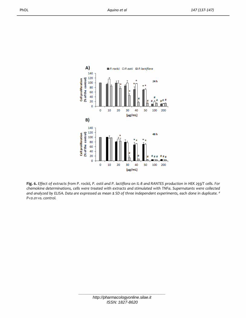
gene of transcription such as NFKB1A, encodingIκBα (Prota et al., 2011). Therefore, in order tofurther evaluate anti-inflammatory properties ofPaeonia extracts, their effects on HumanEmbryonic Kidney (HEK 293/T) cells, transfectedwith a NF-κB reporter plasmid having five tandemcopies of human NF-κB binding site fused toluciferase gene (Gautheron et al., 2010), weredetermined. Particularly, the experiments wereconducted in the presence or not of the potentTNFα pro-inflammatory agent. Preliminary studieswere carried out to determine the higher notcytotoxic concentrations of each Paeonia extract.Figure 4 shows cell proliferation curves of HEK293/T after the treatments with increasingconcentrations of each peony extract.Results demonstrate that P. rockii and P. ostiiwere not cytotoxic until 30 µg/mL in HEK 293/Tcells, while P. lactiflora induced its effect alreadyat 20 µg/mL. All the extracts determined a strongreduction of cell growth (90% of inhibition) atconcentrations higher than 50 µg /mL. As shown,there is a substantial difference with respect toresults obtained in bronchial epithelial CuFi1 andNuLi1 cells, since higher concentrations arerequired to induce any effect on cell growth inHEK 293/T cell line. On the other hands, MTT assaydid not give comparable results showing nosignificant reduction of cell viability up to 100µg/mL (data not shown) (Mencherini et al., 2011).All the above data indicate that Paeonia extractsreduced cell proliferation without affecting HEK293/T cells viability suggesting that the extractsmight cause a cell cycle arrest rather than celldeath. Anyway this needs further investigations.To directly evaluate NF-κB activity, sub-toxicconcentrations of each extract were used to treatTNFα stimulated and unstimulated HEK 293/T cellstransiently transfected as reported in section 2.8.Results (Fig. 5) indicated that all Paeonia extractswere able to reduce TNFα induced activation ofNF-κB, with P. ostii being the most effective in thiscell model system.Furthermore, we determined chemokineproduction in the same cell model possessing anormal expression and activation of NF- Bsignaling. A marked decrease in IL-8 and RANTESproduction following the treatments with Paeoniaextracts was found (Fig. 6A and B). The inhibitorytrend of the chemokines production wascomparable to NF-κB-dependent transcription
_______________________________________
http://pharmacologyonline.silae.it
ISSN: 1827-8620
PhOL Aquino et al 142 (137-147)
activation (Fig. 5).Therefore, the results obtained indicate thatPaeonia extracts possess clear anti-inflammatoryproperty and this activity might be ascribable toreduced NF-κB activation leading to an inhibition ofpro-inflammatory mediators, particularly, thechemokine RANTES.
ConclusionAmong the extracts from three Paeonia speciesexamined in the present study, similar compositionand bioactivity is shown by the morphologicallyand cytologically closely related tree Peonies,P.rockii and P. ostii, belonging to the sectionMoutan DC. The anti-inflammatory potency of theherbaceous species P. lactiflora appears higher andcorrelated to a higher polyphenol content. P.lactiflora is confirmed to be an anti-inflammatoryremedy able to reduce the expression of pro-inflammatory mediators such as RANTES and IL-8proteins, over-expressed in inflammation and,particularly, in lung inflammation of CF patients.Interestingly, all three Paeonia extracts possess aselective activity on RANTES release, chemokinewhich is involved in various severe diseases(Henriquet et al., 2007; Elsner et al., 2004).Therefore, the anti-inflammatory traditional activityreported for Radix Paeoniae (P. lactiflora) in thetraditional Chinese medicine seems to be partiallyshared with other species of the section Moutan .
References1. Aquino, R. P., Prota, L., Auriemma, G., Santoro, A.,
Mencherini, T., Colombo, G., Russo, P. 2012. Dry powderinhalers of gentamicin and leucine: formulationparameters, aerosol performance and in vitro toxicity onCuFi1 cells. International Journal of Pharmaceutics 426,100-107.
2. Borgatti, M., Mancini, I., Bianchi, N., Guerrini, A.,Lampronti, I., Rossi, D., Sacchetti, G., Gambari, R., 2011.Bergamot (Citrus bergamia Risso) fruit extracts andidentified components alter expression of interleukin 8gene in cystic fibrosis bronchial pithelial cell lines. BMCBiochemistry 12:15.
3. Chen, J. J., Cho, J. Y., Hwang, T. L., Chen, I. S., 2008.Benzoic Acid Derivatives, Acetophenones, and Anti-inflammatory Constituents from Melicope semecarpifolia.Journal of Natural Products 71, 71–75.
4. Elsner, J., Escher, S. E., Forssmann, U., 2004. Chemokinereceptor antagonists: a novel therapeutic approach inallergic diseases. Allergy 59, 1243–1258.
5. Flume, P. A., Van Devanter, D. R., 2012. State of progressin treating cystic fibrosis respiratory disease. BMCMedicine, 10, 1-12.
6. Gautheron, J., Pescatore, A., Fusco, F., Esposito, E.,Yamaoka, S., Agou, F., Ursini, M. V., Courtois, G., 2010.Identification of a new NEMO/TRAF6 interface affectedin incontinentia pigmenti pathology. Human MolecularGenetics 19, 3138–3149.
7. Guo, B. L., Basang D., Xiao P. G., Hong D. Y., 2002.Research on the quality of original plants and materialmedicine of Cortex Paeoniae. China journal of Chinesemateria medica 27, 654-657.
8. He, C. N., Peng, Y., Zhang, Y. C., Xu, L. J., Gu, J., Xiao, P.G., 2010. Phytochemical and biological studies ofPaeoniaceae. Chemistry and Biodiversity 7, 805–838.
9. He, D. Y., Dai, S. M., 2011. Anti-inflammatory andimmunomodulatory effects of Paeonia lactiflora Pall., atraditional Chinese herbal medicine. Frontiers inPharmacology 2, 1-5.
10. Henriquet, C., Gougat, C., Combes, A., Lazennec, G.,Mathieu, M., 2007. Differential regulation of RANTESand IL-8 expression in lung adenocarcinoma cells. LungCancer 56, 167—174.
11. Jacquot, J., Tabary, O., Le Rouzic, P., Clement, A., 2008.Airway epithelial cell inflammatory signalling in cysticfibrosis. International Journal of Biochemistry and CellBiology 40, 1703–1715.
12. Jiang, D., Chen, Y., Hou, X., Xu, J., Mua, X., Chen, W.,2011. Influence of Paeonia lactiflora roots extract oncAMP-phosphodiesterase activity and related anti-inflammatory action. Journal of Ethnopharmacology 137,914– 920.
13. Jiang, S., Xu, R., Yan, L., 2009. Traditional Chinesemedicinal granules containing Flos and Scutellaria andothers for treating upper respiratory tract infection. CNPatent 101480451.
14. Kim, L.D., Ha, B.J., 2009. Paeoniflorin protects RAW264.7 macrophages from LPS-induced liver inflammatoryreactions. Archives of Pharmaceutical Research 33, 959-966.
15. Lee, H. U., Kim, I., H., Jung, J. H., 2010. Method formanufacturing medicinal herb extracts for treatinginflammatory skin diseases. KR Patent 2010004215.
16. Li, J., Chen, C. X., Shen, Y. H., 2011. Effects of totalglucosides from paeony (Paeonia lactiflora Pall) roots onexperimental atherosclerosis in rats. Journal ofEthnopharmacology 135, 469–475.
17. Li, W., Huang, S., Wang, R., 2012. Advances in researchon pharmacological actions and quality control ofPaeoniae Radix Alba. Yaoxue Fuwu Yu Yanjiu, 12, 118-122.
18. Liu, B., Xiong, J., Yang, J., Cao, J., Zhao, M., Feng, B.,Guo, Y., 2011. Chinese medicinal oral preparationscontaining Dictamnus and Paeonia and others fortreating dermatitis. CN Patent 102100776.
19. Liu, M., Ma, X., 2006. Chinese medicinal tablet fortreating upper airway infection and preparation methodthereof. CN Patent 1824073.
20. Lyczak, J. B., Cannon, C. L., Pier, G. B., 2002. Lunginfections associated with cystic fibrosis. ClinicalMicrobiology Reviews 15, 194–222.
21. Ma, Y., Sun, J., Wang, J., 2008. Chinese medicinal oralliquid containing Rehmannia and Scrophularia andothers for treating lung and throat diseases. CN Patent101181513.
_______________________________________
http://pharmacologyonline.silae.it
ISSN: 1827-8620
PhOL Aquino et al 143 (137-147)
22. Mehler, E. L., Gerhards, J., 1987. Electronic determinantsof the anti-inflammatory action of benzoic and salicylicacids. Molecular Pharmacology 31, 284-293.
23. Mencherini, T., Picerno, P., Festa, M., Russo, P., Capasso,A., Aquino, R., 2011. Triterpenoid Constituents from theRoots of Paeonia rockii ssp. rockii. Journal of NaturalProducts 74, 2116–2121.
24. Nichols, D., Chmiel, J., Berger, M., 2008. Chronicinflammation in the Cystic Fibrosis Lung: Alterations ininter- and Intracellular signaling. Clinical Reviews inAllergy & Immunology 34, 146-162.
25. Nicolis, E., Lampronti, I., Dechecchi, M. C., Borgatti, M.,Tamanini, A., Bianchi, N., Bezzerri, V., Mancini, I., Giri, M.G., Rizzotti, P., Gambari, R., Cabrini, G., 2008. Pyrogallol,an active compound from the medicinal plant Emblicaofficinalis, regulates expression of pro-inflammatorygenes in bronchial epithelial cells. InternationalImmunopharmacology 8, 1672–1680.
26. Ou, Y. Y., 2008. Research of alcohol extract of Paeonialactiflora pall in anti-inflammatory and analgesic effect.Journal of Mathematical Medicine 21, 600-602.
27. Picerno, P., Mencherini, T., Sansone, F., Del Gaudio, P.,Granata, I., Porta, A., Aquino, R. P., 2011. Screening of apolar extract of Paeonia rockii: Composition andantioxidant and antifungal activities. Journal ofEthnopharmacology 138, 705– 712.
28. Prota, L., Santoro, A., Bifulco, M., Aquino, R. P.,Mencherini, T., Russo, P., 2011. Leucine enhances aerosolperformance of Naringin dry powder and its activity oncystic fibrosis airway epithelial cells. International Journalof Pharmaceutics 412, 8–19.
29. Ratjen, F. A., 2009. Cystic Fibrosis: Pathogenesis andFuture Treatment Strategies. Respiratory Care 54, 595-605.
30. Singh, I. P., Gurtu, S., Kumar, A., Sinha, J. N., Bhargava, K.P., Shanker, K., 1984. Antiinflammatory activities ofcompounds derived from salicylic and benzoic acids.Archiv der Pharmazie 317, 609-614.
31. Verhaeghe, C., Remouchamps, C., Hennuy, B.,Vanderplasschen, A., Chariot, A., Tabruyn, S. P., Oury, C.,Bours, V., 2007. Role of IKK and ERK pathways in intrinsicinflammation of cystic fibrosis airways. BiochemicalPharmacology 73, 1982–1994.
32. Wu, J., Chai, J., 2010. Chinese medicinal compositioncontaining collagen and Saururus and Smilax and othersfor treating gynecological diseases. CN Patent 101648003.
33. Wu, S. H., Wu, D. G., Che, Y. W., 2010. Chemicalconstituents and bioactivities of plant from the genusPaeonia. Chemistry and Biodiversity 7, 90–104.
34. Xu, H. M., Wei, W., Jia, X. Y., Chang, Y., Zhang, L., 2007.Effects and mechanisms of total glucosides of paeony onadjuvant arthritis in rats. Journal of Ethnopharmacology109, 442–448.
35. Zhang, W., Dai, S. H., 2012. Mechanisms involved in thetherapeutic effects of Paeonia lactiflora Pallas inrheumatoid arthritis. International Immunopharmacology14, 27–31.
36. Zhang, Y., Fang, Y., Wang, Y., 2008. Research progress inpharmacological action of total glucosides of RadixPaeoniae alba on treating rheumatoid arthritis. XiandaiZhongxiyi Jiehe Zazhi, 17, 4364-4365.

37. Zhao, T., 2011. Method for manufacturing traditionalChinese medicine preparation containing Atractylodesand Ligusticum and others for treating gynecologicalinflammations. CN Patent 102139072.
38. Zhao, X., Zhou, Z. Q., Lin, Q. B., Pan, K. Y., Li, M. Y., 2008.Phylogenetic analysis of Paeonia sect. Moutan
_______________________________________
http://pharmacologyonline.silae.it
ISSN: 1827-8620
PhOL Aquino et al 144 (137-147)
(Paeoniaceae) based on multiple DNA fragments andmorphological data. Journal of Systematics and Evolution46, 563–572.
39. Zhong, Q., 2013. Preparation method of a traditionalChinese medicinal spray for treating cough. CN Patent102920898.
Fig. 1. Cell proliferation analysis in CuFi1 (A) and NuLi1 (B) cells treated with different concentrations of Paeoniaextracts. Cell proliferation was determined for 24 (upper panels) and 48 (lower panels) h by using a colorimetricbromodeoxyuridine (BrdU) cell proliferation assay. The histograms report the percentage of growing cells comparedto controls (100% proliferation). All data are shown as mean ± SD of three independent experiments each done induplicate. * P<0.05 and # P<0.01 vs. control.

_______________________________________
http://pharmacologyonline.silae.it
ISSN: 1827-8620
PhOL Aquino et al 145 (137-147)
Fig. 2. Effect of extracts from P. rockii, P. ostii and P. lactiflora on the release of IL-8 (A), IL-6 (B) and RANTES (C) in CuFi1supernatants. Briefly cells were treated with each extract at indicated concentrations and then stimulated with TNF-α. Afteradditional 14 h of incubation, supernatants were collected and cytokines production was detected by ELISA. Data areexpressed as mean ± SD of three independent experiments, each done in duplicate. * P<0.05 and # P<0.01 vs. thecorresponding control.
Fig. 3. Effect of extracts from P. rockii, P. ostii and P. lactiflora on the release of IL- 8 (A), IL-6 (B) and RANTES (C) in NuLi1supernatants. Cells were treated with each extract at indicated concentrations and then stimulated with TNFα. Afteradditional 14 h of incubation, supernatants were collected and cytokines production was detected by ELISA. Data areexpressed as mean ± SD of three independent experiments, each done in duplicate. * P<0.05 and # P<0.01 vs. thecorresponding control.

_______________________________________
http://pharmacologyonline.silae.it
ISSN: 1827-8620
PhOL Aquino et al 146 (137-147)
Fig. 4. Effect of P. rockii, P. ostii and P. lactiflora on HEK 293/T proliferation at 24 (A) and 48 (B) h, by using BrdU cellproliferation assay. Results are shown as mean ± SD of three independent experiment each done in duplicate. * P<0.05 and #P<0.01 vs. control.
Fig. 5. Effect of extracts from P. rockii, P. ostii and P. lactiflora on NF-κB transcriptional activation in HEK 293/T cells. Cellswere treated with each extract at indicated concentrations and then stimulated with TNFα. NF- κB activation was measuredusinga luciferase assay as described in section 2.8. Fold activation is shown. #P<0.01 vs. control.
_______________________________________
http://pharmacologyonline.silae.it
ISSN: 1827-8620
PhOL Aquino et al 147 (137-147)
Fig. 6. Effect of extracts from P. rockii, P. ostii and P. lactiflora on IL-8 and RANTES production in HEK 293/T cells. Forchemokine determinations, cells were treated with extracts and stimulated with TNFα. Supernatants were collectedand analyzed by ELISA. Data are expressed as mean ± SD of three independent experiments, each done in duplicate. #
P<0.01 vs. control.