Embed Size (px)
Citation preview
Complication Rate and Pitfalls of Temporary Bridging External Fixator in Periarticular
Communited FracturesJong-Keon Oh, MD, Jin-Ho Hwang, MD*, Dipit Sahu, MD, Seung-Hyub Jun, MD†
Department of Orthopaedic Surgery, Korea University School of Medicine,*Department of Orthopaedic Surgery, Yonsei University College of Medicine,
†Department of Orthopaedic Surgery, Konkuk University School of Medicine, Seoul, Korea
Original Article Clinics in Orthopedic Surgery 2011;3:62-68 • doi:10.4055/cios.2011.3.1.62
Copyright © 2011 by Th e Korean Orthopaedic AssociationTh is is an Open Access article distributed under the terms of the Creative Commons Attribution Non-Commercial License (http://creativecommons.org/licenses/by-nc/3.0)
which permits unrestricted non-commercial use, distribution, and reproduction in any medium, provided the original work is properly cited.Clinics in Orthopedic Surgery • pISSN 2005-291X eISSN 2005-4408
Received December 28, 2009; Accepted April 1, 2010Correspondence to: Jin-Ho Hwang, MDDepartment of Orthopedic Surgery, Yonsei University College of Medicine,250 Seongsan-no (134 Sinchon-dong), Seodaemun-gu, Seoul 120-752, KoreaTel: +82-2-2228-2185, Fax: +82-2-363-1139E-mail: [email protected]
using open reduction and internal fi xation. However, the most widely used treatment is presently a staged operation of open reduction and internal fi xation with plating aft er settling down the soft tissues using a temporary bridging external fi xator (TBEF).
A TBEF is the basis of a staged operation, and easy, fast and simple procedure with many advantages. TBEF provides relative stability to fractured areas while the soft tissues around the fractured area heal before performing formal open reduction and internal fi xation. Such relative stability provides patients with mobility and thus this is considered as “portable traction.”1,2) Th e mobility given to
Background: A second staged operation using temporary bridging external fi xation (TBEF) has been widely used in patients with periarticular complex fracture, yet few papers have been published on the related complications. The purpose of this study was to report the complication rate and pitfalls directly related to TBEF through a retrospective study and to suggest some solutions.Methods: Fifty-nine cases that were treated by using TBEF were studied among 195 periarticular complex fractures. We ret-rospectively collected the clinical and radiological data and then the study data was evaluated for 1) cases with unsatisfactory restoration of length, 2) cases with deep infection caused by half pins invading the zone of defi nitive fi xation, and 3) neurovascular injuries related to half pins.Results: Complications were observed in 7/59 cases (11%). Problems related to the achievement of length were observed in one case of distal tibia fracture and 2 cases of distal femur fracture. Half pin related infection was observed in 2 cases of distal femur fracture. Neurovascular injury (medial calcaneal nerve injury in a distal tibia fracture) was observed in 2 cases. Among 7 complica-tions, four were related to using TBEF in distal femur fracture. This is because the abundant leg muscles have strong deforming force and infection might be increased due to frequent irritation by the half pins.Conclusions: TBEF is a simple procedure with several advantages. However, complications might be observed if certain prin-ciples are not followed. It is thought that many complications due to TBEF can be reduced if the half pins are not inserted in the zone of injury, restoration of length is fully achieved and the neurovascular characteristics are carefully considered. In particular, much more caution is needed in the distal femur, which has abundant muscles surrounding it.Keywords: Periarticular fractures, Bridging external fi xator, Complication
There are various treatments for periarticular complex fracture and these range from minimally invasive external fixation with percutaneous pinning to invasive methods
63
Oh et al. Temporary Bridging External Fixator Clinics in Orthopedic Surgery • Vol. 3, No. 1, 2011 • www.ecios.org
patients reduces the movement of the fractured areas and it subsequently reduces the pain and the following infl am-matory reactions of neighboring tissues, and so it quickly helps soft tissue to heal. The nursing care also becomes easier. Early mobilization can reduce the cardiopulmonary complication in patients with periarticular complex frac-ture.
With these advantages, the staged operation using TBEF has been widely used and its excellent results have been reported, but there are few studies on the complica-tion rate or pitfalls of using TBEF.
We performed a retrospective study on the patients with periarticular complex fracture (distal femur, proximal tibia, distal tibia) and who were treated with TBEF to de-termine the complication rate and the commonly related problems and also to suggest some solutions for these pit-falls.
METHODS
This study is not intended to report the results of staged operations, but rather, to discuss the problems observed during the definitive fixation after soft tissue healing around the fractured areas with the use of a TBEF, and the complications and problems that developed between the time of surgery and the fi rst postoperative outpatient visit. Th e study was conducted on 59 cases for which de-fi nitive plating was performed with a TBEF as the initial treatment, among the 195 fractures (distal femur, 58 cases; proximal tibia, 85 cases; distal tibia, 52 cases) that were treated between January of 2003 and June of 2008.
According to the Orthopaedic Trauma Associa-tion (OTA) classification, distal femur fracture included 5 cases of C1, 4 cases of C2, and 9 cases of C3; distal tibia fracture included 2 cases of C1, 3 cases of C2, and 6 cases of C3. Proximal tibia fracture, according to Shazker’s type, included 9 cases of type IV and 21 cases of type VI. Th e patients included 46 males and 13 females, and the aver-age age was 48 years. The involved mechanism of injury was mostly high-energy injuries, with 35 cases of motor vehicle accidents, 16 cases of falls and 8 cases of industrial accidents. A total of 19 cases were open fractures, among which there were 2 cases of type I, 6 cases of type II, 4 cases of type IIIa and 5 cases of type IIIb. Two out of 19 cases were type IIIc fractures and both were proximal tibia Schazker type VI. Two cases of type IIIc were excluded due to the diffi culties of determining the relationship be-tween a half pin and infection. Among these 19 cases of open fracture, 9 cases were transferred from local private hospitals to our hospital aft er performing TBEF (3 distal
femur fractures with 1 C2 and 2 C3, 4 proximal tibia frac-tures with 2 IV and 2 V and 2 distal tibia fractures with 1 C2 and 1 C3).
Surgical TechniqueTh e technique of TBEF and staged treatment was applied in the cases with severe trauma to the soft tissue around the fractured area (severe swelling, ecchymosis or blister-ing). In cases of open fractures, debridement was per-formed separately from the TBEF procedure if possible. Soft tissue debridement and irrigation were performed and then a bead pouch was made.
Half pins were inserted, using the safe corridor technique, through the region with less neurovascular risk.3) Half pin insertion was performed in a percutaneous fashion with the use of soft tissue sleeves, and pre-drilling was done to avoid thermal necrosis and soft tissue damage to the underlying bone. Half pins were inserted away from the intended site of prospective plating when converting to open reduction and internal fi xation. When the patient was hemodynamically unstable, only damage control sur-gery was performed, and TBEF was applied by focusing on maintaining the length of the limb and fractured areas. When the patient was stable, eff orts were made to provide 3-dimensional alignment of fractured areas if possible. Reduction was performed by using ligamentotaxis only, and no other procedures were performed on the fractured areas. All half pins were inserted with avoiding zones of fracture and soft tissue injury, and the prospective site of defi nitive surgery if possible.
Th e time period between TBEF and plating should be not more than 2 weeks, but there were cases with a time period of more than 2 weeks aft er TBEF such as for patients transferred from other hospitals, or wherever pin site infection was suspected, then the pin site debridement was performed later along with administration of antibiot-ics for 1 week, and then the surgery was performed.
The TBEF was kept in place until all the soft tis-sue envelopes stabilized, which usually took 7 to 10 days. When skin winkles and no pitting edema were observed, this was considered as an indication for converting to the defi nitive surgery. Th e TBEF was removed before the surgery depending on the alignment of the fractured area, and if the alignment was satisfactory, then the site was disinfected with 95% isopropyl alcohol, iodine prep scrub and iodine spray, and the reduction was maintained or TBEF was used as a reduction device in the surgical fi eld.
Knee Spanning FramesKnee spanning frames were applied for complex, unstable
64
Oh et al. Temporary Bridging External Fixator Clinics in Orthopedic Surgery • Vol. 3, No. 1, 2011 • www.ecios.org
distal femur and proximal tibia fractures. Half pins were inserted by considering the future intended location of plates in the defi nitive surgery. Two half pins were inserted in the anterior femur and 2 half pins were inserted in the anteromedial tibia. Various types of external fi xator devices were used, and most of which were modular type external fi xators. Th e advantages of this device include convenient 3-dimensional reduction that can be obtained by only us-ing bars, which were connected to the half pins inserted to each fractured area. In such cases, double-stacked anterior bars were used to increase the stability of the fractured areas. Particularly in the case of the distal femur, it is bet-ter to maintain the alignment during the TBEF procedure because of the bulky muscles in the fractured area and the characteristic displacement after injury. When a patient was hemodynamically stable, correcting the length, ex-ternal rotation and fl exion deformity were attempted sev-eral times under fl uoroscopic guidance. It is thought that maintenance of length is a very important factor. However, if the vascularity of a limb in question was suspicious, then overdistraction could be dangerous. Th erefore, the circula-tion was always checked aft er distraction.
Ankle Spanning FramesThese frames were used in cases of pilon fractures with severely compromised soft tissues. Lateral fi bular plating was not performed in the same surgery along with TBEF. In most cases, a modular external fixator was used. Two half pins were inserted in the proximal tibia and for the distal part, 1 half pin was inserted in the lateral calcaneus and the other one was inserted in the 1st metatarsal base. Bars and medial calcaneus pin were applied in good order to increase the frame stability. If possible, a medial pin was inserted last because of possible damage to the medial
calcaneal branch.4) Blunt dissection and sleeves were used prior to half pin insertion.
When a patient was hemodynamically stable, reduc-tion was performed by ligamentotaxis under fl uoroscopic guidance to preferentially obtain the lengthening of the fracture sites and to achieve ankle joint stability. This is because if the ankle joint has no stability, then soft tissue healing can be very slow. When the circulation was doubt-ful, overdistraction was avoided and the circulation was checked aft er the surgery.
EvaluationThe patients were evaluated clinically and radiologically for the following three possible TBEF related problems.1. Cases with unsatisfactory achievement of length. Fail-
ure of lengthening was defi ned as cases in which any procedure such as the push technique was performed or a femoral distractor was used to obtain lengthen-ing, but it did not achieve the desired length.
2. Cases with deep infection caused by half pins invading the zone of defi nitive fi xation without considering the plating position for definitive fixation. Delayed deep infection that developed in the fracture zone without being invaded by half pins was excluded. Also, deep infection of fractured areas related to open fracture was excluded.
3. Neurovascular injuries observed aft er TBEF fi xation.
RESULTS
TBEF related problems were observed in 7 out of 59 cases (11%). Among these 7 cases, failure to achieve length was observed in 3 cases, including 1 case in distal tibia fracture and 2 cases in distal femur fracture. Neurovascular injuries
Table 1. Summary of the Patients with Complications
No. Gender/Age Classifi cation Complications Location Open fracture Hospital
applying TBEF Final results Comments
1 M/21 33 C1 Length Distal tibia No Author’s Restoration Needs an additional procedure to get the proper length, which makes the operation time longer.
2 M/56 33 C2 Length Distal femur No Other’s Restoration
3 F/43 33 C2 Length Distal femur No Author’s Restoration
4 M/65 33 C3 Infection Distal femur Yes (the other site) Other’s Knee fusion
5 F/41 33 C2 Infection Distal femur No Author’s Revision surgery
6 M/25 43 B2 Neurovascular injury Distal tibia No Author’s Partial recovery
7 M/39 43 C2 Neurovascular injury Distal tibia Yes Author’s Partial recovery
TBEF: temporary bridging external fi xation.
65
Oh et al. Temporary Bridging External Fixator Clinics in Orthopedic Surgery • Vol. 3, No. 1, 2011 • www.ecios.org
were observed in 2 out of 7 cases. Both cases were medial calcaneal nerve injury. Half pin site related deep infec-tion was observed in 2 cases, and both were cases of distal femur. Four out of the 7 cases of complications were cases of distal femur fractures. Two of the the nine were trans-ferred from another local private hospital.
The average time for definitive plating after TBEF was 15.3 days (range, 4 to 81 days). If the cases with deep infection were excluded, then the actual time was 11.3 days.
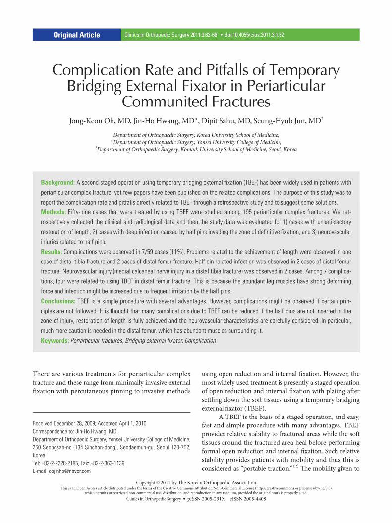
Table 1 summarizes the complications. Two cases among the cases with lengthening related problems were distal femur fractures. Achieving the lengthening in the management of distal femur fracture is easier than that for other deformities. When surgeons see just the AP image of C-arm fluoroscopy during the procedures, they make mistakes such as shortening because of fl exion deformity of the distal femur. Fig. 1 showed a 45-year male patient who was transferred from a local private hospital after the application of initial TBEF for severe soft tissue com-promise. Proximal half pins were involved not only in the
zone of defi nitive surgery, but also in the zone of soft tis-sue injury. In addition, a distal femur articular fragment showed typical deformity such as internal rotation, fl exion and shortening. Fortunately, no infection was observed in the proximal half pin site and the second surgery could be done at the 12th day aft er surgery without debridement of the pin site, and no specifi c infection was observed.
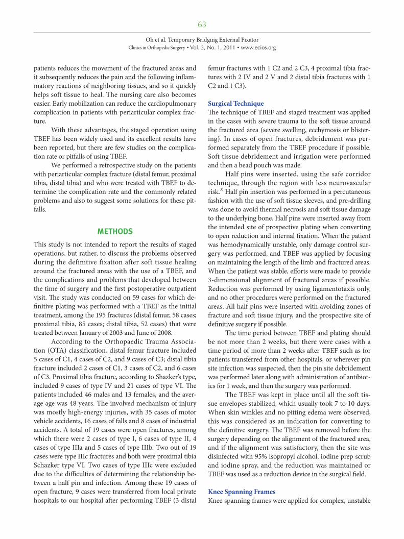
Neurovascular injury was observed in 2 out of 7 cases and both were medial calcaneal nerve injuries. Fig. 2 showed a 40-year male patient who received TBEF for soft tissue swelling and blister formation after pilon fracture. Th e patient did complain about continuous paresthesia in the heel area.
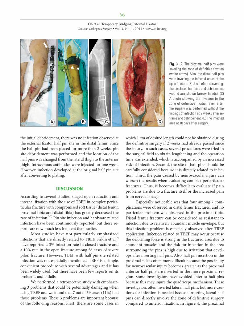
All the TBEF related deep infections on the half pins sites developed in distal femur fractures. A 60-year patient who had a tibia open fracture, including a distal femur fracture, from a traffi c accident was transferred from a lo-cal hospital after 4 weeks of treatment (Fig. 3). Infection was found in the tibia, but this was controlled by perform-ing radical debridement and muscle transfer, and proximal half pin was re-applied. aft er debridement. At that time of
Fig. 1. (A) Radiographs showing the severe external rotation of the proximal femur (white arrow), shortening and flexion deformity of the distal femur (arrow head) and valgus deformity of the proximal tibia (star). The proximal and distal half pins were placed without considering the definitive fixation. (B) A fluoroscopy image shows obtaining lengthening with the use of the push technique during the operation. (C) Restoration of all alignments after surgery.
Fig. 2. (A) Distal half pins were inserted in the medial calcaneal side and the 1st metatarsal base. (B, C) A medial calcaneal pin was inserted almost at the end of the heel.
66
Oh et al. Temporary Bridging External Fixator Clinics in Orthopedic Surgery • Vol. 3, No. 1, 2011 • www.ecios.org
the initial debridement, there was no infection observed at the external fi xator half pin site in the distal femur. Since the half pin had been placed for more than 2 weeks, pin site debridement was performed and the location of the half pins was changed from the lateral thigh to the anterior thigh. Intravenous antibiotics were injected for one week. However, infection developed at the original half pin site aft er converting to plating.
DISCUSSION
According to several studies, staged open reduction and internal fi xation with the use of TBEF in complex periar-ticular fracture with compromised soft tissue (distal femur, proximal tibia and distal tibia) has greatly decreased the rate of infection.5-7) Pin site infection and hardware related infection have been continuously reported, but these re-ports are now much less frequent than earlier.
Most studies have not particularly emphasized infections that are directly related to TBEF. Sirkin et al.7) have reported a 3% infection rate in closed fracture and a 10% rate in the open fracture among 56 cases of severe pilon fracture. However, TBEF with half pin site related infection was not especially mentioned. TBEF is a simple, convenient procedure with several advantages and it has been widely used, but there have been few reports on its problems and pitfalls.
We performed a retrospective study with emphasiz-ing 3 problems that could be potentially damaging when using TBEF and we found that 7 out of 59 cases (11%) had those problems. Th ese 3 problems are important because of the following reasons. First, there are some cases in
which 1 cm of desired length could not be obtained during the defi nitive surgery if 2 weeks had already passed since the injury. In such cases, several procedures were tried in the surgical fi eld to obtain lengthening and the operation time was extended, which is accompanied by an increased risk of infection. Second, the site of half pins should be carefully considered because it is directly related to infec-tion. Third, the pain caused by neurovascular injury can worsen the results when evaluating complex periarticular fractures. Thus, it becomes difficult to evaluate if pain problems are due to a fracture itself or the increased pain from nerve damage.
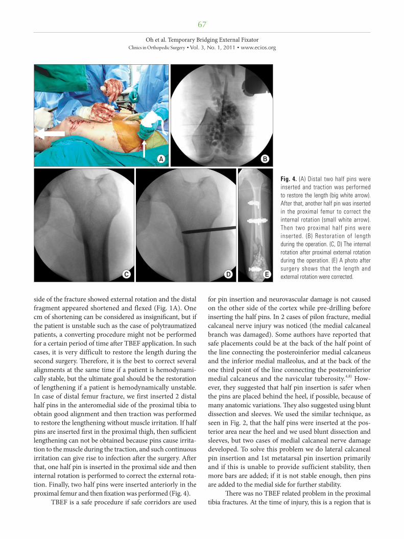
Especially noticeable was that four among 7 com-plications were observed in distal femur fractures, and no particular problem was observed in the proximal tibia. Distal femur fracture can be considered as resistant to infection due to relatively abundant muscle envelope, but this infection problem is especially observed after TBEF application. Infection related to TBEF may occur because the deforming force is strong in the fractured area due to abundant muscles and the risk for infection in the area surrounding the pins is high due to irritation that devel-ops aft er inserting half pins. Also, half pin insertion in the proximal side is oft en more diffi cult because the possibility for neurovascular injury becomes greater as the proximal anterior half pins are inserted in the more proximal re-gion. Some investigators have avoided anterior half pins because this may injure the quadriceps mechanism. Th ese investigators oft en inserted lateral half pins, but more cau-tions for infection is needed because inserting lateral half pins can directly involve the zone of definitive surgery compared to anterior fixation. In figure 4, the proximal
Fig. 3. (A) The proximal half pins were invading the zone of definitive fixation (white arrow). Also, the distal half pins were invading the infected areas of the open fracture. (B) Just before converting, the displaced half pins and debridement wound are shown (arrow heads). (C) A photo showing the invasion to the zone of definitive fixation even after the surgery was performed without the fi ndings of infection at 2 weeks after re-frame and debridement. (D) The infected area at 10 days after surgery.
67
Oh et al. Temporary Bridging External Fixator Clinics in Orthopedic Surgery • Vol. 3, No. 1, 2011 • www.ecios.org
side of the fracture showed external rotation and the distal fragment appeared shortened and flexed (Fig. 1A). One cm of shortening can be considered as insignifi cant, but if the patient is unstable such as the case of polytraumatized patients, a converting procedure might not be performed for a certain period of time aft er TBEF application. In such cases, it is very difficult to restore the length during the second surgery. Th erefore, it is the best to correct several alignments at the same time if a patient is hemodynami-cally stable, but the ultimate goal should be the restoration of lengthening if a patient is hemodynamically unstable. In case of distal femur fracture, we first inserted 2 distal half pins in the anteromedial side of the proximal tibia to obtain good alignment and then traction was performed to restore the lengthening without muscle irritation. If half pins are inserted fi rst in the proximal thigh, then suffi cient lengthening can not be obtained because pins cause irrita-tion to the muscle during the traction, and such continuous irritation can give rise to infection aft er the surgery. Aft er that, one half pin is inserted in the proximal side and then internal rotation is performed to correct the external rota-tion. Finally, two half pins were inserted anteriorly in the proximal femur and then fi xation was performed (Fig. 4).
TBEF is a safe procedure if safe corridors are used
for pin insertion and neurovascular damage is not caused on the other side of the cortex while pre-drilling before inserting the half pins. In 2 cases of pilon fracture, medial calcaneal nerve injury was noticed (the medial calcaneal branch was damaged). Some authors have reported that safe placements could be at the back of the half point of the line connecting the posteroinferior medial calcaneus and the inferior medial malleolus, and at the back of the one third point of the line connecting the posteroinferior medial calcaneus and the navicular tuberosity.4,8) How-ever, they suggested that half pin insertion is safer when the pins are placed behind the heel, if possible, because of many anatomic variations. Th ey also suggested using blunt dissection and sleeves. We used the similar technique, as seen in Fig. 2, that the half pins were inserted at the pos-terior area near the heel and we used blunt dissection and sleeves, but two cases of medial calcaneal nerve damage developed. To solve this problem we do lateral calcaneal pin insertion and 1st metatarsal pin insertion primarily and if this is unable to provide sufficient stability, then more bars are added; if it is not stable enough, then pins are added to the medial side for further stability.
Th ere was no TBEF related problem in the proximal tibia fractures. At the time of injury, this is a region that is
Fig. 4. (A) Distal two half pins were inserted and traction was performed to restore the length (big white arrow). After that, another half pin was inserted in the proximal femur to correct the internal rotation (small white arrow). Then two proximal half pins were inserted. (B) Restoration of length during the operation. (C, D) The internal rotation after proximal external rotation during the operation. (E) A photo after surgery shows that the length and external rotation were corrected.
68
Oh et al. Temporary Bridging External Fixator Clinics in Orthopedic Surgery • Vol. 3, No. 1, 2011 • www.ecios.org
REFERENCES
1. Watson JT, Coufal C. Treatment of complex lateral plateau fractures using Ilizarov techniques. Clin Orthop Relat Res. 1998;(353):97-106.
2. Watson JT, Moed BR, Karges DE, Cramer KE. Pilon frac-tures. Treatment protocol based on severity of soft tissue injury. Clin Orthop Relat Res. 2000;(375):78-90.
3. Behrens F. General theory and principles of external fixa-tion. Clin Orthop Relat Res. 1989;(241):15-23.
4. Casey D, McConnell T, Parekh S, Tornetta P 3rd. Percuta-neous pin placement in the medial calcaneus: is anywhere safe? J Orthop Trauma. 2002;16(1):26-9.
5. Blauth M, Bastian L, Krettek C, Knop C, Evans S. Surgical
options for the treatment of severe tibial pilon fractures: a study of three techniques. J Orthop Trauma. 2001;15(3):153-60.
6. Collinge C, Sanders R, DiPasquale T. Treatment of complex tibial periarticular fractures using percutaneous techniques. Clin Orthop Relat Res. 2000;(375):69-77.
7. Sirkin MS, Bono CM, Reilly MC, Behrens FF. Percutaneous methods of tibial plateau fixation. Clin Orthop Relat Res. 2000;(375):60-8.
8. Santi MD, Botte MJ. External fi xation of the calcaneus and talus: an anatomical study for safe pin insertion. J Orthop Trauma. 1996;10(7):487-91.
easily damaged due to open fracture, but this region seems to have less problems for TBEF application, in which proximal half pins are easily inserted in the distal femur, because it is not a fi xation zone and also the pins are rela-tively easily inserted in the distal part to the ankle area because the anteromedial side is skin on bone.
Although our results were obtained from a retro-spective study and the number of total cases was small, this is the fi rst study on the TBEF problems that most or-thopedic surgeons easily fail to notice. Also, it is thought that a rate of complications over 10% can suffi ciently aff ect the fi nal results of complex periarticular fracture. Th ere-fore, we feel this study on the application of TBEF will catch the attention of diligent and thoughtful surgeons.
In conclusion, TBEF is a basic procedure for the staged operation technique and this technique has recently
been widely used. It is considered to be a relatively easy technique, but its importance has been less emphasized than definitive plating. However, special concerns are needed because the frequency of TBEF related complica-tions is high in cases of distal femur fracture. Th us, better results will be obtained if TBEF is performed with giving full consideration to the fixation zone, the restoration of length and the neurovascular structures. Much caution is needed when performing TBEF and particularly for distal femur fractures.
CONFLICT OF INTEREST
No potential confl ict of interest relevant to this article was reported.