Embed Size (px)
Citation preview

COMPLEX STROKE: Care Overview, Case Studies and Complications
Stroke Coordinators Boot Camp 9/18/2014

Presenters:
9/18/2014 C. Artuso - V. Johnson Stroke Coordinators Boot Camp 2
Christie E. Artuso, ED.D., RN, CNRN, SCRN
Director, Neuroscience Services and Interim Director, Cancer Center
Comprehensive Stroke Center Providence Medical Center
Anchorage, AK
Vicki Johnson, DNP, MHSEd., ARNP
Manager and Nurse Practitioner
Comprehensive Stroke Center at Harborview Medical Center
Seattle, WA

What will we cover today?
9/18/2014 C. Artuso - V. Johnson Stroke Coordinators Boot Camp 3
I. Introduction & Overview: Complex
Stroke
II. The First 36 Hours of Care
III. Basic Brain Imaging
IV. Critical Transitions of Care:
- admission to discharge and beyond
V. Complex Stroke Cases & Imaging
- malignant MCA ischemic stroke
- complications >>>>>>>>>>>>>>>>>>
V. Questions

Introduction and Overview
9/18/2014 C. Artuso - V. Johnson Stroke Coordinators Boot Camp 4
I. Introduction & Overview: Complex
Stroke
II. The First 36 Hours of Care
III. Basic Brain Imaging
IV. Critical Transitions of Care:
- admission to discharge and beyond
V. Complex Stroke Cases & Imaging
- malignant MCA ischemic stroke
- complications >>>>>>>>>>>>>>>>>>
V. Questions

CARE OF THE PATIENT WITH ACUTE STROKE
• Early assessment
• Rapid recognition of symptoms
• Activation of emergency services / appropriate intervention(s)
• Ongoing assessment
• Knowledgeable, multidisciplinary care team approach
COMPLEX STROKE PATIENTS – THE FIRST 36 HOURS
9/5/2014 ©2010, American Heart Association 5

NIH-Recommended ED Response Times1-3
What is the average door-to-needle time at your institution?
*
6
The “golden hour” for evaluating and treating acute ischemic stroke

Pre-hospital assessment scales
• Cincinnati Prehospital Stroke Scale
• Los Angeles Prehospital Stroke Screen
• Miami Emergency Neurologic Deficit Checklist
Pre-hospital actions
• Document “Time last seen normal”
• Consider transport to nearest stroke center
• Perform finger stick to obtain blood glucose
• Obtain vital signs including blood pressure
• Facility pre-notification (code stroke/stroke team alert)
.
1. Jauch EC, et al. Stroke. 2013;44:870-947.
77
Pre-Hospital Assessment/Actions For Potential Stroke: Dispatch and Delivery1

Recommended Evaluation Elements
• Patient history
• Physical exam and formal stroke scale (e.g., NIHSS)
• Stat non-contrast CT scan of the brain
• Consider ordering the following diagnostic tests
– Blood glucose
– Serum electrolytes / renal function tests
– Markers of cardiac ischemia
– Complete blood count, including platelet count
– Activated partial thromboplastin time
– Prothrombin time/international normalized ratio
– Electrocardiogram
– Oxygen saturation
88
Initial Clinical Evaluation of Potential Stroke:Door and Data1
1. Jauch EC, et al. Stroke. 2013;44:870-947.

1. Thomas SH, et al. N Engl J Med. 2006;354:2263-2271.
2. Heiss W-D. J Cereb Blood Flow Metab. 2000;20:1276-1293.
3. Heiss W-D. Stroke. 1983;14:329-331.
• Cells of the ischemic penumbra are
metabolically active and potentially
salvageable with timely assessment and
management1,2
• The infarction expands in the penumbra
over time, increasing the area of
irreversible brain damage2
• Restoration of blood flow to the affected
area may interrupt this process3
Time Is Brain: Damage to the Brain DuringAIS Is a Rapid and Progressive Process
99
Image is for
illustrative
purposes only
Penumbra
Area of infarct
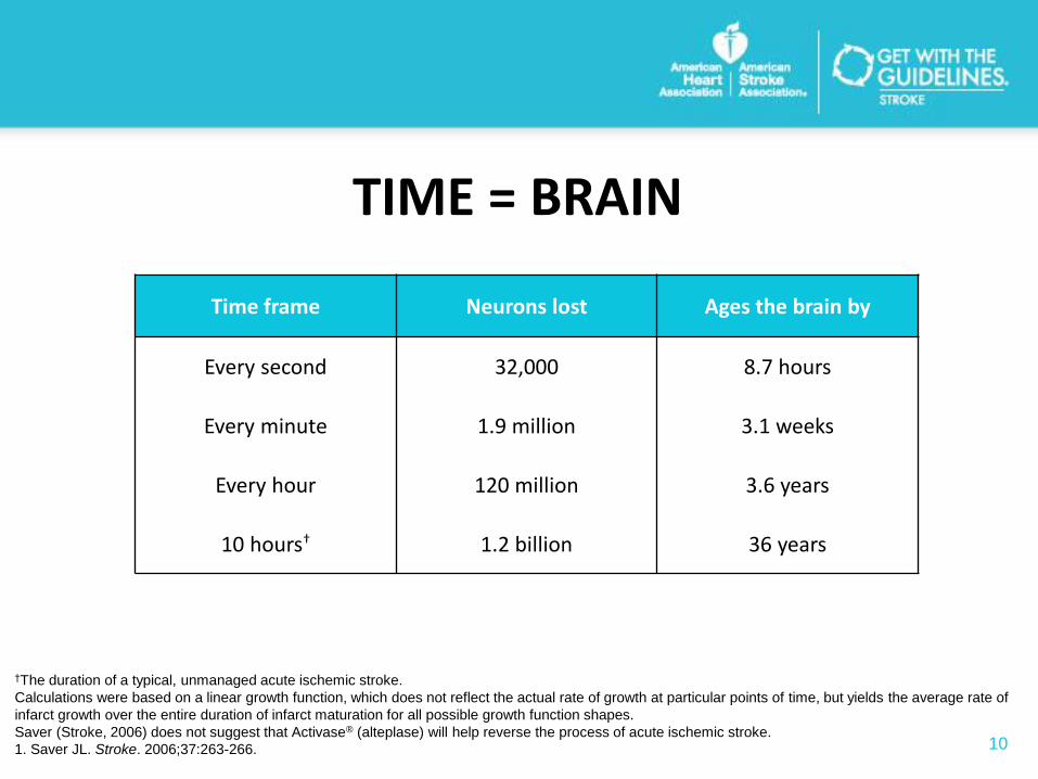
TIME = BRAIN
101. Saver JL. Stroke. 2006;37:263-266.
Time frame Neurons lost Ages the brain by
Every second 32,000 8.7 hours
Every minute 1.9 million 3.1 weeks
Every hour 120 million 3.6 years
10 hours† 1.2 billion 36 years
†The duration of a typical, unmanaged acute ischemic stroke.
Calculations were based on a linear growth function, which does not reflect the actual rate of growth at particular points of time, but yields the average rate of
infarct growth over the entire duration of infarct maturation for all possible growth function shapes.
Saver (Stroke, 2006) does not suggest that Activase® (alteplase) will help reverse the process of acute ischemic stroke.

Clinical Presentation of AIS and Conditions That May Mimic Stroke1,2
11
Clinical Presentation of Acute Ischemic Stroke
Aphasia
Ataxia
Dysarthria
Diplopia
Cranial nerve palsies
Hemianopia
Hemiparesis
Loss of sensation
Quadriparesis
Visual field disturbances
Conditions That May Mimic Stroke
Alcoholic intoxication Metabolic disorders
Cerebral infections Migraines
Drug Overdose Conversion disorder
Epidural hematoma Seizure and post-seizure
Hypoglycemia Tumors
Neuropathies(e.g., Bell’s Palsy)
Hypertensive encephalopathy
1. Jauch EC, et al. Stroke. 2013;44:870-947.
2. Katz MJ, et al. http://www.nursingceu.com/courses/301/index_nceu.html Accessed April 1, 2013.

• Cerebral edema
• Hemorrhagic conversion
• Pneumonia
• Infection
• Seizures
• Deep vein thrombosis
COMPLICATIONS DURING THE FIRST 36 HOURS
9/5/2014 ©2010, American Heart Association 12

• Risk for IntraCranialHemorrhage 0.5% to 2% per year
• Anticoagulation increases risk 10 X
• Risk of cerebral emboli in patients with major
cariodembolic sources (prosthetic valve, cardiac
thrombus, AF) is high (without anticoagulation)
– 20-30%
HEMORRHAGIC STROKE RISK
9/5/2014 ©2010, American Heart Association 13

Relationships With Early Clinical Deterioration and 3-Month
Outcome in the European Cooperative Acute Stroke Study I
(ECASS I) Cohort
RESULTS: Risk of early neurological deterioration and of 3-
month death was severely increased after PH2, indicating
that large hematoma is the only type of hemorrhagic
transformation that may alter the clinical course of ischemic
stroke.
HEMORRHAGIC TRANSFORMATION WITHIN 36
HOURS OF CEREBRAL INFARCT
9/5/2014 ©2010, American Heart Association 14

HEMORRHAGIC CONVERSION
• The presence of petechiae or confluent petechial hemorrhage confined to the ischemic zone
CEREBRAL EDEMA
• Accumulation of fluid in the intracellular or extracellular spaces of the brain;
• Occurs during first 24-48 hours
• Large hemispheric strokes – greater risk
• Often associated with hemorrhagic strokes or a hemorrhagic conversion
VASOSPASM
• More common with subarachnoid hemorrhage related to aneurysm
PNEUMONIA
• Often related to dysphagia and aspiration
COMPLEX STROKE PATIENTS – THE FIRST 36 HOURS
9/5/2014 ©2010, American Heart Association 15
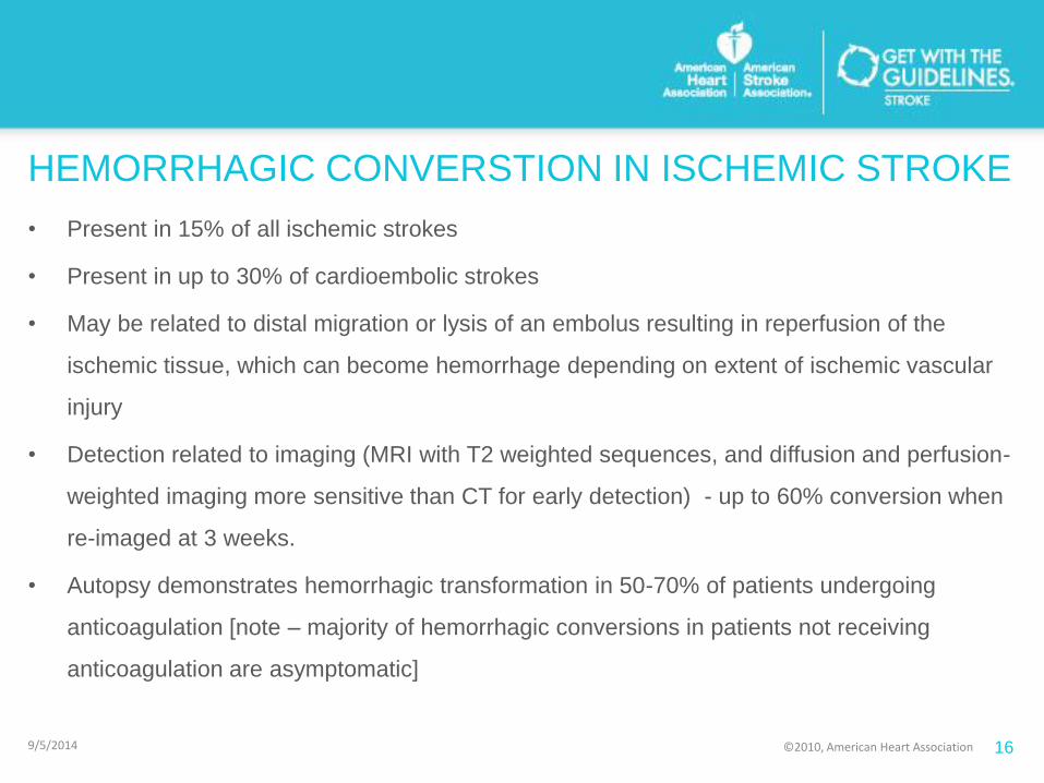
• Present in 15% of all ischemic strokes
• Present in up to 30% of cardioembolic strokes
• May be related to distal migration or lysis of an embolus resulting in reperfusion of the
ischemic tissue, which can become hemorrhage depending on extent of ischemic vascular
injury
• Detection related to imaging (MRI with T2 weighted sequences, and diffusion and perfusion-
weighted imaging more sensitive than CT for early detection) - up to 60% conversion when
re-imaged at 3 weeks.
• Autopsy demonstrates hemorrhagic transformation in 50-70% of patients undergoing
anticoagulation [note – majority of hemorrhagic conversions in patients not receiving
anticoagulation are asymptomatic]
HEMORRHAGIC CONVERSTION IN ISCHEMIC STROKE
9/5/2014 ©2010, American Heart Association 16

• Visualization in patient with ischemic stroke may provide insight into
underlying mechanism of stroke
– May influence ongoing therapy
• Early anticoagulation in ischemic stroke adds risk to hemorrhagic
conversion
• Risk:Benefit ratio of early anticoagulation influenced by the
cardioembolic source of stroke as well as size of infarct [current data
indicates the risk for recurrent stroke within first 5-7 days after ischemic
event 5-8% without anticoagulation {patient with AF}]
– Although heparin reduces the risk of recurrence of cardioembolic stroke – risk of
symptomatic ICH offsets benefits
PEARLS RELATED TO HEMORRHAGIC CONVERSION
9/5/2014 ©2010, American Heart Association 17

• Occurrence and timing of hemorrhagic conversion not impacted by
anticoagulation
– Magnitude and clinical impact of hemorrhagic conversion has higher association
– Large infarct
– Excessive anticoagulation
– Higher risk for symptomatic hemorrhagic transformation
• Current recommendation – delaying anticoagulation for 1-2 weeks (7-10
days)
– Oral warfarin (without heparin) may be used in patients with nonvalvular AF or recent MI
• Typical patient with ischemia and AF is unlikely to benefit from
anticoagulation within the first 1-2 days, especially with large stroke
ANTICOAGULATION AND HEMORRHAGIC CONVERSION
9/5/2014 ©2010, American Heart Association 18

• Patients who may benefit from early
anticoagulation
– Documeted thrombi in left atrium
– Mechanical prosthetic valve
– Intracardiac thrombus
– Congestive heart failure
• Consider other risk factors for brain hemorrhage
ANTICOAGULATION AND HEMORRHAGIC CONVERSION
9/5/2014 ©2010, American Heart Association 19

Introduction and Overview
9/18/2014 C. Artuso - V. Johnson Stroke Coordinators Boot Camp 20
I. Introduction & Overview: Complex
Stroke
II. The First 36 Hours of Care
III. Basic Brain Imaging
IV. Critical Transitions of Care:
- admission to discharge and beyond
V. Complex Stroke Cases & Imaging
- malignant MCA ischemic stroke
- complications >>>>>>>>>>>>>>>>>>
V. Questions

Stroke Types
Normal IschemicStroke
IntracerebralHemorrhage
SubarachnoidHemorrhage 9/1
8/2014 C. Artuso - V. Joh
Stroke Coordinators Boot Camp 2

CT-Angiography
Rapidly images large vessels in the
neck and many first- and second-
order arteries in the brain
Non-contrast CT
Is the most practical and
least time consuming initial
brain imaging test for
evaluation of potential stroke
and can rule out hemorrhage
CT-Perfusion
Provides cerebral blood flow, cerebral blood
volume, and mean transit time maps
1. Love A, et al. Stroke Res Treat. 2011;2011:726573.
2. Jauch EC, et al. Stroke. 2013;44:870-947.
Imaging for Stroke Assessment1,2
2
222
What type of neuro-imaging modalities does your hospital use?
Basic Brain Imaging

Basic Brain Imaging: CT
• X-ray beams (photons) pass through a patient’s body and are
collected by computed tomography “CT” detector.
• A gray scale is created showing different shades of gray
depending on the degree of absorption of the X-ray beams.
• TERMINOLOGY: a structure or lesion is “hyperdense”
(LIGHTER shade of gray/white) or “hypodense” (DARKER
shade of gray)
9/18/2014 C. Artuso - V. Johnson Stroke Coordinators Boot Camp 23

Basic Brain Imaging: Houndsfield Units (“HU’s”)
CT absorption scale is measured in Houndfield units (“HU’s”). The scale is +1000 to - 1000. The higher the HU’s, the more hyperdense. So bone is white, air is black.
Substance HU
Bone 1000 (white)
Calcification 140-200 (white)
Acute blood 56-76 (white)
Gray matter 32-41 (actually whiter than “white” matter)
White matter 23-34
CSF 0
Fat -30 -100 (darker)
Air -1000 (black)
Neuroradiology: The Requisites, Grossman and Yousem;
9/18/2014 C. Artuso - V. Johnson Stroke Coordinators Boot Camp 24

Basic Brain Imaging
Right Left There are 3 views in neuro-
imaging: axial, coronal and
sagittal. Most head CT’s will
show axial cuts only, like the
one to the left.
MRI will usually show all 3
views: axial, coronal and
sagittal view.
The convention is that the
patient’s right side of the brain
will be shown on the left side
of the image. In an axial view,
picture the patient lying in the
scanner with the feet sticking
out at you. 9/18/2014 C. Artuso - V. Johnson Stroke Coordinators Boot Camp 25
Coronal MRI
SAGITAL MRIAXIAL CT

Basic Brain Imaging: Tips for Reading a CT
Pay attention to what a normal head CT’s look like.
There is a WIDE range of normal and a wide range in quality of exams.
As with reading chest x-rays,
• symmetry is your friend;
• it is helpful that there are 2 halves to every brain;
• side by side comparison can reveal a lesion or give assurance that the study is normal.
The most basic head CT will have approximately 32 axial cuts measuring 0.5 cm each. Get used to going through a study the same way every time (bottom to top or top to bottom).
9/18/2014 C. Artuso - V. JohnsonStroke Coordinators Boot Camp 26

Basic Brain Imaging: Tips for Reading a CT
Experiment with viewing different numbers of axial cuts at a time – you can scroll through a study looking at just a single image at a time or you can look at 2, 4 or 6 images at a time.
Many people find it valuable to look at more than one cut at a time -- to get the whole picture and to see if a lesion extends beyond a single cut. However you often may want to focus on a single image to get the best detail of a potential abnormality.
Pay attention to structure density. Look for gray-white differentiation.
Pay attention to ventricle size. Again this will help to give you a sense of the broad range of normal. If the ventricles seem large, do the sulci also seem large?
9/18/2014 C. Artuso - V. Johnson Stroke Coordinators Boot Camp 27

Basic Brain Imaging
9/18/2014 C. Artuso - V. Johnson Stroke Coordinators Boot Camp 28

Basic Brain Imaging
9/18/2014 C. Artuso - V. Johnson Stroke Coordinators Boot Camp 29

Basic Brain Imaging
HEAD CT OPTIONS:
Plain CT
CT with contrast
CT Angiogram (“CTA”)
Head
Neck
CT Perfusion
9/18/2014 C. Artuso - V. Johnson Stroke Coordinators Boot Camp 30
NORMAL

Basic Brain Imaging
HEAD CT OPTIONS:
Plain CT
CT with contrast
CT Angiogram (“CTA”)
Head &/or Neck
CT Perfusion
9/18/2014 C. Artuso - V. Johnson Stroke Coordinators Boot Camp 31
Plan Head CT Best for:
Trauma: skull/face/orbit fracture
Detecting acute blood
subarachnoid hemorrhage
intraparenchymal hemorrhage
subdural hematoma
Detecting calcification
------------------------------------------
Lesions commonly missed on CT:
Acute ischemic stroke
Tumor
Abscess
Demyelinating lesion

Basic Brain Imaging
HEAD CT OPTIONS:
Plain CT
CT with contrast
CT Angiogram (“CTA”)
Head &/or Neck
CT Perfusion
9/18/2014 C. Artuso - V. Johnson Stroke Coordinators Boot Camp 32
CONTRAST= IODINE• administered via 18 ga or larger
ante-cubital IV• flows through cerebral vessels and
leaks out in areas of blood-brain barrier breakdown
• order a CT with contrast when you’re worried about a focal lesion such as: tumor, abscess or other focal infection; you still miss lesions that can be seen only w/MRI but will increase yield over plain CT
• Terminology: area of contrast outline is “enhancing”
CONTRAINDICATIONS • Creatinine >1.5 (there are exceptions)
• Iodine allergy (there are exceptions)
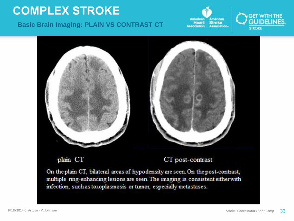
Basic Brain Imaging: PLAIN VS CONTRAST CT
9/18/2014 C. Artuso - V. Johnson Stroke Coordinators Boot Camp 33
Basic Brain Imaging
HEAD CT OPTIONS:
Plain CT
CT with contrast
CT Angiogram
(“CTA”)
Head &/or Neck
CT Perfusion
9/18/2014 C. Artuso - V. Johnson Stroke Coordinators Boot Camp 34
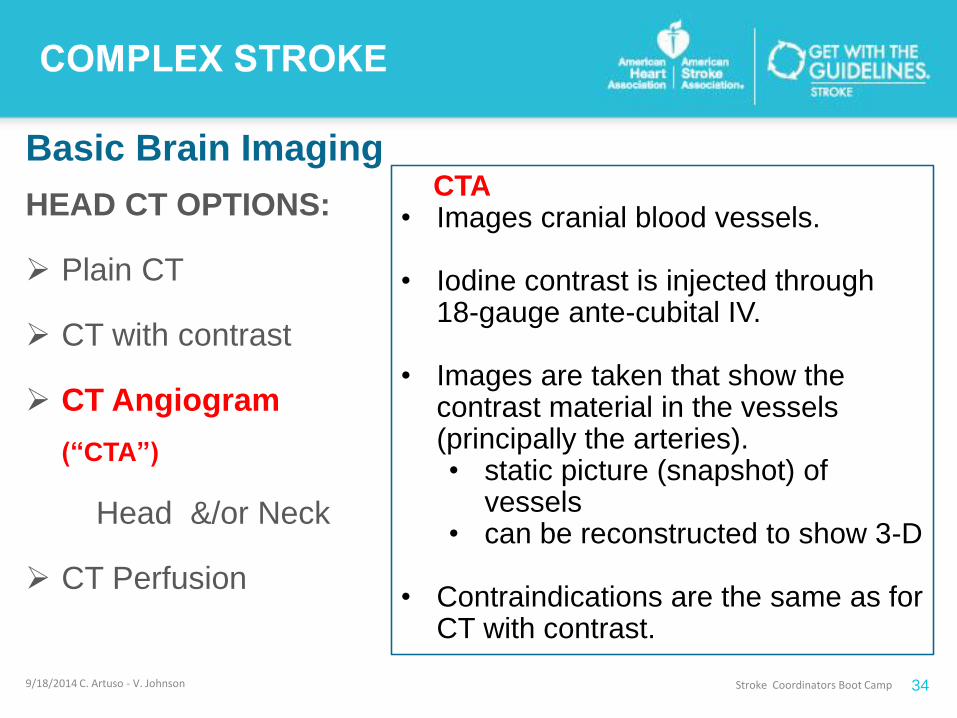
CTA• Images cranial blood vessels.
• Iodine contrast is injected through 18-gauge ante-cubital IV.
• Images are taken that show the contrast material in the vessels (principally the arteries).• static picture (snapshot) of
vessels• can be reconstructed to show 3-D
• Contraindications are the same as for CT with contrast.
Basic Brain Imaging
HEAD CT OPTIONS:
Plain CT
CT with contrast
CT Angiogram
(“CTA”)
Head &/or Neck
CT Perfusion
9/18/2014 C. Artuso - V. Johnson Stroke Coordinators Boot Camp 35
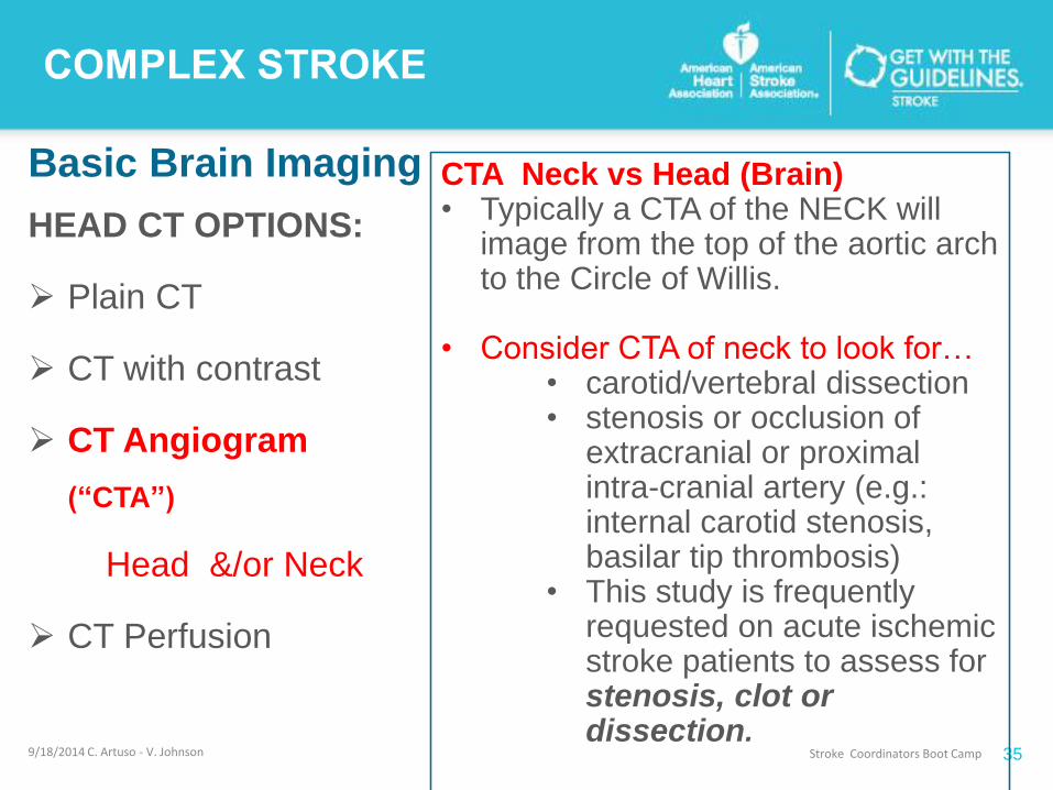
CTA Neck vs Head (Brain)• Typically a CTA of the NECK will
image from the top of the aortic arch to the Circle of Willis.
• Consider CTA of neck to look for… • carotid/vertebral dissection• stenosis or occlusion of
extracranial or proximal intra-cranial artery (e.g.: internal carotid stenosis, basilar tip thrombosis)
• This study is frequently requested on acute ischemic stroke patients to assess for stenosis, clot or dissection.
Basic Brain Imaging: CTA NECK
9/18/2014 C. Artuso - V. Johnson Stroke Coordinators Boot Camp 36
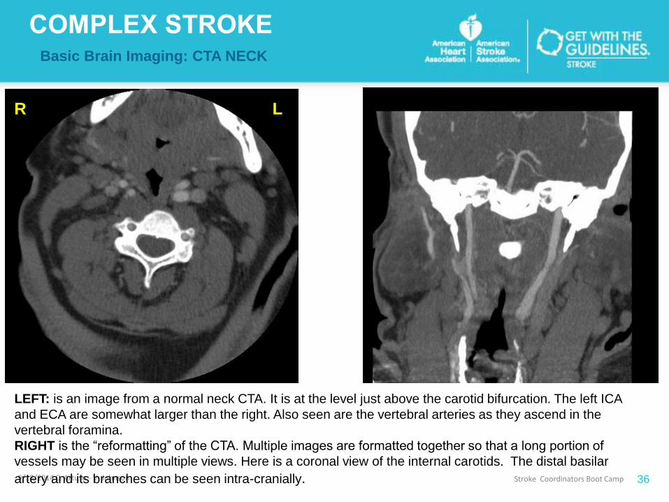
LEFT: is an image from a normal neck CTA. It is at the level just above the carotid bifurcation. The left ICA
and ECA are somewhat larger than the right. Also seen are the vertebral arteries as they ascend in the
vertebral foramina.
RIGHT is the “reformatting” of the CTA. Multiple images are formatted together so that a long portion of
vessels may be seen in multiple views. Here is a coronal view of the internal carotids. The distal basilar
artery and its branches can be seen intra-cranially.
R L

Basic Brain Imaging
HEAD CT OPTIONS:
Plain CT
CT with contrast
CT Angiogram
(“CTA”)
Head &/or Neck
CT Perfusion
9/18/2014 C. Artuso - V. Johnson Stroke Coordinators Boot Camp 37
CTA Neck vs Head (Brain) cont. • CTA of the BRAIN enhances
vessels from the skull base to the vertex – it will not give a good view of extra-cranial vessels.
• Consider CTA of brain to evaluate for suspected:• aneurysm• AVM or other vascular anomaly
• It is important to specify “CTA neck” or “CTA brain” – if you ask for both, the radiologist may protest and/or the study may be technically sub-optimal.

9/18/2014 C. Artuso - V. Johnson Stroke Coordinators Boot Camp 38
From left to right: plain CT, CTA, CTA reconstruction. The cause of this sub-arachnoid
hemorrhage is evident even on the plain CT. There is an aneurysm at the basilar tip. It is more
sharply outlined in the CTA. The CTA reconstruction shows the Circle of Willis with the large
aneurysm very nicely. When you suspect a subarachnoid hemorrhage on plain CT, you should
order a CT-angiogram of the brain to assess for aneurysm.
Basic Brain Imaging: CTA, SAH & Aneurysm

Basic Brain Imaging
HEAD CT OPTIONS:
Plain CT
CT with contrast
CT Angiogram (“CTA”)
Head &/or Neck
CT Perfusion
9/18/2014 C. Artuso - V. Johnson Stroke Coordinators Boot Camp 39
CT Perfusion Study• Iodine or xenon is infused/inhaled
(respectively).
• Cerebral blood flow is measured.
• Hypo-perfused areas are differentiated from normal brain and from infarcted brain.
• Sometimes performed in setting of acute CVA to discern if there is salvageable tissue.
• Really need experience neuro-radiologist to read 2/2 variability in studies from center to center.

HEAD CT OPTIONS:
Plain CT
CT with contrast
CT Angiogram (“CTA”)
Head &/or Neck
CT Perfusion
Basic Brain Imaging
9/18/2014 C. Artuso - V. Johnson Stroke Coordinators Boot Camp 40
CT Perfusion may help ID penumbra
How can it be useful?graphic of penumbra

Stroke Coordinators Boot Camp 41
Basic Brain Imaging: CT- Perfusion Study
CT- Perfusion
Helps determine
cerebral blood flow
and which areas
are at risk.
Stroke Coordinators Boot Camp 42
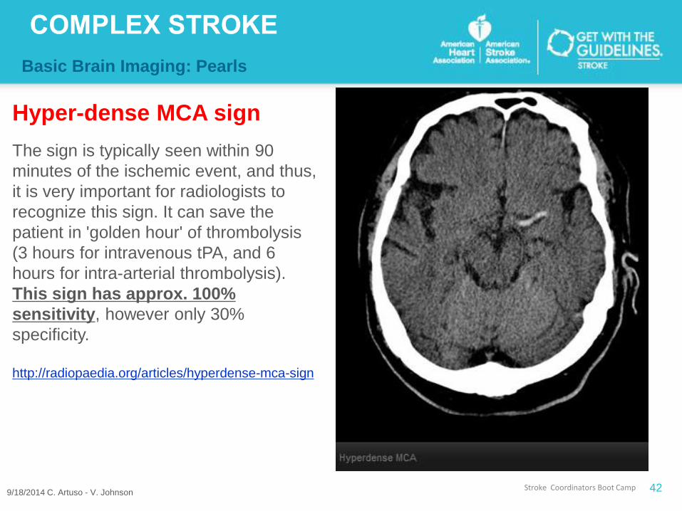
Basic Brain Imaging: Pearls
Hyper-dense MCA sign
The sign is typically seen within 90
minutes of the ischemic event, and thus,
it is very important for radiologists to
recognize this sign. It can save the
patient in 'golden hour' of thrombolysis
(3 hours for intravenous tPA, and 6
hours for intra-arterial thrombolysis).
This sign has approx. 100%
sensitivity, however only 30%
specificity.
http://radiopaedia.org/articles/hyperdense-mca-sign
9/18/2014 C. Artuso - V. Johnson
Stroke Coordinators Boot Camp 43
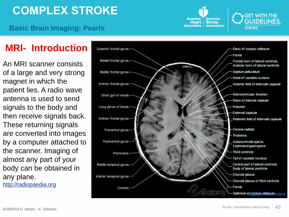
Basic Brain Imaging: Pearls
MRI- Introduction
An MRI scanner consists
of a large and very strong
magnet in which the
patient lies. A radio wave
antenna is used to send
signals to the body and
then receive signals back.
These returning signals
are converted into images
by a computer attached to
the scanner. Imaging of
almost any part of your
body can be obtained in
any plane.http://radiopaedia.org
9/18/2014 C. Artuso - V. Johnson

Stroke Coordinators Boot Camp 44
Basic Brain Imaging: MRI- Introduction
Advantages
• Visualize early ischemic changes
• Ability to image without the use of
ionizing radiation (x-ray) unlike CT
scanning
• multiple plane imaging (Axial, Sagittal,
Coronal, or Oblique) without moving the
patient.
• Ability to be reconstructed in multiple
planes
• Superior soft tissue contrast films
making it ideal for the brain, spine, joints
and other soft tissue body parts
• Some images can be obtained without
contrast, unlike CT or conventional
angiography
• Advanced techniques for visualization of
both brain activity and the underlying
networks
9/18/2014 C. Artuso - V. Johnson
Disadvantages
• There are a number of disadvantages
and challenges to implementing MRI
scanning.
• MRI scans are more expensive than
CT scans and take longer to acquire
so patient comfort is sometimes an
issue. Additionally images are subject
to unique artifacts that must be
recognized and abated
• MRI scanning is not safe for patients
with metal implants and foreign
bodies.
• Careful attention to safety measures
to avoid serious injury and requires
special MRI compatible equipment
and stringent adherence to safety
protocols
http://radiopaedia.org/articles/

Stroke Coordinators Boot Camp 45
Basic Brain Imaging:
MRI- Diffusion Weighted Imaging
• early identification of ischemic
stroke
• differentiation of acute vs
chronic stroke
• differentiation of acute stroke
vs mimics
• differentiation of epidermoid
cyst from arachnoid cyst
• differentiation of abscess from
necrotic tumors
http://radiopaedia.org/articles/diffusion-weighted-imaging-1
9/18/2014 C. Artuso - V. Johnson
• assessment of cortical
lesions in CJD
• differentiation of herpes
encephalitis from diffuse
temporal gliomas
• grading of gliomas and
meningiomas (need
further study)
• assessment of
active MS plaque (old
plaques will not be bright)
DWI major role in the following clinical situations

Stroke Coordinators Boot Camp 46
Basic Brain Imaging:
MRI- Diffusion Weighted Imaging
DWI EARLY (acute) ischemic stroke
9/18/2014 C. Artuso - V. Johnson
DWI OLD (subacute) ischemic
stroke.
Basic Brain Imaging
Resources for neuroanatomy & imaging reviews:
UCLA Neuroradiology has compiled “A collection of the best educational sites”; the website is http://www.neuropat.dote.hu/nrad2.htm
E-anatomy. http://www.imaios.com/en/e-Anatomy/Head-and-Neck/Brain-MRI-in-axial-slices
UBM Medica Network http://radiopaedia.org/
9/18/2014 C. Artuso - V. Johnson Stroke Coordinators Boot Camp 47

Introduction and Overview
9/18/2014 C. Artuso - V. Johnson Stroke Coordinators Boot Camp 48
I. Introduction & Overview: Complex
Stroke
II. The First 36 Hours of Care
III. Basic Brain Imaging
IV.Critical Transitions of Care:
pre-admission to discharge and
beyond
V. Complex Stroke Cases & Imaging
- malignant MCA ischemic stroke
- complications >>>>>>>>>>>>>>>>>>
V. Questions

What are transitions of care?
“Transitions of care”
the movement of patients between health care staff and
practitioners, settings, and home as their condition and care
needs change.
PCP• PCP or specialist in an outpatient setting
HOSP
• to a hospital physician and nursing team during an inpatient admission
SNF• yet another care team at a skilled nursing
facility
PCP• Back home to make appointment with PCP

Critical Transitions of Care:
9/18/2014 C. Artuso - V. Johnson Stroke Coordinators Boot Camp 50
Critical Transitions of Care:
9/18/2014 C. Artuso - V. Johnson Stroke Coordinators Boot Camp 51
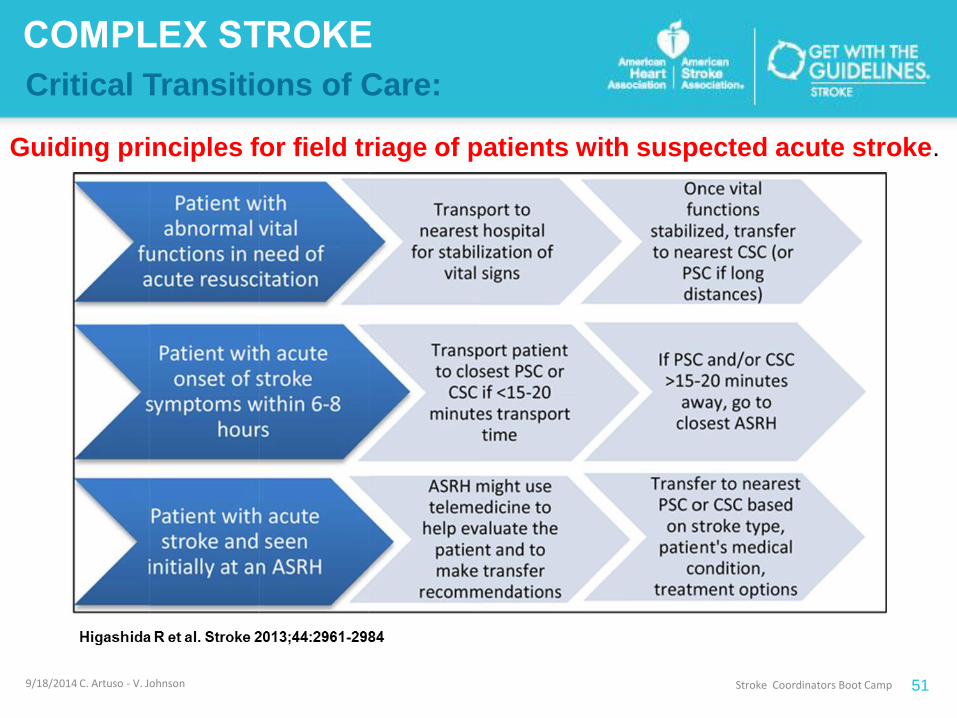
Guiding principles for field triage of patients with suspected acute stroke.

Critical Transitions of Care:
9/18/2014 C. Artuso - V. Johnson Stroke Coordinators Boot Camp 52
Examples of care transitions among staff, specialists, and care areas:
HYPERACUTE ISCHEMIC STROKE PATIENT

Critical Transitions of Care:
9/18/2014 C. Artuso - V. Johnson Stroke Coordinators Boot Camp 53
Examples of care transitions among staff, specialists, and care areas:
HYPERACUTE HEMORRHAGIC PATIENT: IPH/ SAH

Critical Transitions of Care:
9/18/2014 C. Artuso - V. Johnson
Stroke Coordinators Boot Camp
54
Examples of care transitions among staff, specialists, and care areas:
HYPERACUTE ISCHEMIC STROKE …..
POSSIBLE INTERVENTION (ENDOVASCULAR OR SURGICAL)
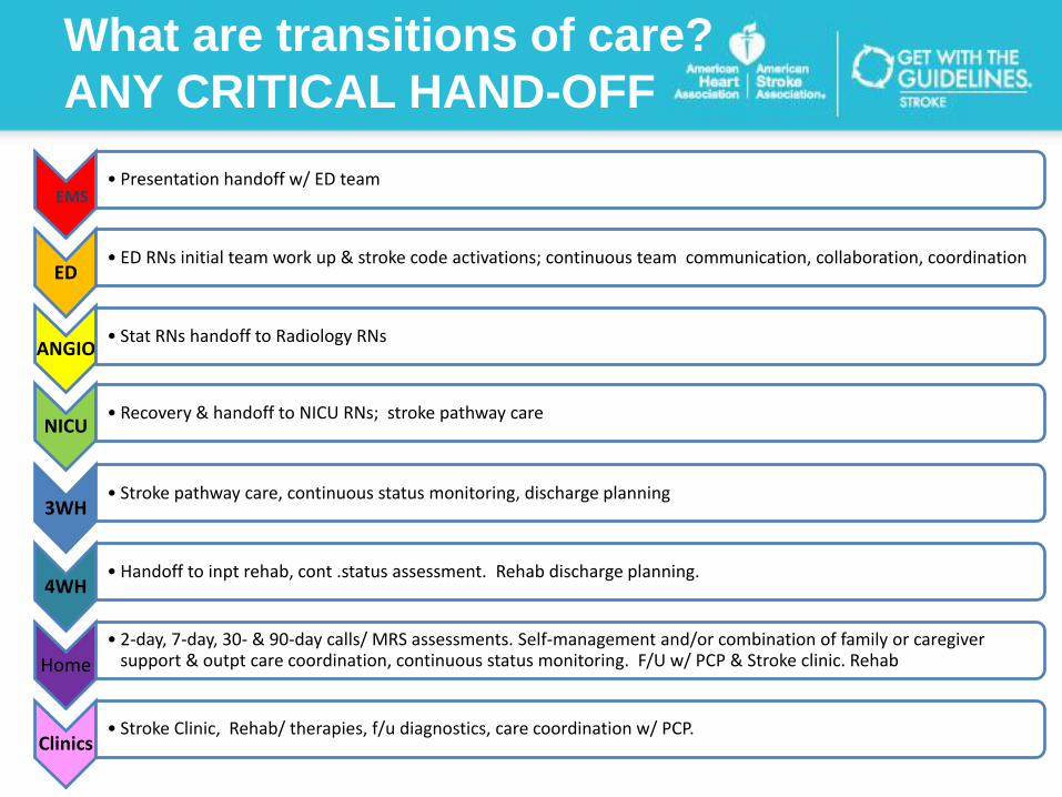
What are transitions of care?
ANY CRITICAL HAND-OFF
EMS• Presentation handoff w/ ED team
ED• ED RNs initial team work up & stroke code activations; continuous team communication, collaboration, coordination
ANGIO• Stat RNs handoff to Radiology RNs
NICU• Recovery & handoff to NICU RNs; stroke pathway care
3WH• Stroke pathway care, continuous status monitoring, discharge planning
4WH• Handoff to inpt rehab, cont .status assessment. Rehab discharge planning.
Home
• 2-day, 7-day, 30- & 90-day calls/ MRS assessments. Self-management and/or combination of family or caregiver support & outpt care coordination, continuous status monitoring. F/U w/ PCP & Stroke clinic. Rehab
Clinics• Stroke Clinic, Rehab/ therapies, f/u diagnostics, care coordination w/ PCP.
What is the mandate?
REGULATORY MANDATE:
To reduce both readmission rates and adverse events, hospitals have the
mandate to improve the effectiveness of transitions of care in which they play a
role. (CMS & Joint Commission Stroke)
CONSEQUENCES: Failure to meet standards
1. Poor patient outcomes.
2. Penalties: Hospitals with unacceptably high readmission rates for Medicare
and Medicaid patients will soon face financial penalties under the Patient
Protection and Affordable Care Act.
3. Denial of Comp. Stroke Certification (TJC)
3. Forster AJ, et al: Adverse drug events occurring following hospital discharge. Journal of General Internal Medicine, April 2005;20(4):317-23

The problem:
Ineffective transitions of careIneffective care transitions lead to preventable adverse events3,4 , higher
hospital readmission rates and avoidable costs. 4
A study of physician-to-physician communication estimated that 80% of serious
medical errors involve miscommunication during the hand-off between medical
providers.5
“falling through the cracks”, occurs in every type of health care setting, but it
is especially problematic when patients leave the hospital to receive care in
another setting or in discharge to home.
3. Forster AJ, et al: Adverse drug events occurring following hospital discharge. Journal of General Internal Medicine, April 2005;20(4):317-23
4. Medicare Payment Advisory Commission, Report to the Congress: Reforming the Delivery System, Washington, D.C.: MedPAC, June 2008
5. Solet DJ, et al: Lost in translation: challenges and opportunities in physician-to-physician communication during patient hand-offs. Academic
Medicine, 2005;80:1094-9

Transitions of Care
Lessons Learned: Check the safety brakes before you let go!
Critical Transitions of Care:
9/18/2014 C. Artuso - V. Johnson Stroke Coordinators Boot Camp 59
Wide variation in symptoms to identify & monitor
Neurologic assessment continuity (variation of assessment
skills)
Documentation: EMR systems & content complex
Acuity: Very High
Balancing multiple high risk health problems
Complex Pathophysiology
Coordinating complicated care with resource limitations
Multiple disciplines, services, and hand-offs involved
Nursing Challenges to Consider

Best-evidence
transitional care practices 6,9,10,12,14,15,20,21,23,24,26,27
• Multidisciplinary communication, collaboration and
coordination – including patient/caregiver education –
from admission through transition. A care team –
• Clinician involvement and shared accountability during
all points of transition
• Comprehensive planning and risk assessment
throughout hospital stay.
Best-evidence
transitional care practices 6,9,10,12,14,15,20,21,23,24,26,27
• Standardized transition plans, procedures and forms.
• Standardized training.
• Timely follow-up, support and coordination after the
patient leaves a care setting.
• If a patient is readmitted within 30 days, gain an
understanding of why.
• Evaluation of transitions of care measures. (surveys,
HAND-OFFs & DOCUMENTATION
BEST PRACTICE MUST HAVE…
Documentation showing tracking of patient status between ALL
transfers with comparable assessments!
9/18/2014 C. Artuso - V. Johnson Stroke Coordinators Boot Camp 62
Post-TPA and Post-procedure Monitoring Challenges

Critical Transitions of Care:
9/18/2014 C. Artuso - V. Johnson Stroke Coordinators Boot Camp 63
Post-TPA and Post-procedure Monitoring Challenges
Post-tPA:
Q 15 min X 2 hours, then q 30 minutes X 6 hours, then q 1
hour X 16 hours, then per unit protocol.
Document on post-procedure tracking tools:
tPA started, completed and each of the timed VS/Neuro/Post-
Angio checks (where applicable)
Post-Intervention & Post-Angiography Monitoring:
limited evidence to guide the “BEST PRACTICE” for exact
assessment & monitoring protocol.

Sample
Post-Angio
Tracking
Sheet.
Front &
Back
9/18/2014 C. Artuso - V. Johnson Stroke Coordinators Boot Camp 64
9/5/2014 ©2010, American Heart Association 65

Introduction and Overview
9/18/2014 C. Artuso - V. Johnson Stroke Coordinators Boot Camp 66
I. Introduction & Overview: Complex
Stroke
II. The First 36 Hours of Care
III. Basic Brain Imaging
IV. Critical Transitions of Care:
- admission to discharge and beyond
V. Complex Stroke Cases
- malignant MCA ischemic stroke
- complications >>>>>>>>>>>>>>>>>>
V. Questions

• Peak edema usually at about 3 days, but can range from 1-5
• Peak edema from hemorrhagic stroke can occur later
• Large strokes require careful monitoring for neurologic deterioration
• Decompressive hemicraniectomy may be needed
Malignant Edema
9/18/2014 C. Artuso - V. Johnson Stroke Coordinators Boot Camp 67

Decreases the risk
of death in cases of
malignant edema
with large, complex
ischemic strokes.
Hemicraniectomy
9/18/2014 C. Artuso - V. Johnson Stroke Coordinators Boot Camp 68

Hemicraniectomy: outcome comparison
9/18/2014 C. Artuso - V. Johnson Stroke Coordinators Boot Camp 69

56 year old male patient, found down at home, left side paresis
• Returned from work at 2 AM; loud thump heard in AM – several hours to gain access to
home
• History of hypertension; has not seen physician in many years
• Smokes 1 ppd, social alcohol use; family hx heart disease
• Initial CT – large right MCA infarct
Large right MCA infarct on CT’
VS: Temp: 36 °C (96.8 °F) HR: 69 RRR: 14 SpO2: 96 % BP: 188/120 mmHg
[BP treated with labetolol in ED]
CASE STUDY: MCA with Unstable Edema,
Hematoma, Bone Flap [7/1]
9/5/2014 ©2010, American Heart Association 70

Well developed, well-nourished male patient; 81.65 kg
Pupils equal, reactive to light; dense left-sided hemiparesis; able to converse with some
hesitation; c/o right side headache;
7/01 CT – Subacute right middle cerebral arterial distribution infarction with moderate cerebral
edema and mass effect on the right lateral ventricle.
Carotid dopplers – occluded right ICA
Admitted to general neurology floor – admission medications included Aspirin, Lipitor, Lovenox,
Pepcid, Insulin, Ativan; IV NSS @ 100 ml/hr
PHYSICAL EXAM
9/5/2014 ©2010, American Heart Association 71

WBC – 17.1 (4.5-11.0uL)
Glucose 125 mg/dL (65-99)
PT 12.6 sec (12.9-15 sec)
Admission VS: Temp: 36 °C (96.8 °F) HR: 62 RR: 35 SpO2: 95 % BP: 191/117 mmHg
ECG - NSR
Admitting Diagnosis: Stroke: multiple risk factors including HTN, smoking, family history.
Currently sinus rhythm. Right MCA distribution with vasogenic edema.
OVERVIEW and ADMISSION
9/5/2014 ©2010, American Heart Association 72

Patient c/o persistent headache – focused on right side; increasingly lethargic progressing to
obtunded
Stat imaging reveals increased right sided edema; midline shift, slight hemorrhage, symmetry
reperfusion
NEUROSURGERY CONSULTATION – prepared for craniectomy
PREOPERATIVE DIAGNOSIS: Malignant middle cerebral artery syndrome secondary to
massive right middle cerebral artery infarct with mass effect, midline shift, and neurologic
deterioration.
POSTOPERATIVE DIAGNOSIS: same as pre-operative dx
OPERATION: Right frontotemporal parietal hemicraniectomy with dural enhancement with graft.
Day 2 – hospital admission
9/5/2014 ©2010, American Heart Association 73
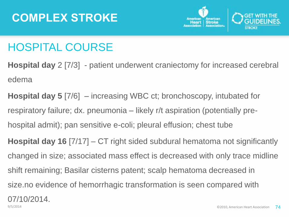
Hospital day 2 [7/3] - patient underwent craniectomy for increased cerebral
edema
Hospital day 5 [7/6] – increasing WBC ct; bronchoscopy, intubated for
respiratory failure; dx. pneumonia – likely r/t aspiration (potentially pre-
hospital admit); pan sensitive e-coli; pleural effusion; chest tube
Hospital day 16 [7/17] – CT right sided subdural hematoma not significantly
changed in size; associated mass effect is decreased with only trace midline
shift remaining; Basilar cisterns patent; scalp hematoma decreased in
size.no evidence of hemorrhagic transformation is seen compared with
07/10/2014.
HOSPITAL COURSE
9/5/2014 ©2010, American Heart Association 74

Respiratory Failure – intubated; tracheostomy performed
Cardiac – slightly elevated troponin; high risk for cardiac cath; recommended medical
management; aortic atherosclerosis
Dysphagia – PEG placed
Elevated LFTs – statin changed to pravastatin
HOSPITAL COURSE [continued]
9/5/2014 ©2010, American Heart Association 75

Discharge DX:
Acute right MCA stroke; unstable cerebral edema; s/p hemicranectomy; left-sided
hemiparesis; PEG for nutrition; pneumonia (resolving); s/p pleural effusions; s/p chest
tubes for evacuation; subdural hematoma (resolving); s/p midline shift (resolving);
compromised airway (trach)
DC Medications: ASA 81mg po od; Lisinopril 5 mg bid; metoprolol 50 mg bid; pravastatin 80
mg od;
Discharged to LTAC on hospital day 28
DISCHARGE SUMMARY
9/5/2014 ©2010, American Heart Association 76

53 year old Asian female patient transferred from distant community hospital after telestroke
evaluation and treatment
Patient’s husband reports that he was awakened when she tried to get out of bed at 0330 AM
and fell to the floor; patient has slurred speech with facial droop, left-side hemiparesis
Past medical history – significant for atrial fibrillation
Evaluated via telemedicine; initial CT WNL; cervical spine films WNL
Treated with rt-PA at 0630; elective intubation for airway protection
CASE STUDY: MCA STROKE WITH
HEMORRHAGIC TRANSFORMATION POST tPA
9/5/2014 ©2010, American Heart Association 77

CBC and CMP were normal except glucose 237. Digoxin level 0.8. INR was 1.0,
troponin normal at 0.11.
EKG showed non-specific ST changes in lead III.
The patient was intubated, sedated on propofol drip, and mechanically ventilated
upon arrival
Sedation D/C to assess neuro status.
Patient is awake and pointing to her ET tube with her R hand; left side flaccid
Repeat CT without contrast – no change at 6 hours, some subjective fullness of the R
MCA Patient is bradycardic in the 40s-50s, BP in the 120s-140s systolic, oxygen
saturation 100%. ABG 7.42/35/204/100. CXR is clear.
INITIAL EVALUATION
9/5/2014 ©2010, American Heart Association 78

• Current Medications
– Lisinopril
– Digoxin
– Coreg
– Spironolactone
– Insulin- novalog
• No allergies
• Denes smoking, rare alcohol use
• Vital signs on admission: BP 160/64; HR; 63; RR 17
• Physical exam after rt-PA – increased ability to move left leg; no sensation left side; left arm
flaccid
INITIAL EVALUATION cont.
9/5/2014 ©2010, American Heart Association 79

• Hospital Day 2 – increasing cerebral edema; evidence of hemorrhage on CT scan;
deteriorating neurologic condition
– Craniectomy performed
– Improved neurologic condition
• Patient made steady improvement with therapy; residual dysarthria; dysphagia; right dense
hemiparesis (U > L); rate controlled atrial fibrillation; on Coumadin - therapeutic
• Fever unknown origin – found associated with CVL – central line discontinued; broad
spectrum antibiotics continued x 10 days; WBC normal; fever WNL
• DM – A1C 8.8
• Discharged home, Metformin 1000 mg bid; Lisinopril 20 mg od; aldactone 50 mg od;
.Novolag with meals; Crestor 5 mg od; Coreg 80 mg od; lanoxin 250 ug od
HOSPITAL COURSE
9/5/2014 ©2010, American Heart Association 80

IMAGING REVIEW
9/5/2014 ©2010, American Heart Association 81
• 80 y.o. female with hx of dementia, depression/anxiety, hypertension,
hyperlipidemia, paroxysmal atrial.fib, breast cancer s/p mastectomy
• Onset of left leg weakness, left facial droop and slurred speech today
• At around 2:15pm, patient was walking around the block with a walker. The
caregiver noticed that the patient was dragging her left foot with walking, had
slurred speech, and left facial droop.
• Daughter was notified, who came home and noticed the above findings. She
quickly assessed that the patient had sensation in all her extremities.
CASE STUDY: CEREBELLAR STROKE – STABLE
HEMORRHAGIC TRANSFORMATION
9/5/2014 ©2010, American Heart Association 82
• Head CT negative for bleed; Frontal lobe predominant cerebral atrophy,
small vessel ischemic changes and cerebral white matter
• Symptoms improving; tPA not administered
IMAGING RESULTS
9/5/2014 ©2010, American Heart Association 83

IMAGING REVIEW
9/5/2014 ©2010, American Heart Association 84

Admission VS: Temp: 36.4 °C (97.5 °F) HR: 63 RR: 24 SpO2: 94 % BP: 118/60 mmHg
Weight: 98.88 kg
Laboratory Results: (abnormal only)
Glucose: 109 mg/dL
BUN: 31 (8-26 mg/dL)
Creatinine: 1.27 (0.6 – 1.1 mg/dL)
Protime 12.6 (12.9 – 15 sec)
EKG: Normal Sinus Rhythm
Carotid U/S reveal bilateral PE
ED AND HOSPITAL COURSE
9/5/2014 ©2010, American Heart Association 85

Patient was admitted and placed on full anticoagulation. She had complete resolution of her
neurologic symptoms and MRI failed to demonstrate acute ischemic event. She remained in
sinus rhythm on telemetry; discharged 3 days later with final diagnosis of transient ischemic
attack. Remains on Coumadin and Lovenox.
Patient re-evaluated in the ED 9 days post discharge with persistent episodes of vomiting and
lack of interest in food. No acute neurologic changes were noted. Patient evaluated by PMD;
video swallow study performed [unremarkable]; MRI ordered.
Patient readmitted post MRI with dx. Hemorrhagic conversion post ischemic stroke.
HOSPITAL COURSE
9/5/2014 ©2010, American Heart Association 86

MRI: generalized cerebral atrophy, with prominence of the ventricles and sulci.
New heterogeneous, T2 hypointense, T1 hyperintense lesion within the right
cerebellar hemisphere, with loss of signal on gradient echo sequences, consistent
with an intraparenchymal hemorrhage. This measures 4.2 x 3.8 x 2.3 cm. Mass effect
from the hemorrhage severely narrows the 4th ventricle, although the lateral
ventricles have increased
only minimally in size, previously 5.1 cm right to left, currently 5.3 cm
right to left. A third ventricle measures 13 mm, not significantly
changed from 12 mm previously.
IMAGING RESULTS
9/5/2014 ©2010, American Heart Association 87

IMAGING REVIEW
9/5/2014 ©2010, American Heart Association 88

Patient discharged to assisted living
Remains on ASA only – accepted risk of devastating embolic
event r/t pulmonary embolus
Family reviewed risks/benefits of anticoagulation, however
given patient’s advanced dementia and risk of additional
neurologic deterioration opted for ASA.
D/C Status
9/5/2014 ©2010, American Heart Association 89

CASE STUDY
9/18/2014 C. Artuso - V. Johnson Stroke Coordinators Boot Camp 90
Malignant Edema & Hemicraniectomy, plus…
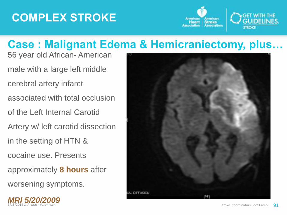
Case : Malignant Edema & Hemicraniectomy, plus…56 year old African- American
male with a large left middle
cerebral artery infarct
associated with total occlusion
of the Left Internal Carotid
Artery w/ left carotid dissection
in the setting of HTN &
cocaine use. Presents
approximately 8 hours after
worsening symptoms.
MRI 5/20/2009
Captions for photo’s and other elements go here.
This is 9pt, Italic and in program color
9/18/2014 C. Artuso - V. Johnson Stroke Coordinators Boot Camp 91
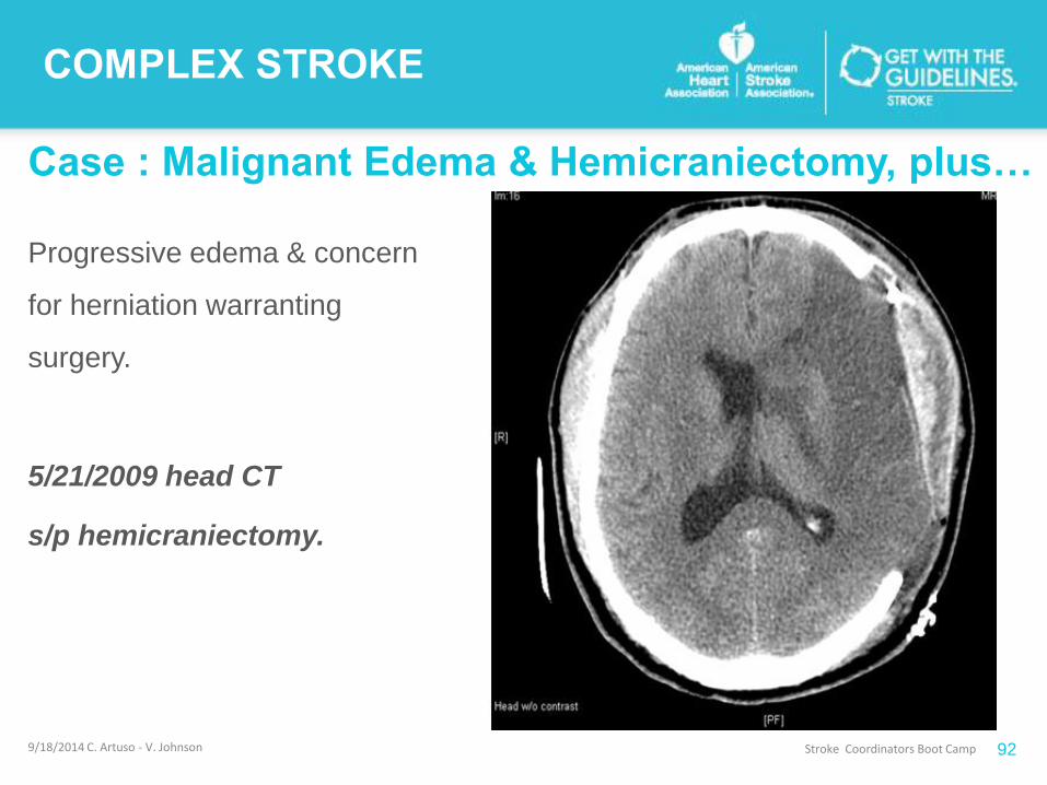
Case : Malignant Edema & Hemicraniectomy, plus…
Progressive edema & concern
for herniation warranting
surgery.
5/21/2009 head CT
s/p hemicraniectomy.
Captions for photo’s and other elements go here.
This is 9pt, Italic and in program color
9/18/2014 C. Artuso - V. Johnson Stroke Coordinators Boot Camp 92

Case : Malignant Edema & Hemicraniectomy, plus…
Captions for photo’s and other elements go here.
This is 9pt, Italic and in program color
9/18/2014 C. Artuso - V. Johnson Stroke Coordinators Boot Camp 93
Case: Malignant Edema & Hemicraniectomy, plus…
9/18/2014 C. Artuso - V. Johnson Stroke Coordinators Boot Camp 94
Let’s Brainstorm…
STROKE NURSING & TRANSITIONAL CARE CONSIDERATIONS.

CASE STUDY
9/18/2014 C. Artuso - V. Johnson Stroke Coordinators Boot Camp 95
Hemorrhagic Presentations…
What’s Hiding?

Case: Hemorrhage presentation
61 yo M flying to Seattle en route developed severe HA, AMS, N/V. Head
CT at OSH ED revealed a R thalamic/BG bleed with intraventricular
extension and hydrocephalus. Typical Hypertensive CT appearance?
Captions for photo’s and other elements go here.
This is 9pt, Italic and in program color
9/18/2014 C. Artuso - V. Johnson Stroke Coordinators Boot Camp 96

Case: Hemorrhage Presentation & Finding
Brain Metastases
FLAIRDWI
GRE
9/18/2014 C. Artuso - V. Johnson Stroke Coordinators Boot Camp 97

Case: Hemorrhage Presentation & Finding
Brain Metastases
9/18/2014 C. Artuso - V. Johnson Stroke Coordinators Boot Camp 98
Let’s Brainstorm…
STROKE NURSING & TRANSITIONAL CARE CONSIDERATIONS.
CASE STUDY
9/18/2014 C. Artuso - V. Johnson Stroke Coordinators Boot Camp 99
Hemorrhage Presentation…
True Hypertensive Bleed
CASE STUDY: Hemorrhage Presentation…
True Hypertensive Bleed
9/18/2014 C. Artuso - V. Johnson Stroke Coordinators Boot Camp 10
0
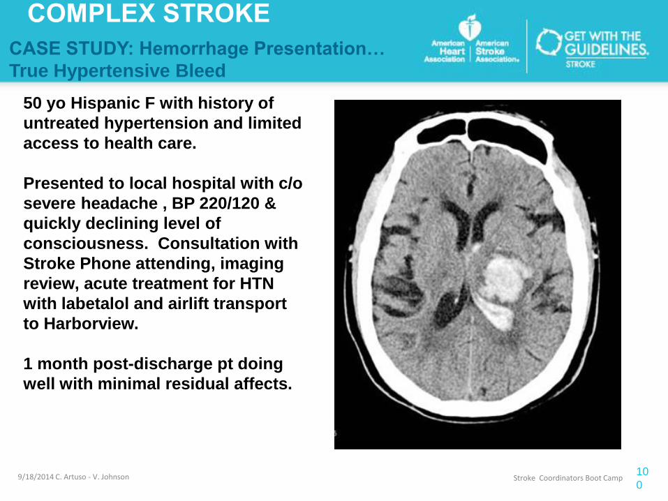
50 yo Hispanic F with history of
untreated hypertension and limited
access to health care.
Presented to local hospital with c/o
severe headache , BP 220/120 &
quickly declining level of
consciousness. Consultation with
Stroke Phone attending, imaging
review, acute treatment for HTN
with labetalol and airlift transport
to Harborview.
1 month post-discharge pt doing
well with minimal residual affects.
9/18/2014 C. Artuso - V. Johnson Stroke Coordinators Boot Camp 10
1
CASE STUDY: Hemorrhage Presentation…
True Hypertensive Bleed
Hypertensive Intracranial (intraparenchymal) Hemorrhage:
HTN most common cause of IPHs
Typically round or oval hyerdense appearing mass
80% Mortality with large IPHs with interventricular extension.
Acute Treatment: control hypertension and hydocephalus.
Typical location:
Putamen/external capsul (65%)
Thalamus (25%)
Pons/cerebellum (10%) www.headneckbrainspine.com
Let’s Brainstorm…STROKE NURSING & TRANSITIONAL CARE CONSIDERATIONS.

CASE STUDY
9/18/2014 C. Artuso - V. Johnson Stroke Coordinators Boot Camp 10
2
Hemorrhage Presentation…
Now What?

Case : Hemorrhage Presentation… Now What?
HISTORY 86 y/o F living independently. PMH: h/o hypertension.
• Hospitalized 5 days ago at an OSH with a UTI and mild confusion. Re-admitted at OSH 2
days ago 2/2 increased confusion. Head CT showed Large L frontal IPH. Transfer to HMC.
EXAM: intermittently opens eyes to voice, can say her name and "hospital" but not city. Follows
command to lift up arms. couldn't get her to lift legs but she moves them symmetrically in bed.
IMAGING MRI reviewed with Neuro-radiologist
• large L frontal bleed shows both subacute and acute ischemic & hemorrhagic findings.
• the ACA territory, there is a DWI positive area and evidence of laminar necrosis -- most
consistent with relatively recent ischemic infarct.
• Intraventricular extension
• IPH in the R temporal-occipital area.
9/18/2014 C. Artuso - V. Johnson Stroke Coordinators Boot Camp 10
3

Case : Hemorrhage Presentation… Now What?
Attending’s Comment:
“The imaging is puzzling. Difficult to explain 2 IPH's
without h/o significant trauma -- and the L sided lesion is
most consistent with hemorrhagic transformation of
ischemic stroke. Hemorrhagic metastases are also
possible -- though the radiographic picture is not typical
for that either.”
9/18/2014 C. Artuso - V. Johnson Stroke Coordinators Boot Camp 10
4

Case : Hemorrhage Presentation… Now What?
9/18/2014 C. Artuso - V. Johnson Stroke Coordinators Boot Camp 10
5
Final Impression:
1. Stable appearance of large left IPH
and IPH along the trigone of the R
lateral ventricle compared to the exam
done 6 hours earlier. There is
associated mass effect with partial
compression of the frontal horns of
the lateral ventricles.
2. IVH is now demonstrated, layering
in the occipital horns of the lateral
ventricles. There is mild dilation of the
temporal horns of the lateral
ventricles, but unchanged from the
exam done 6 hours earlier.
3. No evidence of vascular
abnormality underlying hemorrhages.
4. Heterogeneous right thyroid lobe
nodule measuring 3.1 cm in diameter.
Further evaluation with ultrasound is
recommended

Case : Hemorrhage Presentation… Now What?Discharge Summary Comments:
Diagnosis: Hemorrhagic Stroke, MRI revealed two locations of IPH: L. frontal and R.
temporal, without clear etiology.
Differentials for her Hemorrhagic Stroke(s) include…
• Hypertensive vs.
• Amyloid Angiopathy causing multiple macro-bleeds vs.
• Malignancy (thyroid nodules found on CT, so some possibility of thyroids metastises) vs
• Hemorrhagic Transformation of ischemic lesions…Less likely as TTE was negative for a
thrombus/PFO. CTA performed as part of comprehensive stroke workup revealed
normal vessels of the head and neck (unlikely artery to artery embolic/
atherosclerosis)
9/18/2014 C. Artuso - V. Johnson Stroke Coordinators Boot Camp 10
6

Case : Hemorrhage Presentation… Now What?Discharge Summary Comments: cont…
Follow Up:
• repeat MRI brain w&w/o contrast in 8 weeks to further investigate etiology.
• “Her home Aspirin was held and will continue to be held until she follows up
in Stroke Clinic given concern for macro-bleed type of cerebral amyloid
angiopathy, would likely not restart unless there was a definite indication”.
What is cerebral amyloid angiopathy?
9/18/2014 C. Artuso - V. Johnson Stroke Coordinators Boot Camp 10
7
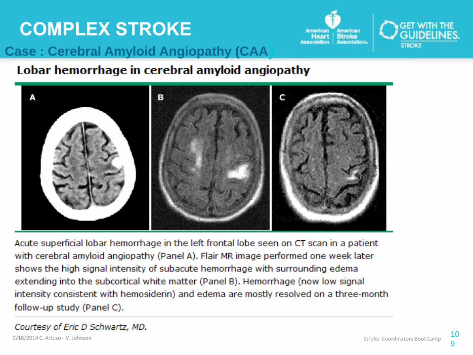
Case : Cerebral Amyloid Angiopathy (CAA) pearls
Description.
●CAA usually manifests with a spontaneous lobar hemorrhage. This location helps distinguish CAA-related
ICH from hypertensive ICH that more commonly arises in the putamen, thalamus, and pons.
●Transient neurologic symptoms are another manifestation of CAA. These are recurrent, brief (minutes),
often stereotyped spells of weakness, numbness, paresthesias, or other cortical symptoms that can spread
smoothly over contiguous body parts. The pathogenesis of these spells is not certain; they are not believed to
be transient ischemic attacks in most cases.
●CAA-related inflammation is a distinct disease subtype characterized by subacute cognitive decline,
seizures and unifocal or multifocal white matter MRI T2 hyperintensities extending to the subcortical white
matter or sulci. Clinical and radiographic improvement can occur with immunosuppressive therapy.
●CAA and Alzheimer disease frequently co-exist. CAA may also be associated with a vascular dementia.
9/18/2014 C. Artuso - V. Johnson
Stroke Coordinators Boot Camp
10
8
Case : Cerebral Amyloid Angiopathy (CAA)
9/18/2014 C. Artuso - V. Johnson Stroke Coordinators Boot Camp 10
9

Case : Hemorrhage Presentation… Now What?
9/18/2014 C. Artuso - V. Johnson Stroke Coordinators Boot Camp 11
0
Looking back at our case of the 86
yo female with hemorrhage.
Hemorrhage presentation
with Cerebral Amyloid
Angiopathy suspected or
confirmed.
Let’s Brainstorm…
STROKE NURSING &
TRANSITIONAL CARE
CONSIDERATIONS.
Case: Cerebral Vasculitis
9/18/2014 C. Artuso - V. Johnson Stroke Coordinators Boot Camp 11
1
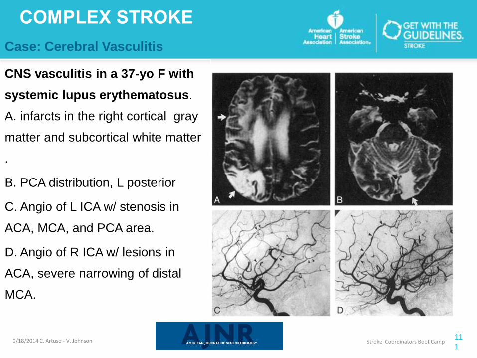
CNS vasculitis in a 37-yo F with
systemic lupus erythematosus.
A. infarcts in the right cortical gray
matter and subcortical white matter
.
B. PCA distribution, L posterior
C. Angio of L ICA w/ stenosis in
ACA, MCA, and PCA area.
D. Angio of R ICA w/ lesions in
ACA, severe narrowing of distal
MCA.
www.uwmedicine.org

Case: Cerebral Vasculitis
9/18/2014 C. Artuso - V. Johnson Stroke Coordinators Boot Camp 11
2
CNS vasculitis in a
37-yo F with systemic
lupus erythematosus.
severe narrowing of
distal MCA.

Case: Cerebral Vasculitis
9/18/2014 C. Artuso - V. Johnson Stroke Coordinators Boot Camp 11
3
The vasculitis affects any part of the CNS, causing the
clinical manifestations to be highly variable and
nonspecific [1]. PACNS should be suspected when
strokes, frequently recurrent, occur in young patients
with no identifiable cardiovascular or hypercoagulable
risk factors;
OR in the setting of chronic meningitis, recurrent focal
neurologic symptoms, unexplained diffuse neurologic
dysfunction, or unexplained spinal cord dysfunction
not associated with systemic disease or any other
process
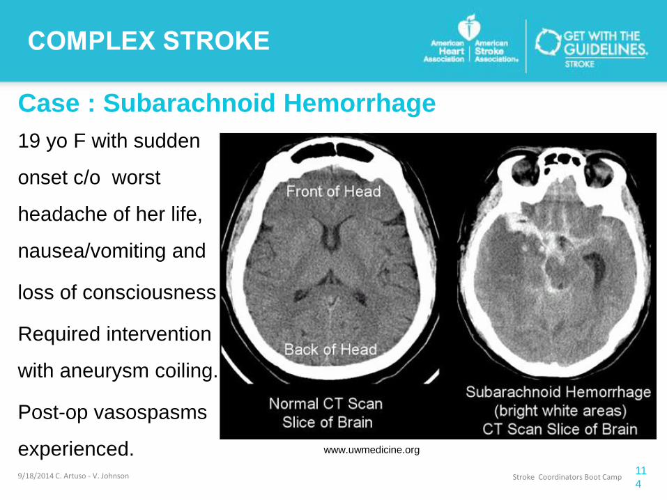
Case : Subarachnoid Hemorrhage
19 yo F with sudden
onset c/o worst
headache of her life,
nausea/vomiting and
loss of consciousness.
Required intervention
with aneurysm coiling.
Post-op vasospasms
experienced.
Captions for photo’s and other elements go here.
This is 9pt, Italic and in program color
9/18/2014 C. Artuso - V. Johnson Stroke Coordinators Boot Camp 11
4
www.uwmedicine.org

Case: Subarachnoid Hemorrhage w/ aneurysm
9/18/2014 C. Artuso - V. Johnson Stroke Coordinators Boot Camp 11
5
Subarachnoid hemorrhage results from the bleeding of an artery around the base of
the brain. It is the least common type of stroke, accounting for about 5 percent of all
strokes
www.uwmedicine.org
Introduction and Overview
9/18/2014 C. Artuso - V. Johnson Stroke Coordinators Boot Camp 11
6
I. Introduction & Overview: Complex
Stroke
II. The First 36 Hours of Care
III. Basic Brain Imaging
IV. Critical Transitions of Care:
- admission to discharge and beyond
V. Complex Stroke Cases & Imaging
- malignant MCA ischemic stroke
- complications >>>>>>>>>>>>>>>>>>
V. Questions