Embed Size (px)
Citation preview

CASE
STUDY
1 & 2
Courtesy of: Mitchell S.V. Elkind, MD, Columbia University and Shadi Yaghi, MD. Brown University

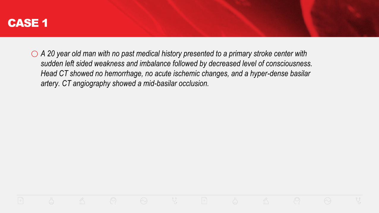
CASE 1
CASE 1
A 20 year old man with no past medical history presented to a primary stroke center with
sudden left sided weakness and imbalance followed by decreased level of consciousness.
Head CT showed no hemorrhage, no acute ischemic changes, and a hyper-dense basilar
artery. CT angiography showed a mid-basilar occlusion.
INFORMATION
FOR PATIENTS
AND FAMILIES
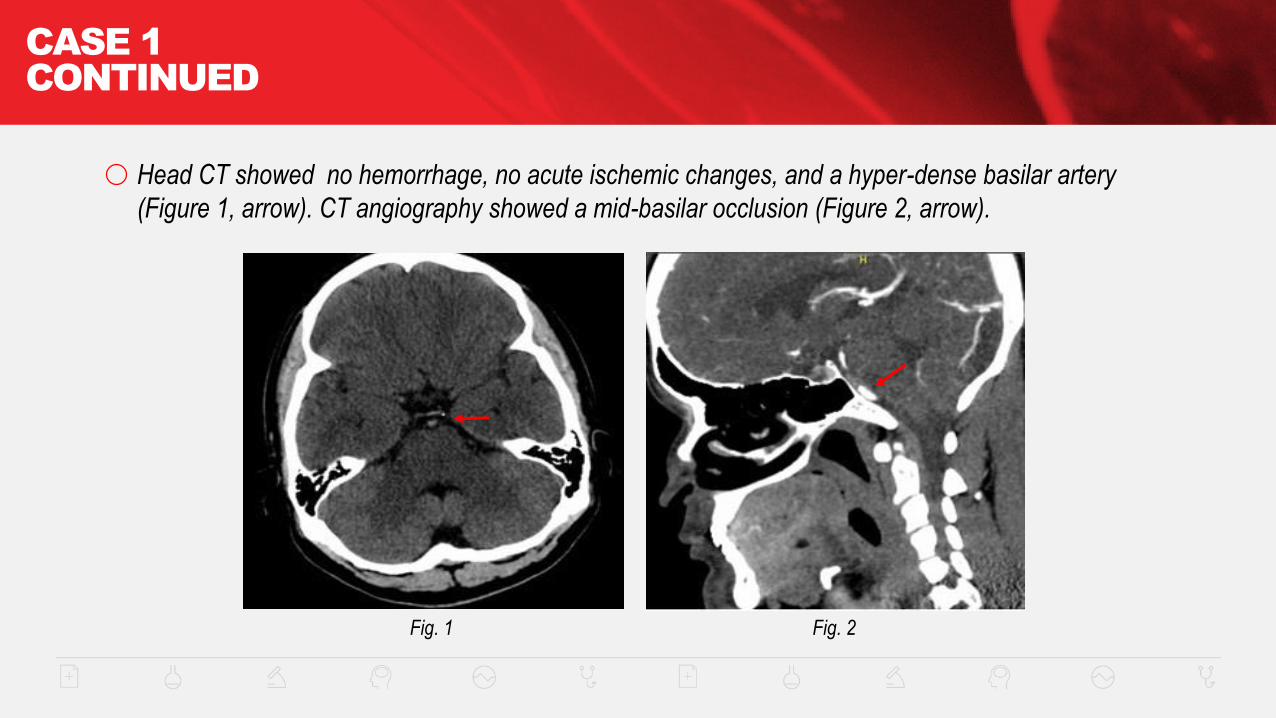
CASE 1
CONTINUED
Head CT showed no hemorrhage, no acute ischemic changes, and a hyper-dense basilar artery
(Figure 1, arrow). CT angiography showed a mid-basilar occlusion (Figure 2, arrow).
Fig. 1 Fig. 2
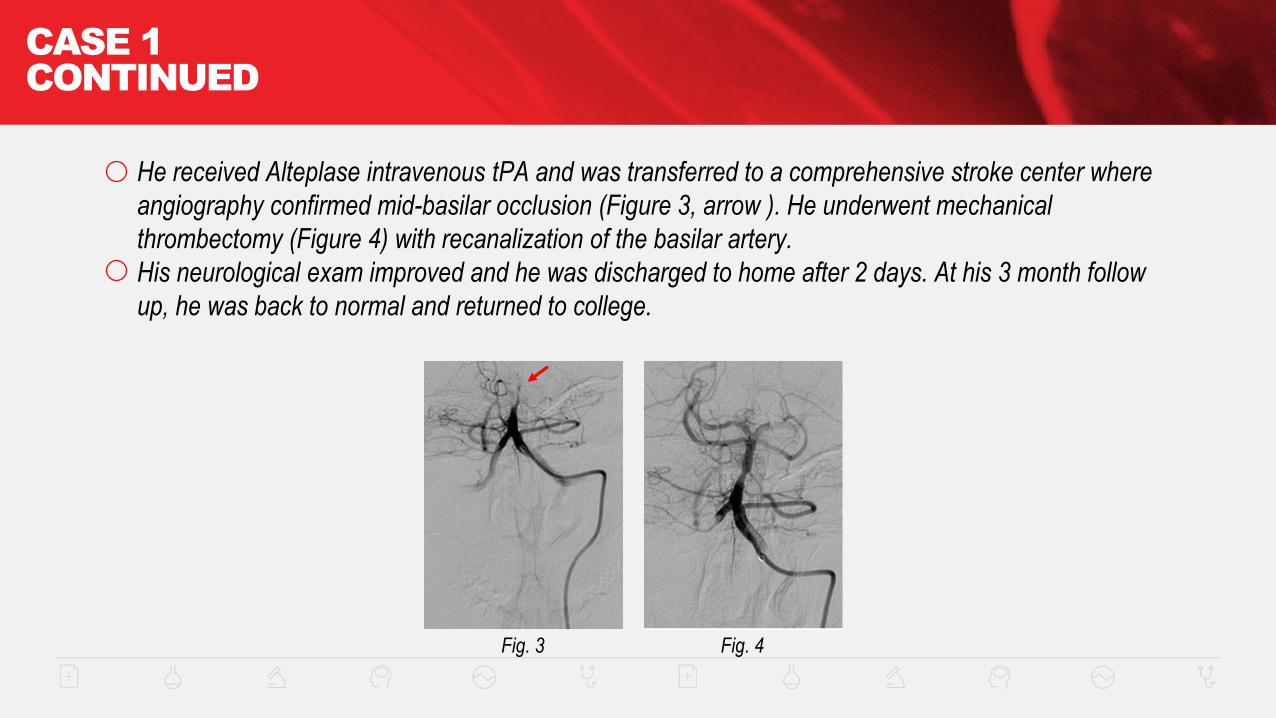
CASE 1
CONTINUED
He received Alteplase intravenous tPA and was transferred to a comprehensive stroke center where
angiography confirmed mid-basilar occlusion (Figure 3, arrow ). He underwent mechanical
thrombectomy (Figure 4) with recanalization of the basilar artery.
His neurological exam improved and he was discharged to home after 2 days. At his 3 month follow
up, he was back to normal and returned to college.
Fig. 3 Fig. 4

CASE 2
CASE 2
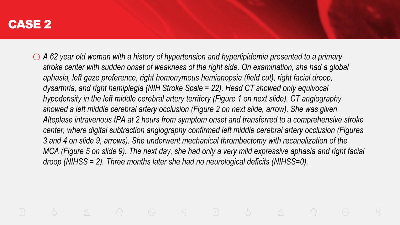
A 62 year old woman with a history of hypertension and hyperlipidemia presented to a primary
stroke center with sudden onset of weakness of the right side. On examination, she had a global
aphasia, left gaze preference, right homonymous hemianopsia (field cut), right facial droop,
dysarthria, and right hemiplegia (NIH Stroke Scale = 22). Head CT showed only equivocal
hypodensity in the left middle cerebral artery territory (Figure 1 on next slide). CT angiography
showed a left middle cerebral artery occlusion (Figure 2 on next slide, arrow). She was given
Alteplase intravenous tPA at 2 hours from symptom onset and transferred to a comprehensive stroke
center, where digital subtraction angiography confirmed left middle cerebral artery occlusion (Figures
3 and 4 on slide 9, arrows). She underwent mechanical thrombectomy with recanalization of the
MCA (Figure 5 on slide 9). The next day, she had only a very mild expressive aphasia and right facial
droop (NIHSS = 2). Three months later she had no neurological deficits (NIHSS=0).

CASE 2
CONTINUED
Fig. 1 Fig. 2
CASE 2
CONTINUED
Fig. 3 Fig. 4 Fig. 5

CASE
STUDY
3 & 4
Courtesy of: Brian L. Hoh, MD, University of Florida

CASE 3
CASE 3: ACUTE LEFT M1 OCCLUSION TREATED
WITH MECHANICAL THROMBECTOMY WITH NO IV TPA
The patient is a 65 year old woman who had a laparoscopic cholecystectomy 3 days prior.
She was last seen normal at 10pm before sleep.
She awoke at 2am and was discovered by her husband to have aphasia and right hemiplegia.
She was brought by EMS to the ED at 3:15 am.
She was not eligible for Alteplase IV tPA because of her wakeup stroke and recent surgery.
Her NIHSS was 19.
CASE 3
CONTINUED
Fig. 1
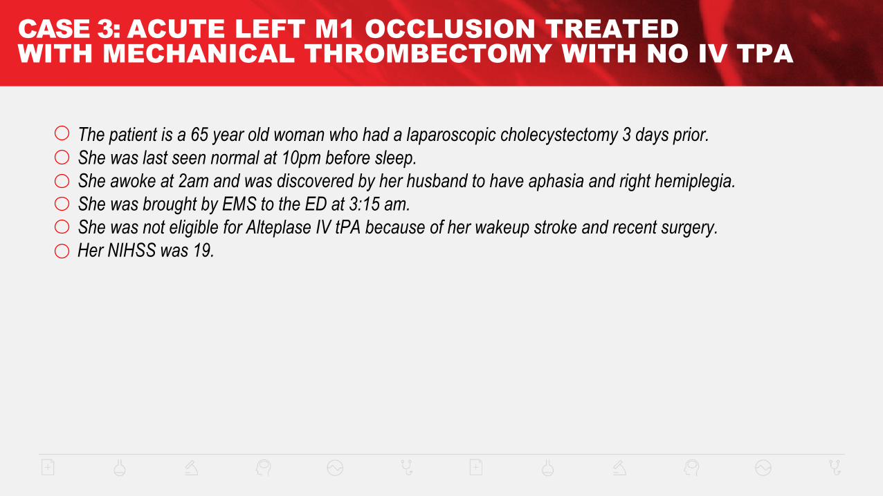
The CTA shows an
occlusion of the left
MCA stem.
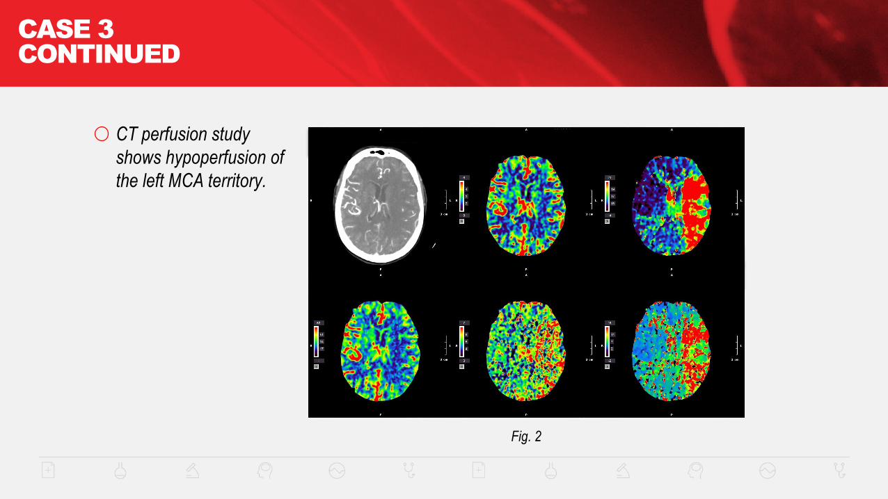
CASE 3
CONTINUED
Fig. 2
CT perfusion study
shows hypoperfusion of
the left MCA territory.
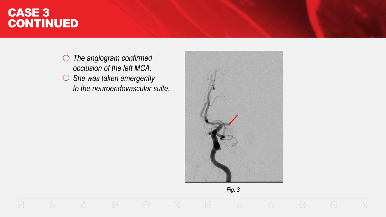
CASE 3
CONTINUED
Fig. 3
The angiogram confirmed
occlusion of the left MCA.
She was taken emergently
to the neuroendovascular suite.
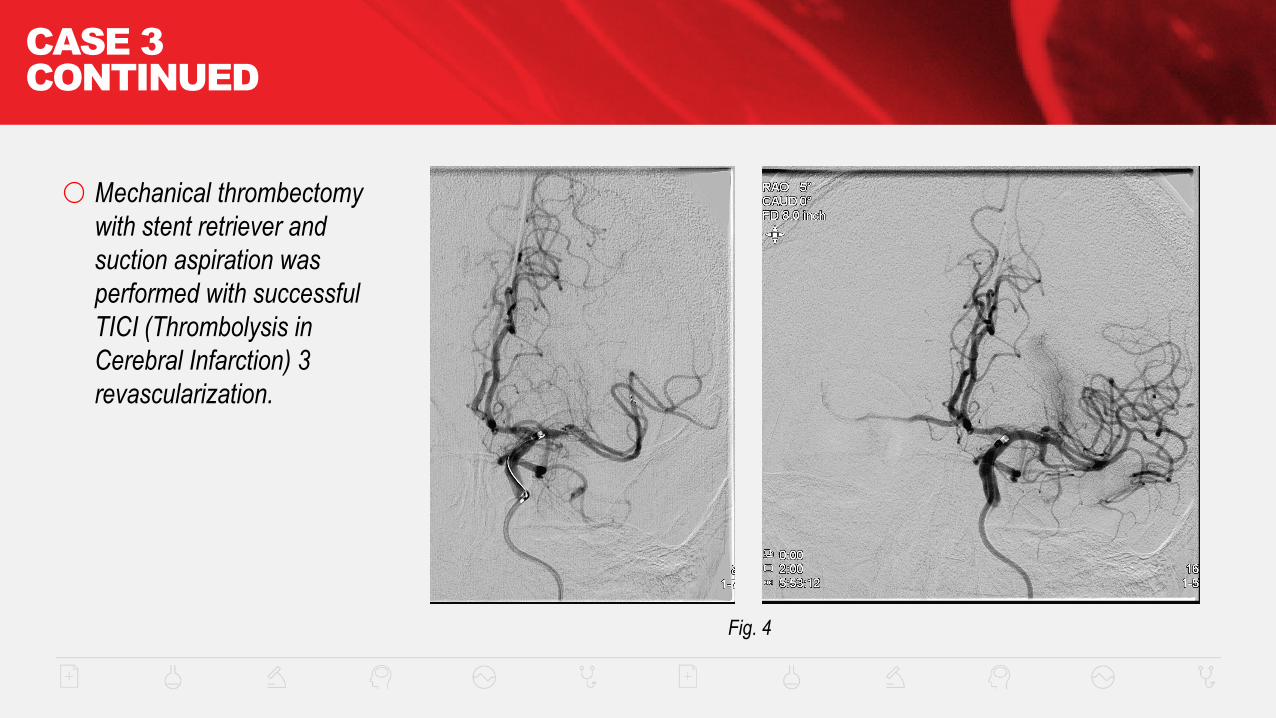
CASE 3
CONTINUED
Fig. 4
Mechanical thrombectomy
with stent retriever and
suction aspiration was
performed with successful
TICI (Thrombolysis in
Cerebral Infarction) 3
revascularization.
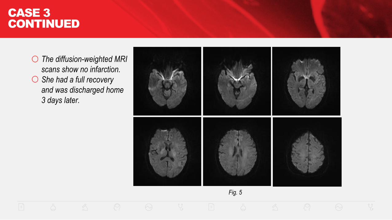
CASE 3
CONTINUED
Fig. 5
The diffusion-weighted MRI
scans show no infarction.
She had a full recovery
and was discharged home
3 days later.

CASE 4
CASE 4: ACUTE RIGHT M1 OCCLUSION TREATED WITH
MECHANICAL THROMBECTOMY AFTER “DRIP & SHIP” IV TPA
The patient is a 38 year old man who developed sudden left hemiparesis.
He was taken to his local hospital ED.
After a telephone consultation with a stroke neurologist, he was given Alteplase IV tPA. Needle
time was 1 hour 30 min after symptom onset.
He was then transferred to a comprehensive stroke center (“drip and ship”).
On arrival to our ED, his NIHSS was 11.
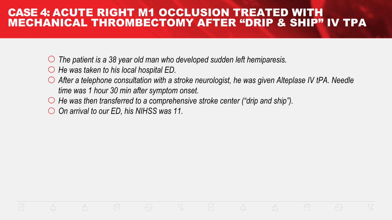
CASE 4
CONTINUED
Fig. 1
CTA showed right
MCA occlusion.
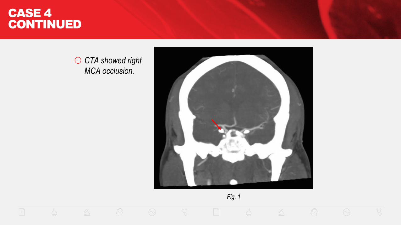
CASE 4
CONTINUED
CT perfusion images
show hypoperfusion to
the right hemisphere.
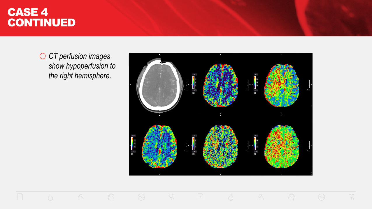
CASE 4
CONTINUED
Fig. 3
Angiography confirmed right
MCA occlusion.
He was taken emergently to
the neuroendovascular suite.
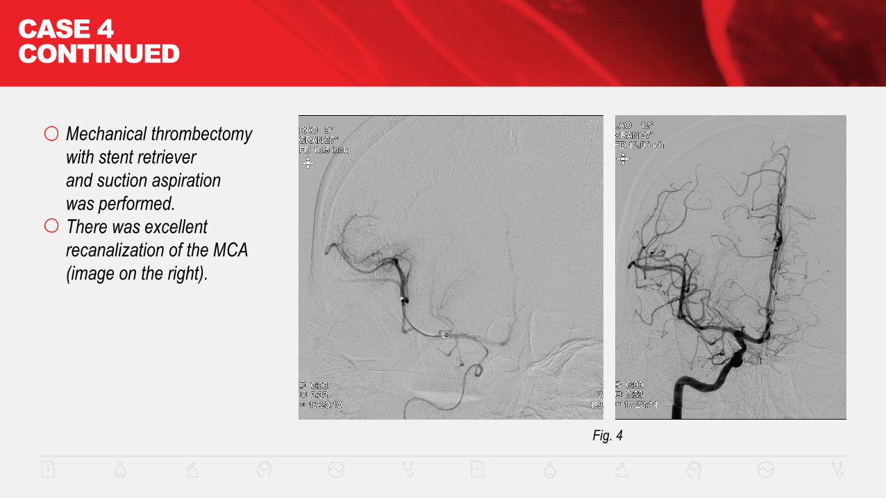
CASE 4
CONTINUED
Fig. 4
Mechanical thrombectomy
with stent retriever
and suction aspiration
was performed.
There was excellent
recanalization of the MCA
(image on the right).
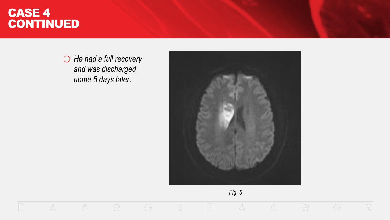
CASE 4
CONTINUED
Fig. 5
He had a full recovery
and was discharged
home 5 days later.

CASE
STUDY 5
Courtesy of: Donald Frei, MD, Michelle Whaley, MSN, CNS, Swedish Medical Center
CASE 5: LEFT INTERNAL
CAROTID OCCLUSION
This patient is a 66-year-old man, living in a rural community without hospital-based emergency
services, who experienced sudden onset aphasia and dysarthria that was witnessed by his
daughter. Local EMS arrived on the scene within 15 minutes, recognized the signs of stroke,
and requested flight transport to a comprehensive stroke center (CSC). Initial NIHSS was
assessed by the flight team as 3, but the patient deteriorated to a NIHSS of 22. The patient
arrived to the CSC on a Saturday, 1 hour and 37 minutes from symptom onset. On examination,
he had global aphasia, right homonymous hemianopsia, left gaze preference, and right-sided
hemiplegia. The patient was rapidly transported to CT for advanced imaging. After a non-
contrast CT, head was deemed normal. He was treated with intravenous alteplase IV r-tPA with
a door-to-needle time of 17 minutes.
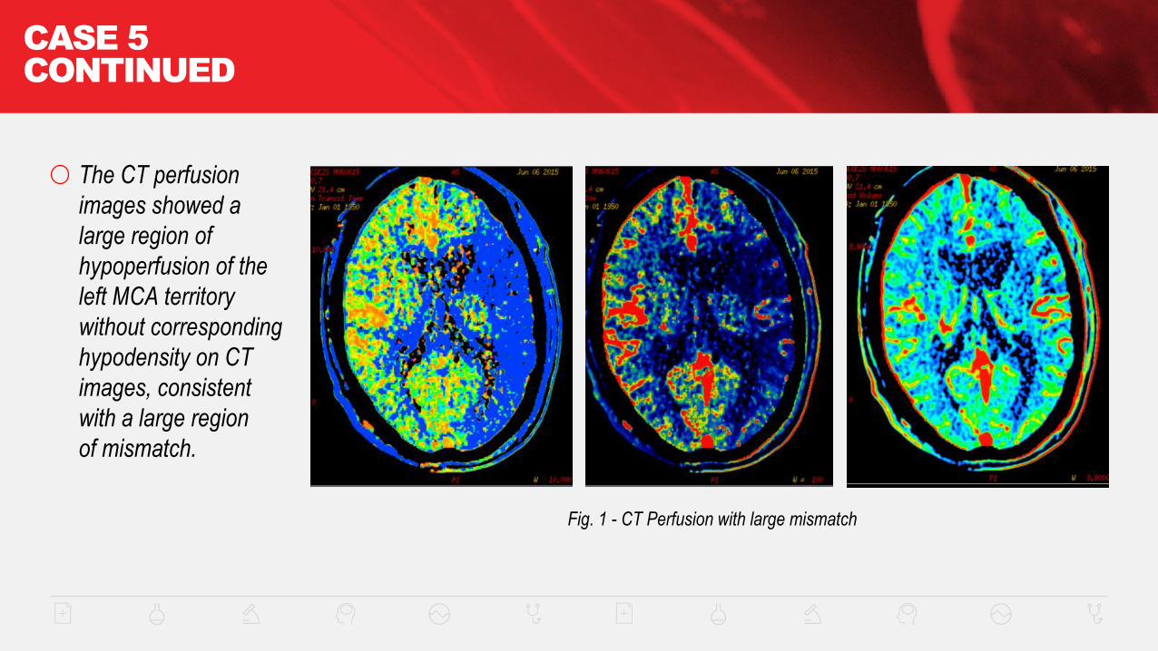
CASE 5
CONTINUED
Fig. 1 - CT Perfusion with large mismatch
The CT perfusion
images showed a
large region of
hypoperfusion of the
left MCA territory
without corresponding
hypodensity on CT
images, consistent
with a large region
of mismatch.
CASE 5
CONTINUED
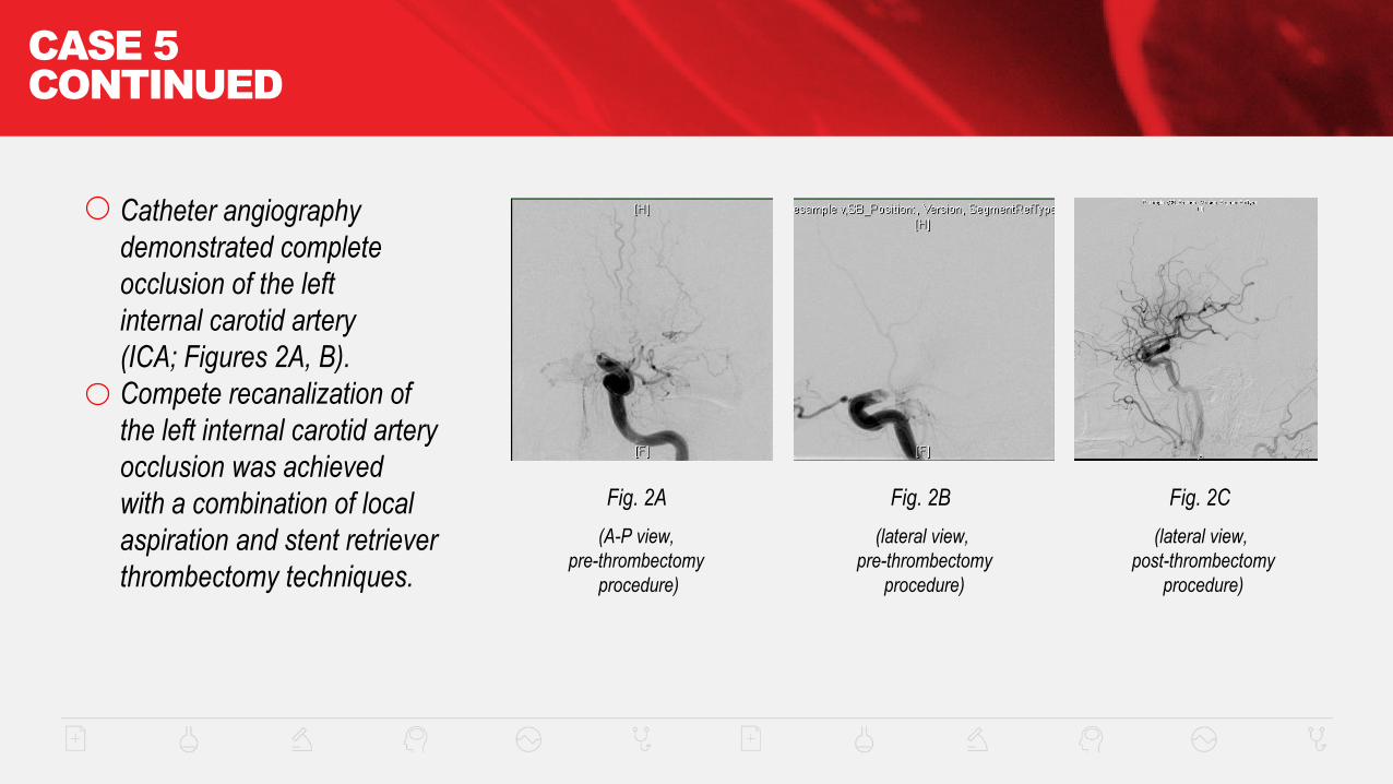
Fig. 2A
Catheter angiography
demonstrated complete
occlusion of the left
internal carotid artery
(ICA; Figures 2A, B).
Compete recanalization of
the left internal carotid artery
occlusion was achieved
with a combination of local
aspiration and stent retriever
thrombectomy techniques.
Fig. 2B Fig. 2C
(A-P view,
pre-thrombectomy
procedure)
(lateral view,
pre-thrombectomy
procedure)
(lateral view,
post-thrombectomy
procedure)
CASE 5
CONTINUED
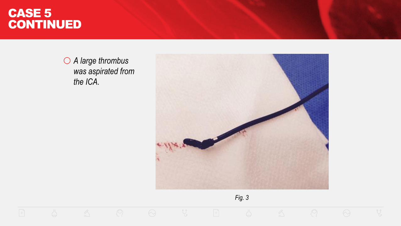
Fig. 3
A large thrombus
was aspirated from
the ICA.
CASE 5
CONTINUED
NIHSS upon arrival to NICU was 9. The patient experienced a dramatic improvement
in symptoms with only mild aphasia and right facial weakness 24 hours post treatment.
NIHSS 24 hours post treatment was 2. On hospital day 3, the patient was diagnosed
with new onset atrial fibrillation. He was discharged home on hospital day 4 on warfarin
and plans for outpatient speech therapy. At 90 days, the patient was nearly back to
normal with a modified Rankin score of 1.
CASE 5
CONTINUED
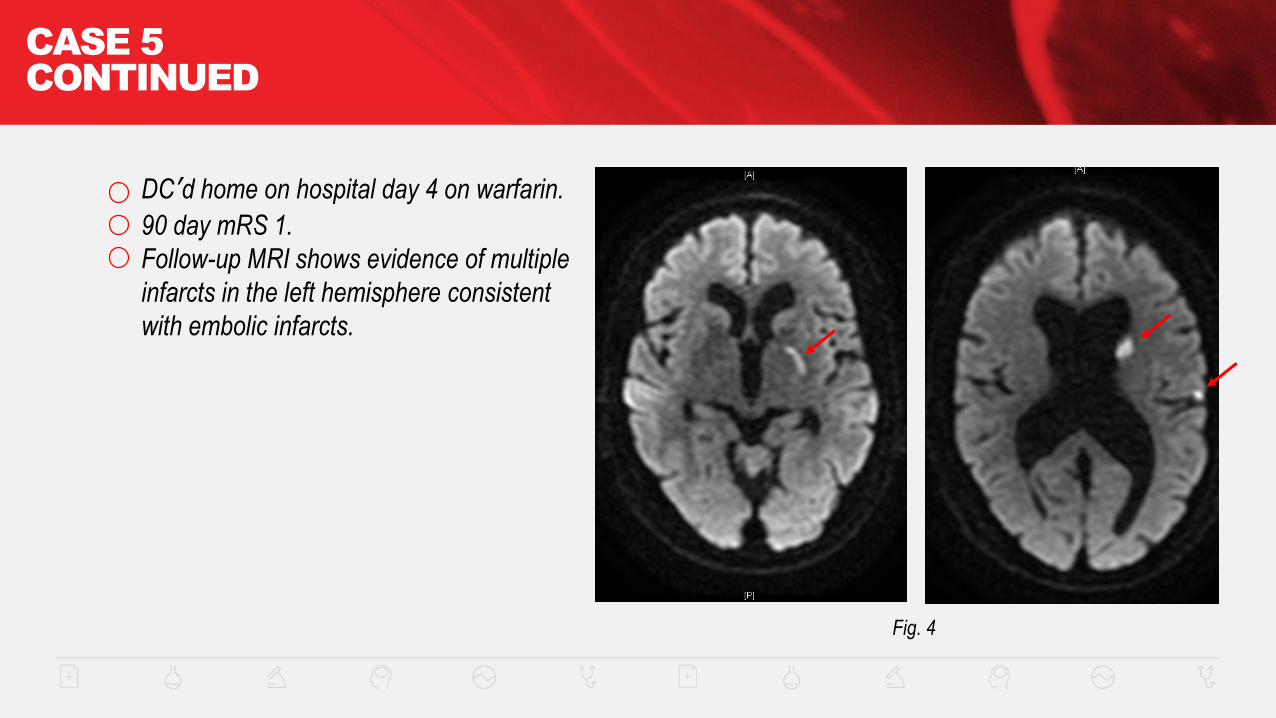
Fig. 4
DC’d home on hospital day 4 on warfarin.
90 day mRS 1.
Follow-up MRI shows evidence of multiple
infarcts in the left hemisphere consistent
with embolic infarcts.
CASE 5
TIMELINE
Door to neurologist – 0 minutes
Door to CT first slice – 10 minutes
Door to needle – 17 minutes
Door to groin puncture – 52 minutes
Door to recanalization – 113 minutes
Symptom onset to recanalization – 205 minutes
Actual Times of Treatment (Military Time) Time Intervals
Symptom onset – 11:15
Local EMS calls flight – 11:37
Flight arrives at 12:15 – Departs scene at 12:32
Arrives to CSC at 12:47
IV Alteplase started 13:04
Arrives to INR (International Normalised Ratio) suite at 13:08
Procedure time out 13:10
Groin stick at 13:39
TICI (Thrombolysis in Cerebral Infarction) 3 Recanalization at 14:40

Columbia University Medical Center
LVO
CASE
STUDIES

CASE 6
CASE 6
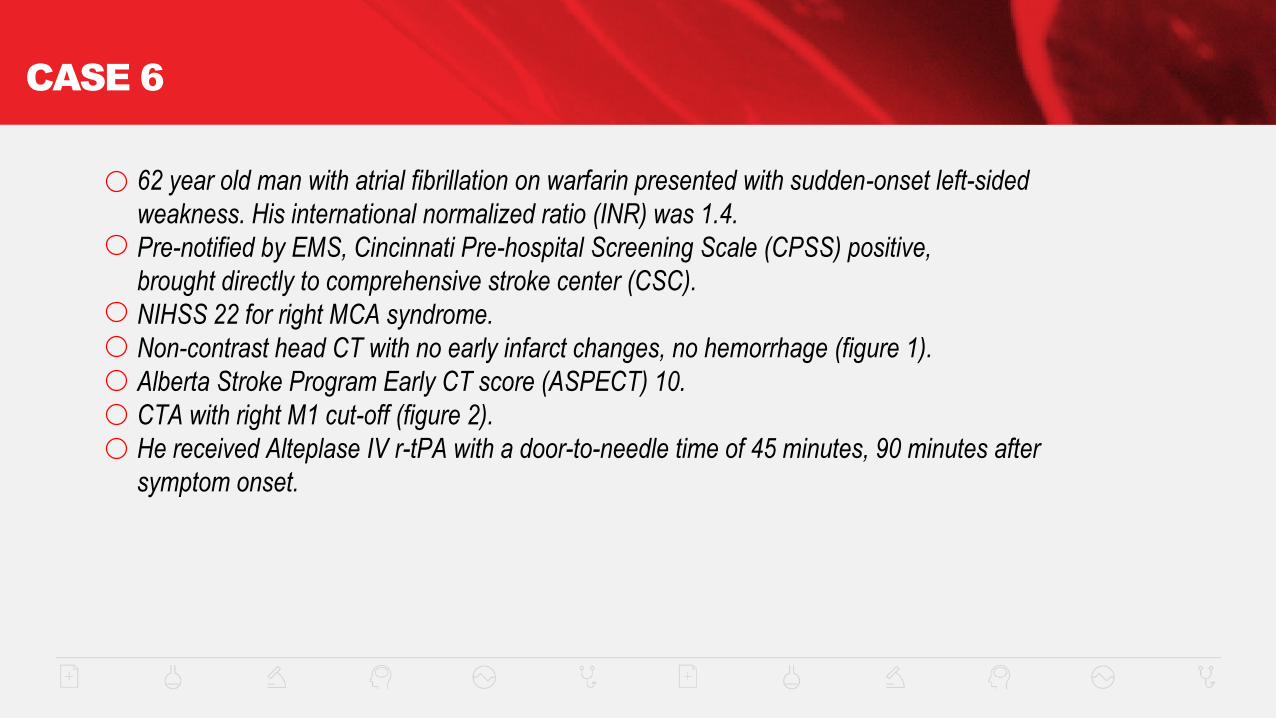
62 year old man with atrial fibrillation on warfarin presented with sudden-onset left-sided
weakness. His international normalized ratio (INR) was 1.4.
Pre-notified by EMS, Cincinnati Pre-hospital Screening Scale (CPSS) positive,
brought directly to comprehensive stroke center (CSC).
NIHSS 22 for right MCA syndrome.
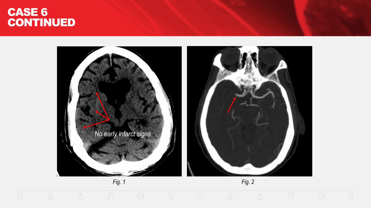
Non-contrast head CT with no early infarct changes, no hemorrhage (figure 1).
Alberta Stroke Program Early CT score (ASPECT) 10.
CTA with right M1 cut-off (figure 2).
He received Alteplase IV r-tPA with a door-to-needle time of 45 minutes, 90 minutes after
symptom onset.
INFORMATION
FOR PATIENTS
AND FAMILIES
CASE 6
CONTINUED
Fig. 1 Fig. 2
No early infarct signs
CASE 6
CONTINUED
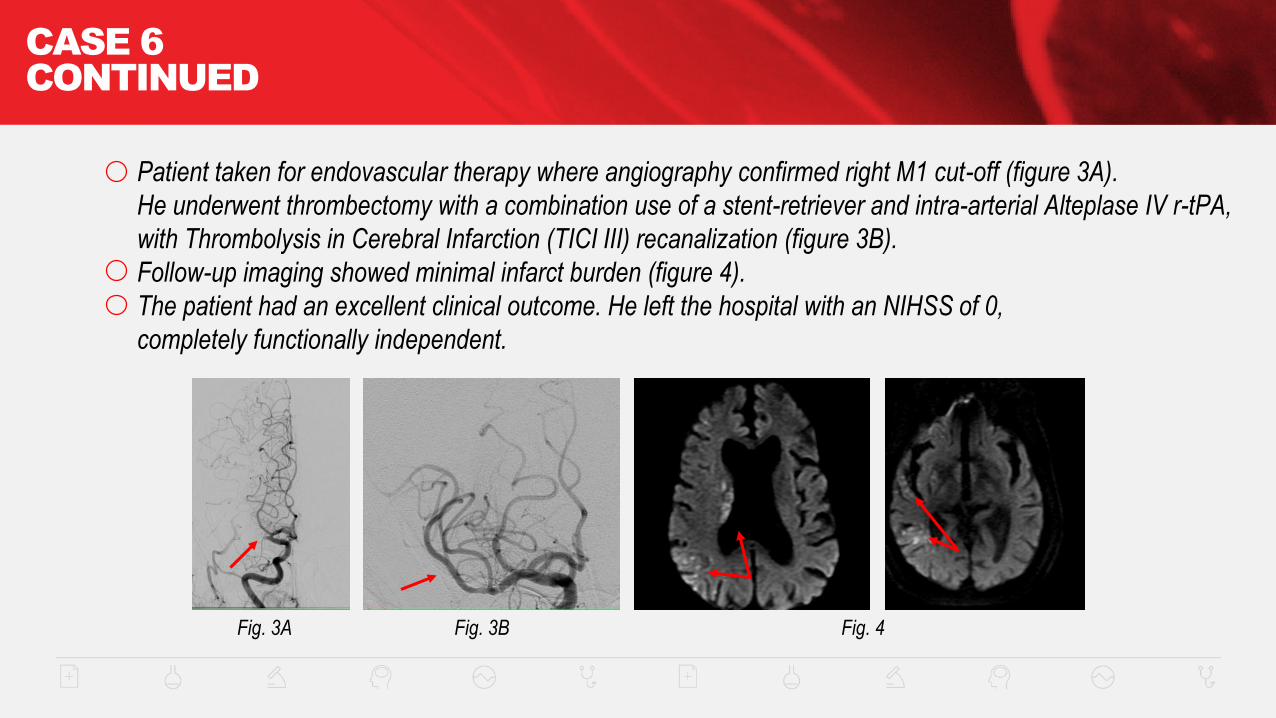
Patient taken for endovascular therapy where angiography confirmed right M1 cut-off (figure 3A).
He underwent thrombectomy with a combination use of a stent-retriever and intra-arterial Alteplase IV r-tPA,
with Thrombolysis in Cerebral Infarction (TICI III) recanalization (figure 3B).
Follow-up imaging showed minimal infarct burden (figure 4).
The patient had an excellent clinical outcome. He left the hospital with an NIHSS of 0,
completely functionally independent.
Fig. 3A Fig. 4Fig. 3B

CASE 7
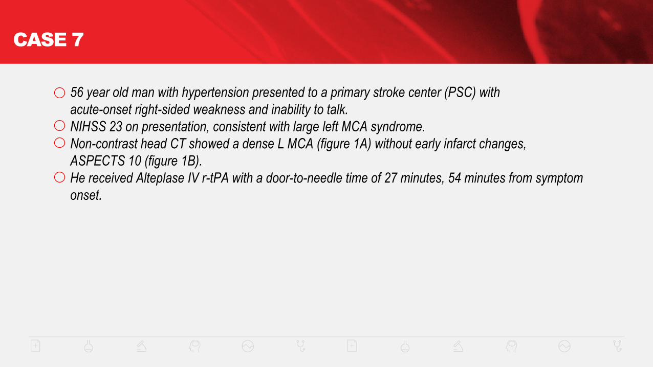
CASE 7
56 year old man with hypertension presented to a primary stroke center (PSC) with
acute-onset right-sided weakness and inability to talk.
NIHSS 23 on presentation, consistent with large left MCA syndrome.
Non-contrast head CT showed a dense L MCA (figure 1A) without early infarct changes,
ASPECTS 10 (figure 1B).
He received Alteplase IV r-tPA with a door-to-needle time of 27 minutes, 54 minutes from symptom
onset.
CASE 7
CONTINUED
Fig. 1A Fig. 1B
No early infarct changes
CASE 7
CONTINUED
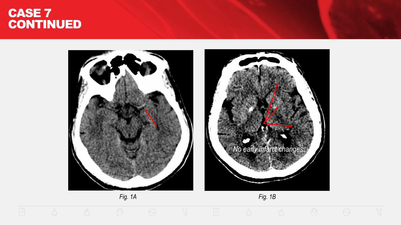
Transfer activated to comprehensive stroke center (CSC) given suspicion for large vessel syndrome.
CTA head obtained at PSC while waiting for transfer showed a proximal L M2 cut-off (figure 2).
Fig. 2
CASE 7
CONTINUED
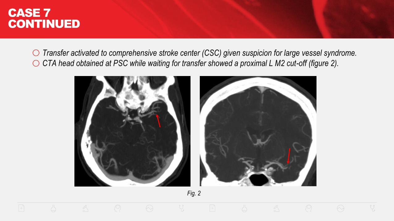
Patient taken directly to angio suite on arrival at CSC.
Proximal left internal carotid artery stenosis (figure 3A) and left M2 cut-off visualized (figure 3B).
Thrombectomy achieved TICI IIb re-canalization of left MCA (figure 4).
Fig. 3A Fig. 3B Fig. 4
CASE 7
CONTINUED
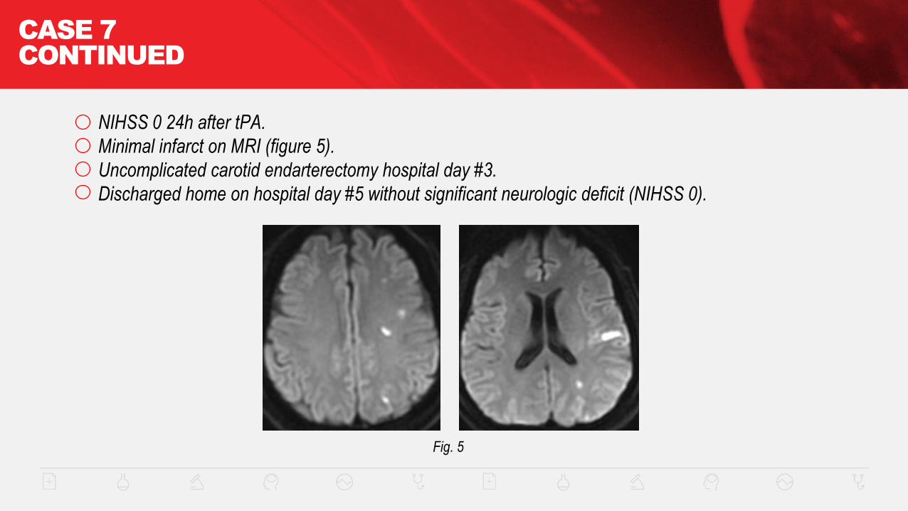
NIHSS 0 24h after tPA.
Minimal infarct on MRI (figure 5).
Uncomplicated carotid endarterectomy hospital day #3.
Discharged home on hospital day #5 without significant neurologic deficit (NIHSS 0).
Fig. 5

CASE 8
CASE 8
85 year old woman with atrial fibrillation off anticoagulation presented with sudden-onset
left-sided weakness and confusion.
Pre-notified by EMS, NIHSS 19 on ED arrival.
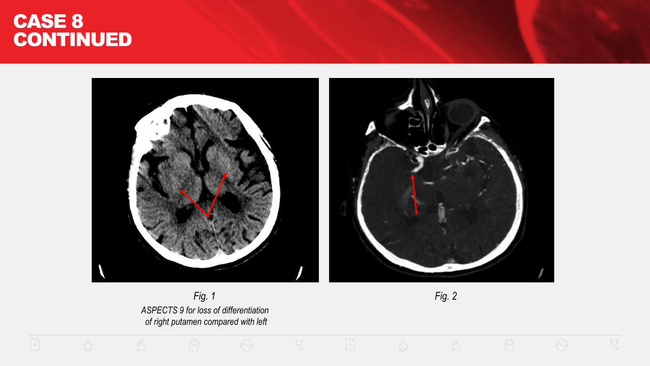
Non-contrast head CT showed ASPECTS 9 with early ischemic changes in basal ganglia (figure 1).
CTA showed R M1 cut-off (figure 2).
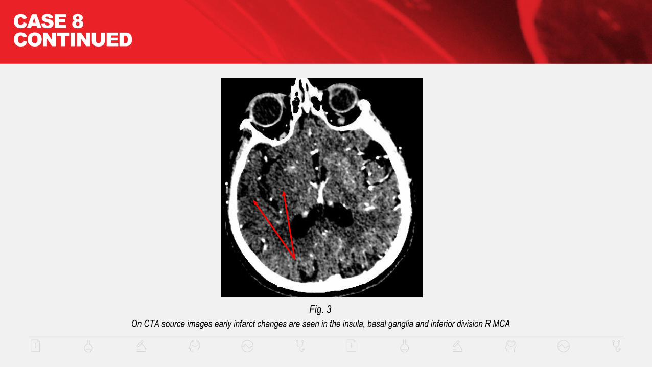
CTA source images showed ASPECTS 7 (figure 3).
CASE 8
CONTINUED
Fig. 1 Fig. 2
ASPECTS 9 for loss of differentiation
of right putamen compared with left
CASE 8
CONTINUED
Fig. 3
On CTA source images early infarct changes are seen in the insula, basal ganglia and inferior division R MCA
CASE 8
CONTINUED
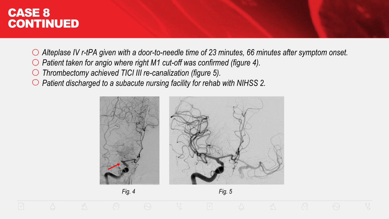
Alteplase IV r-tPA given with a door-to-needle time of 23 minutes, 66 minutes after symptom onset.
Patient taken for angio where right M1 cut-off was confirmed (figure 4).
Thrombectomy achieved TICI III re-canalization (figure 5).
Patient discharged to a subacute nursing facility for rehab with NIHSS 2.
Fig. 5Fig. 4

For more information on
Acute Ischemic Stroke treatment, go to
StrokeAssociation.org/AISToolkit