Embed Size (px)
Citation preview
Comparison of Quality and Cost-Effectiveness in the Evaluation of Symptomatic Cholelithiasis With Different Approaches to Ultrasound Availability in the ED
WILLIAM DURSTON, MD,*I MICHAEL L. CARL, MD,*I" WAYNE GUERRA, MD,:I: ABIGAIL EATON, MPH,§ LYNN ACKERSON, PHD,# TRIS RIELAND, MD,1- BOBBLE SCHAUER, MD,1- EVAN CHISUM, MD,II MAI-I- HARRISON, BS,* AND MISTY L. NAVARRO, BA*
Ultrasound is the imaging study of choice for the detection of gallstones, but ultrasound through medical imaging departments (MI Sono) is not readily available on an immediate basis in many emergency departments (EDs). Several studies have shown that emergency physicians can per- form ultrasound themselves (ED Sono) to rule out gallstones with ac- ceptable accuracy after relatively brief training periods, but there have been no studies to date specifically addressing the effect of ED Sono of the gallbladder on quality and cost-effectiveness in the ED. In this study, we investigated measures of quality and cost-effectiveness in evaluating patients with suspected symptomatic cholelithiasis during three differ- ent years with distinctly different approaches to ultrasound availability. The study retrospectively identified a total of 418 patients who were admitted for cholecystectomy or for a complication of cholelithiasis within 6 months of an ED visit for possible biliary colic. The percentage of patients who had gallstones documented at the first ED visit improved from 28% in 1993, when there was limited availability of ultrasound through the Medical Imaging Department (MI Sono), to 56% in 1995, when MI Sono was readily available, to 70% in 1997, when both MI Sono and ED Sono were readily available (P < .001). There were also significant dif- ferences over the 3 years in the mean number of days from the first ED visit to documentation of gallstones (19.7 in 1993, 10.7 in 1995, 7.4 in 1997, P < .001); the mean number of return visits for possible biliary colic before documentation of gallstones (1.67 in 1993, 1,24 in 1995, and 1,25 in 1997, P < .001); and the incidence of complications of cholelithiasis in the interval between the first ED visit for possible biliary colic and the date of documentation of cholelithiasis (6.8% in 1993, 5.9% in 1995, 1.5% in 1997, P = .049). The number of MI Sonos ordered by emergency physicians per case of symptomatic cholelithiasis identified increased from 1.7 in 1993 to 2.5 in 1995 and dropped back to 1.7 in 1997, when 4.2 ED Sonos per study case were also done. The cost of ED Sonos was
From the *Kaiser Foundation Hospital, South Sacramento, CA; the 1-Division of Emergency Medicine, University of California, Davis, Sacramento, CA; the :l:Porter-Littleton Hospital, Denver, CO; the §Department of Quality Utilization, Kaiser Foundation Hospitals, Oakland, CA; #Division of Research, Kaiser Permanente Medical Care Program, Oakland, CA; and the IIUniversity Hospitals, Depart- ment of Emergency Medicine, State University of New York, Stony Brook, NY.
Manuscript received October 6, 2000; accepted November 22, 2000.
Supported by grant 960030 from the Kaiser Innovations Program. Address reprint requests to William Durston, MD, Emergency
Department, Kaiser Foundation Hospital, 6600 Bruceville Road, Sacramento, CA 95823.
Key Words: Emergency, ultrasonography, cholelithiasis, quality, accuracy, cost-effectiveness.
Copyright © 2001 by W.B. Saunders Company 0735-6757/01/1904-0002535.00/0 doi:l 0.1053/ajem.2001.22660
260
more than offset by savings in avoiding calling in ultrasound technicians after regular Medical Imaging Department hours. The indeterminate rate for ED Sonos was 18%. Excluding indeterminates, the sensitivity of ED Sono for detection of gallstones was 88.6% (95% CI 83.1-92.8%), the specificity 98.2% (95% CI 96.0-99.3%), and the accuracy 94.8% (95% CI 92.5-96.5%). We conclude that greater availability of MI Sono in the ED was associated with improved quality in the evaluation of patients with suspected symptomatic cholelithiasis but also with increased ultra- sound costs. The availability of ED Sono in addition to readily available MI Sono was associated with further improved quality and decreased costs. The indeterminate rate for ED Sono was relatively high, but ex- cluding indeterminates, the accuracy of ED Sono was comparable with published reports of MI Sono. (Am J Emerg Med 2001;19:260-269. Copyright © 2001 by W.B. Saunders Company)
Cholelithiasis is relatively common in the adult popula- tion in the United States. The 10 year prevalence of chole- lithiasis in adults ages 30 to 62 in the Framingham study was 8.2%. 1 In Hispanic women, the prevalence of choleli- thiasis has been reported to be as high as 40%. 2 It has been estimated that from 2.5% to 12% of emergency department (ED) visits for abdominal pain are related to cholelithia- sis, 3-6 although the frequency with which biliary colic is either missed or overdiagnosed in the ED has not been systematically studied. Biliary colic is usually self-limited, but it is frequently recurrent and may cause severe patient discomfort. The annual incidence of serious complications in patients with initially asymptomatic or minimally symp- tomatic cholelithiasis is estimated to be 1% to 3%. 7 The presence or absence of gallstones is difficult to predict on the basis of the patient's clinical signs and symptoms, particularly in the elderlyY Multiple studies have shown that symptoms such as fatty food intolerance, postprandial epi- gastric pain, radiation of pain to the back, and even right upper quadrant pain have poor predictive value for deter- mining the presence or absence of cholelithiasis. 9-11 Labo- ratory tests, including white blood count and liver function tests, are normal in most patients with uncomplicated biliary colic and in about a third of patients with acute cholecys- titis.12,13
Over the past 2 decades, ultrasonography has been the imaging study of choice for determining the presence or absence of cholelithiasis. 14 In many EDs, however, ultra- sound studies through medical imaging departments (MI Sonos) are not routinely available on an immediate basis. 15 Recently, there has been growing interest among emergency
DURSTON ET AL • ED ULTRASOUND IN SYMPTOMATIC CHOLELITHIASIS 261
physicians in performing ultrasound studies themselves (ED Sonos). Several relatively small studies have suggested that emergency physicians can perform ED Sonos of the gall- bladder with reasonably high accuracy after relatively brief training periods. 16-2o Concerns have been raised, though, both by emergency physicians 21 and by the Medical Imag- ing community, 22.23 that emergency physicians may be more likely to make errors in ultrasound interpretation than sonographers in Medical Imaging, and that such errors could lead to adverse patient outcomes. The effect of ED Sono on quality and cost-effectiveness in patients with sus- pected biliary tract pain has not been systematically studied.
At our own medical facility, ultrasound availability in the ED was relatively limited until 1994, when emergency physicians proposed developing a program for training and credentialing in ED Sono. The Medical Imaging Depart- ment opposed this proposal but agreed to make ultrasound examinations more readily available to the ED. In 1996, the ED succeeded in passing an ED Sono protocol through the hospital 's Privileges and Credentials Committee, and emer- gency physicians began performing ED Sono for a variety of indications, including the evaluation of patients with suspected symptomatic cholelithiasis. As part of the ED Sono project, a study was begun to compare quality and cost-effectiveness in the ED in the evaluation of suspected biliary tract pain with different approaches to ultrasound availability. In this report, we present the results of this study.
MATERIALS AND METHODS
The setting for this study and the ED Sono training and creden- tialing protocol have been previously described in detail. 24 Briefly, the study was done in the ED of a staff model HMO with an annual census of approximately 30,000 patients. The study period con- sisted of three 1-year epochs: 1993, 1995, and 1997. During these years, the ED staff was composed of 10 or 11 emergency physi- cians, all of whom were either board-certified or residency-trained in emergency medicine. Two physicians left the staff and one joined during the study period.
During 1993, ultrasound examinations were available through Medical Imaging, but ultrasound technicians were not present at the hospital after regular Medical Imaging Department hours. Ultrasound technicians were usually available on call at night and on weekends, but emergency physicians were discouraged from ordering ultrasounds of the gallbladder without first obtaining surgical consultation. During 1995, ultrasound technicians were present in the hospital from 8 AM to 10 PM on weekdays and available on call at all other times. In 1995, emergency physicians could order MI Sonos at their own discretion. During 1997, an ultrasound machine was available in the ED and all emergency physicians were trained to perform ED Sonos, though MI Sonos were still available as in 1995.
MI Sonos were done in the ultrasound suite of the Medical Imaging Department, which is immediately adjacent to the ED, by full-time ultrasound technicians using an Acuson XP-128 ultra- sound machine (Acuson Corp., Mountain View, CA). The ultra- sound machine used for ED Sonos was a General Electric Logiq 400 equipped with a 3.5 MHz curved transducer (General Electric Corporation, Milwaukee, WI) and a Sony UP-870MD black and white page printer (Sony Corporation of America, New York, NY). Emergency physicians prospectively recorded the "rule out" indication for each ED Sono, and whether the study was positive, negative, or indeterminate for the presence of gallstones. Under an agreement with the Surgery and Medical Imaging departments,
emergency physicians ordered confirmatory outpatient MI Sonos on all patients referred for elective surgical consultation after a positive ED Sono for gallstones.
An initial list of patients potentially eligible for the study was generated by a computerized search for all patients who had a cholecystectomy or who were admitted to the facility with a possible complication of biliary tract disease, including cholecys- titis, cholangitis, or pancreatitis, within 6 months of an ED visit in 1993, 1995, or 1997. Medical records of patients identified by this computerized search were then reviewed by research assistants using explicit review criteria and preprinted data abstraction forms. Cases were included in the study if the patient had an ED visit for possible biliary colic within 6 months of a cholecystectomy or an admission for a possible complication of cholelithiasis; and if gallstones were documented on a pathology report or on an imag- ing study. For the purpose of the study, possible biliary colic was defined as any upper or right-sided abdominal pain for which no cause other than biliary tract disease or pancreatitis was specifi- cally documented. Cases of acalculous cholecystitis and pancre- atitis without cholelithiasis or choledocholithiasis were excluded. Cases were also excluded if the patient had previously documented cholelithiasis before the first ED visit for possible biliary colic. Cases with false-positive imaging studies for cholelithiasis were included.
The presence of gallstones was considered to be confirmed if gallstones were described as being present in the pathologist's report; if the surgeon described opening the gallbladder and find- ing stones in the operative report; or in cases in which the patient did not have surgery, if 2 independent imaging studies (including at least 1 study done in the Medical Imaging Department) docu- mented the presence of gallstones. Pancreatitis, gram negative bacteremia, or cholangitis were considered to be present if the hospital admission history and physical or discharge summary included these diagnoses. Acute cholecystitis was considered to be present only if this diagnosis was documented in the pathology report.
Primary and secondary quality indicators were defined before the beginning of data analysis. The percentage of patients docu- mented to have cholelithiasis at the first ED visit was designated as the primary quality indicator in the study. Secondary quality indi- cators included the number of days from the first ED visit for possible biliary colic to documentation of the presence of chole- lithiasis: the number of unscheduled return visits for possible biliary colic in the interim between the first ED visit and docu- mentation of the presence of cholelithiasis; and the incidence of complications of cholelithiasis in the interim between the first ED visit and documentation of the presence of gallstones. The number of return visits in the 6 months after cholecystectomy for abdom- inal pain that was not directly attributable to a complication of the surgery or a retained common bile duct stone was also included as a secondary quality indicator to serve as a potential measure of the number of inappropriate cholecystectomies.
The accuracy of all ED Sonos done in 1997 to rule out gall- stones was assessed by comparing ED Sono interpretations with surgical pathology, with repeat MI Sonos or other imaging studies, or with clinical follow-up. Accuracy of ultrasound interpretation was judged solely on whether the presence or absence of gallstones was correctly identified. Clinical follow-up was done by reviewing either the patient's paper medical record or computerized summa- ries of the medical record. Computerized summaries included data not only on visits to our own facility, but also on visits to all other related health plan facilities in a region with a radius of approxi- mately 150 miles. The presence of gallstones could not be con- firmed by clinical follow-up alone. The absence of gallstones was considered to be confirmed by clinical follow-up if the patient was followed in the health plan for at least 2 years after an ED Sono and had no imaging studies or surgery showing cholelithiasis over that time period.
262 AMERICAN JOURNAL OF EMERGENCY MEDICINE • Volume 19, Number 4 • July 2001
Data on the number of abdominal MI Sonos ordered by emer- gency physicians were obtained by computerized search of the health plan's laboratory utilization database which includes the type and number of ultrasound studies ordered by each physician in the facility. The cost of calling in an ultrasound technician after regular Medical Imaging Department hours and the charges for nonhealth plan members for ultrasound studies were obtained from the manager of the Medical Imaging Department. The cost of leasing and maintaining the ED ultrasound machine and the cost of physician training were obtained from the hospital administrator. It was estimated that 32% of all ED Sonos were done for the purpose of ruling out gallstones based on a previous study of ED Sono at our facility. 24
Statistical analysis of the data was done using the statistical packages included with EpiInfo version 6 (Centers for Disease Control, Atlanta, GA), Microsoft Excel 97 (Microsoft Corporation, Redmond, WA), and SAS version 6.11 (SAS Institute, Cary, NC). The Kruskall-Wallace test was used to determine the significance of differences in numeric data that were not normally distributed. Ages were compared using ANOVA. Nominal variables were compared using Chi-square or the 2-tailed Fisher exact test when cell sizes were small. For comparisons of data across the three epochs, a P value less than .05 was considered statistically signif- icant. For comparisons between any 2 epochs, a P value less than .0167 (.05/3) was considered significant, in accordance with the Bonferroni correction.
RESULTS
Patient Characteristics
Initial computerized review identified 550 potential cases for inclusion in the study over the 3 epochs of the study period. After chart review, 418 cases (76%) met criteria for inclusion in the final analysis of quality indicators. The number of cases excluded in each epoch and the reasons for excluding them are shown in Table 1. There was no signif- icant difference across the 3 epochs in the number of cases excluded or in the reasons for exclusion. Most cases which were excluded on the basis that the patient had no docu- mented gallbladder disease were cases of alcoholic or idio- pathic pancreatitis. Most cases excluded on the basis that the patient had no ED visits with possible bil iary colic were cases in which patients were seen in the ED for a condition other than abdominal pain within 6 months of a cholecys- tectomy.
The characteristics of the 418 patients included in the final data analysis are shown by epoch in Table 2. The percentage of patients included in the study relative to the total number of patients seen in the ED increased signifi- cantly in each subsequent epoch. The percentage of patients having an ultrasound done at the first ED visit also increased significantly in each subsequent epoch. There were no sig-
nificant differences across the three epochs in the age or sex of patients or in the percentages of patients presenting with a complication of cholelithiasis at their first ED visit.
Quality Indicators and Other Outcome Measures
Primary and secondary quality indicators are shown in Table 3. In one case in 1995 and in two cases in 1997, preoperative sonograms were reported to show gallstones, but no gallstones were documented at surgery. These cases were included in the denominator but not in the numerator in calculations of the primary quality indicator, the percent- age of patients correctly documented to have gallstones at the first ED visit. These cases were excluded from calcula- tions of secondary quality indicators.
There was significant improvement in each subse- quent epoch in the pr imary qual i ty indicator , the percent- age of patients documented to have gal ls tones at the first ED visit. There was also significant improvement in each subsequent epoch in mean number of days from the first ED visit for poss ib le b i l ia ry colic to the data of documenta t ion of gal ls tones. The mean number of un- scheduled visi ts for poss ible b i l ia ry colic before docu- mentat ion of gal ls tones was s ignif icant ly lower in 1995 and 1997 than in 1993. The inc idence of compl ica t ions of cholel i th ias is occurr ing be tween the first ED visi t and the date of documenta t ion of gal ls tones was statisti- cal ly different across the 3 epochs, with a t rend toward the highest rate of compl ica t ions in 1993 and the lowest rate in 1997, though the di f ference be tween any 2 ep- ochs was not s tat is t ical ly significant at the level of P < .016. There was no s ta t is t ical ly significant d i f ference across the 3 epochs in the rate of return visits for non- b i l iary abdominal pain in the 6 months after cholecys tec- tomy.
Accuracy of ED Sono Interpretation
During 1997, emergency physicians performed a total of 802 ED Sonos to rule out gallstones. In 36 cases, ED Sonos confirmed the results of a prior ED Sono on the same patient. In 12 cases, ED Sonos confirmed the results of a prior MI Sono. Of the remaining 754 ED Sonos that were not repeat studies, 185 (24.5%) were interpreted as showing gallstones to be present, 429 (56.9%) were interpreted as showing gallstones to be absent, and 140 (18.6%) were indeterminate. The results of follow-up to confirm the ac- curacy in the 614 nonindeterminate ED Sonos that were not preceded by prior imaging studies are shown in Table 4. ED Sonos confirmed by more than one method are listed only once in Table 4, under the most stringent method, with
TABLE 1. Basis for Excluding Cases Identified by Initial Computerized Search From the Final Data Analysis
1993 1995 1997 P Value
Total potential cases identified Number (%) of cases meeting inclusion criteria Number (%) of cases excluded and basis for exclusion
Acalculous cholecystitis Gallstones previously documented No gallbladder disease No ED visits for possible biliary colic
120 183 247 88 (73%) 135 (74%) 195 (79%)
2 (2%) 5 (3%) 5 (2%) 2 (2%) 6 (3%) 5 (2%) 5 (4%) 13 (7%) 8 (3%)
23 (t9%) 24 (13%) 34 (14%)
.34
.54
DURSTON ET AL • ED ULTRASOUND IN SYMPTOMATIC CHOLELITHIASIS 263
TABLE 2. Characteristics of Patients Included in Study
1993 1995 1997 P Value
ED Census 31,044 31,912 32,456 Number of study cases 88 135 195
% of total ED cases .28%1-:~ .42%1-§ .60%:~§ Age
Mean 47.7 47.5 47.9 Median 45.5 46 47 Range 16-86 14-87 16-90
% Female 69% 71% 76% Number (%) patients having ultrasound at first visit 26:1: (30%) 81:1: (60%) 165:~ (85%)
MI Sono only 26 81 7 ED Sono only 0 0 111 Both 0 0 47
Number (%) of patients with complication(s) at first visit* 22 (25%) 41 (30%) 40 (21%) Acute cholecystitis 13 23 25 Pancreatitis 9 17 14 Cholangitis or bacteremia 1 4 2 Perforation or gangrene 3 8 9 Death 0 0 0
<.001 .98
.30 <.001
.12
* Some patients had more than one complication. P values for pairwise comparisons of data in same row with same superscripts are as follows: 1 P = .003; :1: P < .001 ; § II P = .005. For all other pairwise comparisons in same row, P -> .016.
P = .002; # P < .001 ;
surgical pathology being considered most stringent and clinical follow-up least stringent. For the purpose of further calculations, all cases in which ED Sono results were dis- parate either with surgical pathology or with repeat medical imaging studies, if surgical pathology was not available,
were considered to be errors in ED Sono interpretations. Excluding indeterminate scans, scans confirming prior im- aging studies, and scans in which follow-up was unavailable or could not confirm the accuracy of the ED Sono, the sensitivity of ED Sono for detecting the presence or absence
TABLE 3. Primary and Secondary Quality Indicators
1993 1995 1997 (n = 88) (n = 135) (n = 195) P Value
Primary quality indicator Number (%) of patients correctly documented to have gallstones at
first ED visit Secondary quality indicators
1. Number of days from first ED visit to documentation of gallstones Mean 19.7:1:§ Median 2 Range 0-179
2. Number of unscheduled visits for possible biliary colic from first ED visit to documentation of gallstones Mean 1.67:1:§ Median 1 Range 1-6
3. Incidence of complications between first visit and documentation 6 (6.8%) of gallstones* Acute cholecystitis 2 Pancreatitis 4 Cholangitis or bacteremia 2 Perforation or gangrene of gallbladder 1 Death 0
4. Number of visits for ongoing abdominal pain in 6 months following cholecystectomyl- Mean 0.21 Median 0 Range 0-3
25 (28%):1:§ 76 (56%)~# 137 (70%)§# <.001
10.7~11 7.4§1t 0 0
0-164 0-152
1.24:~ 1.25§ 1 1
1-5 1-5 8 (5.9%) 3 (1.5%)
5 3 2 0 2 0 1 0 0 0
0.16 0.25 0 0
0-3 0-3
<.001
<.001
.047
0.54
* Some patients had more than one complication. 1 Abdominal pain not directly attributable to biliary tract disease or a complication of surgery. :~ P < .001 ; § P < .001; # P = .011; II P = .008. For all other pairwise comparisons in same row, P -> .016.
264 AMERICAN JOURNAL OF EMERGENCY MEDICINE • Volume 19, Number 4 • July 2001
TABLE 4. Results of Follow-Up to Confirm Accuracy of all ED Sonos of the Gallbladder Done in 1997 Which Were not Indeterminate and not Repeat Studies
Positive Negative Scans Scans
(Gallstone (Gallstone Confirmation Status Present) Absent) Totals
Confirmed by surgical pathology 132 3 135 Confirmed by repeat imaging study 31 95 126 Confirmed by clinical followup 0 235 235 Unconfirmed despite followup 9 3 12 Lost to followup 7 72 79 Disparate ED Sono and repeat 4 3 7
imaging study Disparate ED Sono and surgical 2 18 20
pathology Totals 185 429 614
of gallstones was 88.6% (95% CI 83.1-92.8%), the speci- ficity 98.2% (95% CI 96.0-99.3%), and the accuracy 94.8% (95% CI 92.5-96.5%).
Cost-Effectiveness
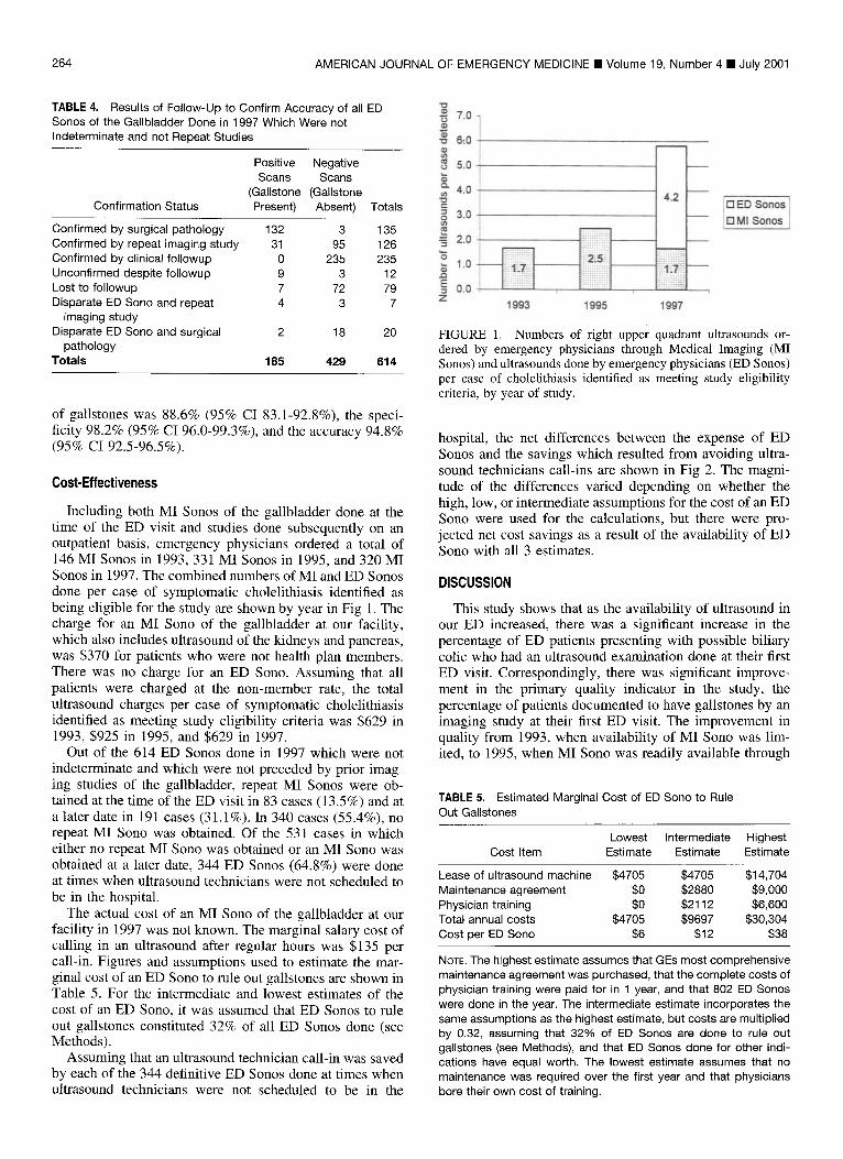
Including both MI Sonos of the gallbladder done at the time of the ED visit and studies done subsequently on an outpatient basis, emergency physicians ordered a total of 146 MI Sonos in 1993, 331 MI Sonos in 1995, and 320 MI Sonos in 1997. The combined numbers of MI and ED Sonos done per case of symptomatic cholelithiasis identified as being eligible for the study are shown by year in Fig 1. The charge for an MI Sono of the gallbladder at our facility, which also includes ultrasound of the kidneys and pancreas, was $370 for patients who were not health plan members. There was no charge for an ED Sono. Assuming that all patients were charged at the non-member rate, the total ultrasound charges per case of symptomatic cholelithiasis identified as meeting study eligibility criteria was $629 in 1993, $925 in 1995, and $629 in 1997.
Out of the 614 ED Sonos done in 1997 which were not indeterminate and which were not preceded by prior imag- ing studies of the gallbladder, repeat MI Sonos were ob- tained at the time of the ED visit in 83 cases (13.5%) and at a later date in 191 cases (31.1%). In 340 cases (55.4%), no repeat MI Sono was obtained. Of the 531 cases in which either no repeat MI Sono was obtained or an MI Sono was obtained at a later date, 344 ED Sonos (64.8%) were done at times when ultrasound technicians were not scheduled to be in the hospital.
The actual cost of an MI Sono of the gallbladder at our facility in 1997 was not known. The marginal salary cost of calling in an ultrasound after regular hours was $135 per call-in. Figures and assumptions used to estimate the mar- ginal cost of an ED Sono to rule out gallstones are shown in Table 5. For the intermediate and lowest estimates of the cost of an ED Sono, it was assumed that ED Sonos to rule out gallstones constituted 32% of all ED Sonos done (see Methods).
Assuming that an ultrasound technician call-in was saved by each of the 344 definitive ED Sonos done at times when ultrasound technicians were not scheduled to be in the
7.0-
"~ 6:0
8 s . o
o. 4.0
3.0
= 2.0
,- 1.0 e ~
0.0 Z
4.2
. . . . . . . . . . . . : . : . : _ . . + _ . _ . : + : . . . . . . : : : . . . . . + : + . _
1993 1995 1997
[] ED Sonos El M Sonos
FIGURE 1. Numbers of right upper quadrant ultrasounds or- dered by emergency physicians through Medical Imaging (MI Sonos) and ultrasounds done by emergency physicians (ED Sonos) per case of cholelithiasis identified as meeting study eligibility criteria, by year of study.
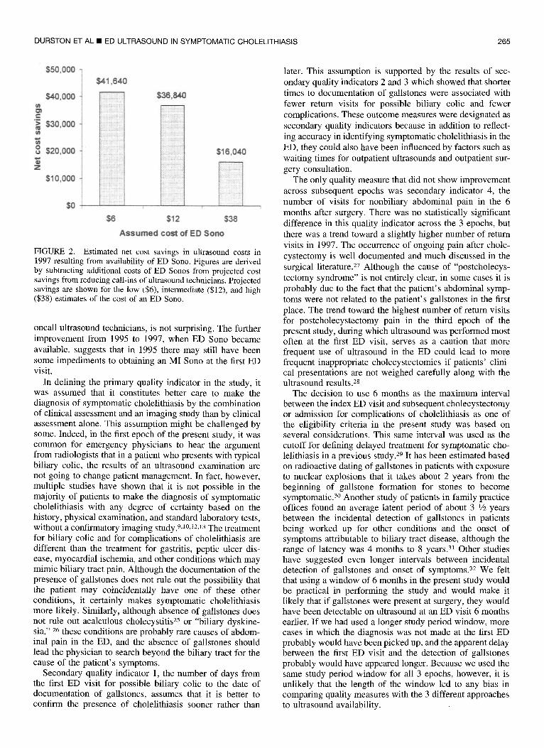
hospital, the net differences between the expense of ED Sonos and the savings which resulted from avoiding ultra- sound technicians call-ins are shown in Fig 2. The magni- tude of the differences varied depending on whether the high, low, or intermediate assumptions for the cost of an ED Sono were used for the calculations, but there were pro- jected net cost savings as a result of the availability of ED Sono with all 3 estimates.
DISCUSSION
This study shows that as the availability of ultrasound in our ED increased, there was a significant increase in the percentage of ED patients presenting with possible biliary colic who had an ultrasound examination done at their first ED visit. Correspondingly, there was significant improve- ment in the primary quality indicator in the study, the percentage of patients documented to have gallstones by an imaging study at their first ED visit. The improvement in quality from 1993, when availability of MI Sono was lim- ited, to 1995, when MI Sono was readily available through
TABLE 5. Estimated Marginal Cost of ED Sono to Rule Out Gallstones
Lowest Intermediate Highest Cost Item Estimate Estimate Estimate
Lease of ultrasound machine $4705 $4705 $14,704 Maintenance agreement $0 $2880 $9,000 Physician training $0 $2112 $6,600 Total annual costs $4705 $9697 $30,304 Cost per ED Sono $6 $12 $38
NOTE. The highest estimate assumes that GEs most comprehensive maintenance agreement was purchased, that the complete costs of physician training were paid for in 1 year, and that 802 ED Sonos were done in the year. The intermediate estimate incorporates the same assumptions as the highest estimate, but costs are multiplied by 0.32, assuming that 32% of ED Sonos are done to rule out gallstones (see Methods), and that ED Sonos done for other indi- cations have equal worth. The lowest estimate assumes that no maintenance was required over the first year and that physicians bore their own cost of training.
DURSTON ET AL • ED ULTRASOUND IN SYMPTOMATIC CHOLELITHIASIS 265
$41,640
$40,000
e., , w
$30,000
o $20,000
Z
$10,000
$0
i!i i i iiiiiiiiii'i!i!,!l iii~i!!!i!i~iii!iiiii~ilil : : : :%: : : : : : : : : : - - !
. . . : . . : . . . + : . .
i:::iii::::::i[i::i . . . . . . . . . . . . . !2! ! !i i:i2iiii~.~ E i i . . . . . . . . . . . . . . : . : : . : : : : : : : : :
i::i i:::::: :: g:i2:: :#: ! ! : i~ i : i i : ! ! ;~ i i:i ~:ii i2 i : i i i~! . . . . . . , . . . . , . , . . . . . . . . . . . . .
i~i:i:.::i~:~i::i::? ~ii i i i : i[:i:!! :
? i ? i ~ i i i i i i ? i : : : : : : : : : : : :
$50,000
i iiiiiiiiii'i!i!!l i i . i ; i ! i i : ~ ! [ ! i i
? 2 : : : : : : : : : : : . . . . . . . . . . . . .
: 2 : : : : : : : : : : : : : : : : : : : : : x : : : : : : . : : : : : : - : . . . . . . . . . . . : : : : : : : - : : : : : :
: : : : : : : : . : : : : : : : : : : : : : : : : : : : : : : : : : : : : : : : : : : : : : : ~ : : : : : . . . . . . . . . . . . . : : : : : : : : : : : : : : : : : : : : : : : : : : : : : : : : : : : : : : : : : : : : : : : : : : : : : : : :
: : : : : : : : : 5 : : : : : : - : : : : : : : : : : : : : : : : : : : : : : : : . : : : : : : : : : : : :
: : : : : : : : : : : : :
: : : . : i : : : : ' : : : : :
i
$6 $12
$16,040
i
$38
Assumed cost of ED Sono
FIGURE 2. Estimated net cost savings in ultrasound costs in 1997 resulting from availability of ED Sono. Figures are derived by subtracting additional costs of ED Sonos from projected cost savings from reducing call-ins of ultrasound technicians. Projected savings are shown for the low ($6), intermediate ($12), and high ($38) estimates of the cost of an ED Sono.
oncall ultrasound technicians, is not surprising. The further improvement from 1995 to 1997, when ED Sono became available, suggests that in 1995 there may still have been some impediments to obtaining an MI Sono at the first ED visit.
In defining the primary quality indicator in the study, it was assumed that it constitutes better care to make the diagnosis of symptomatic cholelithiasis by the combination of clinical assessment and an imaging study than by clinical assessment alone. This assumption might be challenged by some. Indeed, in the first epoch of the present study, it was common for emergency physicians to hear the argument from radiologists that in a patient who presents with typical biliary colic, the results of an ultrasound examination are not going to change patient management. In fact, however, multiple studies have shown that it is not possible in the majority of patients to make the diagnosis of symptomatic cholelithiasis with any degree of certainty based on the history, physical examination, and standard laboratory tests, without a confirmatory imaging study. 9.1o,12-13 The treatment for biliary colic and for complications of cholelithiasis are different than the treatment for gastritis, peptic ulcer dis- ease, myocardial ischemia, and other conditions which may mimic biliary tract pain. Although the documentation of the presence of gallstones does not rule out the possibility that the patient may coincidentally have one of these other conditions, it certainly makes symptomatic cholelithiasis more likely. Similarly, although absence of gallstones does not rule out acalculous cholecystitis 25 or "biliary dyskine- sia," 26 these conditions are probably rare causes of abdom- inal pain in the ED, and the absence of gallstones should lead the physician to search beyond the biliary tract for the cause of the patient's symptoms.
Secondary quality indicator 1, the number of days from the first ED visit for possible biliary colic to the date of documentation of gallstones, assumes that it is better to confirm the presence of cholelithiasis sooner rather than
later. This assumption is supported by the results of sec- ondary quality indicators 2 and 3 which showed that shorter times to documentation of gallstones were associated with fewer return visits for possible biliary colic and fewer complications. These outcome measures were designated as secondary quality indicators because in addition to reflect- ing accuracy in identifying symptomatic cholelithiasis in the ED, they could also have been influenced by factors such as waiting times for outpatient ultrasounds and outpatient sur- gery consultation.
The only quality measure that did not show improvement across subsequent epochs was secondary indicator 4, the number of visits for nonbiliary abdominal pain in the 6 months after surgery. There was no statistically significant difference in this quality indicator across the 3 epochs, but there was a trend toward a slightly higher number of return visits in 1997. The occurrence of ongoing pain after chole- cystectomy is well documented and much discussed in the surgical literature. 27 Although the cause of "postcholecys- tectomy syndrome" is not entirely clear, in some cases it is probably due to the fact that the patient's abdominal symp- toms were not related to the patient's gallstones in the first place. The trend toward the highest number of return visits for postcholecystectomy pain in the third epoch of the present study, during which ultrasound was performed most often at the first ED visit, serves as a caution that more frequent use of ultrasound in the ED could lead to more frequent inappropriate cholecystectomies if patients' clini- cal presentations are not weighed carefully along with the ultrasound results. 2s
The decision to use 6 months as the maximum interval between the index ED visit and subsequent cholecystectomy or admission for complications of cholelithiasis as one of the eligibility criteria in the present study was based on several considerations. This same interval was used as the cutoff for defining delayed treatment for symptomatic cho- lelithiasis in a previous study. 29 It has been estimated based on radioactive dating of gallstones in patients with exposure to nuclear explosions that it takes about 2 years from the beginning of gallstone formation for stones to become symptomatic. 3° Another study of patients in family practice offices found an average latent period of about 3 1/2 years between the incidental detection of gallstones in patients being worked up for other conditions and the onset of symptoms attributable to biliary tract disease, although the range of latency was 4 months to 8 years. 31 Other studies have suggested even longer intervals between incidental detection of gallstones and onset of symptoms. 32 We felt that using a window of 6 months in the present study would be practical in performing the study and would make it likely that if gallstones were present at surgery, they would have been detectable on ultrasound at an ED visit 6 months earlier. If we had used a longer study period window, more cases in which the diagnosis was not made at the first ED probably would have been picked up, and the apparent delay between the first ED visit and the detection of gallstones probably would have appeared longer. Because we used the same study period window for all 3 epochs, however, it is unlikely that the length of the window led to any bias in comparing quality measures with the 3 different approaches to ultrasound availability.
266 AMERICAN JOURNAL OF EMERGENCY MEDICINE • Volume 19, Number 4 • July 2001
Another potential limitation of this study is uncertainty in determining whether visits for abdominal pain in patients with known cholelithiasis were attributable to their gall- stones or to some other coincidental, undiagnosed pathol- ogy. Some of the visits classified as "possible biliary colic" in the present study were probably not, in fact, related to the patient' s gallstones, while some visits for chest pain, nausea and vomiting, or other nonspecific gastrointestinal com- plaints, which were not included in the study, may have been because of symptomatic cholelithiasis. This uncer- tainty as to whether or not a patient's symptoms are caused by gallstones is a limitation in any study of precision in diagnosing biliary tract pain. 33 Although there was probably some misclassification in the present study of visits for nonbiliary pain as possible biliary colic, and vice versa, because the same criteria were used for all 3 epochs, it is unlikely that any misclassification biased the comparisons across the 3 epochs.
The increase in the percentages of all ED patients who met study inclusion criteria from 1993 to 1997, as shown in Table 2, could be because of an increase in the prevalence of symptomatic cholelithiasis in the study population, more frequent misdiagnosis of biliary colic during ED visits in the earlier epochs, a trend for surgeons to perform cholecystec- tomies more often in the later epochs, or a combination of these factors. An increase in the prevalence of symptomatic cholelithiasis in the study population seems unlikely. It cannot be determined from the available data whether more frequent misdiagnosis of symptomatic cholelithiasis in the ED in the earlier epochs or a more liberal approach to surgery in the later epochs was most important in the observed trend toward a higher cholecystectomy rate over time, but both factors probably contributed. With the in- creasing use of laparoscopic cholecystectomy over the past decade and its lower morbidity as compared with open cholecystectomy, 34 there has been an increase in the chole- cystectomy rate in the United StatesY The question of whether or not prompt cholecystectomy constitutes better treatment than expectant management in patients with symptomatic cholelithiasis has not been conclusively an- swered and remains a point of controversy in the medical literature.36-41
For several reasons, it was not practical to compare the accuracy of ED Sono with the accuracy of MI Sono of the gallbladder at our own facility. Most cases of positive MI Sonos for gallstones were picked up by the computer search for patients admitted for cholecystectomy or a complication of cholelithiasis. It was not practical, though, to identify all cases in which MI Sonos of the gallbladder were done and did not show gallstones. Also, because MI Sono results were used as the "gold standard" for the accuracy of ED Sono in cases in which surgical pathology was not available, any comparison of the relative accuracy of ED Sono and MI Sono at our facility would have been biased in favor of MI Sono. Finally, in 1997, most MI Sonos ordered by emer- gency physicians were done after gallstones had already been identified on ED Sonos, and the results of the ED Sonos were available to the sonographers in Medical Imag- ing.
There have been several, relatively small previous studies reporting that emergency physicians can perform ED Sono for the detection of cholelithiasis with acceptable accuracy
after brief training periods.16-2° In the 2 studies which report the most complete data, the sensitivity of ED Sono was 86% to 95% and specificity 87% to 97%, using MI Sono as the gold standard. 17,18 It is of interest that there is also a report in the surgical literature of ultrasound of the gallbladder by surgeons in a convenience sample of 77 cases with a re- ported sensitivity of 100% and specificity of 95%, using MI Sono as the gold standard. 42
In a previous study of the accuracy of MI Sono in detecting cholelithiasis in 993 patients, the indeterminate rate was reported to be 1.3%, the sensitivity 98%, the specificity 93.5%, and the accuracy (excluding indetermi- nate scans) 96%. 14 The investigators do not state, however, what standard was used to confirm the accuracy of sono- grams interpreted as showing no gallstones. In a meta- analysis of 3 studies on MI Sono with a total of 552 patients, the indeterminate rate for MI Sono was 1%, the sensitivity 97% (95% CI 95-99%) and the specificity 95% (95% CI 88-100%). 43
The sensitivity of a test reflects the rate of falsely nega- tive studies. Most studies of the accuracy of MI Sono in detecting cholelithiasis use surgical pathology as the gold standard for the presence of gallstones. In such studies, the problem of verification bias could make the false negative rate of MI Sonos appear lower than it actually is, as patients with falsely negative studies are less likely to go to surgery. Verification bias would be less likely to affect the false- negative rate of ED Sonos in the present study as patients who did not have a confirmatory imaging study or surgery were followed for a full 2 years, longer than in any of the above cited studies, before a negative study for gallstones was considered to be confirmed. Correcting for verification bias in the meta-analysis cited earlier, the investigators calculated the adjusted sensitivity of MI Sono for the de- tection of cholelithiasis to be 84% (95% CI 76%-92%). 43
In the 802 patients in the present study who had ED Sono to rule out gallstones in 1997, the indeterminate rate (18%) was considerably higher than in previous studies of MI Sono. Excluding indeterminate studies and studies for which confirmation was not available, though, the sensitiv- ity of ED Sono for detecting cholelithiasis in the present study (88.6%) is comparable with the sensitivity adjusted for verification bias (84%) in the meta-analysis of MI Sono cited earlier. 43 Likewise, the specificity (98.2%) and overall accuracy (94.8%) of ED Sono in the present study are comparable to the previously cited reports on MI Sono. 14,43
Besides lack of operator experience or expertise, a num- ber of other factors could contribute to a higher indetermi- nate rate for ED Sono. These factors include the condition of the patient at the time of the study, the environment in which the study is performed, and the equipment used to perform the study. Patients in the ED are often nonfasting. Acute biliary colic is probably caused, in most cases, by stones becoming lodged in the neck of the gallbladder, the cystic duct, or the common bile duct, where they are more difficult to visualize sonographically than while they are lying in the body of the gallbladder. The lighting conditions in the ED and the time available for the study are usually suboptimal, as compared with a scheduled appointment in the ultrasound suite of the Medical Imaging Department. Finally, the resolution of portable ultrasound machines used in the ED is usually not as good as the resolution of larger,
DURSTON ET AL • ED ULTRASOUND IN SYMPTOMATIC CHOLELITHIASIS 267
more expensive machines used in most Medical Imaging Departments. It is of interest that in one small study assess- ing the accuracy of sonography performed for a variety of indications in the ED, the accuracy rates for ED physicians and for trained sonographers were not significantly different when both used the same machine in the ED setting. 44 We believe that an indeterminate rate in the range of 18% is acceptable for ED Sono to rule out cholelithiasis, and that it is preferable to consider scans indeterminate than to risk calling them falsely positive or negative.
In this study, we did not specifically address the accuracy of either ED or MI Sonos in assessing features of gallblad- der disease other than the presence or absence of stones. Factors such as thickening of the gallbladder wall, dilatation of the common bile duct (CBD), the presence of stones greater than 20 millimeters in diameter, and contraction of the gallbladder have been shown to predict the need for conversion from laparoscopic to open cholecystectomy. 45 Also, dilatation of the CBD on ultrasound has been shown to be a reliable predictor of the presence of common bile duct stones and of the need for endoscopic retrograde cholangiopancreatography (ERCP) either before or after laparoscopic cholecystectomy. 46-49 To date, only one small study has addressed the correlation between ED Sono and MI Sono in determining the presence or absence of sono- graphic signs of cholecystitis. 2o This study found only a moderate correlation (K = .46). We are not aware of any studies looking at the correlation between ED Sono and MI Sono in measuring CBD diameter.
When we began our study, the emphasis in the ED literature was for emergency physicians to perform "limited, goal-directed" ED Sonos, 17,50 with the limited goal in patients with suspected biliary colic being to rule out gallstones. The rationale for this approach was that keeping the study limited and goal-directed would reduce the likelihood of errors by relatively inexperienced phy- sicians. On the other hand, failure to identify sonographic signs of cholecystitis or CBD obstruction could poten- tially have an adverse effect on patient management. If signs of cholecystitis or CBD obstruction had been missed on ED Sono in the present study, one might expect a trend toward an increased frequency of patients returning with complications of cholelithiasis in 1997. In fact, however, the trend was toward fewer complications during the ED Sono epoch. Nevertheless, we have mod- ified our approach to ED Sono of the right upper quadrant as we have gained experience, and we now expect our emergency physicians to document not only the presence or absence of gallstones, but also to look for sonographic signs of cholecystitis and CBD dilatation.
To our knowledge, no previous study has directly ad- dressed the relative cost-effectiveness of limited versus lib- eral MI Sono availability or of MI Sono versus ED Sono in suspected symptomatic cholelithiasis. As shown in Fig 1, the number of MI Sonos done per case identified as eligible for the study increased from 1.7 in 1993, when the avail- ability of MI Sono was limited, to 2.5 in 1995, when MI Sono was readily available. The number of MI Sonos per case went back down to 1.7 in 1997, when 4.2 ED Sonos were also done per study case. Since there was no charge for an ED Sono, the projected ultrasound charges per case identified, assuming that all patients were charged at the
same non-member rate ($370), showed a similar rise and fall. From these data, it can be calculated that the total ultrasound charges per case identified in 1997 would have been the same as in 1995 if the charge for an ED Sono had been $70.
If it is assumed that there is no cost to missing the diagnosis of symptomatic cholelithiasis in the ED, then the strategy of limited availability of MI Sono in 1993 would be the most cost-effective. Clearly, however, there are costs to missed or delayed diagnoses, though the exact amount of these costs is difficult to estimate. The medical costs include return visits and treatment for the complications of chole- lithiasis. If cholecystectomy is performed before acute cho- lecystitis develops, there is a greater chance that the proce- dure can be done laparoscopically rather than open, 51-53 with shorter hospitalizations and lower hospital costs. Other costs of missed or delayed diagnosis include patient pain and suffering, missed days of work, and potential medical- legal liability. If it is assumed that the lower rate of return visits for possible biliary colic in 1995 compared with 1993 was a result of the more frequent use of ultrasound at the first visit in 1995, then the additional charges for more frequent MI Sonos in 1995 would have equaled the reduc- tion in charges for return visits if the charge for a return visit had been $698. Considering that there are other costs of delayed or missed diagnoses beyond the charges for return visits, it seems likely that liberal use of MI Sono is more cost-effective than limited availability of MI Sono in the evaluation of patients in the ED with suspected symptom- atic cholelithiasis.
To the extent that ED Sono is less expensive than MI Sono, the substitution of ED Sono for MI Sono should further enhance the cost-effectiveness of liberal ultrasound use. The actual cost of an MI Sono is difficult to determine. A detailed analysis of medical imaging costs in intermediate referral hospitals in Finland in 1996 estimated that the cost of a generic ultrasound study, taking into account physician and nonphysician personnel costs, capital equipment, and administrative and physical plant overhead, was 296 Finn- ish marks, or approximately 54 U.S. dollars per study. 54 In a 1999 U.S. study of the relative cost-effectiveness of com- puted tomography versus ultrasound for suspected appen- dicitis, is was estimated that the cost of an abdominopelvic ultrasound in a medical imaging department was $270. 25 It was not practical to determine the actual cost of an MI Sono at our facility, but the marginal cost of calling in an ultra- sound technician after regular medical imaging department hours was known to be $135 per call-in. The different cost components of an ED Sono were directly identifiable, but even so, estimates of the marginal cost of an ED Sono varied from $6 to $38 per study, depending on which assumptions were made regarding the need for ultrasound machine maintenance, whether the facility or the individual physician bore the cost of training, and whether ED Sonos were done only to rule out cholelithiasis or for other indi- cations as well.
Assuming that the only costs of an MI Sonos were the costs of calling in technicians after regular hours and that each nonindeterminate ED Sono done after regular hours saved a call-in, the availability of ED Sono led to a net savings in 1997 of approximately $16,000 to $42,000 in ultrasound costs, as shown in Fig 2, depending on which
268 AMERICAN JOURNAL OF EMERGENCY MEDICINE • Volume 19, Number 4 • July 2001
estimates were used for the cost of an ED Sono. It could be argued that not all after hours ED Sonos saved a call-in, and that in some cases, an MI Sono could have reasonably been done during regular hours the next weekday. Dividing ED Sono costs by call-in costs gives the "break even" point at which the added cost of ED Sonos would have exactly equaled the cost savings from avoiding calling in ultrasound technicians. Using the intermediate estimate ($12) for the cost of an ED Sono, ED Sono costs would have exactly equaled savings in call-ins if it were assumed that 21% of after hours ED Sonos saved a call-in. Using the high ($38) or low ($6) estimates for the cost of an ED Sono, the "break-even" points would have occurred when 65% or 10% of ED Sonos, respectively, precluded call-ins.
In calculating the cost of an ED Sono, we did not include the time the emergency physician spends in actually per- forming the study. In previous studies addressing the time it takes to perform an ultrasound of the gallbladder, the re- ported times have ranged from 5 minutes for radiologists 14 to 7 minutes for emergency physicians ~8 to 12 minutes for surgeons. 42 In an opinion survey at our facility after 1 year of experience with ED Sono, emergency physicians were equally divided between those who felt ED Sono increased their efficiency and those who felt it reduced it. 56 It has previously been shown that the length of stay in the ED for patients with abdominal pain is approximately an hour less for patients who have ED Sono as compared with patients who have MI Sono. 57 In the present study, for the purpose of comparing the cost-effectiveness of ED Sono versus MI Sono, we assumed that the cost of the emergency physi- cian's time in performing the study was approximately offset by the time saved in getting a more timely diagnosis and disposition.
In conclusion, our study shows that as MI Sono became more readily available in our ED from 1993 to 1995, there was approximately a 50% increase in MI Sono utilization by emergency physicians accompanied by significant improve- ment in measures of quality reflecting the time to diagnosis in the evaluation of patients with symptomatic cholelithia- sis. The addition of ED Sono in 1997 was associated with further improvements in quality and a reduction in MI Sono utilization to 1993 levels. The cost of the ED Sono program was more than offset by savings in avoiding call-ins of ultrasound technicians after regular Medical Imaging De- partment hours. The indeterminate rate for ED Sonos was relatively high, but excluding indeterminates, the sensitiv- ity, specificity, and accuracy were comparable to published reports for MI Sono. Our study suggests that the best approach for identifying patients with symptomatic chole- lithiasis in the ED is to screen all patients with possible biliary colic with El) Sono, and to have MI Sono readily available for cases in which ED Sono is indeterminate.
The authors are indebted to Kieran Fitzpatrick, MD, and to Robert Bruun, PhD, for administrative support; to Dave Rentz for providing MI Sono utilization data; to Taya Dunn for performing computer searches for eligible patients; to Michael Bennett for obtaining cop- ies of references; and to the emergency physicians in our depart- ment for their enthusiastic participation in the ED Sono project.
REFERENCES 1. Friedman GS, Kannel WB, Dawber TR: The epidemiology of
gallbladder disease: Observations in the Framingham study. J Chron Dis 1966;19:273-292
2. Kratzer W, Mason RA, Kachele V: Prevalence of gallstones in sonographic surveys worldwide. J Clin Ultrasound 1999;27:1-7
3. Janzon L, Ryden CI, Zederfledt B: Acute abdomen in the surgical emergency room. Acta Chir Scand 1982;148:141-148
4. Bugliosi TF, Meloy TD, Vukov LF: Acute abdominal pain in the elderly. Ann Emerg Med 1990;19:1383-1386
5. Wilson DH, Wilson PD, Walmsley RG, et al: Diagnosis of acute abdominal pain in the accident and emergency department. Br J Surg 1977;64:250-254
6. Brewer R J, Golden GT, Hitch DC, et al: Abdominal pain. An analysis of 1000 consecutive cases in a university hospital emer- gency room. Am J Surg 1976;131:219-223
7. Friedman GD: NaturN history of asymptomatic and symptom- atic gallstones. Am J Surg 1993;165:399-404
8. Parker LJ, Vukov LF, Wollan PC: Emergency department eval- uation of geriatric patients with acute cholecystitis. Acad Emerg Med 1997;4:51-55
9. Thijs C, Knipschild P: Abdominal symptoms and food intoler- ance related to gallstones. J Clin Gastroenterol 1998;27:223-231
10. Kraag N, Thijs C, Knipschild P: Dyspepsia: How noisy are gallstones? A meta-analysis of epidemiologic studies of biliary pain, dyspeptic symptoms, and food intolerance. Scand J Gastroenterol 1995;30:411-421
11. Berger MY, van der Velden J JIM, Lijmer JG, et al: Abdominal symptoms: do they predict gallstones? A systematic review. Scand J Gastroenterol 2000;35:70-76
12. Singer AJ, McCracken G, Henry MC, et al: Correlation among clinical, laboratory, and hepatobiliary scanning findings in patients with suspected acute cholecystitis. Ann Emerg Med 1996;28:267- 272
13. Gruber PJ, Silverman RA, Gottesfeld S, et al: Presence of fever and leukocytosis in acute cholecystitis. Ann Emerg Med 1996; 28:273-277
14. Cooperberg PL, Burhenne HJ: Real-time ultrasonography. Diagnostic technique of choice in calculous gallbladder disease. N Engl J Med 1980;302:1277-1279
15. Heller M, Crocco T, Patterson J, et al: Emergency ultrasound services as perceived by directors of radiology and emergency departments. Am J Emerg Med 1995;13:430-431
16. Jehle D, Davis E, Evans T, et al: Emergency department sonography by emergency physicians. Am J Emerg Med 1989;7: 605-611
17. Schlager D, Lazzareschi G, Whitten D, et al: A prospective study of ultrasonography in the ED by emergency physicians. Am J Emerg Med 1994;12:185-189
18. Kendall JL, Shrimp R J: Performance and interpretation of limited right upper quadrant ultrasound by emergency physicians. Acad Emerg Med 1998;5:106 (abstr)
19. Gussow L, Himmelman R, Zalenski R: Portable ultrasound in patients with suspected cholecystitis: Performance and interpreta- tion by emergency physicians. Ann Emerg Med 1989;18:441 (abstr)
20. Rosen CL, Brown DFM, Chang YC, et al: Gallbladder ultra- sonography: Agreement between emergency physicians and radi- ologists. Acad Emerg Med 1998;5:417 (abstr)
21. Abbott J: Emergency department ultrasound. Is it really time for real time? J Emerg Med 1990;8:491-492
22. Bree RL, Berland LL, Charboneau JW, et al: Sonography by the radiologist: Self-referral, turf battles, and marketing. A JR Am J Roentgenol 1995;164:231-233
23. Hemphill RH, Santen SA, Buggs AM: Limitations of ED gall- bladder ultrasound studies. Ann Emerg Med 1997;30:384 (abstr)
24. Durston W, Carl ML, Guerra W: Patient satisfaction and di- agnostic accuracy with ultrasound by emergency physicians. Am J Emerg Med 1999;17:642-646
25. Babb RR: Acute acalculous cholecystitis. A review. J Clin Gastroenterol 1992;15:238-241
26. Goncalves RM, Harris JA, Rivera DE: Biliary dyskinesia: Nat- ural history and surgical results. Am Surg 1998;64:493-497
27. Middelfart HV, Kristensen JU, Laursen CN, et al: Pain and dyspepsia after elective and acute cholecystectomy. Scand J Gas- troenterol 1998;33:10-14
28. Talley N J: Editorial: Gallstones and upper abdominal discom- fort. Innocent bystander or cause of dyspepsia? J Clin Gastroen- terol 1995;20:182-183
DURSTON ET AL • ED ULTRASOUND IN SYMPTOMATIC CHOLELITHIASIS 269
29. Diehl AK, Westwick T J, Badgett RG, et al: Clinical and socio- cultural determinants of gallstone treatment. Am J Med Sci 1993; 305:383-386
30. Mok HYI, Druffel ERM, Rampone WM: Chronology of chole- lithiasis: Dating gallstones from atmospheric radiocarbon produced by nuclear bomb explosions. N Engl J Med 1986;314:1075-1077
31. Zubler J, Markowski G, Yale S, et al: Natural history of asymptomatic gallstones in family practice office practices. Arch Fam Med 1998;7:230-233
32. Friedman GD, Raviola CA, Fireman B: Prognosis of gallstones with mild or no symptoms: 25 years of followup in a health mainte- nance organization. J Clin Epidemiol 1998;42:127-136
33. Sondenaa K, Nesvik J, Solhaug JH, et al: Randomization to surgery or observation in patients with symptomatic gallbladder stone disease. The problem of evidence-based medicine in clinical practice. Scand J Gastroenterol 1997;32:611-616
34. Wu JS, Dunnegan DL, Luttmann DR, et al: The evolution and maturation of laparoscopic cholecystectomy in an academic prac- tice. J Am Coil Surg 1998;186:554-561
35. Steinle EW, VanderMolen RL, Silbergleit A, et al: Impact of laparoscopic cholecystectomy on indications for surgical treatment of gallstones. Surg Endosc 1997;11:933-935
36. Saltzstein EC, Peacock JB, Mercer LC: Early operation for acute biliary tract stone disease. Surgery 1983;94:704-708
37. Ransohoff DF, Gracie WA: Treatment of gallstones. Ann Int Med 1993;119:606-619
38. Patino JF, Quintero GA: Asymptomatic cholelithiasis revis- ited. World J Surg 1998;22:1119-1124
39. Robertson GSM, Wemyss-Holden SA, Maddern G J: The best management for "crescendo biliary colic" is urgent laparoscopic cholecystectomy. Postgrad Med J 1998;74:681-682
40. Schwesinger WH, Diehl AK: Changing indications for laparo- scopic cholecystectomy. Stones without symptoms and symptoms without stones. Surg Clin N A 1996;76:493-504
41. McSherry CK, Ferstenberg H, Calhoun WF, et al: The natural history of diagnosed gallbladder disease in symptomatic and asymptomatic patients. Ann Surg 1985;202:59-63
42. Fang R, Pilcher JA, Putham AT, et al: Accuracy of surgeon- performed gallbladder ultrasound. Am J Surg 1999;178:475-479
43. Shea JA, Berlin JA, Escarce J J, et al: Revised estimates of diagnostic test sensitivity and specificity in suspected biliary tract disease. Arch Intern Med 1994;154:2573-2581
44. Shapiro RA, Nakamoto M: Ultrasonography in Japanese emergency departments. Am J Emerg Med 1990;8:443-445
45. Jansen S, Jorgensen J, Caplehorn J, et al: Preoperative ultrasound to predict conversion in laparoscopic cholecystectomy. Surg Laparosc Endosc 1997;7:121-123
46. Kruis W, Roehrig H, Hardt M, et al: A prospective evaluation of the diagnostic work-up before laparoscopic cholecystectomy. Endoscopy 1997;29:602-608
47. Majeed AW, Ross B, Johnson AG, et al: Common duct di- ameter as an independent predictor of choledocholithiasis: Is it useful? Clinical Radiology 1999;54:170-172
48. Goodwin AT, Tully J, Charlesworth C, et al: Routine use of ultrasound 24 hours before laparoscopic cholecystectomy can pre- dict the need for intraoperative cholangiogram: Results of a 12- month prospective audit. Br J Clin Pract 1997;51:140-143
49. Contractor AA, Boujemla M, Contractor TQ, et al: Abnormal common bile duct sonography: The best predictor of choledocho- lithiasis before laparoscopic cholecystectomy. J Clin Gastroenterol 1997;25:429-432
50. Plummer D: Diagnosis ultrasonography in the emergency department. Ann Emerg Med 1993;22:592-593
51. Liu C, Fan S, Lai ECS, et al: Factors affecting conversion of laparoscopic cholecystectomy to open surgery. Arch Surg 1996; 131:98-101
52. Alponat A, Kum CK, Koh BC, et al: Predictive factors for conversion of laparoscopic cholecystectomy. World J Surg 1997; 21:629-633
53. Eldar S, Sabo E, Nash E, et al: Laparoscopic cholecystec- tomy for acute cholecystitis: Prospective trial. World J Surg 1997; 21:540-545
54. Laaperi AL: Cost accounting of radiological examinations: Cost analysis of radiological examinations of intermediate referral hospitals and general practice. Acta Radiologica 1996;407:1-54 (suppl)
55. Garcia-Pena BM, Taylor GA, Lund D, et al: Effect of com- puted tomography on patient management and costs in children with suspected appendicitis. Pediatrics 1999;104:440-446
56. Carl ML, Durston WE, Guerra WF: Survey of staff opinions on ultrasound by emergency physicians. Am J Emerg Med 2000;18: 340-341
57. Schlager D, Whitten D, Tolan K: Emergency department ul- trasound: Impact on ED stay times. Am J Emerg Med 1997;15:216- 217


![CHOLELITHIASIS [Autosaved]](https://img.pdfslide.us/doc/110x75/577ce5051a28abf1038fa5b3/cholelithiasis-autosaved.jpg)