Embed Size (px)
Citation preview
Original Article
Comparison between a new ultrasoundprobe with a capacitive micromachinedtransducer (CMUT) and a traditional onein musculoskeletal pathology
Ferdinando Draghi1, Pascal Lomoro1 ,Chandra Bortolotto1 , Luca Mastrogirolamo2 andFabrizio Calliada1
AbstractBackground: The capacitive micromachined ultrasound transducer (CMUT) is a new ultrasound (US) probe manufac-
tured by state-of-the-art cutting-edge semi-conductor micromachined electro-mechanical systems (MEMS) technology.
Purpose: To demonstrate the peculiar characteristics of each probe and the limitations that should be improved.
Material and Methods: This study was performed from March to April 2018. The only inclusion criterion was the
presence of disease, so all patients with musculoskeletal, skin, and subcutaneous pathology were included. A total of 66
patients entered this study. The exams of each patient, with both probes, were evaluated retrospectively and indepen-
dently by three radiologists. Panoramicity of the images, the definition of superficial structures (<2 cm of depth), the
definition of deep structures (>2 cm), and Doppler signal were assessed. A 5-point scale was used for each parameter.
Results: A total of 89 pathologies were detected. The mean of score for 4G-CMUT was higher than L64 for the
panoramicity of the images and the definition of the deep structures. Instead, the mean score for L64 was higher than for
4G-CMUT in the evaluation of superficial structures and Doppler signal. A statistically significant difference was found
(P< 0.05).
Conclusion: CMUT is a breakthrough in US technology. It allows the use of a single probe for different US examina-
tions. The musculoskeletal, skin, and subcutaneous US can be evaluated with a piezoelectric linear transducer or CMUT.
In the present study, the overall diagnostic performance was similar. Improvements in CMUT will provide even more
dynamic and flexible imaging capabilities by a transducer, with a wider bandwidth.
Keywords
Ultrasound, ultrasound color Doppler, linear probe, imaging, musculoskeletal ultrasound, capacitive micromachined
ultrasound transducer
Date received: 16 May 2019; accepted: 18 January 2020
Introduction
Ultrasonography (US) is a low-cost, safe, non-invasive,
and ionizing radiation-free imaging technique that is
widespread in medical practice. It is generally used as
a primary diagnostic imaging technique, especially in
children and pregnant women. US uses a transducer or
probe to produce images (1). The probe is composed of
piezoelectric crystals (quartz or leaded titanium zirco-
nate ceramics or barium titanate) that are made up of
countless dipoles. When crystals are excited by an
1Radiology Department, Fondazione IRCCS Policlinico San Matteo,
Universita degli Studi di Pavia, Pavia, Italy2Hitachi Medical Systems Europe Holding AG, Steinhausen, Switzerland
Corresponding author:
Lomoro Pascal, Radiology Department, Fondazione IRCCS Policlinico San
Matteo, Universita degli Studi di Pavia, Viale Camillo Golgi, Pavia, Italy.
Email: [email protected]
Acta Radiologica
0(0) 1–7
! The Foundation Acta Radiologica
2020
Article reuse guidelines:
sagepub.com/journals-permissions
DOI: 10.1177/0284185120907983
journals.sagepub.com/home/acr
electric impulse, a potential difference is createdbetween the two ends of the dipoles causing a mechan-ical deformation of the crystals that produces ultra-sound. Instead, they produce electrical signals whensubjected to mechanical vibrations, resulting in abrightness-mode (B-mode) image display. The crystalsare protected and isolated by an epoxy resin matrix orsimilar material (2).
There are many types of probes. Linear transducersare typically made of 128–256 crystals in a row, whichprovide a rectangular field of view (FOV). The width ofthe picture is approximately equal to the size of theprobe’s head (small FOV, medium<40 mm, andlarge>40 mm). These probes have higher frequencies,which provide a better resolution but lower penetra-tion. Therefore, they are excellent for the viewing ofsuperficial structures like skin, subcutaneous tissue,muscles, tendons, nerves, and blood vessels.
Convex transducers are made of crystals placed on acurvilinear support, which provides a “fan” field ofview. The width of the picture is larger than the oneobtained by the linear probe. These probes have lowerfrequencies, which provide better penetration but lessresolution. For this reason, they are excellent for thestudy of deep structures, such as abdominal and gyne-cological organs.
The capacitive micromachined ultrasound transduc-er (CMUT) is a new US probe manufactured by thestate-of-the-art cutting-edge semi-conductor microma-chined electro-mechanical systems (MEMS) technolo-gy. This transducer does not have any crystals but usesthe new-generation silicon wafer technology composedof innumerable tiny vibration drums cells formed onthe lm scale. Once submitted to electrical signals, eachcell membrane vibrates, generating an ultrasonic signal
with the same acoustic impedance of the human body
(3). The inverse phenomenon is observed when an echo
contacts the cell membrane. Membranes can vibrate at
different frequencies; therefore, CMUT delivers a one-
probe solution for a wide range of US examinations
(4). Hitachi’s brand new CMUT is a fourth-
generation CMUT (4G-CMUT). The present study
shows real-time images from piezoelectric linear array
and compares them to those of 4G-CMUT to demon-
strate the peculiar characteristics of each probe and the
limitations that should be improved.
Material and Methods
The present study was performed in our Department of
Radiology from March to April 2018. Our institution
is a reference center for musculoskeletal disease; for
this reason, we had the opportunity to evaluate both
common and rare pathologies. Therefore, the only
inclusion criterion was the presence of disease, so all
patients with musculoskeletal, subcutaneous, or skin
pathologies were included.Approval was obtained from the local Institutional
Ethical Committee for this study. Informed consent
was obtained from research participants.A total of 66 patients have been recruited into the
study (35 women [53%], 31 men [47%]; mean
age¼ 57.21 years; age range¼ 22–100 years).
Data and statistical analysis
Real-time US was performed for all patients using
a piezoelectric linear array (L64) and 4G-CMUT
probes on a Hitachi Arietta 850 (Hitachi Ltd., Japan).
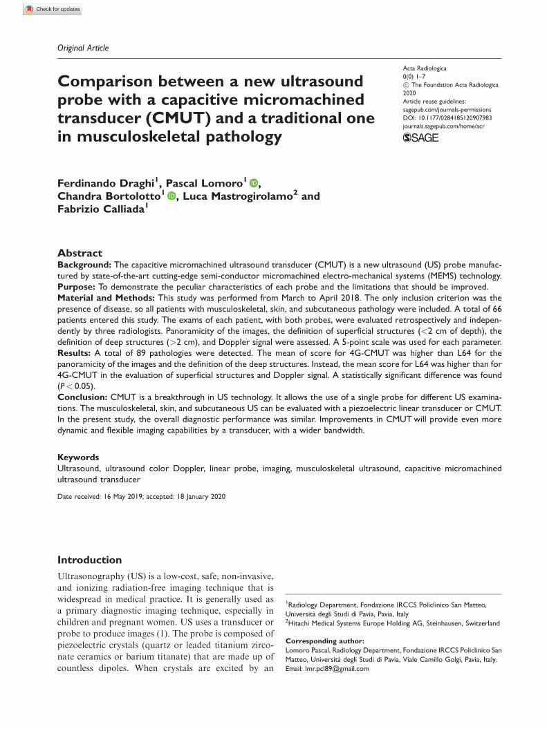
Fig. 1. Complete, acute tear of the supraspinatus tendon (arrows). The diagnostic performance with L64 (a) and CMUT (b) is similar.CMUT, capacitive micromachined ultrasonic transducer.
2 Acta Radiologica 0(0)
The machine was on loan from the company from
March to April 2018.For each exam, B-mode and color-Doppler evalua-
tion were performed and more video clips were saved.
The examinations of each patient with both probes
were evaluated and a comparison of the different
images was performed. The studies were reviewed ret-
rospectively and independently by three radiologists.
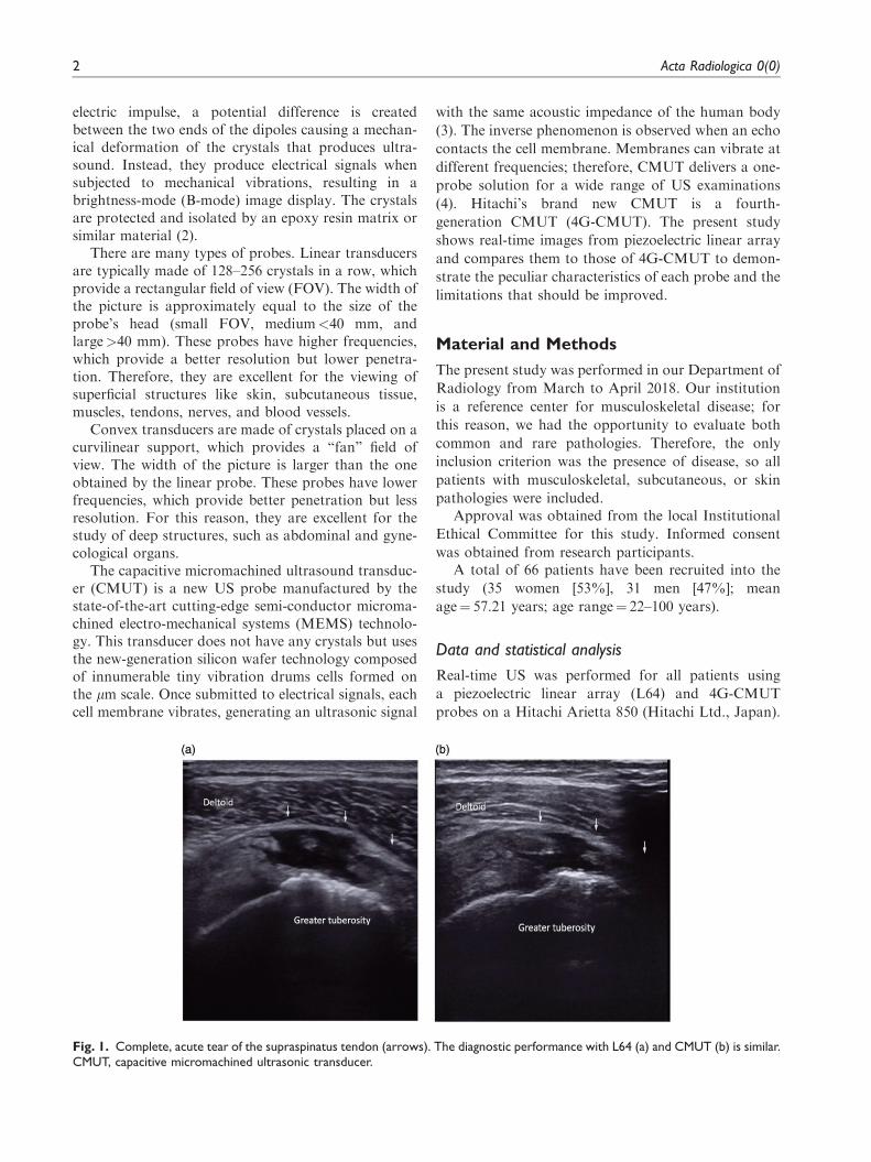
Fig. 2. Strain of vastus lateralis muscle. Coronal proton-density fat-suppressed images show myotendinous strain with muscleretraction of the vastus lateralis (arrows) (a). CMUT (b) has higher panoramicity than L64 (c). CMUT, capacitive micromachinedultrasonic transducer.
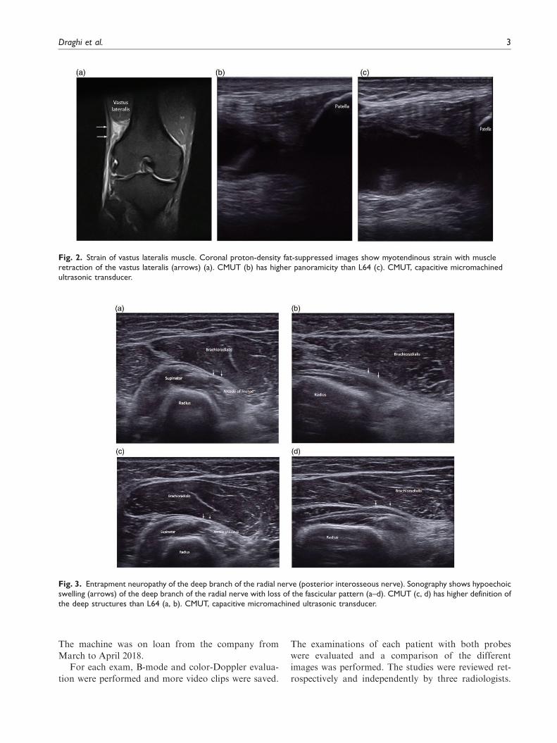
Fig. 3. Entrapment neuropathy of the deep branch of the radial nerve (posterior interosseous nerve). Sonography shows hypoechoicswelling (arrows) of the deep branch of the radial nerve with loss of the fascicular pattern (a–d). CMUT (c, d) has higher definition ofthe deep structures than L64 (a, b). CMUT, capacitive micromachined ultrasonic transducer.
Draghi et al. 3

The scans were performed by a radiologist who did notparticipate in the retrospective evaluation of the imagesin order to minimize the statistical bias. Observers eval-uated the images in three separate sessions, which werelonger than two weeks apart to minimize recall bias. Allradiologist had experience in ultrasound and musculo-skeletal radiology. The images were anonymized andviewed in a random order in each session.
Panoramicity of the images (the ability of theprobe to distinguish both the target lesion and thesurrounding anatomical structures maintaining a highquality images), the definition of superficial structures(<2 cm of depth), the definition of deep structures(>2 cm) (Figs. 1–5), and Doppler signals (Figs. 6 and7) were assessed. A 5-point scale was used for eachparameter.
The mean and median were calculated for L64 and4G-CMUT values, as well as paired Student t-tests todetermine the relative difference between the variablesof opinion between the two probes. A P value<0.05was considered statistically significant (Table 1).
Results
A total of 89 pathologies were evaluated: 48 (72.73%)patients had only one pathology; 16 (24.25%) patientshad two pathologies; 1 (1.51%) patients had fourpathologies; and 1 (1.51%) patient had fivepathologies.
The most frequent regions examined were the shoul-der (Fig. 1) (27.27%), followed by the wrist (15.15%)and ankle (12.12%). The most frequent pathology wascalcific tendinopathy (Cat.) (21.35%) and the supraspi-natus tendon was the mostly common affected(52.63%).
The mean score for 4G-CMUT was higher than thatof L64 for the panoramicity of images and the defini-tion of deep structures (Figs. 2 and 3). Instead, themean score for L64 was higher than that of 4G-CMUT in the evaluation of superficial structures(Figs. 4 and 5) and Doppler signal (Figs. 6 and 7).The comparison of L64 and 4G-CMUT reproducibilityvalues according to different parameters is reported inTable 1. A statistically significant difference was foundin every case with a P value<0.05.
Discussion
The musculoskeletal, skin, and subcutaneous ultra-sound can be performed using a piezoelectric lineartransducer or CMUT. In the present study, the overalldiagnostic performance was similar.
4G-CMUT uses the next-generation silicon wafertechnology composed of innumerable tiny vibrationdrums cells formed on the lm scale. These cells are
Fig. 4. Dupuytren’s disease. Sonography shows hypoechoicnodule (arrow) between the subcutaneous tissues and thesuperficial and deep flexor digitorum tendons (a, b). L64 (a) hashigher definition of the superficial structures than CMUT (b).CMUT, capacitive micromachined ultrasonic transducer.
Fig. 5. Ledderhose disease. Ultrasound shows a well-demar-cated, hypoechoic nodule of the plantar fascia (arrow). L64(a) has higher definition of the superficial structures than CMUT(b). CMUT, capacitive micromachined ultrasonic transducer.
4 Acta Radiologica 0(0)

arranged in a matrix array that improves spatial res-
olution and the frequency bandwidth in the range of
2–22Mhz. It also uses e-Focusing, a dynamic focusing
method that overcomes some of the limitations of
conventional beam-forming. Using fast parallel
beam-forming, it insonifies all or most of the image
fields during each transmission. Several received
echoes are collected dynamically, increasing the
strength of the signal and the frame rate performance.
Homogeneous images are acquired with excellent
signal/noise, significantly improving the sensitivity
and spatial/contrast resolution on the final image.
Thanks to fast parallel beam-forming and
e-Focusing, image definition is achieved in both the
near and far field. Fast parallel beam-forming delivers
uniform lateral resolution in depth, side lobe reduc-
tion and signal/noise improvement. E-Focusing ena-
bles less focus-dependency and improves penetration.
Compared to the old generation, 4G-CMUT has
sufficient power for tissue harmonic imaging techni-
ques which provide excellent images to increase the
signal-to-noise, axial, and lateral resolution (5). It rep-
resents an important tool in the evaluation of deep
structures and in patients with large body habitus.
Thanks to these characteristics, the results of our
study confirmed that 4G-CMUT offered superior pan-
oramicity and a superior diagnostic sensitivity and
accuracy in detecting deep structures compared to pie-
zoelectric linear array. Instead, L64 has a superior
resolution and gives more detail for detecting superfi-
cial structures and the angle of insonation. An
increase in frequency produces an increase in
Doppler frequency and a decrease in penetration (6).
Generally, Doppler is more sensitive to noise com-
pared to B-mode. In our study, Doppler of 4G-
CMUT shows weaknesses in terms of power of
signal, with a better signal for deep structures with
respect to superficial structures, maintaining a good
compromise for signal-to-noise. Instead, the linear
probe Doppler signal is accurate for the evaluation
of blood flow without particular differences between
superficial and deep vessels.
Fig. 6. Epicondylitis. B-mode ultrasound images (a, b) show loss of echogenicity and calcifications of the tendon (arrows), with boneirregularity of the medial epicondyle. The Doppler (c, d) shows hyperemia of the common extensor tendon due to the angiofibro-blastic response. In B-mode, the diagnostic performance of L64 (a) and CMUT (b) are similar, while with color Doppler L64 (c) hashigher definition than CMUT (d). CMUT, capacitive micromachined ultrasonic transducer.
Draghi et al. 5
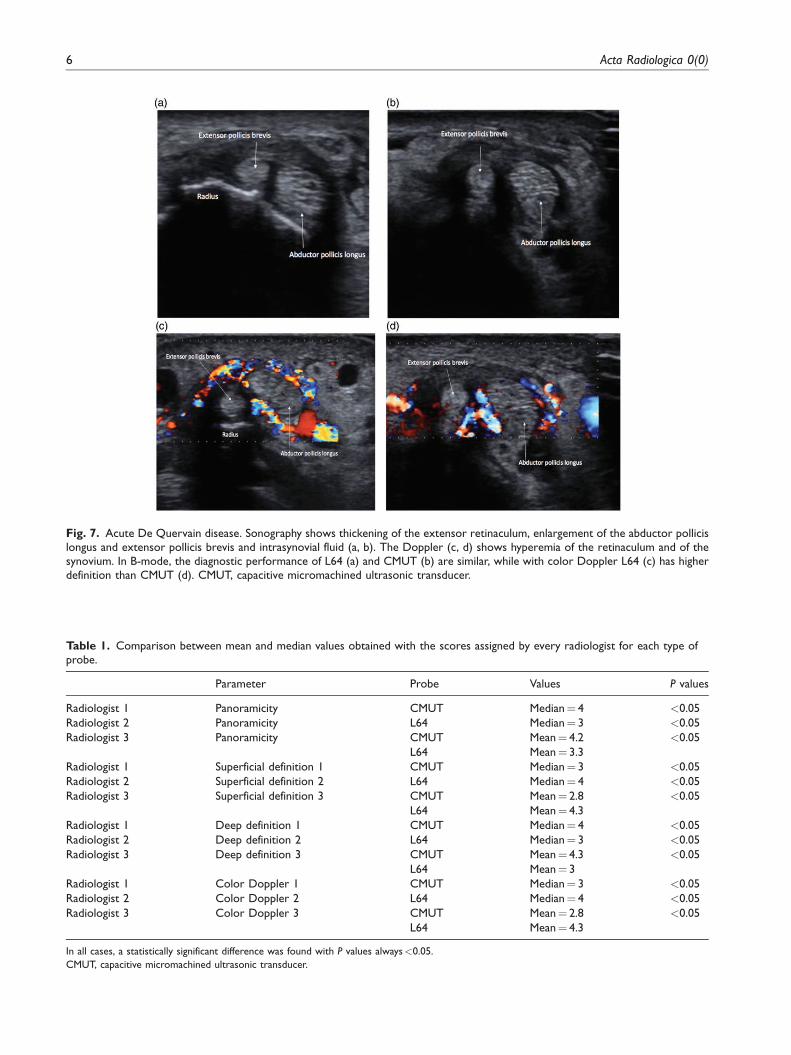
Fig. 7. Acute De Quervain disease. Sonography shows thickening of the extensor retinaculum, enlargement of the abductor pollicislongus and extensor pollicis brevis and intrasynovial fluid (a, b). The Doppler (c, d) shows hyperemia of the retinaculum and of thesynovium. In B-mode, the diagnostic performance of L64 (a) and CMUT (b) are similar, while with color Doppler L64 (c) has higherdefinition than CMUT (d). CMUT, capacitive micromachined ultrasonic transducer.
Table 1. Comparison between mean and median values obtained with the scores assigned by every radiologist for each type ofprobe.
Parameter Probe Values P values
Radiologist 1 Panoramicity CMUT Median¼ 4 <0.05
Radiologist 2 Panoramicity L64 Median¼ 3 <0.05
Radiologist 3 Panoramicity CMUT Mean¼ 4.2 <0.05
L64 Mean¼ 3.3
Radiologist 1 Superficial definition 1 CMUT Median¼ 3 <0.05
Radiologist 2 Superficial definition 2 L64 Median¼ 4 <0.05
Radiologist 3 Superficial definition 3 CMUT Mean¼ 2.8 <0.05
L64 Mean¼ 4.3
Radiologist 1 Deep definition 1 CMUT Median¼ 4 <0.05
Radiologist 2 Deep definition 2 L64 Median¼ 3 <0.05
Radiologist 3 Deep definition 3 CMUT Mean¼ 4.3 <0.05
L64 Mean¼ 3
Radiologist 1 Color Doppler 1 CMUT Median¼ 3 <0.05
Radiologist 2 Color Doppler 2 L64 Median¼ 4 <0.05
Radiologist 3 Color Doppler 3 CMUT Mean¼ 2.8 <0.05
L64 Mean¼ 4.3
In all cases, a statistically significant difference was found with P values always<0.05.
CMUT, capacitive micromachined ultrasonic transducer.
6 Acta Radiologica 0(0)

The present study has some limitations: it is a single-center study and refers to a short period of time.Nevertheless, the main limitation is probably theabsence of a suitable control group. The best optionwould be having a group without pathology, but thenumber of asymptomatic patients in the study periodwas very limited. To overcome this bias, we are work-ing to create a prospective study protocol whichincludes non-pathological patients. These data arenonetheless in an extremely early phase.
In conclusion, the future of ultrasound transducerdevelopment is one of continuing improvement andevolution, with CMUT being a breakthrough in UStechnology. It enables the application of a singleprobe that can be used for different US examinations.The improvement in CMUT will provide even moredynamic and flexible imaging capabilities by a trans-ducer, with a wider bandwidth. We have used 4G-CMUT in the study of musculoskeletal, skin, and sub-cutaneous diseases. In general, we have appreciated theability to provide an excellent panoramicity of theimages and definition of the deep structures. Animprovement in the resolution of superficial tissues,and particularly of the Doppler signal, must beachieved to allow this technology to be a valid replace-ment for lead-based piezoelectric linear probes. Wehope that significant improvements can be obtainedin the very near future to overcome this limitation,increasing the role that sonography plays in medicaldiagnosis and patient management.
Declaration of conflicting interests
The author(s) declared no potential conflicts of interest with
respect to the research, authorship, and/or publication of this
article.
Funding
The author(s) received no financial support for the research,
authorship, and/or publication of this article.
ORCID iDs
Pascal Lomoro https://orcid.org/0000-0002-3614-4706Chandra Bortolotto https://orcid.org/0000-0002-9193-9309
References
1. Carovac A, Smajlovic F, Junuzovic D. Application of ultra-sound in medicine. Acta Inform Med 2011;19:168–171.
2. Williams D. The physics of ultrasound. Anaesth IntensiveCare Med 2012;13:264–268.
3. Yildiz F, Matsunaga T, Haga Y. Fabrication and packag-ing of CMUT using low temperature co-fired ceramic.
Micromachines (Basel) 2018;9:553.4. Vallet M, Varray F, Boutet J, et al. Quantitative compari-
son of PZT and CMUT probes for photoacoustic imaging:Experimental validation. Photoacoustics 2017;8:48–58.
5. Uppal T. Tissue harmonic imaging. Australas JUltrasound Med 2010;13:29–31.
6. Oglat AA, Matjafri MZ, Suardi N, et al. A review of med-ical doppler ultrasonography of blood flow in general andespecially in common carotid artery. J Med Ultrasound2018;26:3–13.
Draghi et al. 7