Embed Size (px)
Citation preview
Comparing Galactan Biosynthesis in Mycobacteriumtuberculosis and Corynebacterium diphtheriae*
Received for publication, September 18, 2016, and in revised form, December 28, 2016 Published, JBC Papers in Press, December 30, 2016, DOI 10.1074/jbc.M116.759340
Darryl A. Wesener‡1,2, Matthew R. Levengood§1,3, and X Laura L. Kiessling‡§4
From the ‡Department of Biochemistry and §Department of Chemistry, University of Wisconsin-Madison,Madison, Wisconsin 53706
Edited by Gerald W. Hart
The suborder Corynebacterineae encompasses species likeCorynebacterium glutamicum, which has been harnessed forindustrial production of amino acids, as well as Corynebacte-rium diphtheriae and Mycobacterium tuberculosis, which causedevastating human diseases. A distinctive component of theCorynebacterineae cell envelope is the mycolyl-arabinogalactan(mAG) complex. The mAG is composed of lipid mycolic acids,and arabinofuranose (Araf) and galactofuranose (Galf) carbohy-drate residues. Elucidating microbe-specific differences in mAGcomposition could advance biotechnological applications andlead to new antimicrobial targets. To this end, we compare andcontrast galactan biosynthesis in C. diphtheriae and M. tuber-culosis. In each species, the galactan is constructed from uridine5�-diphosphate-�-D-galactofuranose (UDP-Galf), which is gen-erated by the enzyme UDP-galactopyranose mutase (UGM orGlf). UGM and the galactan are essential in M. tuberculosis, buttheir importance in Corynebacterium species was not known.We show that small molecule inhibitors of UGM impede C. glu-tamicum growth, suggesting that the galactan is critical incorynebacteria. Previous cell wall analysis data suggest thegalactan polymer is longer in mycobacterial species than coryne-bacterial species. To explore the source of galactan length varia-tion, a C. diphtheriae ortholog of the M. tuberculosis carbohydratepolymerase responsible for the bulk of galactan polymerization,GlfT2, was produced, and its catalytic activity was evaluated. TheC. diphtheriae GlfT2 gave rise to shorter polysaccharides thanthose obtained with the M. tuberculosis GlfT2. These data suggestthat GlfT2 alone can influence galactan length. Our results providetools, both small molecule and genetic, for probing and perturb-ing the assembly of the Corynebacterineae cell envelope.
Mycobacterium tuberculosis and Corynebacterium diphthe-riae, the etiological agents of tuberculosis and diphtheria,respectively, are notorious members of the bacterial suborderCorynebacterineae. This taxon includes other pathogenic bac-terial species, including the causative agents of leprosy, nocar-diosis, and buruli ulcers. One notable feature of Corynebacte-rineae is the cell envelope, which has a unique composition.The Corynebacterineae cell envelope contains mycolic acidsappended to branched polymers of Araf (arabinan),5 which arelinked to peptidoglycan through a linear polymer of Galf (galac-tan) (1, 2). The macromolecular structure that extends beyondthe peptidoglycan is referred to as the mAG complex. Neitherthe mAG nor its individual components are present in hostmammals. Moreover, in M. tuberculosis the mAG complexserves as a barrier to antitubercular drugs and can modulate thehuman immune response in favor of bacterial immune evasion(3, 4). A complete understanding of mAG assembly and varia-tion could yield novel strategies for therapeutic intervention.
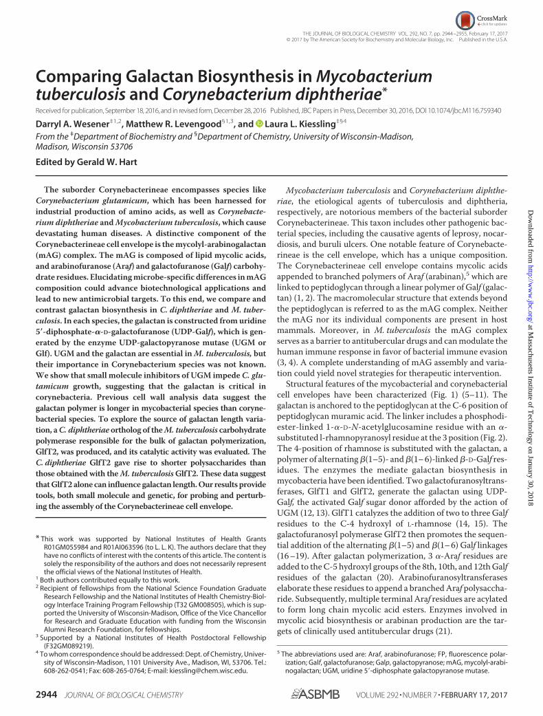
Structural features of the mycobacterial and corynebacterialcell envelopes have been characterized (Fig. 1) (5–11). Thegalactan is anchored to the peptidoglycan at the C-6 position ofpeptidoglycan muramic acid. The linker includes a phosphodi-ester-linked 1-�-D-N-acetylglucosamine residue with an �-substituted l-rhamnopyranosyl residue at the 3 position (Fig. 2).The 4-position of rhamnose is substituted with the galactan, apolymer of alternating �(1–5)- and �(1– 6)-linked �-D-Galf res-idues. The enzymes the mediate galactan biosynthesis inmycobacteria have been identified. Two galactofuranosyltrans-ferases, GlfT1 and GlfT2, generate the galactan using UDP-Galf, the activated Galf sugar donor afforded by the action ofUGM (12, 13). GlfT1 catalyzes the addition of two to three Galfresidues to the C-4 hydroxyl of L-rhamnose (14, 15). Thegalactofuranosyl polymerase GlfT2 then promotes the sequen-tial addition of the alternating �(1–5) and �(1– 6) Galf linkages(16 –19). After galactan polymerization, 3 �-Araf residues areadded to the C-5 hydroxyl groups of the 8th, 10th, and 12th Galfresidues of the galactan (20). Arabinofuranosyltransferaseselaborate these residues to append a branched Araf polysaccha-ride. Subsequently, multiple terminal Araf residues are acylatedto form long chain mycolic acid esters. Enzymes involved inmycolic acid biosynthesis or arabinan production are the tar-gets of clinically used antitubercular drugs (21).
* This work was supported by National Institutes of Health GrantsR01GM055984 and R01AI063596 (to L. L. K). The authors declare that theyhave no conflicts of interest with the contents of this article. The content issolely the responsibility of the authors and does not necessarily representthe official views of the National Institutes of Health.
1 Both authors contributed equally to this work.2 Recipient of fellowships from the National Science Foundation Graduate
Research Fellowship and the National Institutes of Health Chemistry-Biol-ogy Interface Training Program Fellowship (T32 GM008505), which is sup-ported the University of Wisconsin-Madison, Office of the Vice Chancellorfor Research and Graduate Education with funding from the WisconsinAlumni Research Foundation, for fellowships.
3 Supported by a National Institutes of Health Postdoctoral Fellowship(F32GM089219).
4 To whom correspondence should be addressed: Dept. of Chemistry, Univer-sity of Wisconsin-Madison, 1101 University Ave., Madison, WI, 53706. Tel.:608-262-0541; Fax: 608-265-0764; E-mail: [email protected].
5 The abbreviations used are: Araf, arabinofuranose; FP, fluorescence polar-ization; Galf, galactofuranose; Galp, galactopyranose; mAG, mycolyl-arabi-nogalactan; UGM, uridine 5�-diphosphate galactopyranose mutase.
crossmarkTHE JOURNAL OF BIOLOGICAL CHEMISTRY VOL. 292, NO. 7, pp. 2944 –2955, February 17, 2017
© 2017 by The American Society for Biochemistry and Molecular Biology, Inc. Published in the U.S.A.
2944 JOURNAL OF BIOLOGICAL CHEMISTRY VOLUME 292 • NUMBER 7 • FEBRUARY 17, 2017
at Massachusetts Institute of T
echnology on January 30, 2018http://w
ww
.jbc.org/D
ownloaded from
Although mAG biosynthesis and structure is often studied inM. tuberculosis, Corynebacterium species have recently beenused as models to understand mAG assembly. Their advantagesinclude a decreased doubling time, reduced biosafety designa-tion, and the availability of tools for genetic manipulation (22–25). Although most mAG biosynthetic genes are essential inM. tuberculosis (26), their deletion in C. glutamicum oftenyields slow growing but viable mutants. For example, C. glu-tamicum mutants lacking AftA were recently used to deter-
mine that this arabinofuranoysltransferase appends three�(1–5)–Araf residues to the galactan to initiate arabinan bio-synthesis (27). Another C. glutamicum mutant revealed Pks13catalyzes the final step of mycolic acid biosynthesis in coryne-bacteria and mycobacteria (24). Unexpectedly, a C. glutamicummutant lacking the first enzyme required for activated Arafdonor sugar biosynthesis, ubiA, is viable (20). This strain isdevoid of Araf, and that it could be isolated and cultured wassurprising. These examples highlight how experiments using
FIGURE 1. Comparative models of the structure of the mAG complex. Schematic comparison of the mAG complex from M. tuberculosis and C. diphtheriae cellwalls.
FIGURE 2. Proposed enzymatic reactions in galactan biosynthesis in C. diphtheriae.
Corynebacterial Galactan Biosynthesis
FEBRUARY 17, 2017 • VOLUME 292 • NUMBER 7 JOURNAL OF BIOLOGICAL CHEMISTRY 2945
at Massachusetts Institute of T
echnology on January 30, 2018http://w
ww
.jbc.org/D
ownloaded from

Corynebacterium species can provide important insight intocell envelope biosynthesis within the Corynebacterineaesuborder.
Although each Corynebacterineae species possesses an mAGof similar constitution, fine structural features of the mAG canvary. The arabinan of C. diphtheriae lacks the 1,3,5-linked Arafresidues that are responsible for branching, suggesting this ara-binan is less complex than that of other Corynebacterineae (7).Accordingly, arabinan assembly in mycobacteria is mediated bya larger collection of enzymes. Six or more arabinofuranosyl-transferases are involved in mycobacteria, and at least one ofthese enzymes is inhibited by the first-line antitubercular drugethambutol (28 –30). Of the six mycobacterial arabinofurano-syltransferases, three belong to the Emb family of enzymes. Incontrast, corynebacteria encode a single Emb homolog that ismost closely related to M. tuberculosis EmbC (20). Thus, thearabinan of C. diphtheriae is generated using fewer enzymesand is simpler than that of M. tuberculosis. The mycolic acidsfrom Mycobacterium and Corynebacterium species also vary.Mycobacterial mycolic acids possess chains of 70 –90 carbonatoms, whereas the mycolic acids of C. diphtheriae are shorter,with a chain of 30 –36 carbons (7, 31). Additionally, M. tuber-culosis mycolic acids commonly contain functionalities such ascis-cyclopropane that are absent from fast-growing Mycobacte-rium smegmatis or corynebacterial mycolic acids. There alsoare differences in the galactan. Analyses of the galactan length(20) indicate the corynebacterial galactan is shorter than that ofM. tuberculosis (5, 7, 27, 32). Understanding the source of thesedifferences can lend insight into the molecular basis for the cellenvelope properties of specific species.
The M. tuberculosis glycosyltransferase GlfT2 (EC 2.4.1.288)is a bifunctional carbohydrate polymerase that generates thebulk of the galactan (17–19, 33). Studies with chain-terminat-ing glycosyl donors indicate that GlfT2 is sequence-selective,and its fidelity for forming a sequence of alternating �(1–5) and�(1– 6) linkages is high (34, 35). Within the GlfT2 polypeptideis but a single active site (33, 36); site-directed mutagenesisindicates substitution of key amino acids abrogates the forma-tion of both �(1–5) and �(1– 6) linkages (36). Experiments withisotope-labeled acceptors reveal that GlfT2 is processive (37),and its propensity for generating alternating �(1–5) or �(1– 6)Galf linkages is a consequence of processive elongation (38).The enzyme not only controls polymer sequence but also poly-mer length. In a test tube GlfT2 can generate polymers of asimilar length to those obtained from cells (19). With syntheticacceptors, the identity of the anomeric lipid was a critical deter-minant of product polysaccharide length (17–19). Acceptorswith short alkyl anomeric substituents afforded only short olig-omeric saccharides, whereas those with longer lipids on theacceptor afforded polysaccharide products similar to those incells. These findings led to the proposal that M. tuberculosisGlfT2 uses the acceptor lipid as a tether and that polymerlength is controlled by bivalent substrate binding (19). TheDXD motif mediates glycosyltransferase coordination to a diva-lent cation, and M. tuberculosis GlfT2 variants in this DXDmotif afforded truncated oligosaccharide products (36, 39).Thus, GlfT2 is bifunctional with the ability to control the lengthof polymerized M. tuberculosis galactan. Whether these attri-
butes of the M. tuberculosis GlfT2 are preserved in otherCorynebacterineae species is unclear.
To compare and contrast galactan biosynthesis in differentspecies, we examined the galactan biosynthetic enzymes fromC. glutamicum and C. diphtheriae NCTC 13129, includingGlfT2 and UGM. The putative C. diphtheriae UGM (DIP2203)catalyzes the isomerization of UDP-Galp and UDP-Galf with anefficiency similar to other prokaryotic orthologs. This findingsuggests that differences in galactan length are unlikely to arisefrom differences in cellular concentration of UDP-Galf. Toexplore the role of the galactan in cells, we employed smallmolecule UGM inhibitors. These compounds inhibit C. diph-theriae UGM in vitro and prevent the growth of C. glutamicum.These observations support an essential role for the galactan incorynebacteria as well as mycobacteria and suggest that thesmall molecule UGM inhibitors can be used to probe the cellu-lar roles of Galf. With regard to GlfT2, our results indicate thatthe DIP2198 gene product is a bifunctional galactofuranosyl-transferase similar to the M. tuberculosis GlfT2. The coryne-bacterial glycosyltransferase can elongate synthetic acceptorsto afford polysaccharides commensurate in length to those iso-lated from corynebacteria. As with the M. tuberculosis GlfT2,the product polysaccharides attained from variants with aminoacid changes in the proposed donor binding site are truncated.These findings indicate that GlfT2 orthologs have an intrinsicability to control polysaccharide length. This length controlmechanism differs from that employed in O-antigen biosynthe-sis in which length is controlled by an auxiliary protein (40 –42). Our investigations lay a foundation for dissecting themolecular details and mechanisms of galactan biosynthesisusing genetic and chemical genetic tools.
Results
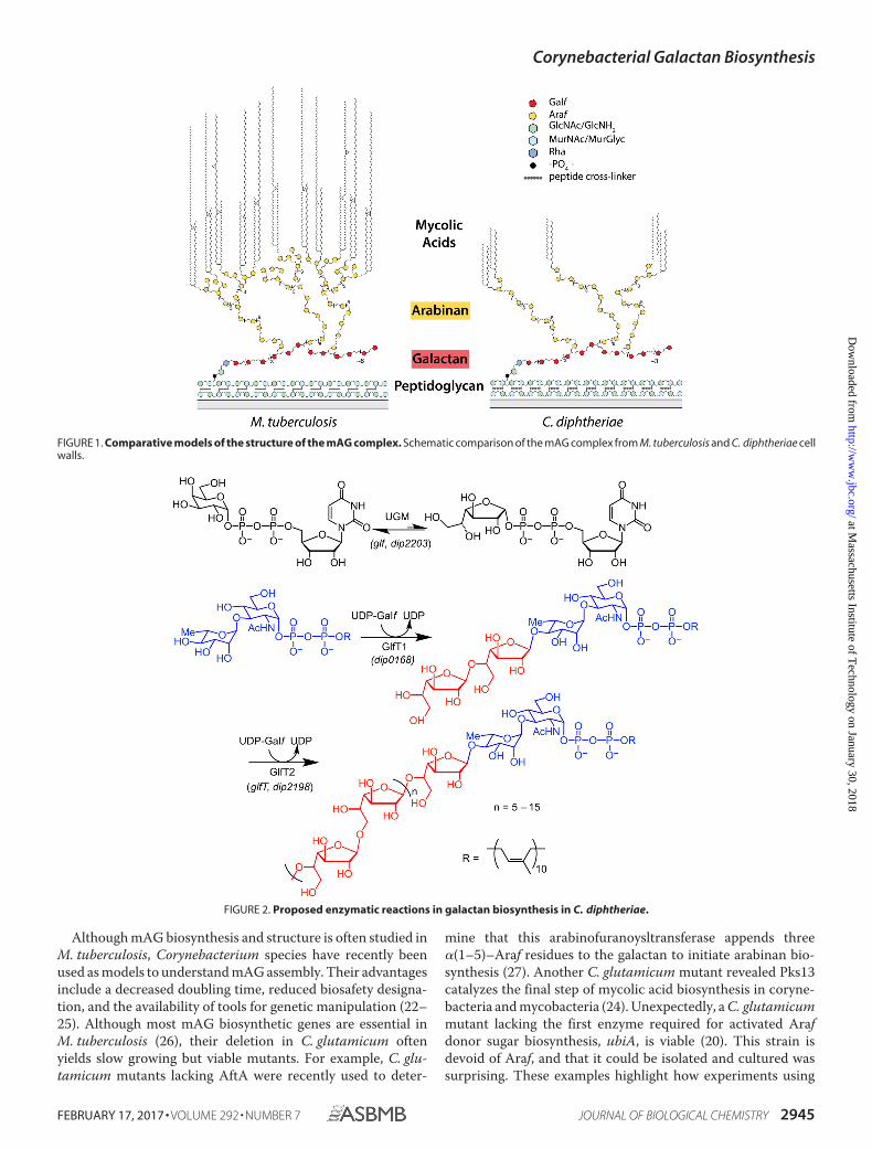
Inhibitors of Galactan Biosynthesis Block CorynebacterialGrowth—Unlike mycobacteria, corynebacteria do not requiremycolic acids (24) or arabinan (20). The survival of corynebac-teria without integral mAG components raises the issue ofwhether these species require the galactan. Small moleculeUGM inhibitors provide the means to address this issue (Fig.2) (43). We previously produced the C. diphtheriae UGM(DIP2203) (44). In this study we characterized the enzyme’scatalytic properties and then identified a C. diphtheriae UGMinhibitor that could be used to probe the role of the galactan.The initial velocities of UDP-Galp production were calculatedfrom reaction mixtures containing a range of UDP-Galf con-centrations (45, 46). Steady-state kinetic parameters weredetermined from the Michaelis-Menten equation, whichafforded a Km of 47 � 5 �M and a kcat value of 20 � 0.6 s�1
(Table 1). The enzymatic efficiency, or kcat/Km, for C. diphthe-riae UGM is 4.3 � 0.5 � 105 M�1s�1. These kinetic constantsare similar to those obtained with UGM orthologs from otherprokaryotes, including those from Klebsiella pneumoniae andEscherichia coli (46, 47).
Our previous experience suggested that inhibitors of M.tuberculosis UGM should act against C. diphtheriae UGM (44).To assess compound affinity for UGM, we validated that a pre-viously described fluorescence polarization (FP) assay (48)could be applied to the C. diphtheriae UGM. The enzyme
Corynebacterial Galactan Biosynthesis
2946 JOURNAL OF BIOLOGICAL CHEMISTRY VOLUME 292 • NUMBER 7 • FEBRUARY 17, 2017
at Massachusetts Institute of T
echnology on January 30, 2018http://w
ww
.jbc.org/D
ownloaded from
bound the fluorescein-bearing UDP derivative with an appar-ent affinity of 24 � 2 nM. Small molecules could then be testedfor their ability to displace the fluorescent probe from theenzyme in a competition assay (49). Only compounds 1 and 2were effective. Their calculated dissociation constants weresimilar, 8 � 1 �M for 1 and 10 � 2 �M for 2. We next evaluatedthe UGM ligands as inhibitors. As expected from the FP assayresults, UDP and compound 3 did not substantially inhibitC. diphtheriae UGM activity at 15 �M, but compounds 1 and 2impeded conversion of UDP-Galf to UDP-Galp by 83 � 2% and69 � 3%, respectively (Fig. 3B). At 50 �M UDP-Galf, a concen-tration near the Km value we report here, the IC50 value ofcompound 1 was determined to be 5 � 1 �M.
We tested whether inhibitor 1 could be used ascertain theconsequences of inhibiting galactan biosynthesis. C. glutami-cum (ATCC 13032) is a non-virulent genetically tractablemodel organism that had been employed to conclude thatcorynebacteria can survive without mycolic acids or arabinan(20, 24). We anticipated the UGM inhibitor would be effectivebecause the sequence similarity of the C. diphtheriae andC. glutamicum UGM is 85%. Indeed, exposure of C. glutami-cum to compound 1 led to growth inhibition (Fig. 3C). Theresults of this chemical genetics experiment suggest that thegalactan is the outermost polysaccharide required for coryne-bacterial growth.
Galactan Polymerization by C. diphtheriae—The C. diphthe-riae GlfT2 (DIP2198 gene) and M. tuberculosis GlfT2 share 57%amino acid identity, but cell wall analysis indicates that thelength of the galactan in corynebacteria is shorter than that inM. tuberculosis. This difference prompted us to compare thecatalytic properties of the GlfT2 orthologs. If GlfT2 can controlgalactan length, the C. diphtheriae enzyme should give rise toshorter polymers than those afforded by M. tuberculosis GlfT2.We produced a His-tagged C. diphtheriae GlfT2 and moni-tored its carbohydrate polymerase activity using matrix-as-sisted laser desorption/ionization (MALDI) mass spectrometry(19, 37). The reaction conditions employed with acceptor 4a(Fig. 4) were similar to those used to assay M. tuberculosisGlfT2 activity. Polysaccharides were attained demonstratingthat the DIP2198 gene product can generate polymers ofgalactofuranose (Fig. 4B, left). On the basis of previous studieswith M. tuberculosis GlfT2 (17, 18, 33, 36), we anticipated thatthe C. diphtheriae GlfT2 product polysaccharides would becomposed of alternating �(1– 6)– and �(1–5)–Galf linkages.We, therefore, also examined elongation from disaccharideacceptor, compound 5, which has a �(1–5)–Galf linkage and,therefore, is a regioisomer of acceptor 4a (36). The C. diphthe-
riae GlfT2 also elongated compound 5 to afford polymers ofsimilar length to those attained with 4a (Fig. 4B, right). Theability of both synthetic acceptors to function as substrates ofC. diphtheriae GlfT2 suggests the enzyme catalyzes formationof both �(1– 6)– and �(1–5)–Galf glycosidic linkages.
Both the M. tuberculosis and C. diphtheriae GlfT2 enzymesgenerate Galf polymers, but mass spectrometry analysis sug-gested that the polymeric products from the C. diphtheriaeortholog are substantially shorter than those from M. tubercu-losis GlfT2 (Fig. 4C). We, therefore, examined the rate of poly-merization by C. diphtheriae GlfT2. We employed compound4a in a coupled continuous enzyme assay that monitors therelease of UDP (50). The data indicate that C. diphtheriae GlfT2is a slower polymerase than is M. tuberculosis GlfT2. Under thesame conditions (1.25 mM UDP-Galf and 0.2 mM 4a), theC. diphtheriae GlfT2 was �17-fold slower than the M. tubercu-losis GlfT2 (0.47 � 0.24 �M/min for C. diphtheriae GlfT2 versus8.2 �M/min for M. tuberculosis GlfT2). The slower rate of theC. diphtheriae enzyme was not a function of decreased proteinstability as the C. diphtheriae GlfT2 was less prone to aggrega-tion and precipitation than M. tuberculosis GlfT2.
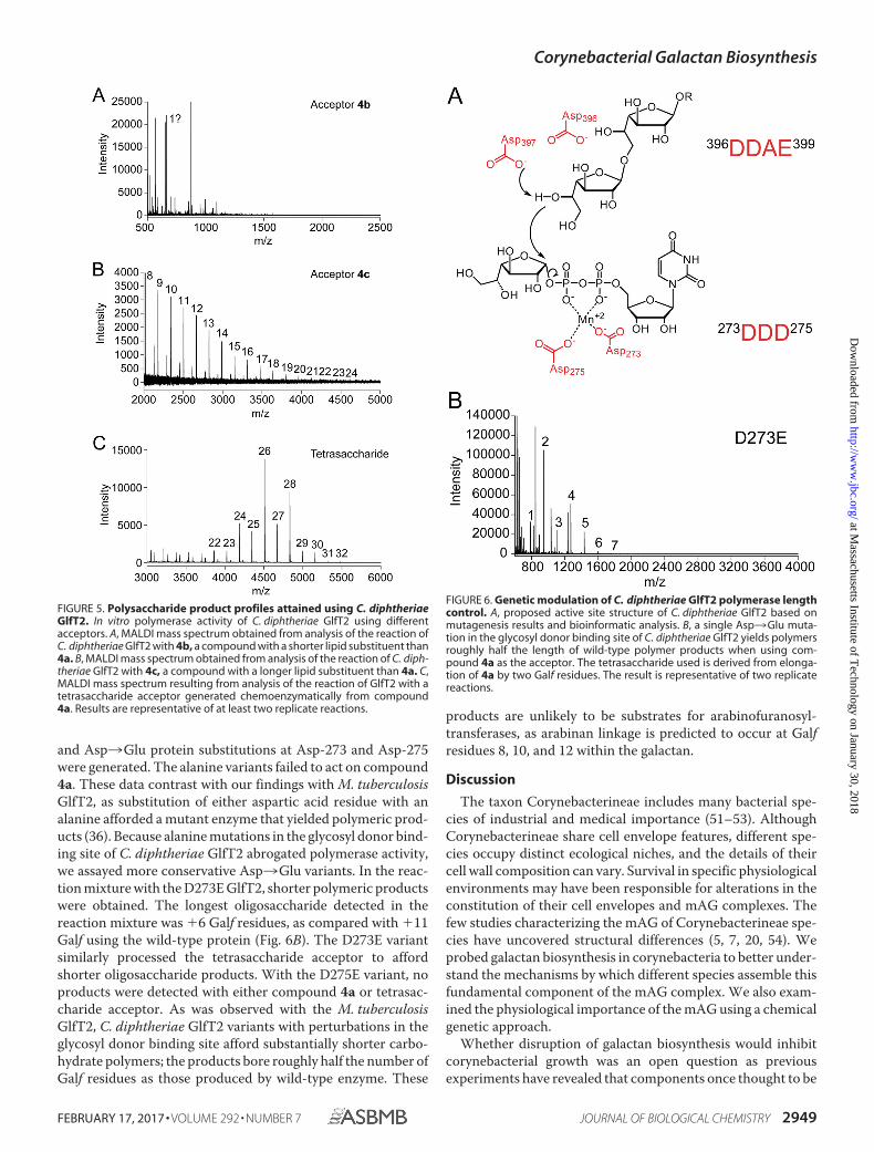
Anomeric Lipid Influences Polysaccharide Length—We foundpreviously that the products of the M. tuberculosis GlfT2depend on the attributes of the anomeric lipid of the acceptor.With longer lipid substituents, longer polysaccharide productswere obtained (19). We tested whether the C. diphtheriaeortholog affords similar results. We used compounds 4b and 4cas substrates; the former has a shorter lipid group than ourbenchmark substrate 4a, and the latter has a longer lipid sub-stituent. When 4b was incubated with C. diphtheriae GlfT2, no
TABLE 1Michaelis-Menten steady state kinetic parameters of prokaryotic UGMproteinsAll activities were measured using UDP-Galf as the substrate in the presence ofsodium dithionite
Species Km kcat kcat/m
�M s�1 105 M�1s�1
C. diphtheriae 47 � 5 20 � 0.6 4.3 � 0.5K. pneumoniaea 43 � 6 5.5 � 0.7 1.3 � 0.2E. colib 27 22 8.1
a From Ref. 47.b From Ref. 46.
FIGURE 3. Chemical inhibition of C. glutamicum growth. A, chemical struc-ture of 2-aminothiazole small molecule UGM inhibitors employed in thisstudy. B, in vitro inhibition of C. diphtheriae UGM activity by the small mole-cules (15 �M) shown in A. Compound 3 has the same chemotype as com-pounds 1 and 2 yet fails to inhibit C. diphtheriae UGM. Data are shown as themean � S.D. (n � 3 technical replicates). C, inhibition of C. glutamicum growthby UGM inhibitor 1. The antibiotic kanamycin was used as a known inhibitorof cell growth. DMSO treatment was used as a vehicle control, and samplestreated with compound 3 were tested as a chemotype control. The concen-tration of a 10-�l solution of 1 added to each well is included below theimage. The result is representative of three biological replicates.
Corynebacterial Galactan Biosynthesis
FEBRUARY 17, 2017 • VOLUME 292 • NUMBER 7 JOURNAL OF BIOLOGICAL CHEMISTRY 2947
at Massachusetts Institute of T
echnology on January 30, 2018http://w
ww
.jbc.org/D
ownloaded from
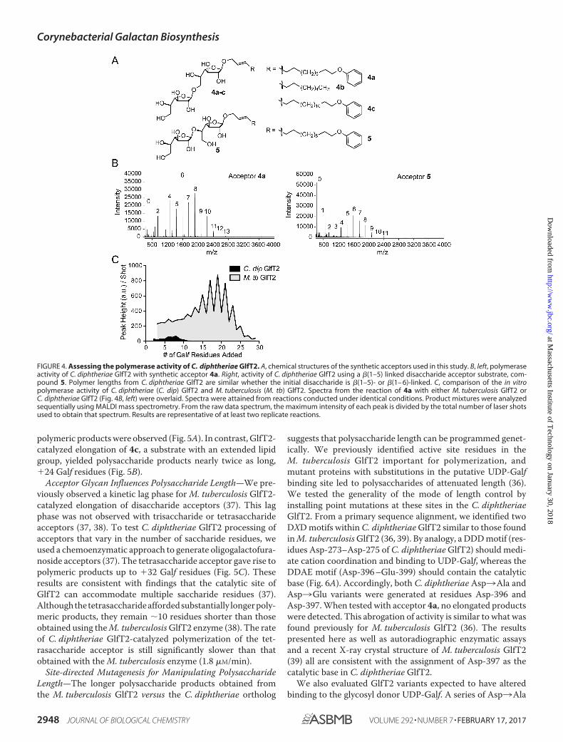
polymeric products were observed (Fig. 5A). In contrast, GlfT2-catalyzed elongation of 4c, a substrate with an extended lipidgroup, yielded polysaccharide products nearly twice as long,�24 Galf residues (Fig. 5B).
Acceptor Glycan Influences Polysaccharide Length—We pre-viously observed a kinetic lag phase for M. tuberculosis GlfT2-catalyzed elongation of disaccharide acceptors (37). This lagphase was not observed with trisaccharide or tetrasaccharideacceptors (37, 38). To test C. diphtheriae GlfT2 processing ofacceptors that vary in the number of saccharide residues, weused a chemoenzymatic approach to generate oligogalactofura-noside acceptors (37). The tetrasaccharide acceptor gave rise topolymeric products up to �32 Galf residues (Fig. 5C). Theseresults are consistent with findings that the catalytic site ofGlfT2 can accommodate multiple saccharide residues (37).Althoughthetetrasaccharideaffordedsubstantially longerpoly-meric products, they remain �10 residues shorter than thoseobtained using the M. tuberculosis GlfT2 enzyme (38). The rateof C. diphtheriae GlfT2-catalyzed polymerization of the tet-rasaccharide acceptor is still significantly slower than thatobtained with the M. tuberculosis enzyme (1.8 �M/min).
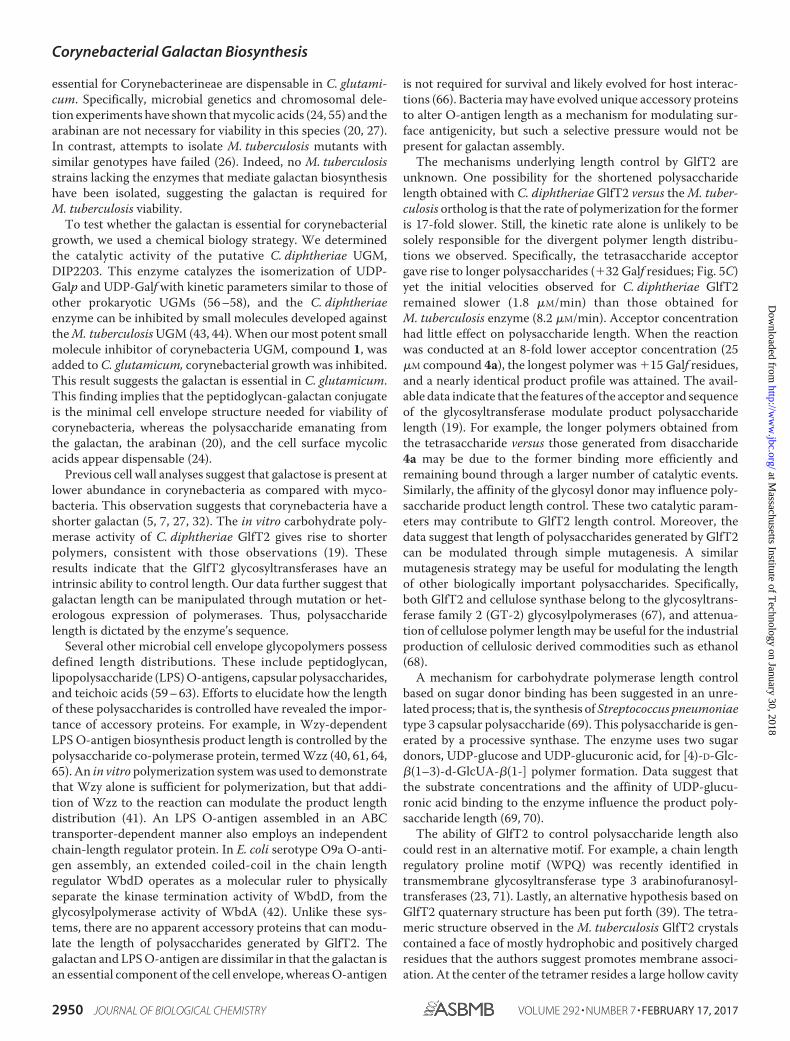
Site-directed Mutagenesis for Manipulating PolysaccharideLength—The longer polysaccharide products obtained fromthe M. tuberculosis GlfT2 versus the C. diphtheriae ortholog
suggests that polysaccharide length can be programmed genet-ically. We previously identified active site residues in theM. tuberculosis GlfT2 important for polymerization, andmutant proteins with substitutions in the putative UDP-Galfbinding site led to polysaccharides of attenuated length (36).We tested the generality of the mode of length control byinstalling point mutations at these sites in the C. diphtheriaeGlfT2. From a primary sequence alignment, we identified twoDXD motifs within C. diphtheriae GlfT2 similar to those foundin M. tuberculosis GlfT2 (36, 39). By analogy, a DDD motif (res-idues Asp-273–Asp-275 of C. diphtheriae GlfT2) should medi-ate cation coordination and binding to UDP-Galf, whereas theDDAE motif (Asp-396 –Glu-399) should contain the catalyticbase (Fig. 6A). Accordingly, both C. diphtheriae Asp3Ala andAsp3Glu variants were generated at residues Asp-396 andAsp-397. When tested with acceptor 4a, no elongated productswere detected. This abrogation of activity is similar to what wasfound previously for M. tuberculosis GlfT2 (36). The resultspresented here as well as autoradiographic enzymatic assaysand a recent X-ray crystal structure of M. tuberculosis GlfT2(39) all are consistent with the assignment of Asp-397 as thecatalytic base in C. diphtheriae GlfT2.
We also evaluated GlfT2 variants expected to have alteredbinding to the glycosyl donor UDP-Galf. A series of Asp3Ala
FIGURE 4. Assessing the polymerase activity of C. diphtheriae GlfT2. A, chemical structures of the synthetic acceptors used in this study. B, left, polymeraseactivity of C. diphtheriae GlfT2 with synthetic acceptor 4a. Right, activity of C. diphtheriae GlfT2 using a �(1–5) linked disaccharide acceptor substrate, com-pound 5. Polymer lengths from C. diphtheriae GlfT2 are similar whether the initial disaccharide is �(1–5)- or �(1– 6)-linked. C, comparison of the in vitropolymerase activity of C. diphtheriae (C. dip) GlfT2 and M. tuberculosis (M. tb) GlfT2. Spectra from the reaction of 4a with either M. tuberculosis GlfT2 orC. diphtheriae GlfT2 (Fig. 4B, left) were overlaid. Spectra were attained from reactions conducted under identical conditions. Product mixtures were analyzedsequentially using MALDI mass spectrometry. From the raw data spectrum, the maximum intensity of each peak is divided by the total number of laser shotsused to obtain that spectrum. Results are representative of at least two replicate reactions.
Corynebacterial Galactan Biosynthesis
2948 JOURNAL OF BIOLOGICAL CHEMISTRY VOLUME 292 • NUMBER 7 • FEBRUARY 17, 2017
at Massachusetts Institute of T
echnology on January 30, 2018http://w
ww
.jbc.org/D
ownloaded from
and Asp3Glu protein substitutions at Asp-273 and Asp-275were generated. The alanine variants failed to act on compound4a. These data contrast with our findings with M. tuberculosisGlfT2, as substitution of either aspartic acid residue with analanine afforded a mutant enzyme that yielded polymeric prod-ucts (36). Because alanine mutations in the glycosyl donor bind-ing site of C. diphtheriae GlfT2 abrogated polymerase activity,we assayed more conservative Asp3Glu variants. In the reac-tion mixture with the D273E GlfT2, shorter polymeric productswere obtained. The longest oligosaccharide detected in thereaction mixture was �6 Galf residues, as compared with �11Galf using the wild-type protein (Fig. 6B). The D273E variantsimilarly processed the tetrasaccharide acceptor to affordshorter oligosaccharide products. With the D275E variant, noproducts were detected with either compound 4a or tetrasac-charide acceptor. As was observed with the M. tuberculosisGlfT2, C. diphtheriae GlfT2 variants with perturbations in theglycosyl donor binding site afford substantially shorter carbo-hydrate polymers; the products bore roughly half the number ofGalf residues as those produced by wild-type enzyme. These
products are unlikely to be substrates for arabinofuranosyl-transferases, as arabinan linkage is predicted to occur at Galfresidues 8, 10, and 12 within the galactan.
Discussion
The taxon Corynebacterineae includes many bacterial spe-cies of industrial and medical importance (51–53). AlthoughCorynebacterineae share cell envelope features, different spe-cies occupy distinct ecological niches, and the details of theircell wall composition can vary. Survival in specific physiologicalenvironments may have been responsible for alterations in theconstitution of their cell envelopes and mAG complexes. Thefew studies characterizing the mAG of Corynebacterineae spe-cies have uncovered structural differences (5, 7, 20, 54). Weprobed galactan biosynthesis in corynebacteria to better under-stand the mechanisms by which different species assemble thisfundamental component of the mAG complex. We also exam-ined the physiological importance of the mAG using a chemicalgenetic approach.
Whether disruption of galactan biosynthesis would inhibitcorynebacterial growth was an open question as previousexperiments have revealed that components once thought to be
FIGURE 5. Polysaccharide product profiles attained using C. diphtheriaeGlfT2. In vitro polymerase activity of C. diphtheriae GlfT2 using differentacceptors. A, MALDI mass spectrum obtained from analysis of the reaction ofC. diphtheriae GlfT2 with 4b, a compound with a shorter lipid substituent than4a. B, MALDI mass spectrum obtained from analysis of the reaction of C. diph-theriae GlfT2 with 4c, a compound with a longer lipid substituent than 4a. C,MALDI mass spectrum resulting from analysis of the reaction of GlfT2 with atetrasaccharide acceptor generated chemoenzymatically from compound4a. Results are representative of at least two replicate reactions.
FIGURE 6. Genetic modulation of C. diphtheriae GlfT2 polymerase lengthcontrol. A, proposed active site structure of C. diphtheriae GlfT2 based onmutagenesis results and bioinformatic analysis. B, a single Asp3Glu muta-tion in the glycosyl donor binding site of C. diphtheriae GlfT2 yields polymersroughly half the length of wild-type polymer products when using com-pound 4a as the acceptor. The tetrasaccharide used is derived from elonga-tion of 4a by two Galf residues. The result is representative of two replicatereactions.
Corynebacterial Galactan Biosynthesis
FEBRUARY 17, 2017 • VOLUME 292 • NUMBER 7 JOURNAL OF BIOLOGICAL CHEMISTRY 2949
at Massachusetts Institute of T
echnology on January 30, 2018http://w
ww
.jbc.org/D
ownloaded from
essential for Corynebacterineae are dispensable in C. glutami-cum. Specifically, microbial genetics and chromosomal dele-tion experiments have shown that mycolic acids (24, 55) and thearabinan are not necessary for viability in this species (20, 27).In contrast, attempts to isolate M. tuberculosis mutants withsimilar genotypes have failed (26). Indeed, no M. tuberculosisstrains lacking the enzymes that mediate galactan biosynthesishave been isolated, suggesting the galactan is required forM. tuberculosis viability.
To test whether the galactan is essential for corynebacterialgrowth, we used a chemical biology strategy. We determinedthe catalytic activity of the putative C. diphtheriae UGM,DIP2203. This enzyme catalyzes the isomerization of UDP-Galp and UDP-Galf with kinetic parameters similar to those ofother prokaryotic UGMs (56 –58), and the C. diphtheriaeenzyme can be inhibited by small molecules developed againstthe M. tuberculosis UGM (43, 44). When our most potent smallmolecule inhibitor of corynebacteria UGM, compound 1, wasadded to C. glutamicum, corynebacterial growth was inhibited.This result suggests the galactan is essential in C. glutamicum.This finding implies that the peptidoglycan-galactan conjugateis the minimal cell envelope structure needed for viability ofcorynebacteria, whereas the polysaccharide emanating fromthe galactan, the arabinan (20), and the cell surface mycolicacids appear dispensable (24).
Previous cell wall analyses suggest that galactose is present atlower abundance in corynebacteria as compared with myco-bacteria. This observation suggests that corynebacteria have ashorter galactan (5, 7, 27, 32). The in vitro carbohydrate poly-merase activity of C. diphtheriae GlfT2 gives rise to shorterpolymers, consistent with those observations (19). Theseresults indicate that the GlfT2 glycosyltransferases have anintrinsic ability to control length. Our data further suggest thatgalactan length can be manipulated through mutation or het-erologous expression of polymerases. Thus, polysaccharidelength is dictated by the enzyme’s sequence.
Several other microbial cell envelope glycopolymers possessdefined length distributions. These include peptidoglycan,lipopolysaccharide (LPS) O-antigens, capsular polysaccharides,and teichoic acids (59 – 63). Efforts to elucidate how the lengthof these polysaccharides is controlled have revealed the impor-tance of accessory proteins. For example, in Wzy-dependentLPS O-antigen biosynthesis product length is controlled by thepolysaccharide co-polymerase protein, termed Wzz (40, 61, 64,65). An in vitro polymerization system was used to demonstratethat Wzy alone is sufficient for polymerization, but that addi-tion of Wzz to the reaction can modulate the product lengthdistribution (41). An LPS O-antigen assembled in an ABCtransporter-dependent manner also employs an independentchain-length regulator protein. In E. coli serotype O9a O-anti-gen assembly, an extended coiled-coil in the chain lengthregulator WbdD operates as a molecular ruler to physicallyseparate the kinase termination activity of WbdD, from theglycosylpolymerase activity of WbdA (42). Unlike these sys-tems, there are no apparent accessory proteins that can modu-late the length of polysaccharides generated by GlfT2. Thegalactan and LPS O-antigen are dissimilar in that the galactan isan essential component of the cell envelope, whereas O-antigen
is not required for survival and likely evolved for host interac-tions (66). Bacteria may have evolved unique accessory proteinsto alter O-antigen length as a mechanism for modulating sur-face antigenicity, but such a selective pressure would not bepresent for galactan assembly.
The mechanisms underlying length control by GlfT2 areunknown. One possibility for the shortened polysaccharidelength obtained with C. diphtheriae GlfT2 versus the M. tuber-culosis ortholog is that the rate of polymerization for the formeris 17-fold slower. Still, the kinetic rate alone is unlikely to besolely responsible for the divergent polymer length distribu-tions we observed. Specifically, the tetrasaccharide acceptorgave rise to longer polysaccharides (�32 Galf residues; Fig. 5C)yet the initial velocities observed for C. diphtheriae GlfT2remained slower (1.8 �M/min) than those obtained forM. tuberculosis enzyme (8.2 �M/min). Acceptor concentrationhad little effect on polysaccharide length. When the reactionwas conducted at an 8-fold lower acceptor concentration (25�M compound 4a), the longest polymer was �15 Galf residues,and a nearly identical product profile was attained. The avail-able data indicate that the features of the acceptor and sequenceof the glycosyltransferase modulate product polysaccharidelength (19). For example, the longer polymers obtained fromthe tetrasaccharide versus those generated from disaccharide4a may be due to the former binding more efficiently andremaining bound through a larger number of catalytic events.Similarly, the affinity of the glycosyl donor may influence poly-saccharide product length control. These two catalytic param-eters may contribute to GlfT2 length control. Moreover, thedata suggest that length of polysaccharides generated by GlfT2can be modulated through simple mutagenesis. A similarmutagenesis strategy may be useful for modulating the lengthof other biologically important polysaccharides. Specifically,both GlfT2 and cellulose synthase belong to the glycosyltrans-ferase family 2 (GT-2) glycosylpolymerases (67), and attenua-tion of cellulose polymer length may be useful for the industrialproduction of cellulosic derived commodities such as ethanol(68).
A mechanism for carbohydrate polymerase length controlbased on sugar donor binding has been suggested in an unre-lated process; that is, the synthesis of Streptococcus pneumoniaetype 3 capsular polysaccharide (69). This polysaccharide is gen-erated by a processive synthase. The enzyme uses two sugardonors, UDP-glucose and UDP-glucuronic acid, for [4)-D-Glc-�(1–3)-d-GlcUA-�(1-] polymer formation. Data suggest thatthe substrate concentrations and the affinity of UDP-glucu-ronic acid binding to the enzyme influence the product poly-saccharide length (69, 70).
The ability of GlfT2 to control polysaccharide length alsocould rest in an alternative motif. For example, a chain lengthregulatory proline motif (WPQ) was recently identified intransmembrane glycosyltransferase type 3 arabinofuranosyl-transferases (23, 71). Lastly, an alternative hypothesis based onGlfT2 quaternary structure has been put forth (39). The tetra-meric structure observed in the M. tuberculosis GlfT2 crystalscontained a face of mostly hydrophobic and positively chargedresidues that the authors suggest promotes membrane associ-ation. At the center of the tetramer resides a large hollow cavity
Corynebacterial Galactan Biosynthesis
2950 JOURNAL OF BIOLOGICAL CHEMISTRY VOLUME 292 • NUMBER 7 • FEBRUARY 17, 2017
at Massachusetts Institute of T
echnology on January 30, 2018http://w
ww
.jbc.org/D
ownloaded from
that the authors suggest functions as an area for growing galac-tan to be displaced into during polymerization, with the size ofthe cavity influencing product polymer length (39).
Several of the aforementioned mechanisms may contributeto regulating galactan length. For example, the cell membranemay function in a similar role as that proposed for the GlfT2acceptor anomeric lipid. Thus, the distance between the GlfT2active site and membrane combined with enzyme binding tosubstrate would influence the length of polymeric products. Weanticipate that future experiments will provide additionalinsight into how galactan length is controlled.
Experimental Procedures
Synthesis of Substrates, Inhibitors, and Fluorescence Polariza-tion Probe—UDP-Galf was prepared as previously reported(72), as were compounds 1, 2, and 3 (43). Acceptor disaccha-rides 4a-c and 5 were synthesized using published procedures(19). The FP probe UDP-10C-fluorescein was synthesized as pre-viously described (48). A tetrasaccharide acceptor for GlfT2derived from compound 4a was generated by chemoenzymaticsynthesis, purified, and characterized as previously described (37).
Cloning of Genes Involved in Galactan Biosynthesis in C.diphtheriae—DIP2198 (glfT2) and DIP2203 (glf) were clonedfrom C. diphtheriae NCTC 13129 genomic DNA (AmericanType Culture Collection (ATCC)). The DIP2198 gene (glfT2)was amplified via the polymerase chain reaction using the for-ward primer 5�-GGGAATTCCATATGCTTACCATGAGT-AAAGCAGTCGAATCACTGC-3� and the reverse primer5�-CGCGGATCCCTAGGACTCCTCAGCCCCTTGTGGC-3�.The forward and reverse primers added an NdeI and BamHIrestriction site, respectively. The purified PCR product andpET-28a(�) vector (EMD Chemicals) were digested with NdeIand BamHI restriction endonucleases (New England Biolabs).The double-digested products were purified using theQIAquick Gel Extraction kit (Qiagen). Digested pET-28a(�)vector and glfT2 insert were ligated with T4 DNA ligase (Fer-mentas). The vector encoded N-terminal hexahistidine tag andin-frame insertion of glfT2 were confirmed by DNA sequenceanalysis at the UW-Madison Biotechnology Center.
The DIP2203 (glf) gene was amplified via the polymerasechain reaction using the forward primer 5�-GGGAATTC-CATATGTTGTGGTGCCTAACCCCCATAAGG-3� and thereverse primer 5�-ATAGTTTAGCGGCCGCTTTCAGG-GCGTCGACAAGC-3�. The forward and reverse primersadded NdeI and NotI restriction sites to the amplicon, respec-tively. Purified PCR product and pET-24a(�) vector (EMDChemicals) were digested with NdeI and NotI restriction endo-nucleases (New England Biolabs). The double-digested prod-ucts were purified using the QIAquick Gel Extraction kit (Qia-
gen). Digested pET-24a(�) vector and glf insert were ligatedwith T4 DNA ligase (Fermentas). The vector encoded C-termi-nal hexahistidine tag, and in-frame insertion of glf was con-firmed by DNA sequence analysis.
C. diphtheriae GlfT2 Mutagenesis—Sequence analysis wasused to identify putative active site resides in C. diphtheriaeGlfT2. QuikChange mutagenesis (Stratagene) was used to gen-erate point mutations in putative active site residues of theglfT2 gene. The primer and its reverse complement used togenerate glfT2 mutants are described in Table 2. The under-lined triplet codon denotes the mutated residue. The pET-28a::His6-glfT2 construct generated in this study was used asthe template for the polymerase chain reaction. Template DNAwas digested with DpnI (Promega) after PCR amplification. TheDpnI-digested mixture was transformed into DH5a E. coli byelectroporation. Plasmid DNA was isolated using the QIAprepSpin Miniprep kit (Qiagen). Mutation of the desired codon wasconfirmed by DNA sequence analysis.
Expression of C. diphtheriae glfT2—The gene encoding C.diphtheriae His6-glfT2 was overexpressed and purified using aprotocol similar to that employed for M. tuberculosis His6-glfT2 (37). Briefly, a pET-28a plasmid encoding His6-glfT2 wastransformed into BL21(DE3) Tuner E. coli cells (Novagen) viaelectroporation. Cultures from a single colony were grown inLuria-Bertani (LB) medium supplemented with 50 �g/ml kana-mycin at 37 °C until A600 � 0.8. At this time cultures wereplaced on ice for 1 h. Protein overexpression was induced by theaddition of isopropyl-�-D-thiogalactopyranoside to 0.3 mM.Cultures were allowed to grow at 18 °C for 18 –20 h, at whichtime cells were harvested by centrifugation (5000 � g), col-lected, and stored at �80 °C until use. Cell pellets were thawedon ice in a lysis buffer (20 ml/liter of growth) that contained 50mM potassium phosphate (pH 7.9), 300 mM sodium chloride(NaCl), 20 mM imidazole, 1:200 protease inhibitor mixture III(Calbiochem) and lysozyme. Cells were disrupted by sonication(Branson Sonifer 450, 5 � 10 s cycles at 90% duty cycle). Lysateswere cleared by centrifugation for 1 h at 22,000 � g. The solublelysate was filtered (0.22 mM, Amicon) before loading onto apre-equilibrated (in lysis buffer) 5-ml HisTrap column (GEHealthcare) at 1.0 ml/min using an AKTA FPLC system (Amer-sham Biosciences). Protein was eluted using a step gradient ofimidazole. Glycerol was added (10% v/v) to 1-ml fractions of90% purity, with purity assessed by sodium dodecyl sulfate-polyacrylamide gel electrophoresis (SDS-PAGE) and stainingwith Coomassie Blue. Samples were vitrified in liquid nitrogen andstored at �80 °C until use. Typical yields were 2 mg/liter culture.When performing enzyme assays, a sample of His6-GlfT2 wasremoved and dialyzed twice against 2 liters of 50 mM HEPES (pH
TABLE 2Primers used to generate mutant glfT2 genes
D273A 5�-GCCTTACATCCTGTACATGGCTGACGACATCGCCATCGAGCCGG-�D273E 5�-GCCTTACATCCTGTACATGGAAGACGACATCGCCATCGAGCCGG-�D275A 5�-GCCTTACATCCTGTACATGGATGACGCCATCGCCATCGAGCCGG-�D275E 5�-GCCTTACATCCTGTACATGGATGACGAAATCGCCATCGAGCCGG-�D396A 5�-GCCACTGTTTATCAAGTGGGCCGATGCCGAATACGGCTTGCGCG-�D396E 5�-GCCACTGTTTATCAAGTGGGAAGATGCCGAATACGGCTTGCGCG-�D397A 5�-GCCACTGTTTATCAAGTGGGACGCTGCCGAATACGGCTTGCGCG-�D397E 5�-GCCACTGTTTATCAAGTGGGACGAAGCCGAATACGGCTTGCGCG-�
Corynebacterial Galactan Biosynthesis
FEBRUARY 17, 2017 • VOLUME 292 • NUMBER 7 JOURNAL OF BIOLOGICAL CHEMISTRY 2951
at Massachusetts Institute of T
echnology on January 30, 2018http://w
ww
.jbc.org/D
ownloaded from
7.4), 100 mM NaCl, and 5 mM EDTA in a 10,000 molecular weightcut-off dialysis cassette (Pierce). Protein concentration was deter-mined with the BCA assay (Pierce) using bovine serum albumin asa standard. The point variants of GlfT2 were expressed and puri-fied identically to the wild-type protein.
Production of C. diphtheriae UGM—The pET-24a::glf-His6plasmid was transformed into BL21(DE3) Tuner E. coli (Nova-gen) by electroporation. Cultures were grown in LB mediumsupplemented with 50 �g/ml kanamycin at 37 °C until A600 0.6.Cells were cooled on ice, and isopropyl-�-D-thiogalactopyrano-side was added to 0.1 mM to induce glf-His6 expression. After18 h at 20 °C, cells were harvested by centrifugation (5000 � g),and cell pellets were stored at �80 °C until use. Cells werethawed on ice and resuspended in 50 mM potassium phosphate(pH 7.4), 300 mM NaCl, and 25 mM imidazole (20 ml/liter ofgrowth). Cells were disrupted by lysozyme, Triton X-100 (0.1%v/v), and sonication (Branson Sonifer 450), and phenylmethyl-sulfonyl fluoride was added to 1 mM. Cellular debris was clearedby centrifugation (22,000 � g, 1 h, 4 °C). The lysate was filtered(0.22 mM, Amicon) and applied to a 1-ml HisTrap column (GEHealthcare) at 0.75 ml/min on an AKTA FPLC (AmershamBiosciences). Protein was eluted with a linear gradient of25– 400 mM imidazole in 50 mM potassium phosphate (pH 7.4),300 mM NaCl. Fractions of 80% pure were combined anddialyzed at 4 °C against 2 liters of 50 mM potassium phosphate(pH 7.0), 300 mM NaCl, and then 2 liters of 50 mM potassiumphosphate (pH 7.0), 5 mM NaCl. The dialyzed solution wasapplied to a 1-ml HiTrap Q HP column (GE Healthcare) on anAKTA FPLC. Purified protein was eluted with a gradient using50 mM potassium phosphate (pH 7.0), 1 M NaCl. Fractions wereanalyzed for purity using SDS-PAGE and Coomassie Blue stain-ing. Fractions 95% pure were collected, pooled, and dialyzedagainst 50 mM potassium phosphate (pH 7.0) and 150 mM NaCl.Typical yields of the two-step purification were 1.5 mg/liter ofculture. Protein concentration was determined by absorbanceof the flavin cofactor at 450 nm (e450 � 11,300 M�1cm�1). Sam-ples were stored at 4 °C until use.
Enzymatic Activity of C. diphtheriae UGM—The enzymaticactivity of UGM was determined 1 day after purification usingan HPLC-based assay (45, 73). Reactions were performed using20 nM C. diphtheriae UGM in 50 mM potassium phosphate (pH7.0) and 20 mM freshly prepared sodium dithionite in a volumeof either 50 �l or 80 �l. Enzyme activity was initiated with therapid addition of chemically synthesized UDP-�-d-Galf at vary-ing concentrations (72). Reactions were quenched by the addi-tion of an equal volume of a 1:1 (v:v) mixture of chloroform:methanol. The aqueous and organic phases were separated bycentrifugation, and the aqueous phase was assayed using a Car-boPac PA-100 column (Dionex) on a Waters HPLC. The addedsubstrate, UDP-Galf, and major product, UDP-Galp, were sep-arated via isocratic elution using 200 mM ammonium acetate(pH 8.0) and monitored via absorbance of the uridine at 262nm. Initial velocities were calculated based on the initial con-centration of substrate and integration of the HPLC chromato-graph. Steady-state kinetic constants were determined by non-linear regression analysis with Prism 4 (Graphpad). Quantifiederror represents the S.D. of triplicate measurements.
Inhibition of C. diphtheriae UGM—Inhibition of UGM wasassayed using the HPLC-based assay described above. Typicalreactions were performed with 20 nM C. diphtheriae UGM, 50�M UDP-Galf, and 20 mM freshly prepared sodium dithionite in50 mM potassium phosphate (pH 7.0) and in a final volume of 80�l. The UDP was dissolved in water and assayed at 15 �M. The2-aminothiazole-based inhibitors 1–3 were dissolved in DMSO(dimethyl sulfoxide) and were all assayed at 15 �M (total con-centration of DMSO equaled 1%). DMSO was used as a vehiclecontrol at 1%. The percent of UDP-Galf converted by theenzyme was determined by integration of the chromatograph,and the data were normalized to the activity of a 50 �M UDP-Galf reaction in the absence of inhibitor or solvent vehicle. Eachdata point was performed in triplicate, and error bars representthe S.D. of the mean.
The IC50 of 1 was determined using the HPLC-based assaydescribed above. The inhibitor concentration was adjusted,whereas the other components were at a constant concentra-tion of 20 nM C. diphtheriae UGM, 50 �M UDP-Galf, and 20 mM
freshly prepared sodium dithionite. The percentage of DMSOin all reactions was 1%. The fraction of UDP-Galf converted toUDP-Galp was determined by integration of the HPLC chro-matograph. Data were plotted using GraphPad Prism 4 and fitusing the one site competition model.
FP to Measure Binding Constants to C. diphtheriae UGM—The affinity of the FP probe for C. diphtheriae UGM was deter-mined by varying UGM concentration (17 nM constant probeconcentration). Binding was measured in 50 mM potassiumphosphate (pH 7.0) using 384-well, black microtiter plates(Corning) with a total volume of 30 �l per well. All points resultfrom assays performed in triplicate, with fluorescence polariza-tion measurements taken on an Infinite M1000 plate reader(Tecan). Data were plotted using GraphPad Prism 4 and fitusing the one site competition model. To measure the apparentaffinity of the small molecules for C. diphtheriae UGM, an assaysimilar to that described above was employed. Specifically, 17nM UDP-10C-fluorescein probe, 120 nM C. diphtheriae UGM,and varying amounts of inhibitor were assayed in 50 mM potas-sium phosphate (pH 7.0). Each data point is the result of mea-surements performed in triplicate; error bars represent the S.D.of the mean. Data were plotted using GraphPad Prism 4 and fitwith the one site competition model. To determine bindingconstants, the equation Kapp � Kd(1 � ([I]/Ki)) was used, where[I] � 17 nM and Ki � 25 nM (as determined in this study).
Bioactivity Plate Assay—C. glutamicum ATCC 13032 wereobtained from the ATCC. C. glutamicum was grown in LBmedia at 30 °C until A600 � 1.5. This culture (400 �l) was usedto seed 40 ml of prewarmed LB agar with bacteria. The seededLB agar was poured into a sterile Omnitray (Nunc), and a sterile96-well PCR plate was placed on top. After solidification of theagar, the PCR rack was removed to leave a plate with liveembedded C. glutamicum and shallow wells for direct compar-ison of small molecule bioactivity. To each well, 10 �l of a solu-tion of kanamycin, AT91, or ED103 were added. Kanamycin,dissolved in water, was assayed in the following concentrations:500 ng/ml, 1 �g/ml, 10 �g/ml, 50 �g/ml, 100 �g/ml, 500 �g/ml.Compounds 1 and 3, dissolved in DMSO, were assayed at thefollowing concentrations: 63 �M, 125 �M, 250 �M, 0.5 mM, 1
Corynebacterial Galactan Biosynthesis
2952 JOURNAL OF BIOLOGICAL CHEMISTRY VOLUME 292 • NUMBER 7 • FEBRUARY 17, 2017
at Massachusetts Institute of T
echnology on January 30, 2018http://w
ww
.jbc.org/D
ownloaded from
mM, 2 mM. The plate was incubated at 25 °C for 45 h and imagedusing a Fotodyne lightbox and camera. Using this assay, inhibi-tion of growth was observed in the wells where 10 �l of 10�g/ml (21 �M) kanamycin was added, and 10 �l of 125 �M
compound 1 was added. No growth inhibition was observedwhen using compound 3.
MALDI Mass Spectrometric Analysis of C. diphtheriae His6-GlfT2 Enzymatic Activity—Reactions were performed in a totalvolume of 60 �l and contained 0.2 �M His6-CdipGlfT2, 200 �M
acceptor substrate, 1 mM UDP-Galf, 50 mM HEPES (pH 7.0), 25mM magnesium chloride (MgCl2), and 100 mM NaCl. Reactionswere typically allowed to proceed for 20 h at 25 °C until theywere quenched with the addition of 60 �l of a 1:1 mixture ofchloroform:methanol to precipitate protein. Samples were con-centrated by evaporation to dryness under vacuum using aSpeedVac SC100 (Varian). Samples were resuspended in 50 �lof 50% acetonitrile in water. Samples were spotted at a 1:3 ratiowith �-cyano-4-hydroxycinnamic acid matrix. Spectra werecollected using a Bruker Ultraflex III mass spectrometer.
Kinetic Assay of C. diphtheriae His6-GlfT2 Activity—A cou-pled enzymatic assay that detects UDP release was employed tomeasure the kinetic activity of His6-CdipGlfT2 (37, 50). Proteinactivity was measured in a total volume of 120 �l using a 1-cmpath length quartz cuvette containing a solution of 50 mM
HEPES (pH 7.0), 25 mM MgCl2, 100 mM NaCl, 300 units ofpyruvate kinase (Sigma), 20 units of lactate dehydrogenase(Sigma), 250 �M reduced nicotinamide adenine dinucleotide(NADH), 500 �M phosphoenolpyruvate, and 0.2 �M His6-CdipGlfT2. The oxidation of NADH to yield NAD� was mon-itored over time using a Cary 50 Bio UV-visible spectro-photometer (Varian) at 340 nm. Once a steady baseline wasobserved, UDP-Galf was added to a final concentration of 1.25mM. Once the baseline again stabilized, acceptor substrate wasadded to the desired concentration. The rate of UDP-Galf con-sumption was determined from the negative slope of the linearportion of NADH absorbance using �340 � 6300 M�1 cm �1.
Author Contributions—M. R. L. and L. L. K. conceived the study.M. R. L. and D. A. W. performed the studies. DAW, M. R. L., andL. L. K. analyzed the data and wrote the manuscript.
Acknowledgments—MALDI-TOF mass spectrometry data wereobtained at the University of Wisconsin (UW)-Madison ChemistryInstrument Center Mass Spectrometry Facility on a Bruker UltraflexIII Instrument that was supported by the National Institutes of Health(Center for Research Resources 1S10RR024601).Additional experi-ments were carried out at the UW-Madison Biophysics Instrumenta-tion Facility, which is supported by UW-Madison, National ScienceFoundation Grant BIR-9512577 and National Institutes of HealthGrant S10 RR13790. We acknowledge R. A. Splain for synthesis of thesynthetic acceptor substrates used in this study and M. A. MartinezFarias for synthesis of the FP probes. We thank H. L. Hodges and A. M.Justen for helpful discussion and help preparing the manuscript.
References1. Dover, L. G., Cerdeño-Tárraga, A. M., Pallen, M. J., Parkhill, J., and Besra,
G. S. (2004) Comparative cell wall core biosynthesis in the mycolatedpathogens, Mycobacterium tuberculosis and Corynebacterium diphthe-riae. FEMS Microbiol. Rev. 28, 225–250
2. Crick, D. C., Mahapatra, S., and Brennan, P. J. (2001) Biosynthesis of thearabinogalactan-peptidoglycan complex of Mycobacterium tuberculosis.Glycobiology 11, 107R–118R
3. Briken, V., Porcelli, S. A., Besra, G. S., and Kremer, L. (2004) Mycobacteriallipoarabinomannan and related lipoglycans: from biogenesis to modula-tion of the immune response. Mol. Microbiol. 53, 391– 403
4. Angala, S. K., Belardinelli, J. M., Huc-Claustre, E., Wheat, W. H., andJackson, M. (2014) The cell envelope glycoconjugates of Mycobacteriumtuberculosis. Crit. Rev. Biochem. Mol. Biol. 49, 361–399
5. Daffe, M., Brennan, P. J., and McNeil, M. (1990) Predominant structuralfeatures of the cell wall arabinogalactan of Mycobacterium tuberculosis asrevealed through characterization of oligoglycosyl alditol fragments by gaschromatography/mass spectrometry and by 1H and 13C NMR analyses.J. Biol. Chem. 265, 6734 – 6743
6. Besra, G. S., Khoo, K. H., McNeil, M. R., Dell, A., Morris, H. R., andBrennan, P. J. (1995) A new interpretation of the structure of the mycolyl-arabinogalactan complex of Mycobacterium tuberculosis as revealedthrough characterization of oligoglycosylalditol fragments by fast-atombombardment mass spectrometry and 1H nuclear magnetic resonancespectroscopy. Biochemistry 34, 4257– 4266
7. Puech, V., Chami, M., Lemassu, A., Lanéelle, M. A., Schiffler, B., Gounon,P., Bayan, N., Benz, R., and Daffé, M. (2001) Structure of the cell envelopeof corynebacteria: importance of the non-covalently bound lipids in theformation of the cell wall permeability barrier and fracture plane. Micro-biology 147, 1365–1382
8. Jankute, M., Grover, S., Birch, H. L., and Besra, G. S. (2014) Geneticsof mycobacterial arabinogalactan and lipoarabinomannan assembly.Microbiol. Spectr. 2, MGM2-0013-2013 (10.1128/microbiolspec.MGM2-0013-2013)
9. Kaur, D., Guerin, M. E., Skovierová, H., Brennan, P. J., and Jackson, M.(2009) Chapter 2: Biogenesis of the cell wall and other glycoconjugates ofMycobacterium tuberculosis. Adv. Appl. Microbiol. 69, 23–78
10. Bansal-Mutalik, R., and Nikaido, H. (2014) Mycobacterial outer mem-brane is a lipid bilayer and the inner membrane is unusually rich in diacylphosphatidylinositol dimannosides. Proc. Natl. Acad. Sci. U.S.A. 111,4958 – 4963
11. Zuber, B., Chami, M., Houssin, C., Dubochet, J., Griffiths, G., and Daffé,M. (2008) Direct visualization of the outer membrane of mycobacteria andcorynebacteria in their native state. J. Bacteriol. 190, 5672–5680
12. Weston, A., Stern, R. J., Lee, R. E., Nassau, P. M., Monsey, D., Martin, S. L.,Scherman, M. S., Besra, G. S., Duncan, K., and McNeil, M. R. (1997) Bio-synthetic origin of mycobacterial cell wall galactofuranosyl residues. Tu-ber. Lung Dis. 78, 123–131
13. Pan, F., Jackson, M., Ma, Y., and McNeil, M. (2001) Cell wall core galacto-furan synthesis is essential for growth of mycobacteria. J. Bacteriol. 183,3991–3998
14. Mikusová, K., Belánová, M., Korduláková, J., Honda, K., McNeil, M. R.,Mahapatra, S., Crick, D. C., and Brennan, P. J. (2006) Identification of anovel galactosyltransferase involved in biosynthesis of the mycobacterialcell wall. J. Bacteriol. 188, 6592– 6598
15. Martinez Farias, M. A., Kincaid, V. A., Annamalai, V. R., and Kiessling,L. L. (2014) Isoprenoid phosphonophosphates as glycosyltransferase ac-ceptor substrates. J. Am. Chem. Soc. 136, 8492– 8495
16. Mikusová, K., Yagi, T., Stern, R., McNeil, M. R., Besra, G. S., Crick, D. C.,and Brennan, P. J. (2000) Biosynthesis of the galactan component of themycobacterial cell wall. J. Biol. Chem. 275, 33890 –33897
17. Kremer, L., Dover, L. G., Morehouse, C., Hitchin, P., Everett, M., Morris,H. R., Dell, A., Brennan, P. J., McNeil, M. R., Flaherty, C., Duncan, K., andBesra, G. S. (2001) Galactan biosynthesis in Mycobacterium tuberculosis:identification of a bifunctional UDP-galactofuranosyltransferase. J. Biol.Chem. 276, 26430 –26440
18. Rose, N. L., Completo, G. C., Lin, S. J., McNeil, M., Palcic, M. M., andLowary, T. L. (2006) Expression, purification, and characterization of agalactofuranosyltransferase involved in Mycobacterium tuberculosis ara-binogalactan biosynthesis. J. Am. Chem. Soc. 128, 6721– 6729
19. May, J. F., Splain, R. A., Brotschi, C., and Kiessling, L. L. (2009) A tetheringmechanism for length control in a processive carbohydrate polymeriza-tion. Proc. Natl. Acad. Sci. U.S.A. 106, 11851–11856
Corynebacterial Galactan Biosynthesis
FEBRUARY 17, 2017 • VOLUME 292 • NUMBER 7 JOURNAL OF BIOLOGICAL CHEMISTRY 2953
at Massachusetts Institute of T
echnology on January 30, 2018http://w
ww
.jbc.org/D
ownloaded from
20. Alderwick, L. J., Radmacher, E., Seidel, M., Gande, R., Hitchen, P. G.,Morris, H. R., Dell, A., Sahm, H., Eggeling, L., and Besra, G. S. (2005)Deletion of Cg-emb in corynebacterianeae leads to a novel truncated cellwall arabinogalactan, whereas inactivation of Cg-ubiA results in an arabi-nan-deficient mutant with a cell wall galactan core. J. Biol. Chem. 280,32362–32371
21. Horsburgh, C. R., Jr, Barry, C. E., 3rd, and Lange, C. (2015) Treatment oftuberculosis. N. Engl. J. Med. 373, 2149 –2160
22. Krawczyk, J., Kohl, T. A., Goesmann, A., Kalinowski, J., and Baumbach, J.(2009) From Corynebacterium glutamicum to Mycobacterium tuberculo-sis: towards transfers of gene regulatory networks and integrated dataanalyses with MycoRegNet. Nucleic Acids Res. 37, e97
23. Seidel, M., Alderwick, L. J., Sahm, H., Besra, G. S., and Eggeling, L. (2007)Topology and mutational analysis of the single Emb arabinofuranosyl-transferase of Corynebacterium glutamicum as a model of Emb proteins ofMycobacterium tuberculosis. Glycobiology 17, 210 –219
24. Portevin, D., De Sousa-D’Auria, C., Houssin, C., Grimaldi, C., Chami, M.,Daffé, M., and Guilhot, C. (2004) A polyketide synthase catalyzes the lastcondensation step of mycolic acid biosynthesis in mycobacteria and re-lated organisms. Proc. Natl. Acad. Sci. U.S.A. 101, 314 –319
25. Seidel, M., Alderwick, L. J., Birch, H. L., Sahm, H., Eggeling, L., and Besra,G. S. (2007) Identification of a novel arabinofuranosyltransferase AftBinvolved in a terminal step of cell wall arabinan biosynthesis in Coryne-bacterineae, such as Corynebacterium glutamicum and Mycobacteriumtuberculosis. J. Biol. Chem. 282, 14729 –14740
26. Sassetti, C. M., Boyd, D. H., and Rubin, E. J. (2003) Genes required formycobacterial growth defined by high density mutagenesis. Mol. Micro-biol. 48, 77– 84
27. Alderwick, L. J., Seidel, M., Sahm, H., Besra, G. S., and Eggeling, L. (2006)Identification of a novel arabinofuranosyltransferase (AftA) involved incell wall arabinan biosynthesis in Mycobacterium tuberculosis. J. Biol.Chem. 281, 15653–15661
28. Jankute, M., Cox, J. A., Harrison, J., and Besra, G. S. (2015) Assembly of themycobacterial cell wall. Annu. Rev. Microbiol. 69, 405– 423
29. Telenti, A., Philipp, W. J., Sreevatsan, S., Bernasconi, C., Stockbauer, K. E.,Wieles, B., Musser, J. M., and Jacobs, W. R., Jr. (1997) The emb operon, agene cluster of Mycobacterium tuberculosis involved in resistance toethambutol. Nat. Med. 3, 567–570
30. Goude, R., Amin, A. G., Chatterjee, D., and Parish, T. (2009) The arabino-syltransferase EmbC is inhibited by ethambutol in Mycobacterium tuber-culosis. Antimicrob. Agents Chemother. 53, 4138 – 4146
31. Minnikin, D. E. (1982) Lipids: complex lipids, their chemistry, biosynthe-sis, and roles. In The Biology of the Mycobacteria (Ratledge, C., and Stan-ford, J., eds.) pp. 95–184, Academic Press Inc., New York
32. Birch, H. L., Alderwick, L. J., Rittmann, D., Krumbach, K., Etterich, H.,Grzegorzewicz, A., McNeil, M. R., Eggeling, L., and Besra, G. S. (2009)Identification of a terminal rhamnopyranosyltransferase (RptA) involvedin Corynebacterium glutamicum cell wall biosynthesis. J. Bacteriol. 191,4879 – 4887
33. Szczepina, M. G., Zheng, R. B., Completo, G. C., Lowary, T. L., and Pinto,B. M. (2009) STD-NMR studies suggest that two acceptor substrates forGlfT2, a bifunctional galactofuranosyltransferase required for the biosyn-thesis of Mycobacterium tuberculosis arabinogalactan, compete for thesame binding site. Chembiochem 10, 2052–2059
34. Brown, C. D., Rusek, M. S., and Kiessling, L. L. (2012) Fluorosugar chaintermination agents as probes of the sequence specificity of a carbohydratepolymerase. J. Am. Chem. Soc. 134, 6552– 6555
35. Peltier, P., Beláňová, M., Dianišková, P., Zhou, R., Zheng, R. B., Pearcey,J. A., Joe, M., Brennan, P. J., Nugier-Chauvin, C., Ferrières, V., Lowary,T. L., Daniellou, R., and Mikušová, K. (2010) Synthetic UDP-furanoses aspotent inhibitors of mycobacterial galactan biogenesis. Chem. Biol. 17,1356 –1366
36. May, J. F., Levengood, M. R., Splain, R. A., Brown, C. D., and Kiessling, L. L.(2012) A processive carbohydrate polymerase that mediates bifunctionalcatalysis using a single active site. Biochemistry 51, 1148 –1159
37. Levengood, M. R., Splain, R. A., and Kiessling, L. L. (2011) Monitoringprocessivity and length control of a carbohydrate polymerase. J. Am.Chem. Soc. 133, 12758 –12766
38. Yamatsugu, K., Splain, R. A., and Kiessling, L. L. (2016) Fidelity and prom-iscuity of a mycobacterial glycosyltransferase. J. Am. Chem. Soc. 138,9205–9211
39. Wheatley, R. W., Zheng, R. B., Richards, M. R., Lowary, T. L., and Ng, K. K.(2012) Tetrameric structure of the GlfT2 galactofuranosyltransferase re-veals a scaffold for the assembly of mycobacterial arabinogalactan. J. Biol.Chem. 287, 28132–28143
40. Bastin, D. A., Stevenson, G., Brown, P. K., Haase, A., and Reeves, P. R.(1993) Repeat unit polysaccharides of bacteria: a model for polymerizationresembling that of ribosomes and fatty acid synthetase, with a novel mech-anism for determining chain length. Mol. Microbiol. 7, 725–734
41. Woodward, R., Yi, W., Li, L., Zhao, G., Eguchi, H., Sridhar, P. R., Guo, H.,Song, J. K., Motari, E., Cai, L., Kelleher, P., Liu, X., Han, W., Zhang, W.,Ding, Y., Li, M., and Wang, P. G. (2010) In vitro bacterial polysaccharidebiosynthesis: defining the functions of Wzy and Wzz. Nat. Chem. Biol. 6,418 – 423
42. Hagelueken, G., Clarke, B. R., Huang, H., Tuukkanen, A., Danciu, I., Sver-gun, D. I., Hussain, R., Liu, H., Whitfield, C., and Naismith, J. H. (2015) Acoiled-coil domain acts as a molecular ruler to regulate O-antigen chainlength in lipopolysaccharide. Nat. Struct. Mol. Biol. 22, 50 –56
43. Dykhuizen, E. C., May, J. F., Tongpenyai, A., and Kiessling, L. L. (2008)Inhibitors of UDP-galactopyranose mutase thwart mycobacterial growth.J. Am. Chem. Soc. 130, 6706 – 6707
44. Kincaid, V. A., London, N., Wangkanont, K., Wesener, D. A., Marcus,S. A., Héroux, A., Nedyalkova, L., Talaat, A. M., Forest, K. T., Shoichet,B. K., and Kiessling, L. L. (2015) Virtual screening for UDP-galactopyra-nose mutase ligands identifies a new class of antimycobacterial agents.ACS Chem. Biol. 10, 2209 –2218
45. Lee, R., Monsey, D., Weston, A., Duncan, K., Rithner, C., and McNeil, M.(1996) Enzymatic synthesis of UDP-galactofuranose and an assay forUDP-galactopyranose mutase based on high-performance liquid chroma-tography. Anal. Biochem. 242, 1–7
46. Zhang, Q., and Liu, H. (2000) Studies of UDP-galactopyranose mutasefrom Escherichia coli: an unusual role of reduced FAD in its catalysis.J. Am. Chem. Soc. 122, 9065–9070 (10.1021/ja001333z)
47. Chad, J. M., Sarathy, K. P., Gruber, T. D., Addala, E., Kiessling, L. L., andSanders, D. A. (2007) Site-directed mutagenesis of UDP-galactopyranosemutase reveals a critical role for the active-site, conserved arginine resi-dues. Biochemistry 46, 6723– 6732
48. Dykhuizen, E. C., and Kiessling, L. L. (2009) Potent ligands for prokaryoticUDP-galactopyranose mutase that exploit an enzyme subsite. Org. Lett.11, 193–196
49. Soltero-Higgin, M., Carlson, E. E., Phillips, J. H., and Kiessling, L. L. (2004)Identification of inhibitors for UDP-galactopyranose mutase. J. Am.Chem. Soc. 126, 10532–10533
50. Rose, N. L., Zheng, R. B., Pearcey, J., Zhou, R., Completo, G. C., andLowary, T. L. (2008) Development of a coupled spectrophotometric assayfor GlfT2, a bifunctional mycobacterial galactofuranosyltransferase. Car-bohydr. Res. 343, 2130 –2139
51. Coyle, M. B., and Lipsky, B. A. (1990) Coryneform bacteria in infectiousdiseases: clinical and laboratory aspects. Clin. Microbiol. Rev. 3, 227–246
52. Funke, G., von Graevenitz, A., Clarridge, J. E., 3rd, and Bernard, K. A.(1997) Clinical microbiology of coryneform bacteria. Clin. Microbiol. Rev.10, 125–159
53. Hermann, T. (2003) Industrial production of amino acids by coryneformbacteria. J. Biotechnol. 104, 155–172
54. Daffe, M., McNeil, M., and Brennan, P. J. (1993) Major structural featuresof the cell wall arabinogalactans of Mycobacterium, Rhodococcus, and No-cardia spp. Carbohydr. Res. 249, 383–398
55. Kacem, R., De Sousa-D’Auria, C., Tropis, M., Chami, M., Gounon, P.,Leblon, G., Houssin, C., and Daffé, M. (2004) Importance of mycoloyl-transferases on the physiology of Corynebacterium glutamicum. Microbi-ology 150, 73– 84
56. Beverley, S. M., Owens, K. L., Showalter, M., Griffith, C. L., Doering, T. L.,Jones, V. C., and McNeil, M. R. (2005) Eukaryotic UDP-galactopyranosemutase (GLF gene) in microbial and metazoal pathogens. Eukaryot. Cell 4,1147–1154
Corynebacterial Galactan Biosynthesis
2954 JOURNAL OF BIOLOGICAL CHEMISTRY VOLUME 292 • NUMBER 7 • FEBRUARY 17, 2017
at Massachusetts Institute of T
echnology on January 30, 2018http://w
ww
.jbc.org/D
ownloaded from
57. Wesener, D. A., May, J. F., Huffman, E. M., and Kiessling, L. L. (2013)UDP-galactopyranose mutase in nematodes. Biochemistry 52, 4391– 4398
58. Soltero-Higgin, M., Carlson, E. E., Gruber, T. D., and Kiessling, L. L. (2004)A unique catalytic mechanism for UDP-galactopyranose mutase. Nat.Struct. Mol. Biol. 11, 539 –543
59. Wang, T. S., Manning, S. A., Walker, S., and Kahne, D. (2008) Isolatedpeptidoglycan glycosyltransferases from different organisms produce dif-ferent glycan chain lengths. J. Am. Chem. Soc. 130, 14068 –14069
60. Glauner, B. (1988) Separation and quantification of muropeptides withhigh-performance liquid chromatography. Anal. Biochem. 172, 451– 464
61. Raetz, C. R., and Whitfield, C. (2002) Lipopolysaccharide endotoxins.Annu. Rev. Biochem. 71, 635–700
62. Yother, J. (2011) Capsules of Streptococcus pneumoniae and other bacte-ria: paradigms for polysaccharide biosynthesis and regulation. Annu. Rev.Microbiol. 65, 563–581
63. Gründling, A., and Schneewind, O. (2007) Synthesis of glycerol phosphatelipoteichoic acid in Staphylococcus aureus. Proc. Natl. Acad. Sci. U.S.A.104, 8478 – 8483
64. Paulsen, I. T., Beness, A. M., and Saier, M. H. (1997) Computer-basedanalyses of the protein constituents of transport systems catalyzing exportof complex carbohydrates in bacteria. Microbiology 143, 2685–2699
65. Tocilj, A., Munger, C., Proteau, A., Morona, R., Purins, L., Ajamian, E.,Wagner, J., Papadopoulos, M., Van Den Bosch, L., Rubinstein, J. L., Fé-thière, J., Matte, A., and Cygler, M. (2008) Bacterial polysaccharide co-po-lymerases share a common framework for control of polymer length. Nat.Struct. Mol. Biol. 15, 130 –138
66. Lerouge, I., and Vanderleyden, J. (2002) O-antigen structural variation:mechanisms and possible roles in animal/plant-microbe interactions.FEMS Microbiol. Rev. 26, 17– 47
67. Cantarel, B. L., Coutinho, P. M., Rancurel, C., Bernard, T., Lombard, V.,and Henrissat, B. (2009) The carbohydrate-active EnZymes database(CAZy): an expert resource for glycogenomics. Nucleic Acids Res. 37,D233–D238
68. Somerville, C. (2006) Cellulose synthesis in higher plants. Annu. Rev. CellDev. Biol. 22, 53–78
69. Ventura, C. L., Cartee, R. T., Forsee, W. T., and Yother, J. (2006) Control ofcapsular polysaccharide chain length by UDP-sugar substrate concentra-tions in Streptococcus pneumoniae. Mol. Microbiol. 61, 723–733
70. Forsee, W. T., Cartee, R. T., and Yother, J. (2009) A kinetic model for chainlength modulation of Streptococcus pneumoniae cellubiuronan capsularpolysaccharide by nucleotide sugar donor concentrations. J. Biol. Chem.284, 11836 –11844
71. Berg, S., Starbuck, J., Torrelles, J. B., Vissa, V. D., Crick, D. C., Chatterjee,D., and Brennan, P. J. (2005) Roles of conserved proline and glycosyltrans-ferase motifs of EmbC in biosynthesis of lipoarabinomannan. J. Biol.Chem. 280, 5651–5663
72. Marlow, A. L., and Kiessling, L. L. (2001) Improved chemical synthesis ofUDP-galactofuranose. Org. Lett. 3, 2517–2519
73. Partha, S. K., Sadeghi-Khomami, A., Slowski, K., Kotake, T., Thomas,N. R., Jakeman, D. L., and Sanders, D. A. (2010) Chemoenzymatic synthe-sis, inhibition studies, and X-ray crystallographic analysis of the phos-phono analog of UDP-Galp as an inhibitor and mechanistic probe forUDP-galactopyranose mutase. J. Mol. Biol. 403, 578 –590
Corynebacterial Galactan Biosynthesis
FEBRUARY 17, 2017 • VOLUME 292 • NUMBER 7 JOURNAL OF BIOLOGICAL CHEMISTRY 2955
at Massachusetts Institute of T
echnology on January 30, 2018http://w
ww
.jbc.org/D
ownloaded from
Darryl A. Wesener, Matthew R. Levengood and Laura L. KiesslingCorynebacterium diphtheriae
and Mycobacterium tuberculosisComparing Galactan Biosynthesis in
doi: 10.1074/jbc.M116.759340 originally published online December 30, 20162017, 292:2944-2955.J. Biol. Chem.
10.1074/jbc.M116.759340Access the most updated version of this article at doi:
Alerts:
When a correction for this article is posted•
When this article is cited•
to choose from all of JBC's e-mail alertsClick here
http://www.jbc.org/content/292/7/2944.full.html#ref-list-1
This article cites 71 references, 21 of which can be accessed free at
at Massachusetts Institute of T
echnology on January 30, 2018http://w
ww
.jbc.org/D
ownloaded from


![[Micro] mycobacterium tuberculosis](https://img.pdfslide.us/doc/110x75/55d6fc67bb61ebfa2a8b47ea/micro-mycobacterium-tuberculosis.jpg)