Embed Size (px)
Citation preview

COMPARATIVE STUDY OF PHYTOPHTHORA FABERI ON COCONUT AND CACAO IN THE PHIUPPINE
ISLANDS.1
OTTO AUGUST RBINKING3
Formerly Collaborator, Office of Crop Physiology and Breeding Investigations, Bureau of Plant Industry, United States Department of Agriculture; Plant Pathologist Philip- pine Agricultural Experiment Station; and Professor of Plant Pathology, College of Agriculture, University of the Philippines
INTRODUCTION
Each year in the Philippine Islands, severe losses result from coconut budrot and from blackrot and canker of cacao. In 1919, the writer (7)3
produced evidence indicating that the organisms causing both diseases were identical. These studies have been continued, and detailed cross inoculation experiments, comparisions of parallel cultures, and more complete physiological and morphological studies have been made.
Coconut budrot is a serious disease of the coconut (Cocos nucifera), affecting the single terminal bud of the tree. Throughout the world various organisms, including bacteria and fungi, have been considered responsible for the disease. In the Philippine Islands there are appar- ently two types of budrot—the really infectious type caused by a Phy- tophthora, and a secondary type following some injury such as beetle injury and caused primarily by the invasion of bacteria in the weak- ened tissue. The earliest symptoms of the infectious type are usually the production of rows of dark-brown spots on the newly unfolded leaves. Later developed leaves then show a severe leaf blighting. Finally, the central group of folded leaves dies and turns brown, indicat- ing the death of the growing point. The bud may not be killed until after 12 months from the time of the first leaf spotting. The usual period, however, is from 2 to 3 months. The fringe of older, healthy, green leaves remains on the tree until it dies a natural death. Infected trees are stunted (PI. 4, B) and the nuts do not develop. Internal symptoms are characterized by a browning of the folded leaves above the growing point. A white mycelial felt may or may not be present on these young leaves. From the growing point, downward and to the sides, the organ- ism advances for a short distance into the woody parts, where the limits of its advance are marked by a dark red to brownish line (PI. 1, A). In advanced cases, after the entrance of bacteria, the soft region about the growing point is changed to a semiliquid, ill-smelling mass.
The blackrot of pods and the canker of cacao (Theobroma cacao) is widely distributed throughout the world in the cacao growing regions. In the Philippines, it has been reported as causing severe losses (6, p. IÇ2-IQ6). The fruit is attacked at any stage of its development. Usually the greatest damage is done to the young fruits. At first a minute black spot is developed on the infected part. This spot gradually
1 Accepted for publication May », 1923. Presented to the graduate school of the University of Wisconsin in partial fulfillment of the requirements for the degree of doctor of philosophy.
1 Plant pathologist of the United Fruit Co. in Honduras and Guatemala. * Reference is made by number (italic) to "literature cited." p. 384.
Journal of Agricultural Research. Vol. XXV, No. 6 Washington, D. C Aug xi. xoaj. air Key No. 0-334
(so;)

268 Journal of Agricultural Research vol. xxv, NO. 6
enlarges until the pod becomes blackened. In excessive damp weather, a mass of mycelium with conidia may be produced on the surface of diseased fruit (PI. 2, B). The mycelium invades the rind, passing into the seed. Diseased pods may fall or remain on the tree, drying up and becoming mummified. Flowers and stems may be attacked and killed by the fungus. The canker is found on young twigs, older branches, and the trunks of trees. Diseased twigs are characterized by browning of the leaves, dying of the tips, and shrivelling of the diseased wood. The first evidence of a canker is the appearance of a darker than normal color on the bark. The infected area may then turn brownish. A shrinking of the diseased area and a definite line of demarcation is usu- ally produced, showing the limits of the infection. In advanced stages the bark may exude an amber fluid and later may crack and scale. The infection has been observed to spread from diseased pods into the branches or trunk. A true cankered condition is not always produced. Internal symptoms are characterized by a browning and blackening of the in- fected bark and wood (PI. 2, A).'
The fungus from coconut, used for the comparative study, was isolated from an infected coconut tree on March 11, 1919. The diseased tree was located in a plantation near Lilio, Laguna Province, P. I., a region in which sporadic epidemics of the disease have been recorded since 1908.
The fungus from cacao was isolated from a diseased cacao pod col- lected in the plantings near Los Baños, Laguna Province, P. I., during 1917. The disease is present there in a severe form throughout the year, causing most damage during the rainy season.
COMPARATIVE STUDY OF THE FUNGUS ON BOTH HOSTS
FmivD CHARACTERISTICS
On coconuts the single terminal bud of the tree is affected. A white mycelial felt may or may not be present on the young, folded, diseased leaves of the bud. Microscopic examination of the infected parts of the leaves, growing point, and surrounding tissue shows the presence of a granular, nonseptate mycelium in the intercellular spaces (PI. 8, A). Fingerlike haustoria are produced in abundance, penetrating the host cells (PI. 8, B). Chlamydospores, in limited numbers, have been ob- served in the infected buds some 60 cm. above the growing point. The writer has never observed the lateral penetration into the tender bud through the leaf bases. The penetration in all cases observed appeared to be a vertical, downward one, along the central leaf sheaths into the bud.
Diseased cacao pods may be covered with a mass of mycelium with conidia, but usually there is no macroscopic evidence of mycelium (Pi. 2, B). Conidia are produced in abundance on the surface of the fruit. Microscopic examination of the interior of diseased pods shows the pres- ence of a granular, nonseptate, intercellular mycelium and usually a mass of chlamydospores (PI. 8, C). In cankered areas the mycelium develops primarily internally in the diseased tissue. Rarely, as under excessive damp conditions, the hyphae are produced on the surface.
INFECTION EXPERIMENTS
The evidence given before by the writer (7) is sufficient to prove that the organism from coconut budrot can produce the disease in coconut seedlings and older trees through injuries. Further infection experi-

Aug. xx, i5K>3 Phytophthora Faberi on Coconut and Cacao 269
ments have been made with both the stab method of inoculation and inoculation of uninjured trees. In practically every instance disease was produced. Out of 17 seedlings inoculated by the stab method, 16 were severely infected (PI. 5). Of 7 trees inoculated by merely pouring a zoospore suspension of the fungus into the bud, 6 contracted the dis- ease and were killed. All controls remained healthy, and reisolations of the fungus were readily made from the infected trees.
One special case of inoculation of a healthy, vigorous tree under control conditions in the field bears further mention. The three trees used for this test were situated on the Faculty Hill of the College of Agriculture at Los Baños, Laguna Province, P. I. The trees, located near the writer's home, were far removed from other coconuts and were extremely healthy, being free from serious fungus attacks and insect infestation. They grew on a hillside where there was a good air movement and, consequently, a location not peculiarly adapted for the best development of the disease. The trees were approximately 3 years old at the time of inoculation. Cultures 1 month and 11 days old growing on corn meal were used for the inoculations. On November 20, 1919, rain water was poured into one culture flask and then as soon as zoospores were produced, the zoo- spore suspension was poured between the young folded leaves of the bud. Another tree was similarly inoculated, except that distilled water was used in place of the rain water. For a control the third tree was used. An equal amount of rain water free from the organism was poured into the bud. On December 19, 1919, the tree inoculated with the rain water suspension showed evidences of disease. The newly unfolded leaves had a series of brown spots on the leaflets forming a concentric ring across the entire leaf. The bud from all outward appearances was en- tirely healthy. This spotting continued to be produced on the newly developed leaves, becoming gradually more pronounced. On April 10, 1920, the spotting was severe. August 6, 1920, the bud apparently was still healthy, but the newly unfolded leaves were badly spotted. A severe leaf blight was observed on October 19, 1920, and the tree showed evidence of being stunted. The bud appeared to be somewhat weakened. On January 8, 1921, the bud was almost dead, being browned, and on January 25, 1921, it was dead. The other two trees were healthy and showed a marked increase in growth over the diseased tree (PI. 4, B). A typical case of budrot was produced in the infected tree (PI. 3, A). Under field conditions in localities where epidemics of the disease are severe, the trees succumb faster, usually within two or three months from the time of infection. The inoculated tree was finally cut down and a longitudinal section through the bud and growing point showed the disease to be typical of the naturally infected cases of budrot (PI. 4, A). The control, as well as the tree inoculated with a zoospore suspen- sion in distilled water, remained healthy.
Besides the coconut palm (Cocos nucífera), other members of the Palmae were also successfully inoculated with the organism, and disease was produced in a severe or medium form. The stab method of inocula- tion was used. A stab with a sterilized scalpel was made through or near the growing point and then the fungus was inserted. In each case the fungus was recovered from the diseased trees. The controls all remained healthy.
The following seedlings were killed by the fungus attack: Archonto- phenix alixandraeae, Dypsis madagascariœnsis, Livistona rotundifolia, Normanbya merrillie* Pinanga insignis.

270 Journal of Agricultural Research V<A. XXV, NO. 6
Livistona rotundifolia appeared to be the most susceptible. A medium to severe infection was produced in the seedlings Phoenix
dactylifera and Ptychosperma mearthurii. Evidence previously given (7) has also proved conclusively that the
Phytophthora isolated from cacao will produce a typical case of coconut budrot in injured and uninjured trees (PI. 3, B). The fungus isolated from cacao and used for the comparative tests was also highly parasitic on the pods and branches of cacao (PI. 2).
In order to determine whether the two fungus strains would both, in a similar manner, attack a variety of hosts, various inoculation experi- ments were conducted. It is realized that these inoculation tests can not alone be used to distinguish between various species of Phytophthora, but taken into consideration with the physiological and morphological characters, it is an added step toward proving the similarity or dis- similarity of two of the strains. In every instance in the inoculation experiments, reisolations of the fungus from the diseased plants were successfully made. All controls remained healthy. The results of the various cross inoculations in which cases both strains of Phytophthora, one from coconut budrot and the other from blackrot of cacao, were used, are summarized as follows :
Seedlings severely infected by both strains— Annona muricata (guanábanos, soursop). Cocos nucifera (coconut). Hevea brasiliensis (Hevea or Para rubber). Theobroma cacao (cacao).
Seedlings slightly infected by both strains— Annona glabra (annonas). Mangifera indica (mango). Sandoricum Koetjape (santol).
Fruitrot produced by both strains— Carica papaya (papaya). Lycopersicum esculentum (tomato). Malus malus (apple). Theobroma cacao (cacao).
Tuber-rot produced by both strains— Solanum tuberosum (Irish potato).
Vegetable blight produced by both strains— Pisum sativum (pea).
The coconut and cacao seedlings were readily killed by inoculation with either strain of the fungus. A severe blight of the Hevea rubber seedlings was produced. Under certain conditions a stemrot of 6 cm. developed in three days. On the soursop a rot 5 to 6 cm. in extent was formed by the attack of both strains.
The rotting of papaya fruit was brought about in the same manner by both. After the first day, the fungus mycelium was well developed on the surface and in the interior of the fruit. In the early stages a white, cottonlike growth was formed (PI. 1, B). The diseased part was slightly discolored, being a darker yellow, especially marked at the boundary of the circular advance of the fungus. Microscopic examination showed the presence of the mycelium within the diseased tissue and the production of numerous spores on the surface. A softrot was finally formed.
Ripe and green tomato fruits were also rotted by both strains of the fungus. The appearance and rapidity of the rot was identical for both

Aug. 11,1923 Phytophthora Faberi on Coconut and Cacao 271
strains. The green fruits were not so rapidly attacked. In two days there was only a slight rot produced on the green fruits, but a severe rot on the ripe ones. Later a dense growth of mycelium developed on the surface, appearing first on the ripe fruits. At the end of nine days, all fruits, both green and ripe, were severely rotted and covered with a mass of white mycelium which arose through the epidermis from within the fruit (PI. 6, A and B). The green fruits were browned and darkened within, while the ripe fruits were only softened. Microscopic examina- tion showed the presence of an intercellular mycelium throughout the entire fruits. A pure culture of the fungus was present on the surface with many chlamydospores and conidia.
Ben Davis apples were easily attacked and rotted by both strains of the fungus. Within five days after the inoculation through the epider- mis, a rot spreading 4 cm. beneath the skin and extending to the core was produced (PI. 6, D and E). The rotted area was brown and rather mealy. Microscopic examination showed an abundance of characterr istic intercellular mycelium throughout the rotted area.
Both strains of the fungus infected cacao pods, developing the charac- teristic blackrot. The strain isolated from cacao appeared to be more virulent.
The rot of potatoes was produced rather slowly by both strains. Irish Cobbler and Rural New Yorker potatoes were used for the inoculations. After 6 days a slight rot developed and within 30 days the rot extended to the center of the tubers. The infected potatoes, in certain cases, showed the symptoms as recorded by Pethybridge (5) on the rotting of potato tubers by a new species of Phytophthora. No definite line of demarcation between the healthy and diseased portions was noted im- mediately after cutting the tubers. A blackish line at the extremity of the infection and following around with the vascular system just beneath the surface then developed. Soon after cutting the tubers, the affected portion appeared somewhat watery, and in 15 to 30 minutes a distinct pinkish to red coloration was formed in the rotted region. Later, on longer exposure to the air, the invaded parts turned a purplish black. Not in all cases of infection did the pinkish or reddish coloration develop. Microscopic examination of the infected areas showed the presence of an intercellular, nonseptate, granular mycelium.
Garden bush pea plants were readily attacked and severely blighted by both strains of the fungus. In six days after inoculation a severe blight was produced. A whitish mass of mycelium formed over the blighted parts. The mycelium was present in the intercellular spaces of the affected tissue.
The consistent similarity in the attack of various hosts by both strains of the fungus shows that in this respect there is a constant likeness. In no case, except probably with the cacao fruit, was there any difference noted in the virulence of the two. Both are omnivorous, capable of attacking a large number of different hosts.
PHYSIOI,OGICAI, CHARACTERISTICS
GROWTH ON VARIOUS MEDIA
The two strains grew well on a variety of media, the character of the growth being similar for each. Since the two grew alike in all partic- ulars, a separate description of each will not be given. The following discussion applies equally well to either. The descriptions were made

272 Journal of A gricultural Research vol. xxv. NO. 6
from cultures growing in the ordinary laboratory light under tropical room temperatures. .
CORN MEAL.—At first the growth on com meal was very scant, being hardly recognized. The mycelium spread slowly and thinly in a circular area over the medium. Few, short, aerial hyphae were produced. In two to four days the growth was sparse and granular, due to the pro- duction of spores. As the culture grew older, the hyphae became more in evidence and a distinct granular appearance was formed. Finally a slight cottony, much granular growth developed over the surface of the com meal. An abundance of conidia and fewer chlamydospores were produced.
LIMA BEAN AGAR (IOO gm. lima beans, 20 gm. agar, 1,000 cc. water).— Growth started with few aerial hyphae that gradually spread over the surface of the agar slant. In three days the mycelium had nearly spread over the surface. It was then characterized by being aerial, white, cottony, and granular. The granular appearance, due to spore pro- duction, was more in evidence on the walls of the glass tube at the edges of the growth. As the culture grew older, there was a gradual thicken- ing of the aerial, mycelium mass.
OAT MEAL AGAR (IOO gm. Quaker Oats, 17 gm. agar, 1,000 cc. water).— At first a few aerial hyphae developed which then gradually spread over the surface of the slant. After four days the mycelium, although still not very thick, had spread over the whole surface of the agar. Many granular bodies developed at this stage. The growth was exceedingly rapid and soon the mycelium formed a thick mass evenly spread over the agar slant, being slightly thicker at the base. As the culture grew older the hyphal mass gradually became more and more dense. Chlamydospores were produced in abundance and conidia were not so much in evidence.
POTATO DEXTROSE AGAR (100 gm. potatoes, 20 gm. dextrose, 15 gm. agar, 1,000 cc. water).—The first growth was characterized by being flocculent and submerged in the medium. On the second day, aerial hyphae appeared along with an increase of the submerged, flocculent growth. The aerial hyphae gradually became more dense and covered the entire slant. After five days the vegetation was dense, white, cottony, and usually thicker on the lower part of the slant. Minute granules formed by the spores were soon in evidence. Chlamydospores appeared to be produced first and in more abundance than the conidia.
RELATION TO UGHT
In all of the cultures after exposure to the ordinary laboratory light for four days spores were produced copiously. If the cultures were kept in a dark chamber or incubator, free from light, only a few spores were formed. Sporulation was therefore hindered to a certain extent by darkness. There were apparently more chlamydospores than conidia produced in the dark. The growth in the light was more granular than that in the dark.
RELATION TO TEMPERATURE
Both strains of the fungus behaved the same in their relation to tem- perature. Altmann controlled temperature incubators ranging from about 40 to 32o were used for these determinations. A parallel series on potato dextrose agar plates was placed at the various tempera- tures and the difference in vegetative growth was determined by measure-

Aug. H, 1923 Phytophthora Faberi on Coconut and Cacao 273
ments of the diameter of the mycelial mass in each case. Very slight growth for both strains was noted at 120. The vegetation gradually in- creased in extent up to between 27o and 30o. From 30o and above there was a gradual decrease. The optimum growth occurred between 27o and 30o, being nearer 27o. Growth still was very good at a temperature of 32o. Below 120 no growth was noted. Neither fungus strain, how- ever, was killed when held at an average temperature of 11.50 for 16 days, for growth took place after removal from the incubator. When kept at an average temperature of 7.5 o, the minimum being about 6° for the same period, they were killed.
In all cases of growth the hyphae grew into the agar. At the lower temperatures the mycelium formed a dense, flat, surface mat with few aerial hyphae. At temperatures of 22o, and above, a tufted, serial mass of hyphae was produced, radiating out from the center. No spores were formed at the lower temperatures, between 120 and 20o. Few conidia and chlamydospores developed at the higher temperatures on the plates kept in the dark. In cases where spores formed in the dark, the chlamy- dospores appeared to predominate. There was a marked contrast be- tween the growth on plates of both strains of the fungus kept in the light at a room temperature of about 22o and that on plates kept in the dark in the incubator at the same temperature. In both cases the vegetative development was approximately the same in extent. In the dark, how- ever, the granular development of aerial hyphae was not produced and few conidia and chlamydospores formed. The growth on plates in the light was tufted, aerial, and granular, with an abundance of both chlamy- dospores and conidia. The development of the coconut strain is clearly shown in Plate 7, A.
Another check on the optimum temperature growth for both strains was obtained in temperature experiments on zoospore production. A suspension of spores from corn meal cultures was made in distilled and tap water. Vials containing 5 cc. of these suspensions were placed at temperatures in the Altmann incubators ranging from 70 to 32o and at room temperature exposed to the light. In no cases during these trials were zoospores produced, but an abundance of direct germination devel- oped. The trials were run for one and three days, respectively. Very slight direct germination of the spores from either strain was noted at a temperature of 70. Germination and growth then increased with the increase in temperature, being best between 20o and 27.5o. The opti- mum temperature for direct germination and growth appeared to be around 27.5o. At 30o germination and growth were not so good. The effect of light and dark at room temperatures showed no difference in the germination.
RELATION TO ACIDITY AND ALKALINITY
The relation of both organisms to acidity and alkalinity was deter- mined by growing them on plates of potato dextrose agar and in Czapeks solution at various hydrogen-ion concentrations ranging from 1.4 to 10+ for the potato dextrose agar and from 2 to 10+ for the Czapeks solution. During the period of the experiments the room temperatures ranged between 21.50 to 27.5o, with an average temperature of approximately 23o. In both cases the vegetative growth was determined by measuring the diameter of the growth at the various concentrations. In all cases

274 Journal of Àgrictdtural Research vol. xxv. No. 6
the strains from coconut and cacao grew so nearly the same that it was impossible to note any marked difference between them.
On potato dextrose agar a distinct curve of growth for each was pro- duced, with no growth at a hydrogen-ion concentration of 2 and below, and slight growth at 2.4. Above this concentration there was a gradual increase in the growth up to 8.8, when a decrease was noted. Between a hydrogen-ion concentration of 5.8 and 8.8 there appeared to be no marked difference in the extent of growth. Growth still took place at a concen- tration of 10+.
The growth between 2.4 and 3.8 was flat, dense, thick, and tough, with the production of few chlamydospores and conidia. A fluffy, dense, thick growth with few to many chlamydospores and with a smaller num- ber of conidia was produced between 4.0 and 5.2. The mass of hyphae was still fluffy between a hydrogen-ion concentration of 5.6 and 7.8, but gradually became thinner, with a surface growth about the edges, above these concentrations. At these concentrations many chlamydospores and conidia were produced. Between 7.8 and 8.8 the aerial, fluffy growth began to disappear. Fewer spores of both kinds were formed. Above a hydrogen-ion concentration of 9 the growth was thin, flat, being on the surface of the agar, and few to no spores were formed. At 10+ it was thin on the surface, and without spores. At all concen- trations except 10+ the hyphae penetrated and grew in the agar as well as upon the surface.
The optimum for growth appeared to be between a hydrogen-ion concentration of 7.4 to 7.8. Good growth was also produced up to 8.8. Slight growth took place at an acidity of 2.4. An alkalinity of 10+ did not prevent the development of either strain.
In the Czapeks solution the difference between the initial hydrogen-ion concentration and that at the end of the experiment was obtained. Control flasks of the solutions without the fungi were also kept. The growth at the different concentrations for both the coconut and cacao strains was so nearly identical that no real difference could be noted. No vegetation was obtained between hydrogen-ion concentrations of 2 and 3.2. Slight growth started at 3.4 and then gradually increased up to 9. Above this the growth gradually decreased, but still was apparent in the flasks with an initial concentration of 10 +. No spores were pro- duced in the liquid media. The optimum growth appeared to be around 7.8.
A marked change in the hydrogen-ion concentration, except in the extremely acid solutions, was noted after the fungus had grown in the media for 20 days. Between 3.4 and 3.6 the initial and final concentra- tions were the same. From 4.6, and above, the final concentration was less than that of the initial. The solutions with an initial concen- tration of from 7.8 to 10+ all were brought down to a final concentra- tion ranging between 7 and 7.9. The controls remained the same except for a slight reduction in the concentrations above 7.8.
These tests show that both strains of the fungus were able to withstand a rather large range of concentrations from acid to alkaline. The best growth, however, appeared to be somewhat above the neutral point, between 7.4 and 8.4. The minimum hydrogen-ion concentration at which growth took place was 2.4, and the maximum was 10 + . Plate 7, B and C, shows the growth at the various hydrogen-ion concentrations.

Aug. n, 19*3 Phytophthora Faberi on Coconut and Cacao 275
MORPHOI^OGICAI, CHARACTKRISTICS
A comparative study from a morphological standpoint was made of both strains. Since no marked differences could be determined, one description only will be given to represent both. The descriptions of the morphological characters given before for the coconut strain by the writer (7) will be followed, but certain additional studies will be included.
MYCEUUM
The mycelium is white, producing a dense mass in pure culture. It contains many nuclei. In young cultures it is nonseptate and granular. In older ones septa may be produced during spore production. Pro- toplasmic streaming is common. The submerged mycelium (PI. 9, A and B) is more or less gnarled, while the aerial is straight. Branching is abundant. The width varies from about 3 to about 8 ¿i. In the host tissue the mycelium is intercellular (PL 8, A). Numerous fingerlike forms of haustoria are produced, penetrating the host cells (PL 8, B). A distinct constriction is formed at the point of entrance into the cell. No cacao material was available at the time that the haustoria studies were made, so it cannot be stated with certainty that they are present in this strain. The similarity of both strains of the fungus in all other respects would lead one to believe that haustoria are also produced by the mycelium and present in the tissues of the cacao.
CONIDIOPHORES
Conidiophores are produced in great abundance in pure culture. Special culture methods must be employed to show them in their best condition. Material showing excellent conidiophore production may be obtained in sterilized Van Tieghem cells, by placing a few spores on a film of plain agar that has been spread over the flamed cover slip. Each conidiophore may bear from 1 to 15 or more conidia. A conidium is produced at the tip of a conidiophore; the latter then continues its growth by pushing the conidium to one side and produces another conidium at its tip. By a continuation of this process a bunch of spores is finally formed (PL 9, C and D). The conidiophores vary in size, ranging approximately from 180 to 645 JJL in length, and from 3 to 6 ¡x in width.
CONIDIA
The conidia are produced terminally as described above. They are elliptical to ovate, and possess very prominent, raised, terminal papillae (PL 8, F and G; pi. 10, A and B). A short stalk may be present The spores are pale yellow to colorless, are densely granular, contain many nuclei, and usually have a large vacuole (PL 12, A and B). The nuclei are clearly shown in prepared sections stained by means of the triple stain (PL 8, D, F, G). Older conidia usually possess more granules that are in groups.
MEASUREMENTS OF conidia.—The measurements conform closely to those of Phytophthora faberi MaubL, using the methods employed by Rosenbaum (8). Measurements were made from oatmeal agar cultures. The coconut culture was 9 to 11 days old, and that of cacao 16 to 19 days old. The lengths and widths of 400 spores were measured. The results are presented in Table I, which gives the class in microns and the number of conidia out of 400, both for length and width, that falls into each class.

276 Journal of Agricultural Research vol. x^y, No. 6
TABLE I.—Summary of measurements of conidia from coconut and cacao
Class (in microns).
9-5 "•5 13-5 15-5 17-5 19-5 21.5 23.5 25-5 27.S 29-5 31.5 33-5 35-5 37-5 39-5 41.5 43-5 45-5 47-5 49-5 51-5 53-5 55-5 57-5 59-5 61.5 63-5 655 67-5 695 71.5 73-5 75-5 77-5 79-5 81.5 83.5 85.5 87.5 89. S
to 11.49 to 13.49 to 15.49 to 17.49 to 19.49 to 21.49, to 23.49. to 25.49, to 27.49, to 29.49, to 31.49 to 33-49 to 35-49 to 37.49 to 39.49. to 41.49. to 43.49, to 45.49, to 47.49, to 49.49, to 51.49 to 53.49 to 55.49 to 57.49 to 59.49 to 61.49 to 63.49. to 65.49 to 67.49, to 69.49. to 71.49, to 73.49 to 75.49 to 77.49 to 79.49 to 81.49 to 83.49 to 85.49, to 87.49 to 89.49 to 91.49
Total.
Number of coconut conidia in each class according to—
lyength.
O
O
O
O
o 3 o 2
3 2
3 13
5 2
14 13 15 24 16 24 46
39 26 36 14 29
30 10
7 16
3 3 5 o 2
3 2
o o o o
400
Width.
O O I
3 3 6
12 12
37 32 64
116 43 27 39
1 2 2 o o o o o o o o o o o o o o o o o o o o o o o
400
Number of cacao conidia in each class according to—
length.
O
O
O
O
O
2
3 2
7 4 6
12
5 5
'7 4
10 29 11
!? 40 36 27 40 l6 21
30 12 12
13 S 3 7 o 1 1 o o I I o
400
Width.
4 4 8 5
23 41 30 73
"3 37 30 27
3 o o o o o o o o o o o o o o a o o a o o o o o o o
400
From Table I it can be readily seen that for the coconut strain the conidia varied in length from 19.5 to 83.49 p and in width from 13.5 to 45.49 /i. In length the majority of the spores fell into the classes between 37.5 and 63.49 ju, the average length being 52.27 ¿t. In width the majority fell into classes between 19.5 and 39.49 ju> the average width being 31.28 M. These measurements made in May, 1922, on oatmeal agar show a greater range than the measurements made in 1919 on corn-meal cultures and reported by the writer (7). The average length and width, however, remained approximately the same.

Aug. H, 1933 Phytophthora Faberi on Coconut and Cacao 277
For the cacao strain the conidia varied in length from 19.5 to 89.49 /¿, and in width from 11.5 to 41.49 ju- In length the majority of the spores fell into the classes between 37.5 and 63.49 ¿t, the average length being 51.92 /*. In width the majority fell into classes between 19.5 and 39.49 /x» the average width being 30.67 /¿.
The measurements of both strains show a remarkable similarity. Figures 1 and 2 present the measurements in graphic form and clearly show the close similarity of both strains.
Qüi
-PU "^Z ■pr; 77/ "T" "~i 1 Is — ->
40
30
20
/O
■ CAC/tO
r \\Á A u ' ' r
\ / \ J \ Á ^ f\ r 1 n w A
¿7 ^ *, ? ¿ í( £ f ^ < f \ ,/; M 1 's V ̂ s ̂ ̂ s. M
V c 8? îî Ü
[>•< ^ 40 íi íí n ^tm^iä lÜH isaiíl! íMWI ¿£A/Gr// OF-COAt/DM. //V Af/CAOM?
FIG. I.—Graph showing the variation in length of conidia.
Table II gives the arrangement in classes of the ratios of the length to the width of the conidia for both strains.
TABIVB II.—Arrangement in classes of the ratios of the length to the width of conidia, show- ing the limits of variation
Class (in microns)-
0.95 to 1.04, 1.05 to 1.14 1.15 to 1.24 1.25 to 1.34 1.35 to 1.44 1.45 to 1.54 1.55 to 1.64 1.65 to 1.74 1.75 to 1.84 1.85 to 1.94 1.95 to 2.04 2.05 to 2.14 2.15 to 2.24 2.25 to 2.34
2.35 to 2.44 2.45 to 2.54 2.55 to 2.64 2.65 to 2.74 2.75 to 2.84 2.85 to 2.94 2.95 to 3.04 3.05 to 3.14. 3.15 to 3.24, 3.25 to 3.34. 3-35 to 3.44. 3-45 to 3.54. 3.55 to 3.64.
Total
Number of coconut spores in
each class.
o I 6
31 43 57 80 53 39 28 i? 24
9 4 3 2 o o o o o I I o o I o
400
Number of cacao
spores in each class.
a 2 7
22 48 54 59 5* 54 35 27 19 8 5 2 5 o o o o I o a o o o o
400

278 Journal of Agricultural Research vol. xxv. No. 6
The class of ratio values into which the greatest number of conidia fell for both strains was 1.25 to 2.14. The average ratio of length to width was 1.68 for both strains. While this ratio is somewhat larger than the measurements showed in the previous work (7), it is not of a sufficient variance, when all other morphological characters are taken into consideration, to warrant making a new species for both strains. Rosenbaum {8) in his description of Phytophihora faberi Maubl. gives a mean ratio of 1.47 and this figure was more clearly obtained by the writer in his previous measurements of the coconut strain (7). Graphic representations of the arrangements in classes of the ratio of the length to the width of conidia, showing the limits of variation for both strains, are given in figures 3 and 4. p GERMINATION 01? THE CONIDIA.—Germination takes place by the pro- duction of either germ tubes or swarm spores. Every conidium is poten-
tially a sporangium; its method of germination is influenced greatly by its environment. Germination by germ tubes is by far the commoner method under cultural conditions. From 1 to 5 germ tubes may be pro- duced ; these apparently may develop from any part of the surface of the conidium (PI. 10, C). Both strains, under favorable conditions, will produce an abundance of zoospores. These conditions are not clearly un- derstood. Attempts to produce zoo- spores from spore suspension of corn- meal cultures in sterilized and tap water by placing vials containing 5 cc. of these suspensions at various tem- peratures in Altmann regulated in- cubators from 40 up to 30o C, failed to produce zoospores. Excellent di- rect germination was obtained. Fre- quently old oatmeal agar cultures,
from which the surface mycelium had been removed and then new growth allowed to start, would produce zoospores by the addition of water. Swarm spores were also obtained in Van Tieghem cells by growing the organisms on very dilute agar or in hanging drops of water placed on sterile cover slips. Just before formation there appeared a rearrange- ment of the protoplasmic granules. The swarm spores were then pro- duced within the sporangium (PI. 11, A and B). The end of the papilla finally broke off or was dissolved and the spores escaped. No vesicle formation has been observed. The spores approach the opening and escape one by one. When produced on dilute agar the zoospores oozed out of the sporangium and remained in a mass at the mouth. Here they soon germinated by sending out germ tubes (PI. 11, C and D). Fre- quently zoospores did not escape from the sporangium and then germi- nation took place within the sporangium (PI. 11, C and D).
The process of emerging from the sporangium in water cultures is ex- tremely interesting. After the dissolution of the tip of the papilla an active movement of the developed zoospores within takes place. As
FIG.
w/ar// orcav/a/zt /A/ M/CXOA/S
2.—Graph showing the variation in width of conidia.

Aug. H, 1923 Phytophthora Faberi on Coconut and Cacao 279
soon as a zoospore finds the opening it comes to rest and gradually oozes out, becoming much elongated in the process and constricted in the middle and bulging out at both ends, due to the smallness of the opening (PL 11, A and B). After the zoospore passes through, it comes to rest for a portion of a second and then swims off actively. While one zoospore is passing out, the rest within swim about against the wall until the open- ing is again free, when one by one they will escape in the same manner. The zoospores are granular, slightly greenish, and have distinct nuclei and one or two vacuoles. From a side view they are kidney shaped, and when observed from the end a groove is seen on one side from tip to tip. The swarm spores swim about for a time by means of two flagella. Just
/OO
/¡>x7vo or^£UsrH TO W/DTH $IG. 3.—Arrangement in classes of the ratios of the length to the width of conidia (coconut), showing: the
limits of variation.
before coming to a permanent rest the spores become sluggish, settle down for a time, then start off, settle down again, and swim away once more, and finally settle down again definitely. No distinct spore wall was noted on the motile spores, but as soon as they came to a permanent rest a wall could be distinguished, which was especially clear when germi- nation started (PL 11, A and B). In the rest stage the spores became spherical, in some cases with a depression on one side, took on a more greenish tinge, became more granular, enlarged somewhat, and then put out germ tubes which grew into a mycelium (PL 11, A and B). In 25 minutes the germ tube of one zoospore on potato dextrose agar was observed to double in length. Frequently in hanging drop slides of water and dilute agar all the zoospores were produced from secondary sporangia that arose from the original spores placed in the drop or on the agar*
54484—23 3

28o Journal of Agricultural Research Vol. XXV, No. 6
The original spore in these cases first germinated by the production of a germ tube that grew into a short mycelium, which in turn became a sporangiophore producing a group of sporangia. Some of the latter would then produce zoospores.
CHLAMYDOSPORES
Chlamydospores are produced directly from the mycelium, usually terminally, but sometimes intercalarily (PI. 12, C and D). They are spherical, granular like the conidia, but with a slightly deeper yellow shade (PI. 8, E). In pure culture they are produced in great abundance on oatmeal or potato dextrose agar. In the diseased rind of cacao fruits many thick-walled chlamydospores are usually produced (PI. 8, F).
1 ¡
Î
I
90
so
TO
60
li/i
%\
,
■PC/
40
30
20
/Si-
<St
II 1 lili
1 ,1 Hill llllii 1 1 ,1 ■
il ¡1 MÍ ïîï* H CO
[¡s i! tí Me 5Ï t"«s
w /Mr/o o/? Ltt/sr// ro w/or//
PIG. 4.—Arrangement in classes of the ratios of the length to the width of conidia (cacao), showing the limits of variation.
MEASUREMENTS OF CHLAMYDOSPORES.—The size of the chlamydospores is one of the criteria by which the species of Phytophthora are separated. Four hundred measurements were made of the diameters of the spores from both coconut and cacao strains grown on oatmeal agar. The coco- nut culture was 9 to 10 days old, and that of cacao was 11 days old. A summary of these measurements, grouped in classes, appears in Table III.
The chlamydospores of the coconut strain varied in diameter from 19.5 to 61.49 /*, the average diameter being 41.62 \i. These measure- ments were made in May, 1922, of spores grown on oatmeal agar and correspond favorably with those made in 1919 grown on com meal (7).
For the cacao strain the chlamydospores varied in diameter from 17.5 to 53.49 /x, the average diameter being 41.06 ¿i-

Aug. iif 1933 Phytophthora Faberi on Coconut and Cacao 281
TABLE III.—Summary of measurements of chlamydospores from coconut and cacao
Class (in microns).
Number of chlamydo- spores in each class from—
Cacao.
15.5 to 17.49 17.5 to 19.49 19.5 to 21.49 21.5 to 23.49 23.5 to 25.49 25.5 to 27.49 27.5 to 29.49 29.5 to 31.49 31.5 to 33.49 33-5 to 35-49 35-5 to 37.49 37-5 to 3949 39.5 to 41.49 41.5 to 43.49 43.5 to 45.49 45.5 to 47.49 47.5 to 49.49 49.5 to 51.49 51.5 to 53.49 53,5 to 55.49 55-5*057.49 57.5 to 59.49 59.5 to 61.49 61.5 to 63.49,
Total.
o 1 o o r 3 3 4 7 8
25 69
69 81 99 17 8 3 2 o o o o o
400 400
The measurements, therefore, for both strains are so nearly identical that from this standpoint both fall under the same species. A graphic representation of the measurements for both strains is given in figure 5.
GERMINATION OF CHLAMYDOSPORSS.—In Van Tieghem cells, prepared with hanging drops of distilled water, corn meal extract, potato dextrose agar, or pure agar, direct germination took place within 24 hours. From i^to 12 germ tubes may arise from one spore.
SEXUAI, BODIES
No sexual bodies have been observed in diseased portions of coconut or cacao trees or in pure cultures of either of these strains. The absence of antheridia places these strains in the Faberi group, in accordance with Rosenbaum (8), "which embraces the forms in which the antheridia are entirely absent or in which the relation of the antheridium to the oogonium is unknown."
TAXONOMY
According to the tentative table offered for the separation of species of Phytophthora and devised by Rosenbaum (8), both strains fall into the Faberi group and in the species Phytophthora faberi Maubl. The discrepancy in the case of the ratio of the length to the width of the conidia, alone, does not warrant the creation of a new species for either strain, especially in the present state of the Phytophthora group. The descriptions and measurements of both strains also correspond to those of the original description of the species by Maublanc (4, p. 314-324)-

282 Journal of Agricultural Research Vol. XXV, No. 6
DISCUSSION
RELATIONSHIP
The two strains of Phytophthora are, therefore, to be considered as identical. Both behave alike in infection experiments. The physio- logical characteristics of each from a standpoint of growth on various media, relation to light, relation to temperature, and relation to acidity and alkalinity are the same. The morphological characteristics are also so nearly alike that no real distinction can be made between them. Both strains of the fungus correspond closely to the Faberi group and to the species Phytophthora faberi Maubl. as determined by Rosenbaum (6*). Antheridia are entirely unknown. The papillae of the conidia are raised and very prominent. The measurements of conidia and chlamydo- spores closely approximate those for this species.. There is a slight variation in the ratio of the length to the width of conidia. This differ-
ence, when taken into con- I "'«3J1 i i i i i i i i i i i » i » i i i i i i i i i s^eratîon wit*1 a^ t*16 other
points of similarity, is not suflScient to warrant the cre- ation of a new species. Ro- senbaum {8) gives i .47 as the mean ratio of length to width of conidia of Phytophthora faberi Maubl. The ratio ob- tained in the last compara- tive measurements for both strains on oatmeal agar was i.6o. Measurements made in 1919 (7) of the coconut strain grown on com meal gave a mean ratio of 1.51, being more nearly that as given by Rosenbaum. The slight change in the form of the spores measured in 1919 and 1922 can not be accounted
for unless it be due to a difference in age and a change in the medium used. The original description of Phytophthora faberi Maubl. as given by
Maublanc {4) in 1909 on cacao (Theobroma cacao) corresponds also very closely to that of the two strains studied by the writer.
In 1907 Butler (2) described a new species of a Phycomycete occurring on various hosts, among which was the coconut (Cocos nucifera), under the name of Pythium palmivorum. Later in 1918 (j), the genus was transferred to that of Phytophthora without redescription. In 1907 Ashby (J), in an article on two diseases of coconut palm in Jamaica, ascribed the cause of the budrot to Phytophthora palmivorum Butl. after having submitted a culture to Butler for comparison. A culture of the fungus from Jamaica (Ashby, subculture of 31-8-20), obtained from Mr. A, Sharpies, mycologist of the Federated Malay States, appears to be very much like that of the Phytophthora on coco- nut and cacao from the Philippines. No accurate comparisons, however, were made. The Phytophthora from the Philippines in all probability is different from that in India, as originally described under Pythium
FIG. 5.—Graph showing the variation in the diameter oí chlamydospores.

Aug. H, 1923 Phytophthom Faberi on Coconut and Cacao 283
palmivorum, and then later referred to Phyiophthora palmivorum, as the latter was reported to produce antheridia and oogonia. The develop- ment of these organs was not, however, determined with certainty. Ashby (1) also observed antheridia and oogonia in his strain from coconut.
In addition to these two hosts, Phytophthoras have been obtained from the following various plants in the Philippines; Two apparently different strains causing the fruitrot and blight of eggplant (Solanum melongena) ; a strain causing the blight of roselle seedlings {Hibiscus sabdariffa); a strain causing the blight of citrus seedlings and buds of budded plants (Citrus spp.) ; a strain producing the rot of abaca suckers (Musa textilis); and a strain causing the blight of Hevea rubber (Hevea brasiliensis). From a general observation some of these strains appear to be identical with those described from coconut and cacao. How- ever, until a complete comparative test is made, no definite statement as to their identity can be given. The determination of the relationship of these various strains is of great importance in.a study of tropical diseases of plants.
SIGNIFICANCE OF SIMILARITY OF BOTH STRAINS
The discovery of the similarity of the strains from coconut and cacao is of extreme consequence from a control standpoint. In certain coco- nut sections it is a common practice to interplant with cacao. As the latter tree is practically always infested with the Phytophthora, this custom will have to be discontinued. It seems highly probable that a number of the other strains listed above may be identical with those on coconut and cacao. A study of these would prove to be of great interest and would bring out new means of combating the diseases.
SUMMARY
Coconut culture is one of the chief industries of the Philippine Islands. Millions of trees have been planted on the various islands of the group. The infectious type of budrot has made its appearance in certain sections, primarily in the extensive coconut regions in Laguna, Batangas, and Tayabas Provinces on the island of Luzon. Here sporadic epidemics of the disease have been authentically reported since 1908,- and thous- ands of dollars of loss has occurred since that time.
Various organisms, including bacteria and fungi, have been considered responsible for the disease. In the Philippine Islands there are appar- ently two types of budrot, the really infectious type caused by a Phytoph- thora, and a secondary type following some injury such as beetle injury and caused, primarily by the invasion of bacteria in the weakened tissue.
The culture of cacao in the Philippine Islands, while not extensive, can be considered as one of the secondary industries. The manner in which the trees are grown by the average farmer subjects the trees to severe fungus attacks. In certain sections frequently one-half of the cacao crop is destroyed. The canker of the branches and trunk, and the rot of the pods are the chief troubles, and they are caused by a Phytophthora. Cacao trees are often interplanted with coconut trees, offering a ready means of transfer of a fungus from one plant to the other.
A careful study of the cross infection possibilities and the physio- logical and morphological characteristics of the fungus strains from the

284 Journal of Agricultural Research vol. xxv. No. 6
coconut and cacao has shown that the coconut budrot and the blackrot and canker of cacao are caused by the same species of Phycomycete, Phytophthora faberi Maubl. The discovery of the identity of these two strains is of utmost importance from a control standpoint. Coconut and cacao trees should not be interplanted, since the fungus can be readily transmitted from one host to the other.
Similar types of coconut budrot in India and Jamaica have been shown by the investigators in those countries to be due to a Phytoph- thora. The similarity of the strains on coconut and cacao has been determined only in the Philippine Islands. A comparative study of the Phytophthoras from India and Jamaica would be of interest.
The presence of other strains of Phytophthora on important eco- nomic plants in the Philippine Islands, with the possibilities of cross inoculation between these, indicates that some may be identical with the strains on coconut and cacao.
LITERATURE CITED (1) ASHBY, S. F.
1920. NOTES ON TWO DISEASES OF THE COCO-NUT PALM IN JAMAICA CAUSED BY FUNGI OF THE GENUS PHYTOPHTHORA. In West Indian Bui., v. 18,
JP- 6l-73- (2) BUTLER, E. J.
1907. AN ACCOUNT OF THE GENUS PYTHIUM AND SOME CHYTRIDIACEAE. In Mem. Dept. Agr. India Bot. Ser., v. 1, no. 5, 160 p., 10 pi. lyiterature, p. 142-147.
(3) 1918. FUNGI AND DISEASE IN PLANTS, iv, 547 p., 2o6 fig. Calcutta. Biblio-
graphy, p. 518-531. (4) MAUBLANC, André.
1909-IO. LES MALADIES DES PLANTES CULTIVÉES DANS LES PAYS CHAUDS. Maladies du cacaoyer. In Agr. Prat. Pays Chauds, ann. 9, p. 314- 324, 393-4071 472-479; Io) P- S1^12* iUus-
(5) PETHYBRIDGE, George H. 1913. ON THE ROTTING OF POTATO TUBERS BY A NEW SPECIES OF PHYTOPHTHORA
HAVING A METHOD OF SEXUAL REPRODUCTION HITHERTO UNDESCRIBED. In Sei. Proc. Roy. Dublin Soc., n. s., v. 13, p. 529-564, pi. 42-44. Bibliography, p. 561-562.
(6) REINKING, Otto A. 1918. PHILIPPINE ECONOMIC-PLANT DISEASES. In Philippine Jour. Sei. Sec.
A., v. 13, p. 165-216, 217-274, 43 fig., 22 pi. (7)
1919. PHYTOPHTHORA FABERI MAUBL.: THE CAUSE OF COCONUT BUD ROT IN THE PHILIPPINES. In Philippine Jour. Sei., v. 14, p. 131-151, 3 pi. Bibliography, p. 149-150.
(8) ROSENBAUM, J. 1917. STUDIES OF THE GENUS PHYTOPHTHORA. In Jour. Agr. Research, v. 8,
p. 233-276, 13 fig., pi. 71-77. Literature cited, p. 273-276.

PLATE i
A.—Typical field case of coconut budrot showing infection of region about growing point and in bud. From this specimen, obtained on March n, 1919, at Lilio, Laguna Province, P. I., the pure culture of the fungus strain used for the coconut experiments was obtained. The photomicrographs (PI. 8) showing the mycelium and haustoria in the tissue were made from sections obtained from this particular specimen.
B.—Early stages in rot of papaya (Carica papaya) produced by inoculation with Phytophthora strain from coconut. A similar infection is produced by the cacao strain.

^A!
Phytophthora faberi on Coconut and Cacao
■■■■■■■■i ■¡¡■■■■■■■Pi.
Journal of Agricultural Research Washington, D. C.

Phytophthora faberi on Coconut and Cacao PLATE 2
Journal of Agricultural Research Washington, D. C.

PLATE a
Canker of cacao branches and blackrot of cacao pods. A.—Canker of cacao branches produced by inoculation with Phytophthora strain
from cacao. B. Blackrot of cacao pod produced by inoculation with Phytophthora strain from
cacao.

PIRATE 3
Typical cases of coconut budrot produced by inoculation with Phytophthora strains from coconut and cacao.
A.—Budrot produced in healthy, vigorously growing coconut tree by merely pouring a zoospore suspension in rain water of the coconut strain between the growing leaves of the bud. Bud killed in 15 to 16 months after inoculation.
B.—Budrot produced in a disease-free coconut tree by inoculation with a zoospore suspension of the cacao strain. The tree was also infested with beetles, as indicated by the leaves with triangular shaped portions cut out of the leafleats. Bud killed 2 to 3 months after inoculation. The rapid production of the rot in this case was undoubtedly due to the beetle injury to the leaves, and also to the fact that the tree was not in a vigorous condition of growth.

Phytophthora faberi on Coconut and Cacao PLATE 3
Journal of Agricultural Research Washington- D. C-

Phytophthora faberi on Coconut and Cacao PLATE 4
Journal of Agricultural Research Washington, D. C.

PLATE 4
Coconut budrot produced by inoculation of healthy tree with Phytophthora strain from coconut.
A.—Longitudinal section of diseased tree shown in Plate 3, A, showing typical rot of region about growing point and in lower part of bud.
B.—Diseased tree, on right, and healthy control tree, on left, 15 to 16 months after inoculation. At time of inoculation both trees were of approximately the same age and height. The stunting of the infected tree is clearly shown.

PLATE s
Coconut seedlings inoculated with Phytophthora strain from coconut. A.—Inoculated tree and control. Stab method of inoculation employed. B.—Typical and severe rot produced in seedling, using stab method of inoculation.

Phytophthora aberi on Coconut and Cacao PLATE 5
Journal of Agricultural Research Washington, D. C.

Phytophthora faberi on Coconut and Cacao PLATE 6
Journal of Agricultural Research Washington, D. C.

PLATE 6
Rot of tomato, potato, and apple produced by inoculation with both strains of Phytophthora.
A.—Rot of ripe tomato produced by inoculation with Phytophthora strain from cacao.
B.—Rot of ripe tomato produced by inoculation with Phytophthora strain from coconut.
C.—Rot of potato produced by inoculation with Phytophthora strain from coconut. Rotted area pinkish m color. A similar rot is produced by Phytophthora strain from cacao.
D.—Rot of Ben Davis apple produced by inoculation with Phytophthora strain from cacao.
B.—Rot of Ben Davis apple produced by inoculation with Phytophthora strain from coconut.

PLATE 7
A.—Growth of coconut strains on potato dextrose agar made in Altman incubators at various temperatures. The effect of light and darkness is clearly shown in the two plates grown at the same temperature: 22.0 in the dark; 22.0L, in the light. A fluffy, granular growth due to sporulation is produced in the light.
B.—Growth on potato dextrose agar at room temperatture of the Phytophthora strain from coconut undçr various hydrogen-ion concentrations. I C.—Growth on potato dextrose agar at room temperature of the Phytophthora strain from cacao under various hydrogen-ion concentrations.

Phytophthora faberi on Coconut and Cacao PLATE 7
Jrv* :^"*
:>(• #(•-
• <#>
Journal of Agricultural Research Washington, D. ;C.

Phytophthora faberi on Coconut and Cacao PLATE 8
v*^^
Journal of Agricultural Research Washington, D. C.

PLATE 8
Photomicrographs of diseased tissues of coconut and cacao: A.-^-Section from diseased tissue near growing point of field case of coconut budrot,
showing intercellular, nonseptate mycelium. Approximately, X 200. Acid fuchsin and light-green stain.
B.—Section from diseased tissue near growing point of field case of coconut budrot, showing fingerlike haustoria penetrating host cells. Approximately, X 750. Acid fuchsin and light-green stain.
C.—Free-hand section of rind of diseased cacao pod, showing chlamydospores. Ap- proximately, X 250. Acid fuchsin and light-green stain.
Photomicrographs of conidia and chlamydospores of both strains of Phytophthora: D.—Conidia of Phytophthora strain from coconut, showing presence of many nuclei.
Sectioned material from pure culture of fungus on oatmeal agar. Approximately, X 750. Triple stain.
E.—Chlamydospores of Phytophthora strain from cacao. Sectioned material from pure culture on oatmeal agar. Approximately, X 750. Triple stain.
F.—Conidium of Phytophthora strain from coconut, showing papilla stained deeper than wall of spore, and numerous nuclei. Sectioned material from pure culture on oatmeal agar. Approximately, X 750. Triple stain.
G.—Conidia of Phytophthora strain from cacao showing presence of many nuclei. Sectioned material from pure culture of fungus on oatmeal agar. Approximately, X 750. Triple stain.

PLATE 9
Mycelium, conidiopliores, and conidia of both strains of Phytophthora. A, B.—Submerged mycelium of both strains of Phytophthora grown on potato agar^
X 400. A, coconut. B, cacao. C, D.—Conidiophores and conidia of both strains of Phytophthora. X 40P* C^
coconut. D, cacao.

Phytophthora faberi on Coconut and Cacao PLATE 9
Journal of Agricultural Research Washington, D. C.

Phytophthora faberi on Coconut and Cacao PLATE 10
Journal of Agricultural Research Washington, D. C.

PLATE lo
A, B.—Prominent, raised, terminal papillae from conidia of both strains of Phy-^ tophthora grown on potato agar. X 400. A, coconut. B, cacao.
C.—Direct germination of conidia of the Phytophthora strain from coconut showing the production of from few to many germ tubes. X 400.
54484—23 4

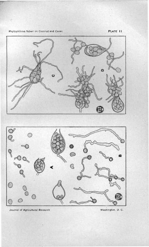
PLATE ii
Zoospore production and gennination in various stages of development of both strain of Phytophmora.
A, B.—Zoospore production showing various stages of development and germina- tion. X 400. A, coconut. B, cacao.
C, D.—Zoospore germination in place within the sporangium and at the mouth of the sporangium. X 400. Produced in Van Tieghem cells on dilute agar. C, coconut. B, cacao.
Phytophthora faberi on Coconut and Cacao PLATE 11
Journal of Agricultural Research Washington, D. C.

Phytophthora faberi on Coconut and Cacao PLATE 12
Journal of Agricultural Research Washington, D. C.

PLATE 12
Various types of conidia and chlamydospores of both strains of Phytophthora. A, B.—Conidia of varions forms of both strains of Phytophthora. X 400. A#
coconut. B, cacao. C, D.—Chlamydospores of various sizes of both strains of Phytophthora. X 400.
C, coconut. D, cacao.

ADDITIONAL COPIES OP THIS PUBLICATION MAY BB PROCURED FROM
TOB SUPßRINTßNDBNT OF DOCUMENTS GOVERNMENT PRINTING OFFICE
WASHINGTON» D, C. AT
io CENTS PER COPY SUBSCRIPTION PRICE, $4-00 PER YEAR
PURCHASER AGREES NOT TO RESELL OR DISTRIBUTE THIS COPY FOR PROFIT.—-PUB, RES. 67, APPROVED MAY 11, 1922