Embed Size (px)
Citation preview

_____________________________________________________________________________________________________ *Corresponding author: E-mail: [email protected];
Ophthalmology Research: An International Journal 13(3): 1-12, 2020; Article no.OR.60140 ISSN: 2321-7227
Comparative Study of Anterior Chamber Angle and Depth Recorded with Pentacam and AS-OCT SD
“Spectralis”
E. Pateras1* and N. Morogiannis2
1Biomedical Department, Course of Optics and Optometry, University of West Attica, Athens, Greece.
2Biomedical Department, Course of Optics and Optometry, Greece.
Authors’ contributions
This work was carried out in collaboration between both authors. Author EP designed the study,
performed the statistical analysis, wrote the protocol and wrote the first draft of the manuscript. Author NM managed the analyses of the study and the literature searches. Both authors read and approved
the final manuscript.
Article Information
DOI: 10.9734/OR/2020/v13i330167 Editor(s):
(1) Dr. Tatsuya Mimura, Tokyo Women's Medical University Medical Center East, Japan. Reviewers:
(1) Faruk Ozturk, Hacettepe University, Turkey. (2) Roland Hollhumer, University of the Witwatersrand, South Africa.
Complete Peer review History: http://www.sdiarticle4.com/review-history/60140
Received 06 June 2020 Accepted 11 August 2020 Published 21 August 2020
ABSTRACT
Aims: To compare the anterior chamber angle values recorded by Pentacam and AS-OCT SD “Spectralis” (Heidelberg Engineering) and present the correlation between the two devices. Sample and Study Design: A total of 50 patients were examined at the Private Ophthalmology Clinic O.M.M.A. Ophthalmological Institute. All participants volunteer to participate in this study where the data was kept anonymous. Patients aged 18-45 years without a pathological history were selected. All of them were emmetropes or with ametropia ranged ±0.75 D. There was no separation between hyperopic, myopic or emmetropic patients. Place and Duration of Study: University of West Attica Dept Biomedical Science Course Optics & Optometry in collaboration with Private Ophthalmology Clinic O.M.M.A. during the period between January 2019 to October 2019. Methodology: In this study, two basic structures of the eye are measured with the help of two devices of different principle of operation. Specifically, the study of the angle of the anterior chamber (ACA) as well as the depth of the chamber (ACD). The two devises are compared. Results: The ACA for both devices had mean difference of -2,004° for the R.E. while the mean difference for L.E. was 1,986°. Pentacam arithmetic mean ACA (R.E.) was 37,638 ± 2,98° and AS-
Original Research Article
Pateras and Morogiannis; OR, 13(3): 1-12, 2020; Article no.OR.60140
2
OCT “Spectralis” 35,766 ± 2,90° with Correlation coefficient 0,7063 (P<0,0001). Pentacam arithmetic mean ACA (L.E.) was 37,638 ± 2,98° and AS-OCT “Spectralis” 35,652 ± 2,79° with Correlation coefficient 0,7569 (P<0,0001). The ACD for both devices had mean difference of -0,3028 for the R.E. while the mean difference for L.E. was -0,2860. Pentacam arithmetic mean ACD (R.E.) was 3,5866 ± 0,20 and AS-OCT “Spectralis” 3,2838 ± 0,20 with Correlation coefficient 0,4201 (P=0,0024). Pentacam arithmetic mean ACD (L.E.) was 3,558 ± 0,21 and AS-OCT “Spectralis” 3,2720 ± 0,20 with Correlation coefficient 0,4023 (P=0,0038). Conclusion: Values of ACA measured by Pentacam and AS-OCT “Spectralis” were similar within the sample population of normal eyes right and left (P<0,0001). ACD measured by Pentacam and AS-OCT “Spectralis” showed also similar results the sample population of normal eyes for the right eye (P=0,0024) and left (P=0,0038).
Keywords: Anterior chamber angle; AS-optical coherence tomography; ”Spectralis”; comparison;
“Pentacam”; aqueous humor; anterior chamber depth.
1. INTRODUCTION The anterior chamber is an area which is delimited by the back surface of the cornea, the endothelium and the anterior surface of the iris and the front capsule of the crystalline lens. The anterior chamber angle is formed peripherally from the end of the cornea to the end of the iris root. The anterior chamber angle contains the trabecular meshwork (TM), the scleral spur (SS), the ciliary body (CB) and the root of the iris. The depth of the anterior chamber is important because it determines the aqueous humor flow which is related to the intraocular pressure (IOP) of the eye [1,2,3,4]. The contents of the anterior chamber are the aqueous humor helps maintain intraocular pressure (IOP) and is involved in the metabolism of the avascular crystalline lens and cornea. It is produced at the ciliary body non-pigmented epithelium (a ring-shaped tissue) at a rate of, 2,4– 3,4 μl/min. There is considerable variation in anterior chamber depth, depending on age, refractive error, and genetics. In general, in hyperopic patient, the central depth of the anterior chamber ranges from 3 mm to 3.5 mm, in emmetropes from 3.1 to 3.6 mm and in myopes from 3.3 to 3.8 mm. [4,5,6,7,8] The depth of the anterior chamber decreases with age, most likely due to thickening of the lens. By the age of 15, the anterior chamber depth is between approximately 3.6-3.65 mm. At the ages of 15 to 35, this depth has been found between 3 mm and 3.7 mm, and between 35 and 55, ranging from 2.8 to 3.3 mm. [9,10,11,12, 13,14].
The intraocular pressure is constant (normal levels 15-20 mm Hg) [15,16]. If unregulated pressure above 20 mmHg causes pressure on the optic nerve causing atrophy of the optic fibers and nerve damage. The anterior chamber angle is an important anatomical structure for differentiating the two types of glaucoma: open-angle glaucoma, which is the most common type of glaucoma, and closed-angle glaucoma. Modern medical treatment of open-angle glaucoma aims to reduce the production of aqueous humor and increase the aqueous outflow.
Perhaps the most common marking system is based on the angle formed between the iris surface and the trabecular meshwork (Shaffer grading system) [17]. The clinical methods for assessing the anterior chamber angle and depth include the Pen torch method, Smith’s method, Van Herrick’s technique, Split limbal technique, Gonioscopy, Scheimpflug corneal topography systems based on Shiflung technique, OCT- Anterior (Optical Coherence Tomography). The first 5 methods are subjective because they rely on the clinician skill. The last two are objective and they should be evaluated for their correlation.
There has been a lot of researches that try to image and record ACA and ACD [18-25]. Different devices and different methodologies for ACA and ACD showed how important it is to know the values of the angle and the depth especially in glaucoma monitoring and treatment [26-31].
2. METHODOLOGY
The sample consists of 50 patients where both their eyes (R.E. & L.E.) were examined at the
Pateras and Morogiannis; OR, 13(3): 1-12, 2020; Article no.OR.60140
3
Private Ophthalmology Clinic OMMA Ophthalmological Institute. All participants volunteer to participate in this study and their data was kept anonymous and they volunteered to participate Patients aged 18-45 years without a pathological history were selected. All of them were emmetropes or with a low ametropia ranged ±0.75 D. There was no separation between hyperopic, myopic or emmetropic patients. Pentacam uses a rotating Scheimpflug
camera and takes multiple images of the anterior segment of the eye. This generates three-dimensional images and calculate measurements of the eye especially anterior segment angle at 360°. The reference point and all the measurements for the anterior chamber angle was taken at the angle 90-270°. In the case of AS-OCT SD “Spectralis” (Heidelberg Engineering) all measurements derived from the manual recording of the operator. In the overview of the shot, the depth of the front chamber (ACD) is given.
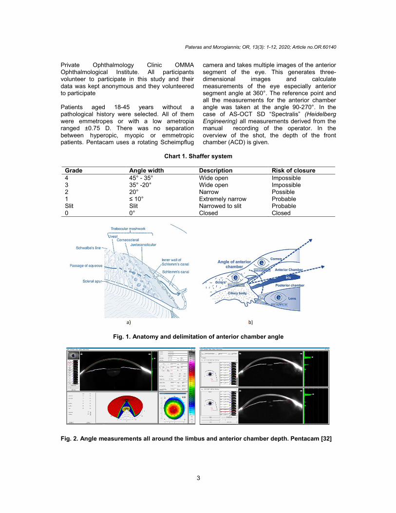
Chart 1. Shaffer system
Grade Angle width Description Risk of closure 4 45° - 35° Wide open Impossible 3 35° -20° Wide open Impossible 2 20° Narrow Possible 1 ≤ 10° Extremely narrow Probable Slit Slit Narrowed to slit Probable 0 0° Closed Closed
Fig. 1. Anatomy and delimitation of anterior chamber angle
Fig. 2. Angle measurements all around the limbus and anterior chamber depth. Pentacam [32]
Pateras and Morogiannis; OR, 13(3): 1-12, 2020; Article no.OR.60140
4
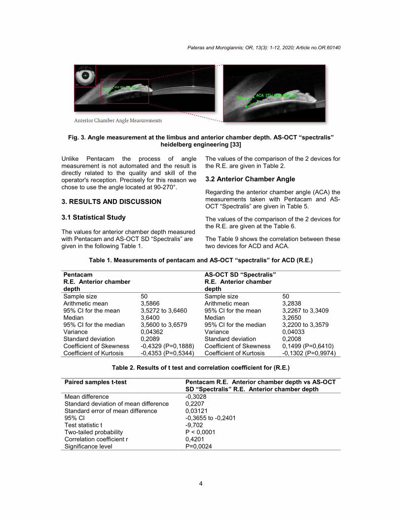
Fig. 3. Angle measurement at the limbus and anterior chamber depth. AS-OCT “spectralis” heidelberg engineering [33]
Unlike Pentacam the process of angle measurement is not automated and the result is directly related to the quality and skill of the operator's reception. Precisely for this reason we chose to use the angle located at 90-270°.
3. RESULTS AND DISCUSSION 3.1 Statistical Study The values for anterior chamber depth measured with Pentacam and AS-OCT SD “Spectralis” are given in the following Table 1.
The values of the comparison of the 2 devices for the R.E. are given in Table 2.
3.2 Anterior Chamber Angle
Regarding the anterior chamber angle (ACA) the measurements taken with Pentacam and AS-OCT “Spectralis” are given in Table 5.
The values of the comparison of the 2 devices for the R.E. are given at the Table 6.
The Table 9 shows the correlation between these two devices for ACD and ACA.
Table 1. Measurements of pentacam and AS-OCT “spectralis” for ACD (R.E.) Pentacam R.E. Anterior chamber depth
AS-OCT SD “Spectralis” R.E. Anterior chamber depth
Sample size 50 Sample size 50 Arithmetic mean 3,5866 Arithmetic mean 3,2838 95% CI for the mean 3,5272 to 3,6460 95% CI for the mean 3,2267 to 3,3409 Median 3,6400 Median 3,2650 95% CI for the median 3,5600 to 3,6579 95% CI for the median 3,2200 to 3,3579 Variance 0,04362 Variance 0,04033 Standard deviation 0,2089 Standard deviation 0,2008 Coefficient of Skewness -0,4329 (P=0,1888) Coefficient of Skewness 0,1499 (P=0,6410) Coefficient of Kurtosis -0,4353 (P=0,5344) Coefficient of Kurtosis -0,1302 (P=0,9974)
Table 2. Results of t test and correlation coefficient for (R.E.)
Paired samples t-test Pentacam R.E. Anterior chamber depth vs AS-OCT
SD “Spectralis” R.E. Anterior chamber depth Mean difference -0,3028 Standard deviation of mean difference 0,2207 Standard error of mean difference 0,03121 95% CI -0,3655 to -0,2401 Test statistic t -9,702 Two-tailed probability P < 0,0001 Correlation coefficient r 0,4201 Significance level P=0,0024
Pateras and Morogiannis; OR, 13(3): 1-12, 2020; Article no.OR.60140
5
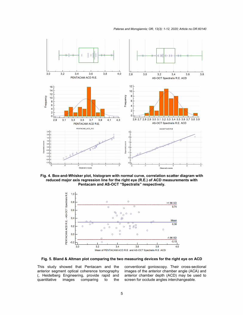
Fig. 4. Box-and-Whisker plot, histogram with normal curve, correlation scatter diagram with reduced major axis regression line for the right eye (R.E.) of ACD measurements with
Pentacam and AS-OCT “Spectralis” respectively.
Fig. 5. Bland & Altman plot comparing the two measuring devices for the right eye on ACD
This study showed that Pentacam and the anterior segment optical coherence tomography (, Heidelberg Engineering, provide rapid and quantitative images comparing to the
conventional gonioscopy. Their cross-sectional images of the anterior chamber angle (ACA) and anterior chamber depth (ACD) may be used to screen for occlude angles interchangeable.

Pateras and Morogiannis; OR, 13(3): 1-12, 2020; Article no.OR.60140
6
Table 3. Measurements of Pentacam and AS-OCT “Spectralis” for ACD
Pentacam
L.E. Anterior chamber depth
AS-OCT SD “Spectralis”
L.E. Anterior chamber depth
Sample size 50 Sample size 50
Arithmetic mean 3,5580 Arithmetic mean 3,2720
95% CI for the mean 3,4960 to 3,6200 95% CI for the mean 3,2138 to 3,3302
Median 3,6000 Median 3,2450
95% CI for the median 3,5321 to 3,6400 95% CI for the median 3,1960 to 3,3400
Variance 0,04762 Variance 0,04191
Standard deviation 0,2182 Standard deviation 0,2047
Coefficient of Skewness -0,2131 (P=0,5090) Coefficient of Skewness 0,1884 (P=0,5587)
Coefficient of Kurtosis -0,5745 (P=0,3353) Coefficient of Kurtosis -0,3051 (P=0,7351)
Fig. 6. Box-and-Whisker plot, histogram with normal curve, correlation scatter diagram with reduced major axis regression line for the left eye (L.E.) of ACD measurements with Pentacam
and AS-OCT “Spectralis” respectively
Pateras and Morogiannis; OR, 13(3): 1-12, 2020; Article no.OR.60140
7
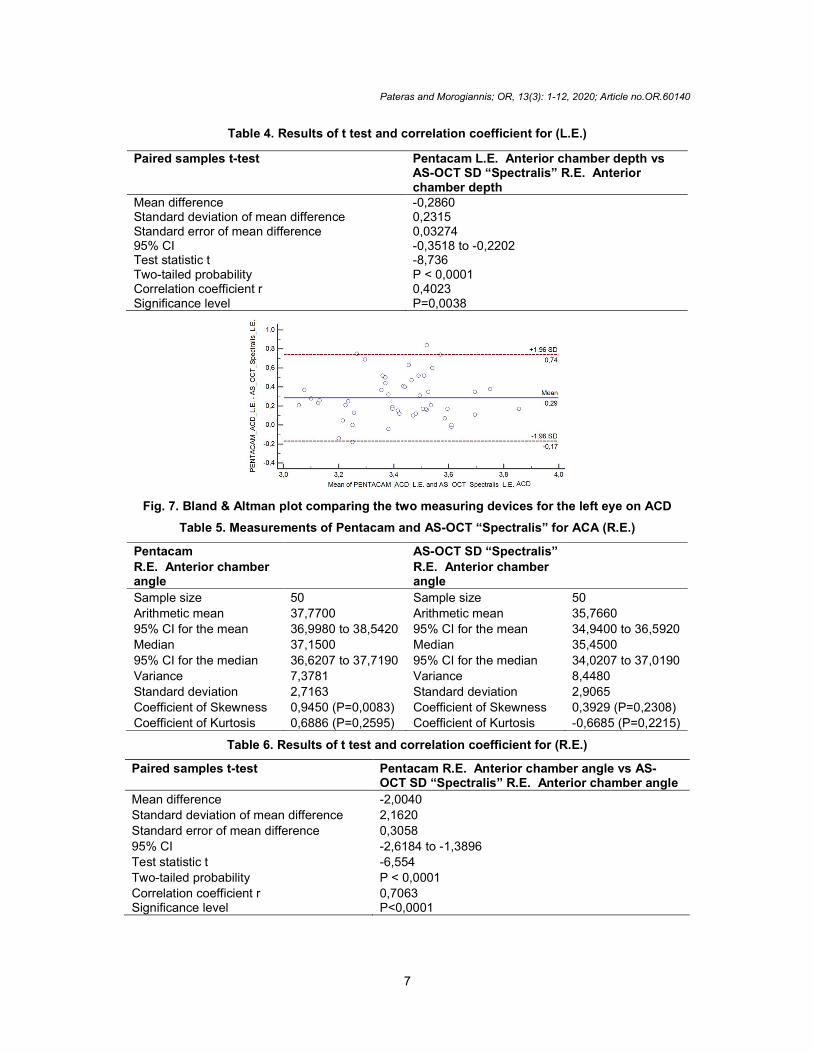
Table 4. Results of t test and correlation coefficient for (L.E.)
Paired samples t-test Pentacam L.E. Anterior chamber depth vs AS-OCT SD “Spectralis” R.E. Anterior chamber depth
Mean difference -0,2860 Standard deviation of mean difference 0,2315 Standard error of mean difference 0,03274 95% CI -0,3518 to -0,2202 Test statistic t -8,736 Two-tailed probability P < 0,0001 Correlation coefficient r 0,4023 Significance level P=0,0038
Fig. 7. Bland & Altman plot comparing the two measuring devices for the left eye on ACD
Table 5. Measurements of Pentacam and AS-OCT “Spectralis” for ACA (R.E.)
Pentacam
R.E. Anterior chamber angle
AS-OCT SD “Spectralis”
R.E. Anterior chamber angle
Sample size 50 Sample size 50
Arithmetic mean 37,7700 Arithmetic mean 35,7660
95% CI for the mean 36,9980 to 38,5420 95% CI for the mean 34,9400 to 36,5920
Median 37,1500 Median 35,4500
95% CI for the median 36,6207 to 37,7190 95% CI for the median 34,0207 to 37,0190
Variance 7,3781 Variance 8,4480
Standard deviation 2,7163 Standard deviation 2,9065
Coefficient of Skewness 0,9450 (P=0,0083) Coefficient of Skewness 0,3929 (P=0,2308)
Coefficient of Kurtosis 0,6886 (P=0,2595) Coefficient of Kurtosis -0,6685 (P=0,2215)
Table 6. Results of t test and correlation coefficient for (R.E.)
Paired samples t-test Pentacam R.E. Anterior chamber angle vs AS-OCT SD “Spectralis” R.E. Anterior chamber angle
Mean difference -2,0040
Standard deviation of mean difference 2,1620
Standard error of mean difference 0,3058
95% CI -2,6184 to -1,3896
Test statistic t -6,554
Two-tailed probability P < 0,0001
Correlation coefficient r 0,7063 Significance level P<0,0001

Pateras and Morogiannis; OR, 13(3): 1-12, 2020; Article no.OR.60140
8
Fig. 8. Box-and-Whisker plot, histogram with normal curve, correlation scatter diagram with reduced major axis regression line for the right eye (R.E.) of ACA measurements with
Pentacam and AS-OCT “Spectralis” respectively
Fig. 9. Bland & Altman plot comparing the two measuring devices for the right eye on ACA

Pateras and Morogiannis; OR, 13(3): 1-12, 2020; Article no.OR.60140
9
Table 7. Measurements of Pentacam and AS-OCT “Spectralis” for ACA (L.E.)
Pentacam L.E. Anterior chamber angle
AS-OCT SD “Spectralis” L.E. Anterior chamber angle
Sample size 50 Sample size 50 Arithmetic mean 37,6380 Arithmetic mean 35,6520 95% CI for the mean 36,7906 to 38,4854 95% CI for the mean 34,8591 to 36,4449 Median 36,8000 Median 35,3500 95% CI for the median 36,4603 to 37,9397 95% CI for the median 34,5603 to 36,2000 Variance 8,8914 Variance 7,7846 Standard deviation 2,9818 Standard deviation 2,7901 Coefficient of Skewness 0,5909 (P=0,0792) Coefficient of Skewness 0,3514 (P=0,2817) Coefficient of Kurtosis 0,6373 (P=0,2840) Coefficient of Kurtosis -0,1184 (P=0,9859)
Table 8. Results of t test and correlation coefficient for (L.E.)
Paired samples t-test Pentacam L.E. Anterior chamber angle vs AS-OCT SD “Spectralis” L.E. Anterior chamber angle
Mean difference -1,9860 Standard deviation of mean difference 2,0205 Standard error of mean difference 0,2857 95% CI -2,5602 to -1,4118 Test statistic t -6,950 Two-tailed probability P < 0,0001 Correlation coefficient r 0,7569 Significance level P<0,0001
Table 9. Correlation between two devices for ACD and ACA
Pentacam AS-OCT "Spectralis" ACA R.E. Arithmetic mean R.E. 37,77 ± 2,71° 35,766 ± 2,90° Mean difference -2,004 Correlation coefficient r 0,7063 Significance level P<0,0001 Two-tailed probability t-test P < 0,0001 Pentacam AS-OCT "Spectralis" ACA L.E. Arithmetic mean L.E. 37,638 ± 2,98° 35,652 ± 2,79° Mean difference -1,986 Correlation coefficient r 0,7569 Significance level P<0,0001 Two-tailed probability t-test P <0,0001 Pentacam AS-OCT "Spectralis" ACD R.E. Arithmetic mean R.E. 3,5866 ± 0,20 3,2838 ± 0,20 Mean difference -0,3028 Correlation coefficient r 0,4201 Significance level P=0,0024 Two-tailed probability t-test P < 0,0001 Pentacam AS-OCT "Spectralis" ACA L.E. Arithmetic mean L.E. 3,558 ± 0,21 3,2720 ± 0,20 Mean difference -0,286 Correlation coefficient r 0,4023 Significance level P=0,0038 Two-tailed probability t-test P < 0,0001

Pateras and Morogiannis; OR, 13(3): 1-12, 2020; Article no.OR.60140
10
Fig. 10. Box-and-Whisker plot, histogram with normal curve, correlation scatter diagram with reduced major axis regression line for the left eye (L.E.) of ACA measurements with Pentacam
and AS-OCT “Spectralis” respectively
Fig. 11. Bland & Altman plot comparing the two measuring devices for the left eye on ACA
Pateras and Morogiannis; OR, 13(3): 1-12, 2020; Article no.OR.60140
11
4. CONCLUSION
In this study only normal subjects with open angle were included with very small refractive errors so any pathology was excluded in our sample. The angle of the anterior chamber is an important anatomical structure for differentiating the two types glaucoma: Open-angle glaucoma, which is the most common type of glaucoma and closed angle glaucoma.
The modern medical treatment open angle glaucoma is accomplished by the reduction of production of aqueous humor and by the increase of the aqueous humor outflow. Also, another possible treatment for closed angle glaucoma is the iridectomy surgery. According to the above gonioscopy is an additional important tool for monitoring and treating glaucoma reducing the acute rise in intraocular pressure.
In this study, measurements of ACA (Anterior chamber angle) and ACD (Anterior chamber angle) were evaluated by two imaging devices Pentacam and AS-OCT “Spectralis”. Their data were similar between them, and showed good reproducibility and agreement between these two methods.
CONSENT AND ETHICAL APPROVAL
As per international standard or university standard guideline participant consent and ethical approval has been collected and preserved by the authors.
COMPETING INTERESTS
Authors have declared that no competing interests exist.
REFERENCES
1. Lee Ann Remington, Denise Goodwin. Clinical Anatomy and Physiology of the Visual System, 3
rd Edition, Butterworth-
Heinemann; 2011. ISBN: 9781437719260.
2. Ansari MW, Nadeem A. Atlas of ocular anatomy. Springer International Publishing; 2016. ISBN: 978-3-319-42781-2.
3. Khurana. Anatomy & physiology of eye. 2nd
Edition. CBS Publisher & Distributors P Ltd; 2011. ASIN: B011DC5DPA
4. Snell Richard S, Lemp Michael A. Clinical anatomy of the eye” 2nd (second) Edition,
Wiley-Blackwell Wiley-Blackwell; 2 edition; 1997. ASIN: B00E6T6T10.
5. Héctor Barajas M. Atlas of the human eye: Anatomy & Biometrics Palibrio; 2015. ISBN - 13:9781506510330
6. Allen L, Burian H, Braley A. The anterior border ring of Schwalbe and the pectinate ligament. Arch Ophthalmol. 1955;53:799.
7. Ashton N, Brini A, Smith R: Anatomical studies of trabecular meshwork of normal human eye. Br J Ophthalmol. 1956;40:257.
8. Holmberg A: Schlemm’s canal and the trabecular meshwork. An electron Microscopic study of the normal structure in man and monkey (cerecopithecus ethiops). Doc Ophthalmol (Den Haag). 1965;19:339.
9. Moses RA, Grodzki WJ Jr: The scleral spur and scleral roll. Invest Ophthalmol Vis Sci. 1977;16:925.
10. Fine B. Structure of the trabecular meshwork and the canal of Schlemm. Trans Am Acad Ophthalmol Otolaryngol. 1966;70:777.
11. Bill A. Scanning electron microscopic studies of the canal of Schlemm. Exp Eye Res. 1970;10:214.
12. Vranka JA, Kelley MJ, Acott TS, Keller KE. Extracellular matrix in the trabecular meshwork: Intraocular pressure regulation and dysregulation in glaucoma. Exp. Eye Res. 2015;133:112-25.
13. Johnson Douglas H, Trabecular Meshwork and Uveoscleral Outflow Models. Journal of Glaucoma. 2005;14(4):308-310. DOI: 10.1097/01.ijg.0000169397.32674.5e
14. Sudha A. Anatomy, physiology, histology and normal cytology of eye. J Cytol. 2007;24:16-9.
15. Derek W, et al. Anatomy and physiology of the cornea. Journal of Cataract & Refractive Surgery. 2011;37(3):588-598.
16. Ernst R. Tamm. The trabecular meshwork outflow pathways: Structural and functional aspects. Experimental Eye Research. 2009;88(4):648-655. Available:https://doi.org/10.1016/j.exer.2009.02.007
17. Wallace LM Alward, Reid A. Longmuir. Color Atlas of Gonioscopy, 2
nd Ed.
American Academy of Ophthalmology; 2008. ISBN-13: 978-1560558965
18. Goldsmith JA, Li Y, Chalita MR, et al. Anterior chamber width measurement by
Pateras and Morogiannis; OR, 13(3): 1-12, 2020; Article no.OR.60140
12
high-speed optical coherence tomography. Ophthalmology; 2005.
19. Radhakrishnan S, Goldsmith J, Huang D, et al. Comparison of optical coherence tomography and ultrasound biomicroscopy for detection of narrow anterior chamber angles. Arch Ophthalmol. 2005;123:1053-1059.
20. Radhakrishnan S, Goldsmith J, Huang D, et al. Optical coherence tomography imaging of the anterior chamber angle. Ophthalmol Clin North Am. 2005;18: 375–381.
21. Lackner B, Schimidger G, Skorpik C. Validity and repeatability of anterior chamber depth measurements with Pentacam and Orbscan. Optom Vis Sci. 2005;82:858–861.
22. Foster PJ, Buhrmann R, Quigley HA, Johnson GJ. The definition and classification of glaucoma in prevalence surveys. Br J Ophthalmol. 2002;86:238–242.
23. Samin Hong, et al. Detection of occludable angles with the pentacam and the anterior segment optical coherence tomography. Yonsei Med J. 2009;50(4): 525–528.
24. Jeong-Ho Yi, et al. Anterior chamber measurements by pentacam and AS-OCT in eyes with normal open angles. Korean J Ophthalmol. 2008;22(4):242–245.
25. Shajari Mehdi, et al. Comparison of corneal diameter and anterior chamber depth measurements using 4 different devices cornea. 2016;35(6):838-842. DOI: 10.1097/ICO.0000000000000840
26. Jing Dong , et al. Comparison of axial length, anterior chamber depth and intraocular lens power between IOL Master and ultrasound in normal, long and short eyes. Plos One; 2018.
Available:https://doi.org/10.1371/journal.pone.0194273
27. Elbaz U, Barkana Y, Gerber Y, Avni I, Zadok D. Comparison of different techniques of anterior chamber depth and keratometric measurements. Am J Ophthalmol. 2007;143(1):48–53.
PMID:17101110
28. Hashemi H, Yazdani K, Mehravaran S, Fotouhi A. Anterior chamber depth measurement with a-scan ultrasonography, Orbscan II, and IOL Master. Optom Vis Sci. 2005;82(10):900–904.
PMID:16276322
29. Porporato N, Baskaran M, Husain R. et al. Recent advances in anterior chamber angle imaging. Eye. 2020;34:51–59.
Available:https://doi.org/10.1038/s41433-019-0655-0
30. Bonomi L, et al. Epidemiology of angle-closure glaucoma: Prevalence, clinical types, and association with peripheral anterior chamber depth in the Egna-Neumarket Glaucoma Study. Ophthalmology. 2000;107:998–1003.
31. Yukiko Shimizu, et al. Comparison of the anterior chamber angle structure between children and adults. Journal of American Association for Pediatric Ophthalmology and Strabismus. 2017;21(1):57-62.
32. Available:https://doi.org/10.1016/j.jaapos.2016.10.005https://www.pentacam.com/fileadmin/use_upload/pentacam.de/downloads/interpretationsleitfaden/interpretation_guideline_3rd_edition_0915.pdf
33. Available:https://business-lounge.heidelbergengineering.com/us/en/products/spectralis/anterior-segment-module/
_________________________________________________________________________________ © 2020 Pateras and Morogiannis; This is an Open Access article distributed under the terms of the Creative Commons Attribution License (http://creativecommons.org/licenses/by/4.0), which permits unrestricted use, distribution, and reproduction in any medium, provided the original work is properly cited.
Peer-review history: The peer review history for this paper can be accessed here:
http://www.sdiarticle4.com/review-history/60140