Embed Size (px)
Citation preview
Hindawi Publishing CorporationISRN OphthalmologyVolume 2013, Article ID 706201, 5 pageshttp://dx.doi.org/10.1155/2013/706201
Clinical StudyAge and Positional Effect on the Anterior Chamber Angle:Assessment by Ultrasound Biomicroscopy
Nicholas P. Bell,1,2 Kundandeep S. Nagi,1,3 Ricardo J. Cumba,1,4 Alice Z. Chuang,1
David A. Lee,1,2 Thomas C. Prager,1,5 Kavita Rao,1 and Robert M. Feldman1,2
1 Ruiz Department of Ophthalmology and Visual Science, The University of Texas Medical School at Houston, 6431 Fannin Street,MSB 7.024, Houston, TX 77030, USA
2 Robert Cizik Eye Clinic, 6400 Fannin Street, Suite 1800, Houston, TX 77030, USA3Department of Ophthalmology, The University of Texas Health Science Center at San Antonio, 7703 Floyd Curl Drive,Mail Code 6230, San Antonio, TX 78229, USA
4Ophthalmology Department, University of Puerto Rico, Medical Sciences Campus, P.O. Box 365067, San Juan, PR 00936, USA5Houston Eye Associates, 2855 Gramercy Street, Houston, TX 77025, USA
Correspondence should be addressed to Nicholas P. Bell; [email protected]
Received 8 March 2013; Accepted 1 April 2013
Academic Editors: U. U. Inan, T. Mimura, and M. Nakazawa
Copyright © 2013 Nicholas P. Bell et al.This is an open access article distributed under the Creative Commons Attribution License,which permits unrestricted use, distribution, and reproduction in any medium, provided the original work is properly cited.
Purpose. To investigate age- and position-related changes of anterior chamber angle anatomy in normal, healthy eyes. Patients andMethods. Thirty subjects were separated into a younger and older cohort. The superior and inferior anterior chamber angles of theeyes were measured in supine and sitting positions by ultrasound biomicroscopy (UBM) with bag/balloon technology. Statisticalanalysis was used to evaluate positional and age-related changes in angle morphology. Results. In the younger cohort, no locationor positional differences in angle anatomy were observed. In the older cohort, the inferior quadrant was significantly narrower thanthe superior quadrant (𝑃 = 0.0186) in the supine position. This cohort also demonstrated an interaction effect between positionand location. In the older cohort, the angle was deeper inferiorly while the subject was sitting but was deeper superiorly whilethe subject was supine. Conclusion. Comparison of positional variations in anterior chamber angle anatomy as measured by UBMhas recently become possible. This study found that age-related positional changes in the anterior chamber angle anatomy exist innormal healthy eyes.
1. Introduction
Ultrasound biomicroscopy (UBM) provides noninvasivehigh-resolution in vivo imaging of the anterior segment.While anterior tissues are readily visualized by conventionalmethods (e.g., slit-lamp biomicroscopy and gonioscopy),structures posterior to the iris are hidden due to absorptionof light by the iris pigment epithelium. Such structures mayalso be undetected by anterior segment optical coherencetomography (ASOCT), given the inability of light to penetratethe iris pigment epithelium. B-scan ultrasonography, thoughextremely useful in imaging posterior ocular structures, isalso not ideal for ciliary body imaging due to the near-field artifact and poorer resolution obtained with lower fre-quencies. However, the higher frequency ultrasound waves,
utilized by UBM, are not obstructed by pigmented tissueand give the required resolution. Thus, UBM can be used toimage not only the anterior chamber angle but also the ciliarybody, peripheral lens, zonules, and the posterior chamberof the eye [1]. UBM has been demonstrated to be useful inevaluating the anterior segment of eyes with the primaryangle closure spectrum of disorders [2]. It also may beused to elucidate the mechanism of malignant glaucoma,pigmentary glaucoma, and anterior scleral disease [3–5].Therefore,UBMhas become the current standard for imagingabnormalities of ciliary body position, as are found in plateauiris configuration and annular choroidal effusions.
UBM technology is now readily available and simple touse with the development of a water-filled bag (ClearScan
2 ISRN Ophthalmology
Cover, ESI, Inc., Plymouth, MN), replacing traditional open-shell immersion techniques.The single-use, sterile bag snuglyfits over the distal end of the probe to form a watertightseal, which when pushed against the eye creates balloon-likepositive pressure [6]. The tip of the UBM probe does notcontact ocular structures, thus overcoming near-field artifactas well as increasing patient comfort. A distinct advantage ofthis technique is the ability to perform UBM examinationswith the subject in any position [6].
Historically, as used clinically, UBM examinations haveonly been possible in the supine position due to the require-ment of an open-shell immersion technique to overcome theacoustic near-field artifact inherent to ultrasonic imaging.There have been scattered reports in the literature of posi-tional measurements using UBM [7–10]. However, the effectsof age and supine to sitting positional change on anteriorchamber anatomy in normal subjects are not known.Changesin laxity of the irido-lenticular-zonular (ILZ) apparatus withage theoretically may lead to alterations of the anteriorchamber angle configuration that may be identified andmeasured with UBM. This study is designed to investigateage- and position-related changes of anterior chamber angleanatomy in normal, healthy eyes.
2. Patients and Methods
This research was approved by the Committee for the Protec-tion for Human Subjects at The University of Texas HealthScience Center at Houston. The protocol and proceduresadhered to the tenets of the Declaration of Helsinki, and thestudy was HIPAA compliant.
2.1. Study Population. Subjects were initially considered forparticipation if they were between 18 and 30 years of age(younger cohort) or older than 45 years of age (older cohort)and could tolerate both the supine and sitting positionsfor at least 15 minutes. Informed consent was obtained,and a screening history and examination were performedconsisting of slit-lamp biomicroscopy, applanation tonome-try, and gonioscopy. Subjects were considered eligible if thefollowing criteria were met: open angles (Schaffer grade 3-4), intraocular pressure (IOP) 8–21mmHg, and no ocularhistory other than refractive error. If both eyes were eligible,the right eye of each participant was selected as the study eye.
2.2. UBM Imaging. The superior and inferior anterior cham-ber angles were imaged at the limbus using the VuMax IIUBM (Sonomed, Lake Success, NY) in high resolution modewith a 35MHz probe tipped with a ClearScan Cover undertopical anesthesia as previously described [6]. Of note, thetechnique was modified by filling the bag to the lower aspectof the sealing collar instead of to the top end. Thus, thebag had a lower internal positive pressure than the eye tominimize compression of the ocular structures. With theprobe’s orientation line facing the cornea, theUBMprobewasheld perpendicular to the segment of angle being measured.
Images of the anterior chamber angle at superior andinferior quadrants were taken in sitting and supine positions
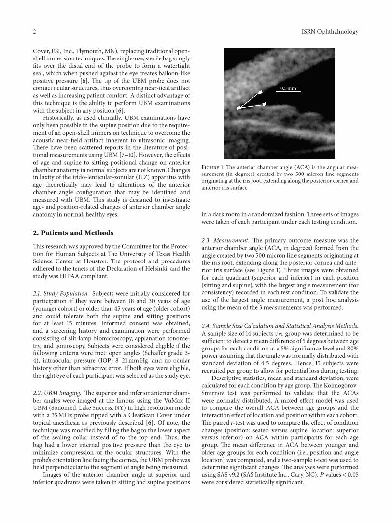
0.5mm
Figure 1: The anterior chamber angle (ACA) is the angular mea-surement (in degrees) created by two 500 micron line segmentsoriginating at the iris root, extending along the posterior cornea andanterior iris surface.
in a dark room in a randomized fashion.Three sets of imageswere taken of each participant under each testing condition.
2.3. Measurement. The primary outcome measure was theanterior chamber angle (ACA, in degrees) formed from theangle created by two 500 micron line segments originating atthe iris root, extending along the posterior cornea and ante-rior iris surface (see Figure 1). Three images were obtainedfor each quadrant (superior and inferior) in each position(sitting and supine), with the largest angle measurement (forconsistency) recorded in each test condition. To validate theuse of the largest angle measurement, a post hoc analysisusing the mean of the 3 measurements was performed.
2.4. Sample Size Calculation and Statistical Analysis Methods.A sample size of 14 subjects per group was determined to besufficient to detect amean difference of 5 degrees between agegroups for each condition at a 5% significance level and 80%power assuming that the angle was normally distributed withstandard deviation of 4.5 degrees. Hence, 15 subjects wererecruited per group to allow for potential loss during testing.
Descriptive statistics, mean and standard deviation, werecalculated for each condition by age group.The Kolmogorov-Smirnov test was performed to validate that the ACAswere normally distributed. A mixed-effect model was usedto compare the overall ACA between age groups and theinteraction effect of location and position within each cohort.The paired t-test was used to compare the effect of conditionchanges (position: seated versus supine; location: superiorversus inferior) on ACA within participants for each agegroup. The mean difference in ACA between younger andolder age groups for each condition (i.e., position and anglelocation) was computed, and a two-sample t-test was used todetermine significant changes. The analyses were performedusing SAS v9.2 (SAS Institute Inc., Cary, NC). 𝑃 values < 0.05were considered statistically significant.
ISRN Ophthalmology 3
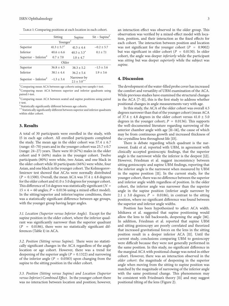
Table 1: Comparing positions at each location in each cohort.
Sitting Supine Sit − Supine§
Younger†
Superior 41.3 ± 5.7∗ 41.5 ± 4.4 −0.2 ± 5.7Inferior 40.6 ± 6.6 40.5 ± 3.3∗ 0.1 ± 7.1Superior − Inferior‡ 0.7 ± 7.0 1.0 ± 4.7
OlderSuperior 36.8 ± 4.5 38.3 ± 5.2 −1.5 ± 3.6Inferior 38.1 ± 4.4 36.2 ± 5.4 1.9 ± 3.6
Superior − Inferior‡ −1.3 ± 3.4 Narrower by2.1 ± 3.0∗∗
†Comparing mean ACA between age cohorts using two sample 𝑡-test.‡Comparing mean ACA between superior and inferior quadrants usingpaired 𝑡-test.§Comparing mean ACA between seated and supine positions using paired𝑡-test.∗Statistically significantly different between age cohorts.∗∗Statistically significantly different between superior and inferior quadrantswithin older cohort.
3. Results
A total of 30 participants were enrolled in the study, with15 in each age cohort. All enrolled participants completedthe study. The mean age in the older cohort was 57.4 ± 6.7(range: 45–70) years and in the younger cohort was 25.7±0.7(range: 24–27) years. There were 10 (67%) males in the oldercohort and 9 (60%) males in the younger cohort. Twelveparticipants (80%) were white, two Asian, and one black inthe older cohort while 10 participants (66%) were white, fourAsian, and one black in the younger cohort.TheKolmogorov-Smirnov test showed that ACAs were normally distributed(𝑃 = 0.1500). Overall, the mean ACA was 37.4 ± 4.8 degreesfor the older cohort and 41.0 ± 5.0degrees for younger cohort.This difference of 3.6 degrees was statistically significant (𝑁 =15 × 4 = 60 angles; 𝑃 = 0.0136 using a mixed-effect model).In the sitting/superior and inferior/supine conditions, therewas a statistically significant difference between age groups,with the younger group having larger angles.
3.1. Location (Superior versus Inferior Angle). Except for thesupine position in the older cohort, where the inferior quad-rant was significantly narrower than the superior quadrant(𝑃 = 0.0186), there were no statistically significant dif-ferences (Table 1) in ACA.
3.2. Position (Sitting versus Supine). There were no statisti-cally significant changes in the ACA regardless of the anglelocation or age cohort. However, there was a trend fordeepening of the superior angle (𝑃 = 0.1322) and narrowingof the inferior angle (𝑃 = 0.0583) upon changing from thesupine to the sitting position in the older cohort.
3.3. Position (Sitting versus Supine) and Location (Superiorversus Inferior) Combined Effect. In the younger cohort therewas no interaction between location and position; however,
an interaction effect was observed in the older group. Thisobservation was verified by a mixed-effect model with loca-tion, position, and their interaction as the fixed effects foreach cohort. The interaction between position and locationwas not significant for the younger cohort (𝑃 = 0.9002)but was significant in older cohort (𝑃 = 0.0130). In oldercohort, the angle was deeper inferiorly while the participantwas sitting but was deeper superiorly while the subject wassupine.
4. Discussion
Thedevelopment of thewater-filled probe cover has increasedthe comfort and versatility of UBM examination of the ACA.While previous studies have examined the structural changesin the ACA [7–11], this is the first study to address whetherpositional changes in angle measurements vary with age.
In this study, the ACA of the older cohort was overall 4.5degrees narrower than that of the younger cohort (meanACAof 37.4 ± 4.8 degrees in the older cohort versus 41.0 ± 5.0degrees in the younger cohort; 𝑃 = 0.0136). This supportsthe well-documented literature regarding narrowing of theanterior chamber angle with age [11–14], the cause of whichmay be from continuous growth and increased thickness ofthe crystalline lens throughout life [15].
There is debate regarding which quadrant is the nar-rowest. Esaki et al. reported with UBM, in agreement withclinically accepted gonioscopic findings, that the superiorangle is the narrowest while the inferior is the deepest [12].However, Friedman et al. suggest inconsistency betweensitting gonioscopic and supine UBM findings, reporting thatthe inferior angle is the narrowest when measured by UBMin the supine position [11]. In the current study, for theyounger cohort, there was no difference between the superiorand inferior angle width regardless of position. In the oldercohort, the inferior angle was narrower than the superiorangle in the supine position (inferior angle narrower by2.1 ± 3.0 degrees; 𝑃 = 0.0186), in contrast to the sittingposition, where no significant difference was found betweenthe superior and inferior angle widths.
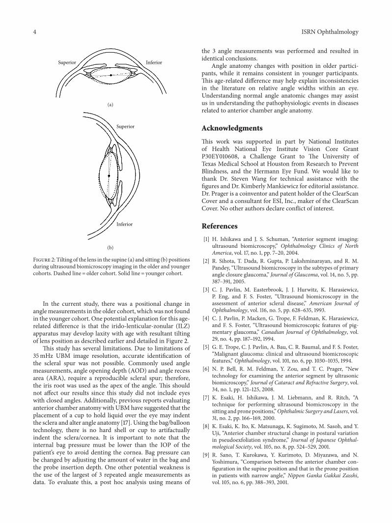
Position has been hypothesized to affect ACA width.Ishikawa et al. suggested that supine positioning wouldallow the lens to fall backwards, deepening the angle [16].In addition, Friedman et al. reported that supine UBMand sitting gonioscopy are poorly correlated and theorizedthat increased gravitational forces on the lens in the sittingposition result in a deeper inferior ACA [11]. Until thecurrent study, conclusions comparing UBM to gonioscopywere difficult because they were not generally performed inthe same position. In this study, no significant difference inthe marginal ACAwith positional change was noted in eithercohort. However, there was an interaction observed in theolder cohort: the magnitude of deepening in the superiorangle when moving from the sitting to supine position wasmatched by the magnitude of narrowing of the inferior anglewith the same positional change. This phenomenon maybe consistent with Friedman’s theory [11] and may suggestpositional tilting of the lens (Figure 2).
4 ISRN Ophthalmology
Superior Inferior
(a)
Superior
Inferior
(b)
Figure 2: Tilting of the lens in the supine (a) and sitting (b) positionsduring ultrasound biomicroscopy imaging in the older and youngercohorts. Dashed line = older cohort. Solid line = younger cohort.
In the current study, there was a positional change inanglemeasurements in the older cohort, whichwas not foundin the younger cohort. One potential explanation for this age-related difference is that the irido-lenticular-zonular (ILZ)apparatus may develop laxity with age with resultant tiltingof lens position as described earlier and detailed in Figure 2.
This study has several limitations. Due to limitations of35mHz UBM image resolution, accurate identification ofthe scleral spur was not possible. Commonly used anglemeasurements, angle opening depth (AOD) and angle recessarea (ARA), require a reproducible scleral spur; therefore,the iris root was used as the apex of the angle. This shouldnot affect our results since this study did not include eyeswith closed angles. Additionally, previous reports evaluatinganterior chamber anatomywithUBMhave suggested that theplacement of a cup to hold liquid over the eye may indentthe sclera and alter angle anatomy [17]. Using the bag/balloontechnology, there is no hard shell or cup to artifactuallyindent the sclera/cornea. It is important to note that theinternal bag pressure must be lower than the IOP of thepatient’s eye to avoid denting the cornea. Bag pressure canbe changed by adjusting the amount of water in the bag andthe probe insertion depth. One other potential weakness isthe use of the largest of 3 repeated angle measurements asdata. To evaluate this, a post hoc analysis using means of
the 3 angle measurements was performed and resulted inidentical conclusions.
Angle anatomy changes with position in older partici-pants, while it remains consistent in younger participants.This age-related difference may help explain inconsistenciesin the literature on relative angle widths within an eye.Understanding normal angle anatomic changes may assistus in understanding the pathophysiologic events in diseasesrelated to anterior chamber angle anatomy.
Acknowledgments
This work was supported in part by National Institutesof Health National Eye Institute Vision Core GrantP30EY010608, a Challenge Grant to The University ofTexas Medical School at Houston from Research to PreventBlindness, and the Hermann Eye Fund. We would like tothank Dr. Steven Wang for technical assistance with thefigures and Dr. Kimberly Mankiewicz for editorial assistance.Dr. Prager is a coinventor and patent holder of the ClearScanCover and a consultant for ESI, Inc., maker of the ClearScanCover. No other authors declare conflict of interest.
References
[1] H. Ishikawa and J. S. Schuman, “Anterior segment imaging:ultrasound biomicroscopy,” Ophthalmology Clinics of NorthAmerica, vol. 17, no. 1, pp. 7–20, 2004.
[2] R. Sihota, T. Dada, R. Gupta, P. Lakshminarayan, and R. M.Pandey, “Ultrasound biomicroscopy in the subtypes of primaryangle closure glaucoma,” Journal of Glaucoma, vol. 14, no. 5, pp.387–391, 2005.
[3] C. J. Pavlin, M. Easterbrook, J. J. Hurwitz, K. Harasiewicz,P. Eng, and F. S. Foster, “Ultrasound biomicroscopy in theassessment of anterior scleral disease,” American Journal ofOphthalmology, vol. 116, no. 5, pp. 628–635, 1993.
[4] C. J. Pavlin, P. Macken, G. Trope, F. Feldman, K. Harasiewicz,and F. S. Foster, “Ultrasound biomicroscopic features of pig-mentary glaucoma,” Canadian Journal of Ophthalmology, vol.29, no. 4, pp. 187–192, 1994.
[5] G. E. Trope, C. J. Pavlin, A. Bau, C. R. Baumal, and F. S. Foster,“Malignant glaucoma: clinical and ultrasound biomicroscopicfeatures,” Ophthalmology, vol. 101, no. 6, pp. 1030–1035, 1994.
[6] N. P. Bell, R. M. Feldman, Y. Zou, and T. C. Prager, “Newtechnology for examining the anterior segment by ultrasonicbiomicroscopy,” Journal of Cataract and Refractive Surgery, vol.34, no. 1, pp. 121–125, 2008.
[7] K. Esaki, H. Ishikawa, J. M. Liebmann, and R. Ritch, “Atechnique for performing ultrasound biomicroscopy in thesitting and prone positions,”Ophthalmic Surgery and Lasers, vol.31, no. 2, pp. 166–169, 2000.
[8] K. Esaki, K. Ito, K. Matsunaga, K. Sugimoto, M. Sasoh, and Y.Uji, “Anterior chamber structural change in postural variationin pseudoexfoliation syndrome,” Journal of Japanese Ophthal-mological Society, vol. 105, no. 8, pp. 524–529, 2001.
[9] R. Sano, T. Kurokawa, Y. Kurimoto, D. Miyazawa, and N.Yoshimura, “Comparison between the anterior chamber con-figuration in the supine position and that in the prone positionin patients with narrow angle,” Nippon Ganka Gakkai Zasshi,vol. 105, no. 6, pp. 388–393, 2001.
ISRN Ophthalmology 5
[10] H. V. Tran, H. Ishikawa, J. M. Liebmann, and R. Ritch, “Anew silicone eyecup for ultrasound biomicroscopy,”OphthalmicSurgery and Lasers, vol. 34, no. 1, pp. 73–75, 2003.
[11] D. S. Friedman, G. Gazzard, C. B. Min et al., “Age and sexvariation in angle findings among normal Chinese subjects: acomparison of UBM, Scheimpflug, and gonioscopic assessmentof the anterior chamber angle,” Journal of Glaucoma, vol. 17, no.1, pp. 5–10, 2008.
[12] K. Esaki, H. Ishikawa, J. M. Liebmann, D. S. Greenfield, Y. Uji,and R. Ritch, “Angle recess area decreases with age in normalJapanese,” Japanese Journal of Ophthalmology, vol. 44, no. 1, pp.46–51, 2000.
[13] Z. Pan, T. Furuya, and K. Kashiwagi, “Longitudinal changesin anterior chamber configuration in eyes with open-angleglaucoma and associated factors,” Journal of Glaucoma, vol. 21,no. 5, pp. 296–301, 2012.
[14] L. Xu, W. F. Cao, Y. X. Wang, C. X. Chen, and J. B. Jonas,“Anterior chamber depth and chamber angle and their associa-tionswith ocular and general parameters: the Beijing Eye Study,”American Journal of Ophthalmology, vol. 145, no. 5, pp. 929–936,2008.
[15] Y. Chen, Y. Z. Bao, and X. T. Pei, “Morphologic changes in theanterior chamber in patients with cortical or nuclear age-relatedcataract,” Journal of Cataract and Refractive Surgery, vol. 37, no.1, pp. 77–82, 2011.
[16] H. Ishikawa, K. Esaki, J. M. Liebmann, Y. Uji, and R. Ritch,“Ultrasound biomicroscopy dark room provocative testing: aquantitative method for estimating anterior chamber anglewidth,” Japanese Journal of Ophthalmology, vol. 43, no. 6, pp.526–534, 1999.
[17] H. Ishikawa, K. Inazumi, J. M. Liebmann, and R. Ritch, “Inad-vertent corneal indentation can cause artifactitious wideningof the iridocorneal angle on ultrasound biomicroscopy,” Oph-thalmic Surgery and Lasers, vol. 31, no. 4, pp. 342–345, 2000.

Submit your manuscripts athttp://www.hindawi.com
Stem CellsInternational
Hindawi Publishing Corporationhttp://www.hindawi.com Volume 2014
Hindawi Publishing Corporationhttp://www.hindawi.com Volume 2014
MEDIATORSINFLAMMATION
of
Hindawi Publishing Corporationhttp://www.hindawi.com Volume 2014
Behavioural Neurology
EndocrinologyInternational Journal of
Hindawi Publishing Corporationhttp://www.hindawi.com Volume 2014
Hindawi Publishing Corporationhttp://www.hindawi.com Volume 2014
Disease Markers
Hindawi Publishing Corporationhttp://www.hindawi.com Volume 2014
BioMed Research International
OncologyJournal of
Hindawi Publishing Corporationhttp://www.hindawi.com Volume 2014
Hindawi Publishing Corporationhttp://www.hindawi.com Volume 2014
Oxidative Medicine and Cellular Longevity
Hindawi Publishing Corporationhttp://www.hindawi.com Volume 2014
PPAR Research
The Scientific World JournalHindawi Publishing Corporation http://www.hindawi.com Volume 2014
Immunology ResearchHindawi Publishing Corporationhttp://www.hindawi.com Volume 2014
Journal of
ObesityJournal of
Hindawi Publishing Corporationhttp://www.hindawi.com Volume 2014
Hindawi Publishing Corporationhttp://www.hindawi.com Volume 2014
Computational and Mathematical Methods in Medicine
OphthalmologyJournal of
Hindawi Publishing Corporationhttp://www.hindawi.com Volume 2014
Diabetes ResearchJournal of
Hindawi Publishing Corporationhttp://www.hindawi.com Volume 2014
Hindawi Publishing Corporationhttp://www.hindawi.com Volume 2014
Research and TreatmentAIDS
Hindawi Publishing Corporationhttp://www.hindawi.com Volume 2014
Gastroenterology Research and Practice
Hindawi Publishing Corporationhttp://www.hindawi.com Volume 2014
Parkinson’s Disease
Evidence-Based Complementary and Alternative Medicine
Volume 2014Hindawi Publishing Corporationhttp://www.hindawi.com