Embed Size (px)
Citation preview
1
The Journal of Phytopharmacology 2015; 4(1): 1-5
Online at: www.phytopharmajournal.com
Research Article
ISSN 2230-480X
JPHYTO 2015; 4(1): 1-5
January- February
© 2015, All rights reserved
Aparna V
PG Scholar, Department of PG
Studies in Dravyaguna, SDM
College of Ayurveda, Kuthpady, Udupi, Karnataka-574118, India
Mallya Suma V
Associate professor, Department of
PG Studies in Dravyaguna, SDM College of Ayurveda, Kuthpady,
Udupi, Karnataka-574118, India
Srikanth P
Professor, Department of PG
Studies in Dravyaguna, SDM College of Ayurveda, Kuthpady,
Udupi, Karnataka-574118, India
Sunil Kumar KN
Senior Research Officer,
Department of Pharmacognosy, SDM Centre for Research in
Ayurveda and Allied Sciences,
Kuthpady, Udupi, Karnataka-574118, India
Correspondence: Sunil Kumar KN
Senior Research Officer,
Department of Pharmacognosy,
SDM Centre for Research in
Ayurveda and Allied Sciences,
Kuthpady, Udupi, Karnataka-
574118, India
Comparative pharmacognosy of two medhya dravyas,
Brahmi (Bacopa monnieri Linn.) and Mandukaparni
(Centella asiatica Linn.)
Aparna V, Mallya Suma V, Srikanth P, Sunil Kumar KN*
Abstract
Background: Brahmi (Bacopa monnieri) and Mandukaparni (Centella asiatica) are the two important distinct
Medhya (Nootropic) drugs mentioned in Indian systems of medicine. Lack of morphological description and
attribution of similar properties led towards confusion in identity of these two sources. Pharmacopoeias suggest
macro-microscopic characterization and chemical profiling of botanical material forms a pilot step in drug
standardization. Hence the detailed macro-microscopic records, along with phytochemical documentation of
these two plants were planned in the present investigation. Methodology: Matured, authenticated plants were
collected from its natural habitat. Macro-microscopic and preliminary phytochemical studies were carried out.
Results: Transverse section of Bacopa monnieri passing through midrib is isobilateral in histology whereas
that of Centella asiatica is dorsiventral. Striated cuticle and few layered spongy parenchyma are the features of
Centella asiatica. Plenty of air cavities with stomatal opening are specific to Bacopa monnieri. Phytochemical
analysis of these two drugs has revealed the presence of alkaloids, saponins, glycosides and tannins. In addition
to this Centella asiatica has shown the presence of flavanoids. Conclusion: The macro-microscopic and
phytochemical tests can be used to differentiate the botanical source of Brahmi/Mandukaparni.
Keywords: Brahmi, Bacopa monnieri, Centella asiatica, Macro-microscopic, Mandukaparni,
Phytochemical.
Introduction
The main object of Ayurveda is to live full length of life which is free from physical and mental
disorders. Medhya rasayana drugs have been claimed to exert a pronounced effect on the mental
capability of a person.1 The potential role of these drugs is on intellectual function and mental
performance. Brahmi and Mandukaparni are the two most popular drugs which are restorative with a
specific effect on the intellectual functions.2 Charaka considers both these drugs as promoters of general
mental ability (medhya).3 Inclusion of these two drugs in Ashtanga ghrita a formulation mentioned in
Asthanga hridaya clearly implies these as two distinct drugs.4 Brihatrayees, the three classical texts of
Ayurveda even supports the same.
Bacopa monnieri of Scrophulariaceae is the accepted source of Brahmi. It is a weak, creeping
herbaceous plant common in marshes and aslong back water and is called Brahmi or Nirbarhmi in
vernacular language.5 The drug is reported to be cold, sweet, astringent, diuretic, laxative and tonic for
the heart and nerves. The whole plant is used in a variety of preparations like Brahmighritam,
Mishrakasneham etc.6
The literal meaning of the term Mandukaparna is a plant having leaves resembling the shape of frogs.
And it also means, the stolons grow in the manner of jumping frog. Centella asiatica is the accepted
source of this herb.3 It is a stoloniferous creeping herb, rooting at nodes. The whole plant is reported to
be a nervine and cardiotonic, astringent and diuretic.7 Charaka includes this under Vayasthapana varga8,
the group of drugs that are capable of maintaining the youthful vigour and strength.
There is, however, some confusion with regard to the drugs Mandukaparni and Brahmi. This may be due
to the lack of description of the two drugs in the texts, attribution of similar properties to them and also
application of the same synonyms.3
Drug standardization or quality assurance forms an essential step before its therapeutic utility.9 The word
standardization should encompass the entire field of study from the birth of a plant to its clinical
application. Most of the pharmacopoeias suggest macro-microscopic characterization and chemical
profiling of botanical material turn out to be a pilot step in drug standardization.10

The Journal of Phytopharmacology
2
Hence the detailed macro-microscopic records, along with
phytochemical documentation of these two plants were planned under
this paper.
Materials and Methods
Fresh plant materials were collected during flowering season from
Udupi District of Karnataka, authenticated referring to regional
floras.11,12 Photograph from natural habitat were taken to record
habitat features. The dried ones were obtained by drying them under
shade, powdered and kept for phytochemical analysis. Fresh plant
material was studied for pharmacognostical characters as differences
in microscopic features are not evident in the dried form. Voucher
specimen and number 558.15013101-02 is deposited in the
Pharmacognosy department of the SDM Centre for Research in
Ayurveda and Allied Sciences, Udupi.
Representative parts from leaves were cut and preserved immediately
after collection. Detailed macroscopic and microscopic studies were
carried out as per standardized methodologies.12,13
The materials were left in a fixative solution Formalin-5ml + Acetic
acid-5ml + 70% Ethyl alcohol 90ml (FAA) for more than 48 hours.
Macroscopic study
Macroscopic characters of whole plant parts were recorded
systematically as prescribed in text book of Pharmacognosy.14
Microscopic study
The preserved specimens were dehydrated with a graded series of
tertiary-butyl alcohol as per the schedule. After dehydration, paraffin
infiltration was carried out till super saturation of tertiary butyl
alcohol was achieved. Following super saturation, materials were
transferred to pure paraffin wax for two times and the materials were
cast into paraffin blocks.14
Dermal features
Small pieces of leaves were mildly heated with Nitric acid and the
droplets of cutin formed were made to dissolve using benzene. Such
peelings stained with safranin were used to study the nature of cells of
the epidermis, stomata etc.14
Histology
The paraffin wax embedded specimens were sectioned with the help
of rotary microtome. The thickness of the sections was 10 to 12 µm.
The sections were stained with toluidine blue as per the method
introduced by O’Brein et al., 1964.15 Toluidine blue is a
polychromatic stain, the staining results were remarkably good; and
some cytochemical reactions were also obtained. The dye rendered
pink colour to the cellulose walls, blue to the lignified cells, dark
green to suberin, violet to the mucilage, blue to the protein bodies.
Wherever necessary, sections were stained with safranin and fast
green.
Quantitative microscopy
The cleared materials were washed thoroughly, stained with safranin
for quantitative microscopic studies. The tests were performed as per
the procedure given by Wallis TE, 1967.13 Few of the leaf constants
such as stomatal number, epidermal cells per sq. mm, stomatal index
etc. were done using micrometers.13,14
Stomatal number
It is the average number of stomata per square mm of the epidermis of
the leaf. A minimum of ten readings were taken from different
locations of the leaflet and the average value was calculated.14
Palisade ratio
It is the average number of palisade cells beneath one epidermal cells
of the lamina. It is determined by counting the palisade cells beneath
four continuous epidermal cells and dividing it by four.13
Photomicrography and description
In order to supplement the descriptive part, photomicrographs in
different magnifications of all necessary cells and tissues were taken
in Zeiss AxioLab trinocular microscope and Zeiss Stemi stereo
microscope. For normal histological purposes, sections were
photographed under bright field light. Magnifications of the figures
are indicated by the scale-bars.
Phytochemical study
Known quantity two dry powdered drugs were extracted with
petroleum ether, chloroform, ethanol and cold water. These extracts
were tested for different constituents.10
Results
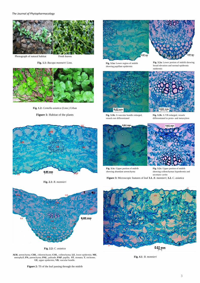
Bacopa monnieri (Linn.) is a juicy, succulent, glabrous herb rooting at
nodes with numerous ascending branches. Leaves simple, opposite,
sessile, entire, fleshy, obscurely veined. Flowers whitish, axillary,
solitary. Centella asiatica is a small creeping herb with slender stem,
rooting at nodes. Leaves simple with elongated petiole and sheathing
leaf base, reniform, crenate, toothed. (Figure 1)
TS of leaf of B. monnieri passing through midrib (Fig. 2.1, 3.1) is
nearly isobilateral in histology, midrib is not differentiated and both
side flat without any elevation (dorsiventral in histology and midrib is
differentiated, shows a broad elevation at the lower side in C. asiatica,
see Fig. 2.2). The leaf is covered by the upper and lower epidermis,
few of the lower epidermal cells being papillate, shows plenty of
stomata on either side (epidermal cells of the midrib region are thick-
walled shows collenchymatous hypodermis in C. asiatica, see Fig.
2.2, and 3.2 a, c). A vascular bundle comprising of xylem elements,
which are not differentiated into protoxylem and metaxylem, and
indistinct patches of phloem surrounding it is placed at the centre
(oval shaped bundle with distinct patches of phloem at the lower side,
vessels differentiated into protoxylem towards the upper side and
metaxylem towards lower side in C. asiatica, see Fig. 3.2b). Beneath
the upper epidermis plenty of air cavities are embedded with stomatal
openings (just above the vascular bundle secretary cavity is placed in
C. asiatica, see Fig. 3.2c).
TS of leaf passing through the lamina in B. monneri (Fig. 4.1) is flat
and without any elevations and is covered by the upper and lower
epidermis, a thick normal cuticle covers the epidermii and stomata are
embedded throughout the upper and lower epidermis (cuticle striated
in C. asiatica, see Fig. 4.2). The mesophyll region is not differentiated
into palisade and spongy parenchyma but few cells beneath the upper
epidermal cells are elongated and embed numerous air cavities (single
layer of palisade without much air space in C. asiatica, see Fig. 4.2).
The spongy parenchyma is many layered with a few trace bundles
(few layered in C. asiatica, see Fig. 4.2)
Stomatal number on upper and lower epidermii of B. monneri was
calculated to be 118 and 130 respectively, while the upper epidermis
of C. asiatica has only 58 stomata per sq. mm. There are no glandular
trichomes on the lower epidermis of C. asiatica. Palisade ratio is 0 on
both the epidermii of B. monneri and lower epidermis of C. asiatica
(Table 1, Figure 5).
Phytochemical analysis of these two drugs has revealed the presence
of alkaloids, saponins, glycosides and tannins. In addition to this C.
asiatica has shown the presence of flavanoids (Table 2 & 3).
The Journal of Phytopharmacology
3
Photograph of natural habitat Fresh leaves
Fig. 1.1: Bacopa monneiri Linn.
Fig. 1.2: Centella asiatica (Linn.) Urban
Figure 1: Habitat of the plants
Fig. 2.1: B. monnieri
Fig. 2.2: C. asiatica
AER, aerenchyma; CHL, chlorenchyam; COL, collenchyma; LE, lower epidermis; ME,
mesophyll; PA, parenchyma; PAL, palisade; PAP, papilla; ST, stomata; T, trichome;
UE, upper epidermis; VB, vascular bundle.
Figure 2: TS of the leaf passing through the midrib
Figure 3: Microscopic features of leaf 3.1. B. monnieri; 3.2. C. asiatica
Fig. 4.1: B. monnieri
Fig. 3.1a: Lower region of midrib
showing papillate epidermis
Fig. 3.2a: Lower portion of midrib showing
broad elevation and normal epidermis
epidermis
Fig. 3.1b: A vascular bundle enlarged,
vessels not differentiated Fig. 3.2b: A VB enlarged, vessels
differentiated to proto- and metaxylem
Fig. 3.1c: Upper portion of midrib
showing abundant aerenchyma Fig. 3.2c: Upper portion of midrib
showing collenchymas hypodermis and
secretary cavity

The Journal of Phytopharmacology
4
Fig. 4.2: C. asiatica
AER, aerenchyma; CS, cuticular striations; LE, lower epidermis; ME, mesophyll; PAL,
palisade; ST, stomata; UE, upper epidermis.
Figure 4: TS of leaf passing through lamina
CS, cuticular striations; EC, epidermal cell; GT, glandular trichome; PAL, palisade; ST,
stomata
Figure 5: Epidermal features of the Plants
Table 1: Quantitative microscopic values of Centella asiatica and Bacopa
monnieri leaf
Per sq. mm. Bacopa monnieri Centella asiatica
UE LE UE LE
Stomatal number 118 130 58 140
Trichome (glandular)
number
25 13 16 0
Palisade ratio 0 0 6.5 0
UE – upper epidermis; LE – lower epidermis
Table 2: Phytochemical study of Bacopa monnieri (Dry form)
S. No Tests Pet. ether Chloroform Ethanol Cold
infusion
1. Proteins
a. Biuret test –ve –ve –ve –ve
b. Ninhydrin test –ve –ve –ve –ve
c. Xanthoproteic test –ve –ve –ve –ve
d. Hopkins-cole test –ve –ve –ve –ve
e. Sulphur test –ve –ve –ve –ve
2. Carbohydrate test for starch
a. Molisch’s test –ve –ve +ve –ve
b. Iodine test –ve +ve +ve -ve
c. Fehling’s test +ve -ve +ve +ve
d. Benedict’s test +ve +ve +ve -ve
3. Tannins
a. Gelatin test -ve –ve –ve +ve
4. Anthrocyanins
a. Aqueous NaOH test –ve –ve –ve –ve
b. Conc. H2SO4 test –ve –ve –ve –ve
5. Glycosides
a. Molisch’s test +ve -ve +ve +ve
b. Conc. H2SO4 test -ve –ve –ve +ve
c. Keller Kiliani test -ve +ve +ve +ve
6. Saponin
a. Foam test +ve -ve +ve –ve
7. Flavanoids
a. Flavanoid test –ve –ve –ve –ve
b. Pew’s test for
Dihydroflavanols
–ve –ve –ve –ve
c. Shinoda test –ve –ve –ve –ve
d. Aqueous NaOH test –ve –ve –ve –ve
e. Conc. H2SO4 test –ve +ve –ve -ve
8. Phenols
a. Phenol test -ve –ve +ve –ve
9. Steroids
a. Salkowski’s test –ve –ve –ve –ve
10 Alkaloids
a. Mayer’s test –ve –ve +ve –ve
b. Dragendroff’s test –ve –ve –ve –ve
Tale 3: Phytochemical study of Centella asiatica (Dry form)
S. No Tests Pet. Ether Chloroform Ethanol Cold
infusion
1. Proteins
a. Biuret test –ve –ve –ve –ve
b. Ninhydrin test –ve –ve –ve –ve
c. Xanthoproteic test –ve –ve –ve –ve
d. Hopkins-cole test –ve –ve –ve –ve
e. Sulphur test –ve –ve –ve –ve
2. Carbohydrate test for starch
a. Molisch’s test –ve –ve +ve –ve
b. Iodine test –ve +ve +ve -ve
c. Fehling’s test +ve -ve +ve +ve
d. Benedict’s test +ve +ve +ve -ve
3. Tannins
a. Gelatin test -ve –ve –ve +ve
4. Anthrocyanins
a. Aqueous NaOH test –ve –ve –ve –ve
b. Conc. H2SO4 test –ve –ve –ve –ve
5. Glycosides
a. Molisch’s test +ve -ve +ve +ve
b. Conc. H2SO4 test -ve –ve –ve +ve
c. Keller Kiliani test -ve +ve –ve +ve
6. Saponin
a. Foam test +ve -ve +ve –ve
7. Flavanoids
a. Flavanoid test –ve –ve –ve –ve
b. Pew’s test for
Dihydroflavanols
–ve –ve –ve –ve
c. Shinoda test –ve –ve –ve –ve
d. Aqueous NaOH test –ve –ve –ve –ve
Fig. 5.1: Epidermal features upper
epidermis of B. monnieri
Fig. 5.2: Epidermal features of
Upper epidermis of C. asiatica
Fig. 5.3: Palisade ratio of C. asiatica

The Journal of Phytopharmacology
5
e. Conc. H2SO4 test +ve +ve –ve -ve
8. Phenols
a. Phenol test -ve –ve +ve –ve
9. Steroids
a. Salkowski’s test –ve –ve –ve –ve
10 Alkaloids
a. Mayer’s test –ve –ve +ve –ve
b. Dragendroff’s test –ve –ve –ve –ve
Discussion
Lack of standards is a major problem associated with herbal medicine,
while macro-microscopic scientific records are essential steps in
quality assurance of a drug. Common pharmaco-therapeutic property
and fewer morphological descriptions available in the texts of
Ayurveda, made Brahmi and Mandukaparni as controversial drugs3.
But their specific inclusion under particular formulations and single
drug usage clarifies these as two separate drugs attributed with a
specific mode of action. Bacopa monnieri and Centella asiatica are
accepted source of Brahmi and Mandukaparni respectively.5
Transverse section of B. monnieri passing through midrib is found to
be nearly isobilateral in histology, midrib is not differentiated and
both sides are flat without any elevation. C. asiatica shows
dorsiventral histology and the midrib is differentiated with broad
elevation at the lower side. Lamina of B. monnieri is flat and without
any elevations and is covered by upper and lower epidermii, a thick
normal cuticle covers the epidermis and stomata are embedded
throughout the upper and lower epidermis. However cuticle layer of
Centella asiatica is striated. Spongy parenchyma is many layered in
Bacopa monnieri while it is few layered in Centella asiatica. Presence
of plenty air cavity embedded with stomatal opening beneath the
upper epidermis evocative of aquatic habitat of Bacopa monnieri.
Quantitative microscopic characters further help in identification of
genuine drug.
Preliminary phytochemical studies are essential to understand the
basic chemical nature of the plant. Presence of flavanoids is found to
be a marked difference of C. asiatica though alkaloids, saponins,
glycosides and tannins occur in both.
Conclusion
The macro-microscoipc profile along with their preliminary
phytochemical data delineated here would be helpful to differentiate
each other and other admixture.
References
1. Brain aging and Ayurveda; New Delhi, Central Council for Research in Ayurevda & Siddha, Dept of Ayush, Min of Health& family welfare, Govt of
India, 2008, P. 14.
2. Singh HK, Dhawan BN, Neuropsychopharmacological effects of of
Ayurvedic nootropic Bacopa monnieri Linn.(Brahmi). Indian J Pharmacol 1997; p 29.
3. Charaka, Charaka samhita, Chikitsa sthana, Varanasi; Choukamba Sanskrit
series, 2002, P. 385.
4. VV Sivaranjan and Indira Balachandran, Ayurvedic drugs and their plant
sources, New Delhi; Published by Oxford & IBH Publishing Co. Pvt. Ltd, 1994, P. 289.
5. Indian Medicinal Plants, Volume 1, Published Orient Longman Private Ltd,
1994, p.235.
6. Priyavat Sharma, Dravya guna Vijnana, Varanasi, Chaukamba Bharati
Academy; Volume 2, 1998, p. 6.
7. Khare CP, Indian Medicinal Plants an Illustrated Dictionary, New Delhi,
Springer; 2007, p.77.
8. Charaka, Charaka samhita, Chikitsa sthana, Varanasi; Choukamba Sanskrit
series, 2009, P. 34.
9. Sunil Kumar KN, Pharmacognostical evaluation of Cinnamomum tamala (Buch.- Ham) Nees and Eberm (Tamala patra) and few of its allied species.
Msc. Thesis, Institute of Ayurvedic Medical Sciences, Gujarat Ayurveda
University, Jamnagar, Gujarat, 2006.
10. Quality Control Methods for Medicinal Plant Materials, WHO, Geneva, 1998, p.16- 27.
11. Nadakarni AK. Dr. KM Nadakarni’s, Indian Materia Medica, Popular
Prakashan, Bomaby, 1976. p579.
12. Gamble JS. Flora of the presidency of Madras. Botanical survey of India,
Calcutta, 1925. Vol.II, p.556.
13. Wallis TE. Textbook of Pharmacognosy. Delhi; CBS Publishers and Distributors; 1985, p572.
14. Evans WC. Trease and Evans Pharmacognosy, London: Bailliere Tindall;
1989, p530.
15. O’Brien TP, Feder N, Mc Cull ME. Polychromatic staining of plant cell
walls by toluidine blue-O. Protoplasma 1964; 59: 364-373.