Embed Size (px)
Citation preview
Governors State UniversityOPUS Open Portal to University Scholarship
All Capstone Projects Student Capstone Projects
Spring 2015
Comparative Literature Analysis of PeripheralVestibular Function Assessment ToolsRiddhi ChinoyGovernors State University
Follow this and additional works at: http://opus.govst.edu/capstones
Part of the Analytical, Diagnostic and Therapeutic Techniques and Equipment Commons, andthe Physical Therapy Commons
For more information about the academic degree, extended learning, and certificate programs of Governors State University, go tohttp://www.govst.edu/Academics/Degree_Programs_and_Certifications/
Visit the Governors State Physical Therapy DepartmentThis Project Summary is brought to you for free and open access by the Student Capstone Projects at OPUS Open Portal to University Scholarship. Ithas been accepted for inclusion in All Capstone Projects by an authorized administrator of OPUS Open Portal to University Scholarship. For moreinformation, please contact [email protected].
Recommended CitationChinoy, Riddhi, "Comparative Literature Analysis of Peripheral Vestibular Function Assessment Tools" (2015). All Capstone Projects.126.http://opus.govst.edu/capstones/126
1
SECTION 1: Introduction
The vestibular system, located in the inner ears, is comprised of a peripheral and a
central component.1 Together these components provide the central nervous system with
information regarding head, body, and eye movements.1 There are three main functions
of the vestibular system: “(1) to stabilize visual images on the fovea of the retina during
head movement to allow clear vision, (2) to maintain postural stability, especially during
movement of the head, and (3) to provide information used for spatial orientation.”1, p822
The peripheral vestibular system is the most common origin for patient signs and
symptoms of vestibular dysfunction1 and will serve as the primary focus of this paper.
The peripheral vestibular system contains two different sensory systems on each
side: three semicircular canals (anterior, posterior, lateral (also referred to as horizontal))
and two otolith organs (saccule and utricle).1 The total six semicircular canals provide
information about head angular velocity (yaw, pitch, roll) which is primarily used for
gaze stability, while the four otolith organs provide information about head tilt and linear
acceleration which is used for postural stability.1,2
The semicircular canals drive the movement of the eyes to stabilize vision during
rapid head movements through the vestibulo-ocular reflex (VOR).1-3
In a typical, intact
vestibular system, as the head moves in one direction, the VOR triggers the eyes to move
in the opposite direction with velocity and amplitude equal to the head movement to
maintain a stable gaze and clear vision of a stationary target.1-3
This relationship of eye
velocity to head velocity is defined as the vestibular gain (eye velocity / head velocity =
1).1-3
The VOR typically operates at head velocities from 60 to as great as 400 degrees
2
per second; less than 60 degrees per second involves another mechanism called smooth
pursuit, while velocities beyond 400 degrees per second reduces the VOR gain and
deteriorates the gaze stability.1
The six semicircular canals work in pairs: the right anterior semicircular canal
pairs with the left posterior semicircular canal, the right posterior with the left anterior,
and the two lateral canals with each other.1 For example, as the head turns to the right,
both lateral semicircular canals are stimulated (the right lateral will have an increased
firing rate of the peripheral vestibular neurons while the left lateral has a decreased firing
rate), causing the VOR to signal both eyes to move at equal speed and distance as the
head to the opposing left side which allows the eyes to maintain clear vision of a target.1-3
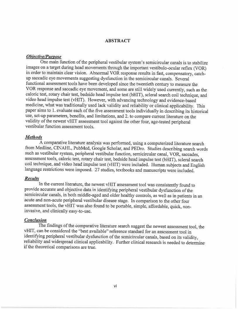
A “normal” VOR response in a typical healthy subject is shown in Figure 1.3 Following
a manually delivered head rotation movement to the right (Figure 1a to 1b), the eyes
reflexively move toward the opposing left side (Figure 1c).3
Figure 1 a, b, c VOR response on a normal healthy subject
From: Curthoys et al., 2011
In a malfunctioning semicircular canal, the VOR presents differently. For
example, if the right lateral semicircular canal loses typical functionality, such as in a
3
peripheral vestibular disorder, turning the head to the right does not stimulate the right
lateral canal to signal the VOR to drive the eyes to the opposite left direction.1,3
Instead,
the eyes would move with the head initially, and the compensatory response to re-fixate
the lost vision back on the target would be a fast eye movement to the opposing left side,
defined as a corrective catch-up saccade.1-3
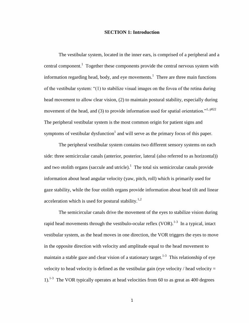
Figure 2 below illustrates this abnormal VOR
response in a patient with right-sided vestibular dysfunction by the presence of a
corrective catch-up saccade (Figure 2f) during a manually delivered head rotation
movement to the right.3
Figure 2 d, e, f Abnormal VOR response with corrective catch-up saccade on a patient with right peripheral vestibular dysfunction
From: Curthoys et al., 2011
If a corrective catch-up saccade occurs at the end of the head movement, it is
known as an overt saccade as it is easily detected during a clinical examination.2,3
If a
corrective catch-up saccade occurs during the head movement, it is known as a covert
saccade and is undetectable by the naked eye, thus missed by the clinician.2,3
Both overt
and covert saccadic eye movements in response to an abnormal VOR indicate a
dysfunctional semicircular canal.1,4
The canal that is diagnosed as dysfunctional is
4
dependent on the plane of paired semicircular canals being tested and in which head
position the presence of a saccadic eye movement is detected.3,4
This interaction is
outside of the scope of this paper. Instead, the purpose of this paper is to describe
methods of objectively and clinically measuring the adequacy of the VOR response and
the presence of saccadic eye movement to determine overall semicircular canal function,
which can be difficult.3
The function of the peripheral vestibular system’s semicircular canals must be
evaluated by clinicians using a thorough patient history, a variety of clinical
examinations, and formal quantitative testing.3,4
Several functional assessment tools
were introduced in early 20th
century and are still currently used to specifically measure
the VOR response and saccadic eye movement.1-5
However, with the advent of
technology and new medical research, the traditional “gold standard” tools may not be as
effective today.3-5
Thus, the aim of this paper is to compare current literature regarding
four commonly used peripheral vestibular function assessment tools-- the caloric test,
rotary chair test, bedside head impulse test (bHIT), and scleral search coil technique-- to
the newest assessment tool, the video head impulse test (vHIT), and to explore the
potentiality of the vHIT becoming the next “gold standard” tool.
5
SECTION 2: Peripheral Vestibular Function Assessment Tools
Five commonly known peripheral vestibular function assessment tools are
currently used clinically to specifically analyze VOR response and saccadic eye
movement to determine the overall functionality of semicircular canals. These
assessment tools are: the caloric test, the rotary chair test, the bedside head impulse test
(bHIT), the sclera search coil technique, and the video head impulse test (vHIT). In this
section, recent literature on these tests will be evaluated, and the tests will be compared
specifically on the characteristics of the historical use, test set-up, benefits, and
limitations (Table 1).
Caloric Test
Caloric testing is historically one of the oldest assessment tools of early 20th
century used to evaluate asymmetric function in the peripheral vestibular system,
specifically of the lateral semicircular canals.5 This test involves irrigation of the external
ear canal with cold and warm water or air. This irrigation stimulates a fluid density
change inside the inner ear triggering endolymph fluid movement of the lateral
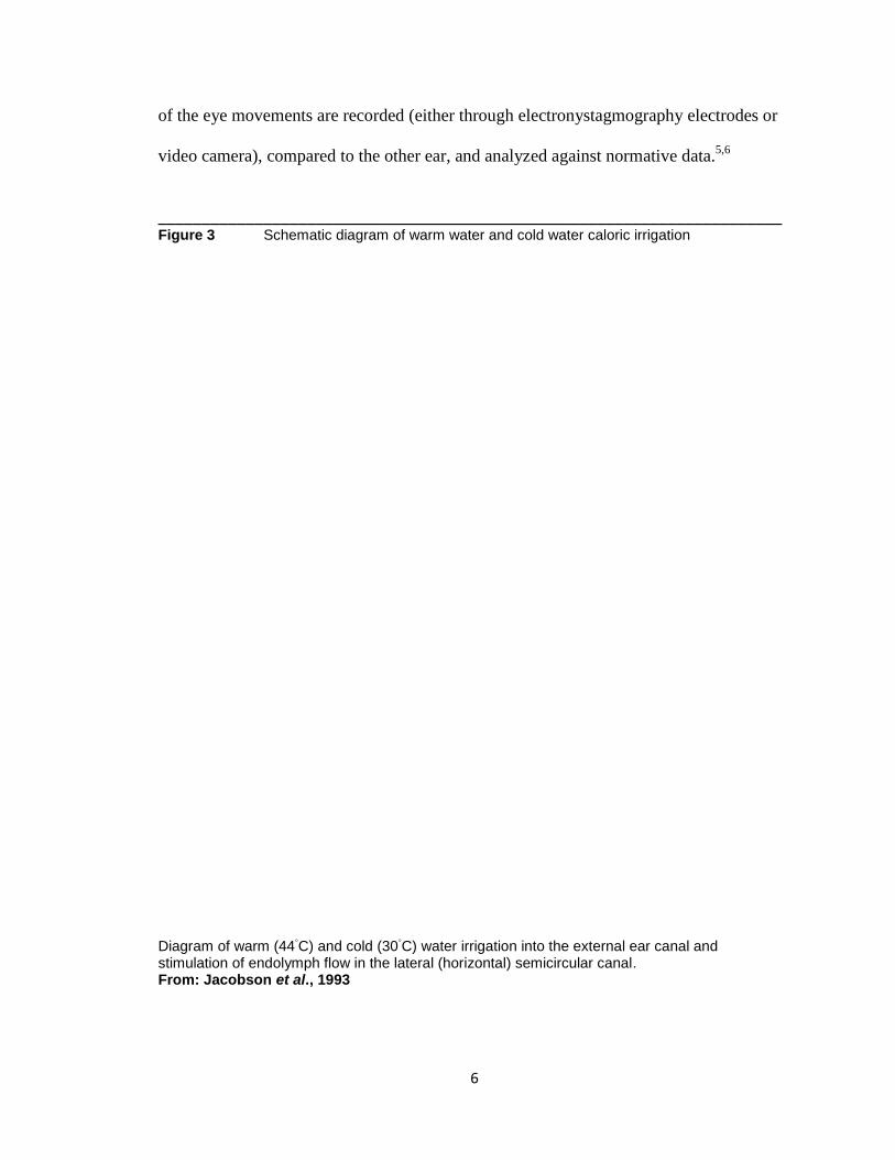
semicircular canal of that ear as shown in Figure 3.5 The endolymph fluid movement
results in fast, side-to-side eye movements called nystagmus, and corrective saccades5,6
Under typical test conditions, cold (30oC) irrigated water will cause fast corrective
saccades away from the stimulated ear, while warm (44oC) irrigated water will cause fast
corrective saccades toward the side of stimulated ear (adhering to the mnemonic word
“COWS:” cold opposite, warm same).5,6
The latency, duration, frequency, and velocity
6
of the eye movements are recorded (either through electronystagmography electrodes or
video camera), compared to the other ear, and analyzed against normative data.5,6
_______________________________________________________________________Figure 3 Schematic diagram of warm water and cold water caloric irrigation
Diagram of warm (44◦C) and cold (30
◦C) water irrigation into the external ear canal and
stimulation of endolymph flow in the lateral (horizontal) semicircular canal. From: Jacobson et al., 1993
7
The caloric test is usually performed with the patient in supine with the head
elevated slightly to 30 degrees to bring the lateral semicircular canals parallel to earth-
vertical axis (or alternatively patient can sit up with head extended 60 degrees).7,8
In a set
sequence, each ear is irrigated for a 20-40 second duration with cold and then warm water
(or air if indicated) at a designated volume with a designated time interval between
irrigations. 5,7,8
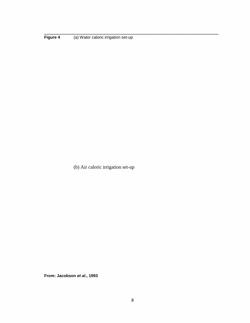
The set-up for the caloric test is shown in Figure 4 specifically for (a)
water and (b) air irrigation.5 The velocity of the eye movements evoked by the irrigation
method is analyzed to determine the presence of unilateral lateral semicircular canal
dysfunction through a mathematical calculation of Jongkees formula,5,7,8
in which canal paresis (CP) is defined as 25% or greater asymmetry between the eye
velocities for the left and right ears.5,7
WR is the recorded eye nystagmus velocity during
warm water irrigation in the right ear, WL for warm water irrigation in the left ear, CR for
cold water irrigation in the right ear, and CL for cold water irrigation in the left ear.5,7,8
8
_______________________________________________________________________Figure 4 (a) Water caloric irrigation set-up
(b) Air caloric irrigation set-up
From: Jacobson et al., 1993
9
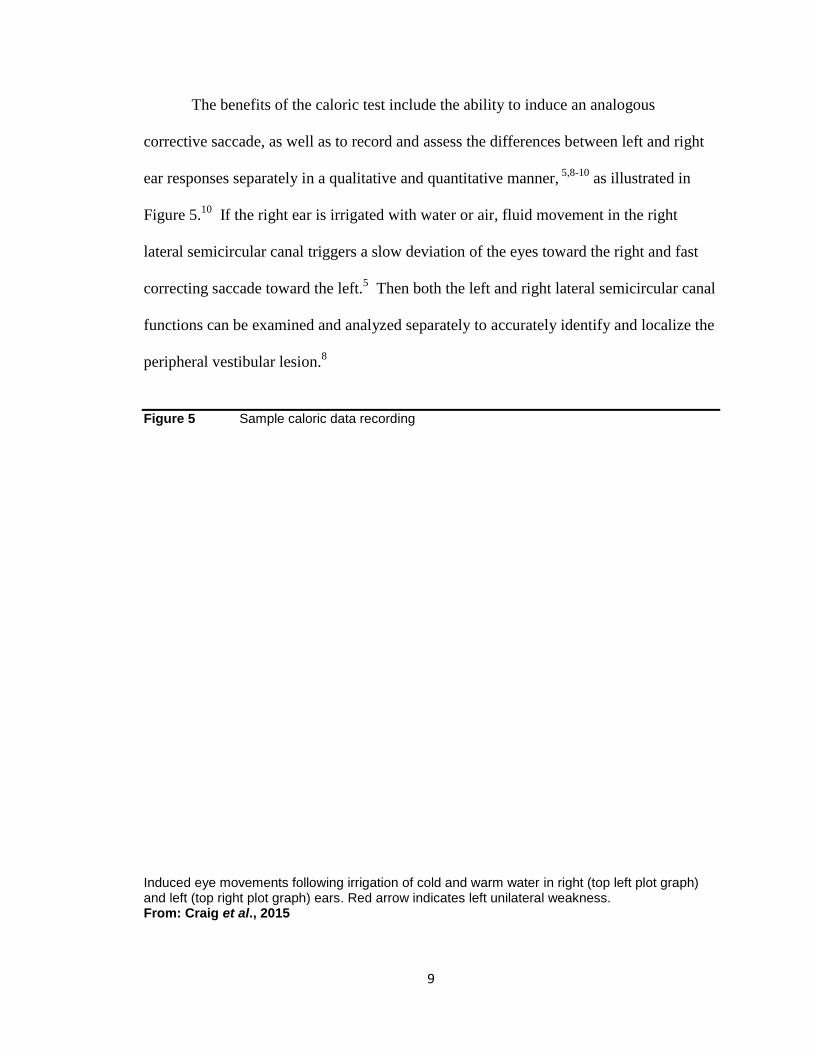
The benefits of the caloric test include the ability to induce an analogous
corrective saccade, as well as to record and assess the differences between left and right
ear responses separately in a qualitative and quantitative manner, 5,8-10
as illustrated in
Figure 5.10
If the right ear is irrigated with water or air, fluid movement in the right
lateral semicircular canal triggers a slow deviation of the eyes toward the right and fast
correcting saccade toward the left.5 Then both the left and right lateral semicircular canal
functions can be examined and analyzed separately to accurately identify and localize the
peripheral vestibular lesion.8
Figure 5 Sample caloric data recording
Induced eye movements following irrigation of cold and warm water in right (top left plot graph) and left (top right plot graph) ears. Red arrow indicates left unilateral weakness. From: Craig et al., 2015
10
Major limitations of this caloric test include lack of normal physiological
response, patient discomfort, and being time-consuming.5,6,8-10
While this test provides
qualitative and quantitative evaluation of peripheral vestibular function by comparing the
left and right lateral semicircular canals, it tests at a low, nonphysiological head rotation
frequency below what is considered normal.5,8,9
A normal peripheral vestibular system
responds to natural head movements covering a wide frequency range of approximately
0.01 to 8 Hz.5 However, the head rotation frequency from a caloric stimulation produces
only 0.003 Hz.5 Also, the quantitative assumption of peripheral vestibular dysfunction is
based only on the evaluation of the lateral semicircular canals, as the caloric test lacks the
ability to measure the other two anterior and posterior canals and the otolith organs.5,10
Patients often report discomfort in the ears during cold or warm water and air
irrigation, and may also experience brief symptoms of vertigo, nausea, and blurred vision
due to the nystagmus provocation as a response of water and air irrigation.6,9
The time to
complete a caloric test exclusively is approximately 30 minutes,10
but this test exists as a
subtest of the standardized electronystagmography (ENG) test battery, which in total can
take up to two-three hours.5,10
Rotary Chair Test
Rotary chair testing is another historically common peripheral vestibular function
assessment tool of early 20th century.11,12
It provides precise, quantitative analysis of the
VOR response by evaluating the vestibular gain (in terms of rotary chair, eye velocity /
chair velocity), phase (timing between eye velocity and head velocity), and asymmetry
(directional preponderance between left and right eye nystagmus movement).11,12
The
11
test also assesses the eyes’ VOR and/or corrective saccade response via physiological
rotational stimuli of the patient’s body and head en bloc - through a computer-controlled
motorized chair.13
The rotational stimuli can span a wide frequency range to simulate a
more natural head rotation of a normal vestibular system.13,14
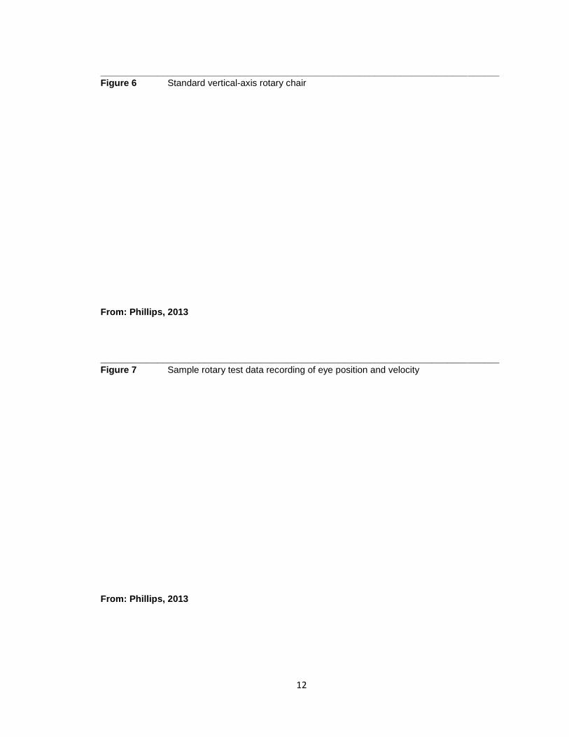
A standard rotary chair
places the patient in a vertical-axis rotation to allow direct assessment of the lateral
semicircular canal function as shown in Figure 6.13
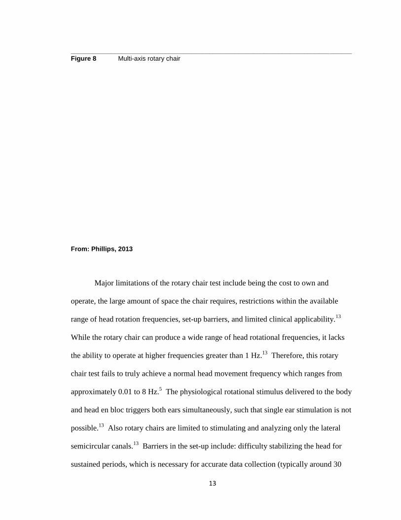
The eye movements generated by the
rotating patient in the chair are recorded by electro-oculography, while a software
program digitally analyzes the objective data which measure vestibular gain, phase, and
asymmetry11,13
as shown in Figure 7.13
The data are then compared to a large set of
normative data for adults to determine any clinical abnormalities of the peripheral
vestibular system.13
The main benefit of the rotary chair testing is the ability to simulate a dynamic
range of head rotational frequencies comparable to a normal vestibular system during
natural head rotation movements.13,14
This rotational frequency range varies by rotary
chair manufacturers, but the most common range is from 0.01 to 0.64 Hz.14
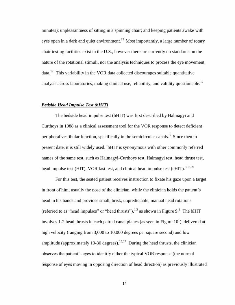
Also, many
manufacturers have produced rotary chairs that can also perform off-vertical axis rotation
to allow assessment of otolith organ function (which is outside of the scope of this paper)
in addition to lateral semicircular canal function.13
Figure 8 provides an example of a
multi-axis rotary chair.13
12
_____________________________________________________________________________Figure 6 Standard vertical-axis rotary chair
From: Phillips, 2013
_____________________________________________________________________________Figure 7 Sample rotary test data recording of eye position and velocity
From: Phillips, 2013
13
_____________________________________________________________________________Figure 8 Multi-axis rotary chair
From: Phillips, 2013
Major limitations of the rotary chair test include being the cost to own and
operate, the large amount of space the chair requires, restrictions within the available
range of head rotation frequencies, set-up barriers, and limited clinical applicability.13
While the rotary chair can produce a wide range of head rotational frequencies, it lacks
the ability to operate at higher frequencies greater than 1 Hz.13
Therefore, this rotary
chair test fails to truly achieve a normal head movement frequency which ranges from
approximately 0.01 to 8 Hz.5 The physiological rotational stimulus delivered to the body
and head en bloc triggers both ears simultaneously, such that single ear stimulation is not
possible.13
Also rotary chairs are limited to stimulating and analyzing only the lateral
semicircular canals.13
Barriers in the set-up include: difficulty stabilizing the head for
sustained periods, which is necessary for accurate data collection (typically around 30
14
minutes); unpleasantness of sitting in a spinning chair; and keeping patients awake with
eyes open in a dark and quiet environment.13
Most importantly, a large number of rotary
chair testing facilities exist in the U.S., however there are currently no standards on the
nature of the rotational stimuli, nor the analysis techniques to process the eye movement
data.12
This variability in the VOR data collected discourages suitable quantitative
analysis across laboratories, making clinical use, reliability, and validity questionable.12
Bedside Head Impulse Test (bHIT)
The bedside head impulse test (bHIT) was first described by Halmagyi and
Curthoys in 1988 as a clinical assessment tool for the VOR response to detect deficient
peripheral vestibular function, specifically in the semicircular canals.3 Since then to
present date, it is still widely used. bHIT is synonymous with other commonly referred
names of the same test, such as Halmagyi-Curthoys test, Halmagyi test, head thrust test,
head impulse test (HIT), VOR fast test, and clinical head impulse test (cHIT).3,15-21
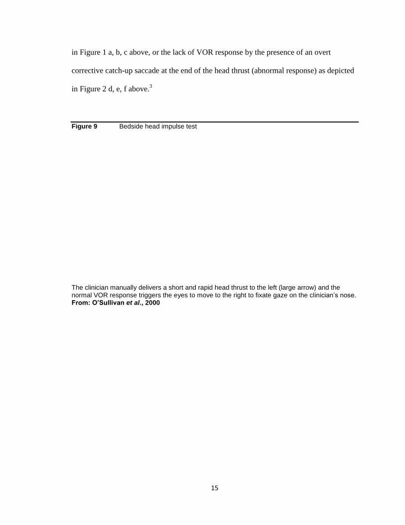
For this test, the seated patient receives instruction to fixate his gaze upon a target
in front of him, usually the nose of the clinician, while the clinician holds the patient’s
head in his hands and provides small, brisk, unpredictable, manual head rotations
(referred to as “head impulses” or “head thrusts”),1,3
as shown in Figure 9.1 The bHIT
involves 1-2 head thrusts in each paired canal planes (as seen in Figure 101), delivered at
high velocity (ranging from 3,000 to 10,000 degrees per square second) and low
amplitude (approximately 10-30 degrees).15,17
During the head thrusts, the clinician
observes the patient’s eyes to identify either the typical VOR response (the normal
response of eyes moving in opposing direction of head direction) as previously illustrated
15
in Figure 1 a, b, c above, or the lack of VOR response by the presence of an overt
corrective catch-up saccade at the end of the head thrust (abnormal response) as depicted
in Figure 2 d, e, f above.3
Figure 9 Bedside head impulse test
The clinician manually delivers a short and rapid head thrust to the left (large arrow) and the normal VOR response triggers the eyes to move to the right to fixate gaze on the clinician’s nose. From: O’Sullivan et al., 2000
16
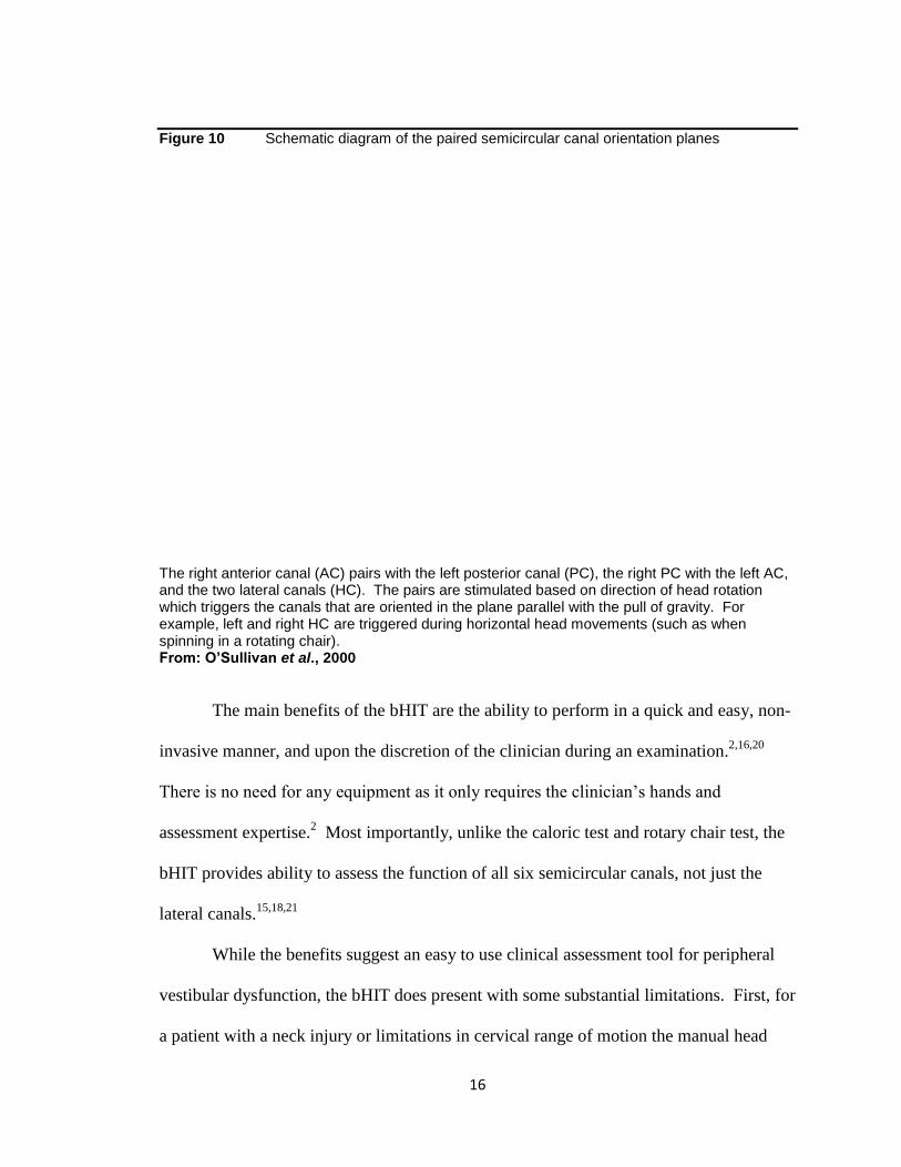
Figure 10 Schematic diagram of the paired semicircular canal orientation planes
The right anterior canal (AC) pairs with the left posterior canal (PC), the right PC with the left AC, and the two lateral canals (HC). The pairs are stimulated based on direction of head rotation which triggers the canals that are oriented in the plane parallel with the pull of gravity. For example, left and right HC are triggered during horizontal head movements (such as when spinning in a rotating chair). From: O’Sullivan et al., 2000
The main benefits of the bHIT are the ability to perform in a quick and easy, non-
invasive manner, and upon the discretion of the clinician during an examination.2,16,20
There is no need for any equipment as it only requires the clinician’s hands and
assessment expertise.2 Most importantly, unlike the caloric test and rotary chair test, the
bHIT provides ability to assess the function of all six semicircular canals, not just the
lateral canals.15,18,21
While the benefits suggest an easy to use clinical assessment tool for peripheral
vestibular dysfunction, the bHIT does present with some substantial limitations. First, for
a patient with a neck injury or limitations in cervical range of motion the manual head
17
thrusts of the bHIT method would be a general clinical precaution or a contraindication.10
Thus, this method would not be applicable for a patient with those types of limitations
and other assessment tools would need to be used.10
Second, an overt saccadic eye
movement is fairly easy to detect at the end of the head impulse, however covert saccades
that occur during the head impulse are not identifiable even by a well trained clinician’s
naked eye; a false-negative result could confound the diagnosis entirely.2,3,16-19
Third, the
bHIT relies on the clinician’s skills and visual acuity to provide the proper manual head
thrust and to detect the small and quick overt corrective saccade which only lasts
approximately 150 ms.17
Fourth, since the bHIT is a subjective test, the velocity and
amplitude provided during the head thrusts can vary greatly among clinicians.16,18,19
Fifth, the bHIT lacks an objective measure of both the VOR gain and the overt corrective
saccade.3,16,18
Finally, the bHIT only relies on a few head thrusts in the planes of each
paired canals but does not give a range of stimuli for generating a stimulus-response
function like that of a natural head rotation.16,19
Scleral Search Coil Technique
The bedside head impulse test (bHIT) contributed to the inception of the scleral
search coil technique, which is currently considered the gold standard for head impulse
test measurements.2,9,16,22,23
Since the VOR response requires coaction between the six
semicircular canals and the twelve extraocular muscles to stabilize gaze on a target, a tool
which provides accurate, objective measurement of head rotations and eye movements is
necessary.24
Unlike the subjective bHIT, the scleral search coil technique provides
quantifiable and recordable data to allow precise assessment of peripheral vestibular
18
dysfunction.2,16,22,24
Moreover, this technique also detects and records the elusive covert
corrective saccades which are undetectable with the bHIT.16,22,23
The scleral search coil technique requires sophisticated instrumentation consisting
of precalibrated dual-search coils which record head and eye positions onto a computer
software-driven device (as shown in Figures 11 and 1224
).22,24
For this test, a patient is
adorned with a head coil secured either on a head mounting band or to a dental
impression bite bar.16,21,22
Search coils mounted on a contact lens are placed in the
patient’s right eye after application of a topical anesthetic eye drop.16,21,22
The patient is
then seated in a chair such that the pupillary axis of the right eye is positioned in the
center of a magnetic field coil frame.16,21,22
After the device set-up, the room is dimly lit
or darkened, and the patient is instructed to fixate forward on a laser dot projected onto a
screen approximately one meter away.16,21,22
Then 20-50 manual head thrusts with
randomized amplitude, velocity, and acceleration are delivered to the patient in the planes
of the three paired semicircular canals.16,21,22
The head velocity and eye movements are
recorded and analyzed for overt and covert corrective saccades as illustrated in Figure
13.16
19
_____________________________________________________________________________Figure 11 Simplified schematic diagram of a scleral field coil
From: Robinson, 1963
Figure 12 Complete schematic diagram of a scleral search coil instrument
From: Robinson, 1963
20
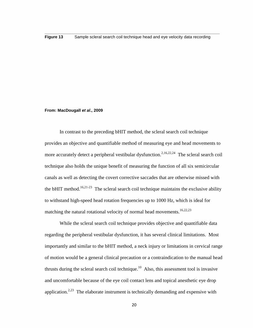
____________________________________________________________________________ Figure 13 Sample scleral search coil technique head and eye velocity data recording
From: MacDougall et al., 2009
In contrast to the preceding bHIT method, the scleral search coil technique
provides an objective and quantifiable method of measuring eye and head movements to
more accurately detect a peripheral vestibular dysfunction.2,16,22,24
The scleral search coil
technique also holds the unique benefit of measuring the function of all six semicircular
canals as well as detecting the covert corrective saccades that are otherwise missed with
the bHIT method.16,21-23
The scleral search coil technique maintains the exclusive ability
to withstand high-speed head rotation frequencies up to 1000 Hz, which is ideal for
matching the natural rotational velocity of normal head movements.16,22,23
While the scleral search coil technique provides objective and quantifiable data
regarding the peripheral vestibular dysfunction, it has several clinical limitations. Most
importantly and similar to the bHIT method, a neck injury or limitations in cervical range
of motion would be a general clinical precaution or a contraindication to the manual head
thrusts during the scleral search coil technique.10
Also, this assessment tool is invasive
and uncomfortable because of the eye coil contact lens and topical anesthetic eye drop
application.2,23
The elaborate instrument is technically demanding and expensive with

21
limited availability and practical use in the clinical field.2,16,22,23
The procedure is time-
intensive from set-up to data recording,16
and some studies have indicated minor eye coil
slippage during eye movement which can result in lower than actual eye velocity and
vestibular gain findings.21,22
Video Head Impulse Test (vHIT)
The lack of broad clinical applicability of the scleral search coil technique led
several researchers to develop the video head impulse test (vHIT) assessment tool.2,16,20
Based on the same principles and manual techniques as the bHIT, with the addition of the
objective, high-speed recordings of ocular and head velocity data as with the scleral
search coil technique, the vHIT assessment tool allows for more practical and widespread
use to quantitatively assess peripheral vestibular dysfunction.2,9,16,20,23
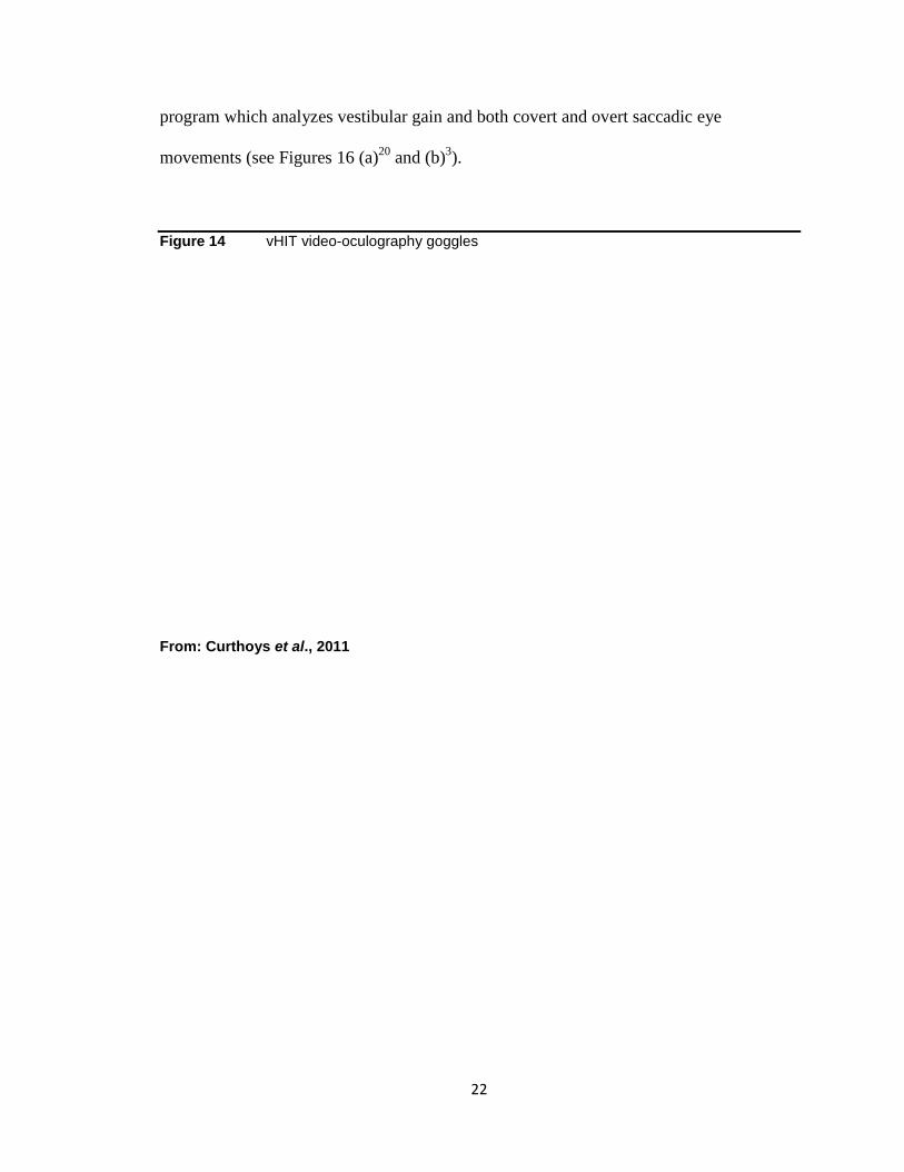
The vHIT entails the use of video-oculography, which is taking measurements of
right eye and head movements by a small, lightweight, high-speed, digital video camera
mounted onto a pair of equally lightweight eye goggles.3,16
A patient dons the eye
goggles and secures the attached elastic strap snug over the head to minimize slippage of
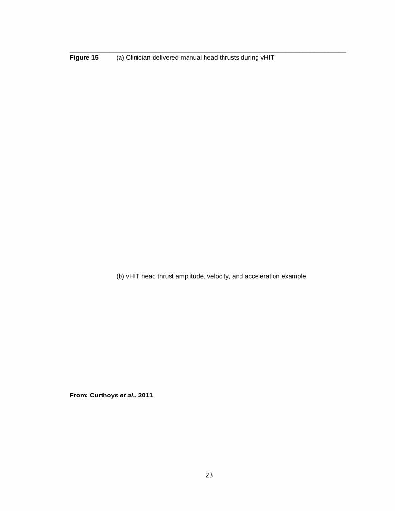
the camera as seen in Figure 14.3 Then, similar to the scleral search coil technique after
set-up, the patient is seated in a chair and instructed to fixate on a dot approximately one
meter away, while 15-20 short range, high-velocity, high-acceleration, unpredictable
head thrusts are manually delivered to the patient (as seen in Figures 15 (a) and (b)3)
) in
the plane of each of the three paired semicircular canals, as described in Figure 10
above.1,3,9,16,21,22
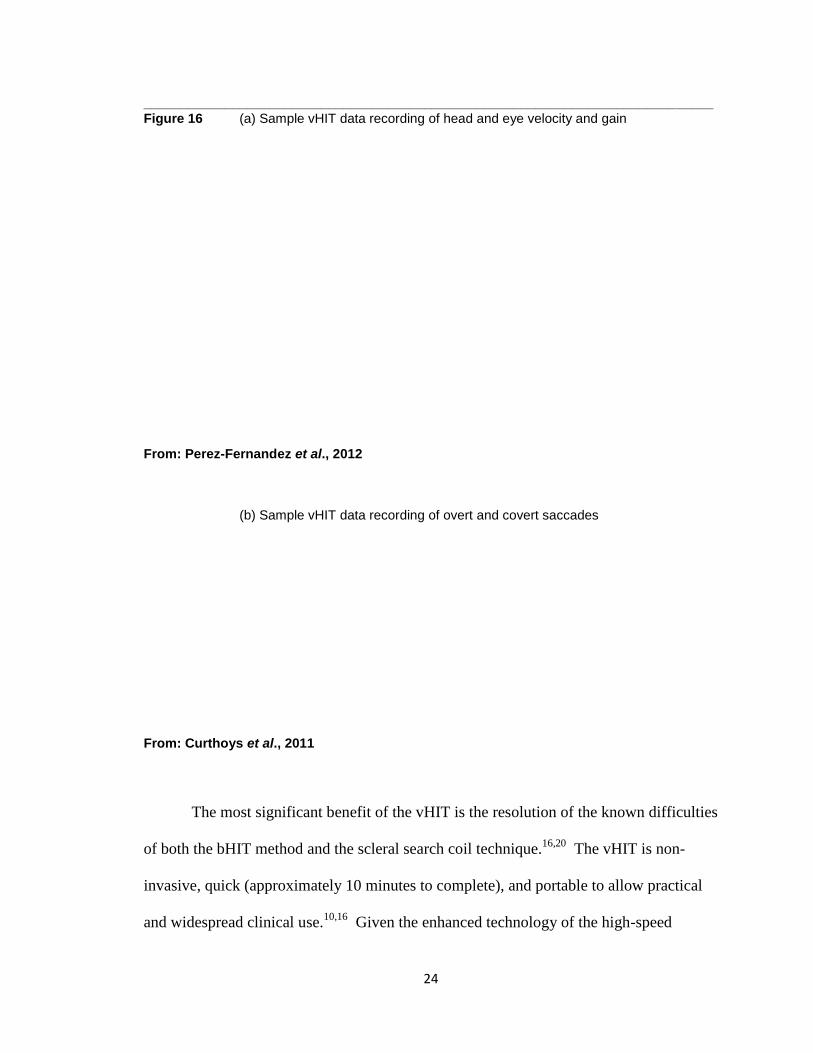
The eye and head movement data are recorded by a computer software
22
program which analyzes vestibular gain and both covert and overt saccadic eye
movements (see Figures 16 (a)20
and (b)3).
Figure 14 vHIT video-oculography goggles
From: Curthoys et al., 2011
23
_____________________________________________________________________________Figure 15 (a) Clinician-delivered manual head thrusts during vHIT
(b) vHIT head thrust amplitude, velocity, and acceleration example
From: Curthoys et al., 2011
24
_____________________________________________________________________________Figure 16 (a) Sample vHIT data recording of head and eye velocity and gain
From: Perez-Fernandez et al., 2012
(b) Sample vHIT data recording of overt and covert saccades
From: Curthoys et al., 2011
The most significant benefit of the vHIT is the resolution of the known difficulties
of both the bHIT method and the scleral search coil technique.16,20
The vHIT is non-
invasive, quick (approximately 10 minutes to complete), and portable to allow practical
and widespread clinical use.10,16
Given the enhanced technology of the high-speed
25
camera, only small 15-20 degree amplitude head thrusts are necessary, which makes the
test more pleasant for the patient.10
Also, due to decreased patient discomfort and
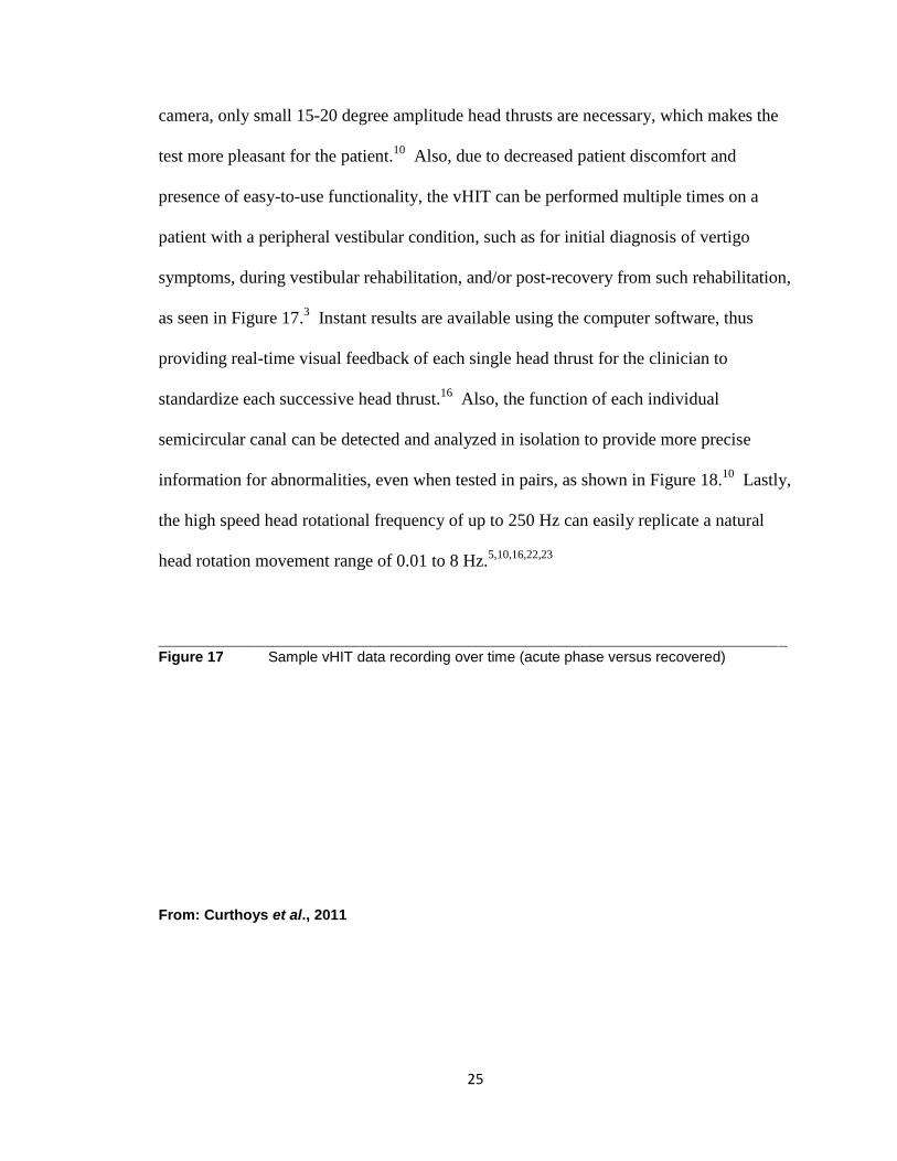
presence of easy-to-use functionality, the vHIT can be performed multiple times on a
patient with a peripheral vestibular condition, such as for initial diagnosis of vertigo
symptoms, during vestibular rehabilitation, and/or post-recovery from such rehabilitation,
as seen in Figure 17.3 Instant results are available using the computer software, thus
providing real-time visual feedback of each single head thrust for the clinician to
standardize each successive head thrust.16
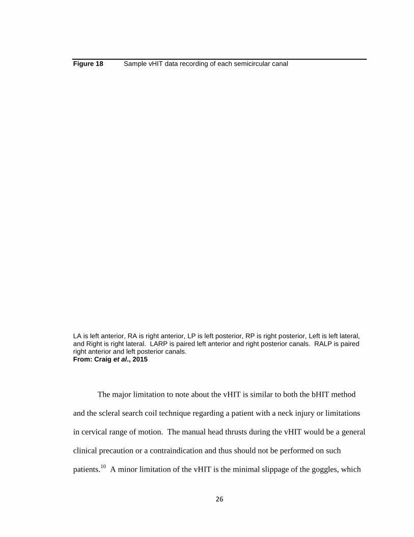
Also, the function of each individual
semicircular canal can be detected and analyzed in isolation to provide more precise
information for abnormalities, even when tested in pairs, as shown in Figure 18.10
Lastly,
the high speed head rotational frequency of up to 250 Hz can easily replicate a natural
head rotation movement range of 0.01 to 8 Hz.5,10,16,22,23
_____________________________________________________________________________Figure 17 Sample vHIT data recording over time (acute phase versus recovered)
From: Curthoys et al., 2011
26
Figure 18 Sample vHIT data recording of each semicircular canal
LA is left anterior, RA is right anterior, LP is left posterior, RP is right posterior, Left is left lateral, and Right is right lateral. LARP is paired left anterior and right posterior canals. RALP is paired right anterior and left posterior canals. From: Craig et al., 2015
The major limitation to note about the vHIT is similar to both the bHIT method
and the scleral search coil technique regarding a patient with a neck injury or limitations
in cervical range of motion. The manual head thrusts during the vHIT would be a general
clinical precaution or a contraindication and thus should not be performed on such
patients.10
A minor limitation of the vHIT is the minimal slippage of the goggles, which
27
usually occurs if there is not a snug fit of the goggles on the head.3,10,16,21,23
This slippage
creates artifactual results as if the eyes have moved off the fixed dot, underestimating the
vestibular gain, however a quick adjustment of the goggles can easily fix this
problem.3,10,16,21,23
The vHIT assessment tool has evolved from the benefits of both the bHIT method
and the scleral search coil technique, while eliminating the major limitations of both.
Given its noteworthy advantages and minimal limitations, the vHIT tool opens the
possibility of potentially being the next “gold standard” for identifying peripheral
vestibular dysfunction of the semicircular canals. The next section will evaluate recent
literature comparing each of the four described peripheral vestibular function assessment
tools-- the caloric test, rotary chair test, bedside head impulse test (bHIT), and scleral
search coil technique-- against the newest tool, the vHIT.
28
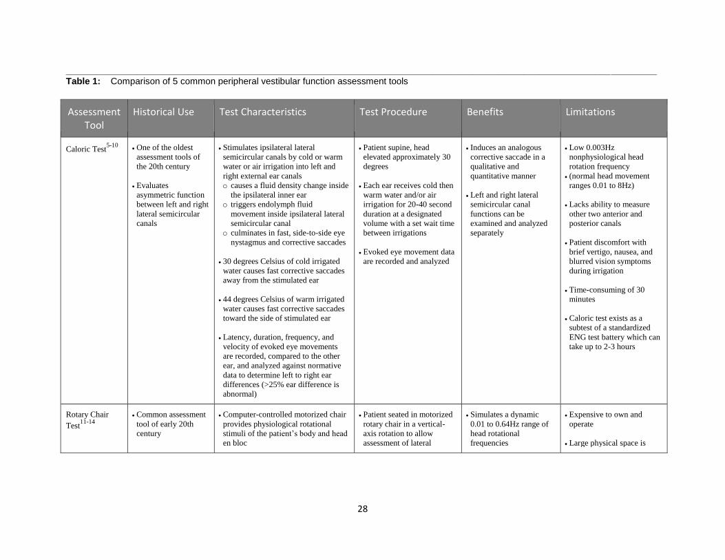
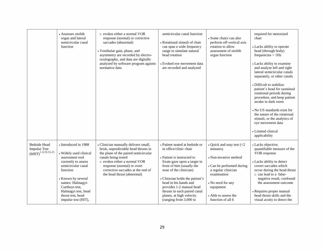
____________________________________________________________________________________________________________________Table 1: Comparison of 5 common peripheral vestibular function assessment tools
Assessment Tool
Historical Use Test Characteristics Test Procedure Benefits Limitations
Caloric Test5-10
One of the oldest
assessment tools of
the 20th century
Evaluates
asymmetric function
between left and right
lateral semicircular
canals
Stimulates ipsilateral lateral
semicircular canals by cold or warm
water or air irrigation into left and
right external ear canals
o causes a fluid density change inside
the ipsilateral inner ear
o triggers endolymph fluid
movement inside ipsilateral lateral
semicircular canal
o culminates in fast, side-to-side eye
nystagmus and corrective saccades
30 degrees Celsius of cold irrigated
water causes fast corrective saccades
away from the stimulated ear
44 degrees Celsius of warm irrigated
water causes fast corrective saccades
toward the side of stimulated ear
Latency, duration, frequency, and
velocity of evoked eye movements
are recorded, compared to the other
ear, and analyzed against normative
data to determine left to right ear
differences (>25% ear difference is
abnormal)
Patient supine, head
elevated approximately 30
degrees
Each ear receives cold then
warm water and/or air
irrigation for 20-40 second
duration at a designated
volume with a set wait time
between irrigations
Evoked eye movement data
are recorded and analyzed
Induces an analogous
corrective saccade in a
qualitative and
quantitative manner
Left and right lateral
semicircular canal
functions can be
examined and analyzed
separately
Low 0.003Hz
nonphysiological head
rotation frequency
(normal head movement
ranges 0.01 to 8Hz)
Lacks ability to measure
other two anterior and
posterior canals
Patient discomfort with
brief vertigo, nausea, and
blurred vision symptoms
during irrigation
Time-consuming of 30
minutes
Caloric test exists as a
subtest of a standardized
ENG test battery which can
take up to 2-3 hours
Rotary Chair
Test11-14
Common assessment
tool of early 20th
century
Computer-controlled motorized chair
provides physiological rotational
stimuli of the patient’s body and head
en bloc
Patient seated in motorized
rotary chair in a vertical-
axis rotation to allow
assessment of lateral
Simulates a dynamic
0.01 to 0.64Hz range of
head rotational
frequencies
Expensive to own and
operate
Large physical space is
29
Assesses otolith
organ and lateral
semicircular canal
function
o evokes either a normal VOR
response (normal) or corrective
saccades (abnormal)
Vestibular gain, phase, and
asymmetry are recorded by electro-
oculography, and data are digitally
analyzed by software program against
normative data
semicircular canal function
Rotational stimuli of chair
can span a wide frequency
range to simulate natural
head rotation
Evoked eye movement data
are recorded and analyzed
Some chairs can also
perform off-vertical axis
rotation to allow
assessment of otolith
organ function
required for motorized
chair
Lacks ability to operate
head (through body)
frequencies > 1Hz
Lacks ability to examine
and analyze left and right
lateral semicircular canals
separately, or other canals
Difficult to stabilize
patient’s head for sustained
rotational periods during
procedure, and keep patient
awake in dark room
No US standards exist for
the nature of the rotational
stimuli, or the analytics of
eye movement data
Limited clinical
applicability
Bedside Head
Impulse Test
(bHIT)1-3,10,15-21
Introduced in 1988
Widely used clinical
assessment tool
currently to assess
semicircular canal
function
Known by several
names: Halmagyi-
Curthoys test,
Halmagyi test, head
thrust test, head
impulse test (HIT),
Clinician manually delivers small,
brisk, unpredictable head thrusts in
the plane of the paired semicircular
canals being tested
o evokes either a normal VOR
response (normal) or overt
corrective saccades at the end of
the head thrust (abnormal)
Patient seated at bedside or
in office/clinic chair
Patient is instructed to
fixate gaze upon a target in
front of him (usually the
nose of the clinician)
Clinician holds the patient’s
head in his hands and
provides 1-2 manual head
thrusts in each paired canal
planes, at high velocity
(ranging from 3,000 to
Quick and easy test (<2
minutes)
Non-invasive method
Can be performed during
a regular clinician
examination
No need for any
equipment
Able to assess the
function of all 6
Lacks objective,
quantifiable measure of the
VOR response
Lacks ability to detect
covert saccades which
occur during the head thrust
o can lead to a false-
negative result, confound
the assessment outcome
Requires proper manual
head thrust skills and the
visual acuity to detect the
30
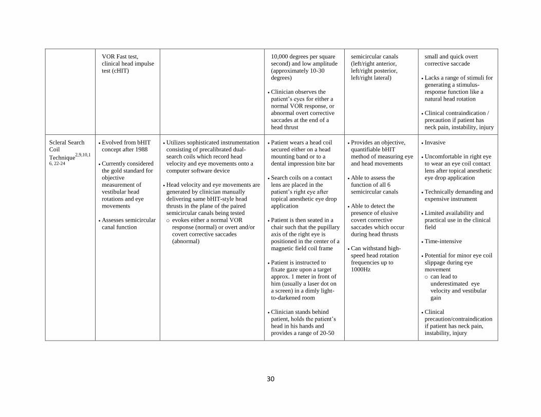
VOR Fast test,
clinical head impulse
test (cHIT)
10,000 degrees per square
second) and low amplitude
(approximately 10-30
degrees)
Clinician observes the
patient’s eyes for either a
normal VOR response, or
abnormal overt corrective
saccades at the end of a
head thrust
semicircular canals
(left/right anterior,
left/right posterior,
left/right lateral)
small and quick overt
corrective saccade
Lacks a range of stimuli for
generating a stimulus-
response function like a
natural head rotation
Clinical contraindication /
precaution if patient has
neck pain, instability, injury
Scleral Search
Coil
Technique2,9,10,1
6, 22-24
Evolved from bHIT
concept after 1988
Currently considered
the gold standard for
objective
measurement of
vestibular head
rotations and eye
movements
Assesses semicircular
canal function
Utilizes sophisticated instrumentation
consisting of precalibrated dual-
search coils which record head
velocity and eye movements onto a
computer software device
Head velocity and eye movements are
generated by clinician manually
delivering same bHIT-style head
thrusts in the plane of the paired
semicircular canals being tested
o evokes either a normal VOR
response (normal) or overt and/or
covert corrective saccades
(abnormal)
Patient wears a head coil
secured either on a head
mounting band or to a
dental impression bite bar
Search coils on a contact
lens are placed in the
patient’s right eye after
topical anesthetic eye drop
application
Patient is then seated in a
chair such that the pupillary
axis of the right eye is
positioned in the center of a
magnetic field coil frame
Patient is instructed to
fixate gaze upon a target
approx. 1 meter in front of
him (usually a laser dot on
a screen) in a dimly light-
to-darkened room
Clinician stands behind
patient, holds the patient’s
head in his hands and
provides a range of 20-50
Provides an objective,
quantifiable bHIT
method of measuring eye
and head movements
Able to assess the
function of all 6
semicircular canals
Able to detect the
presence of elusive
covert corrective
saccades which occur
during head thrusts
Can withstand high-
speed head rotation
frequencies up to
1000Hz
Invasive
Uncomfortable in right eye
to wear an eye coil contact
lens after topical anesthetic
eye drop application
Technically demanding and
expensive instrument
Limited availability and
practical use in the clinical
field
Time-intensive
Potential for minor eye coil
slippage during eye
movement
o can lead to
underestimated eye
velocity and vestibular
gain
Clinical
precaution/contraindication
if patient has neck pain,
instability, injury
31
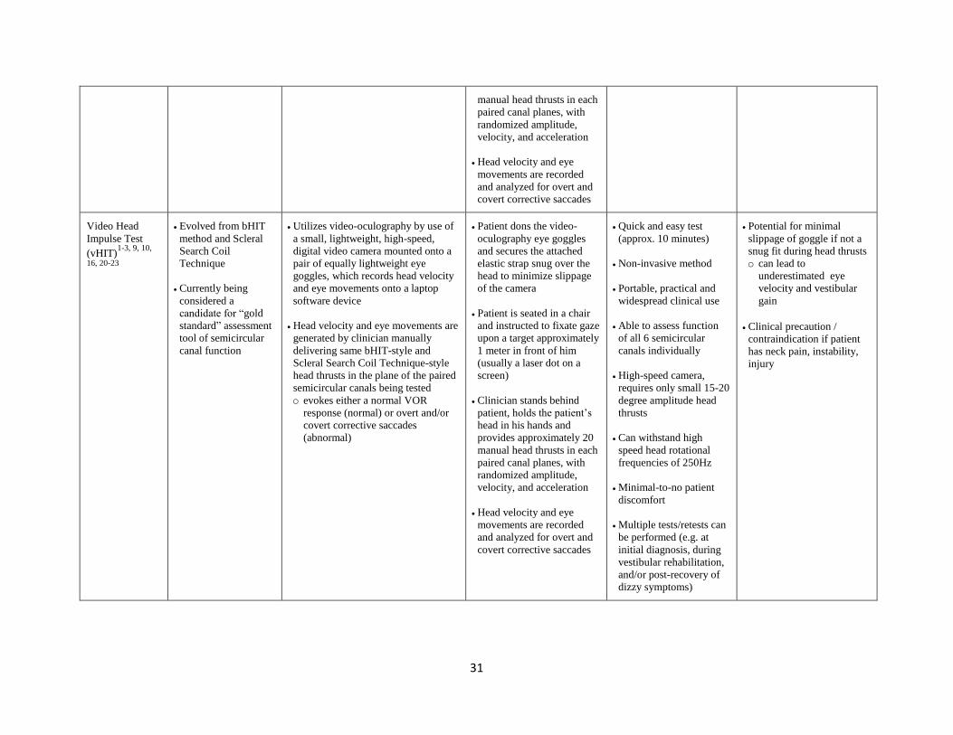
manual head thrusts in each
paired canal planes, with
randomized amplitude,
velocity, and acceleration
Head velocity and eye
movements are recorded
and analyzed for overt and
covert corrective saccades
Video Head
Impulse Test
(vHIT)1-3, 9, 10,
16, 20-23
Evolved from bHIT
method and Scleral
Search Coil
Technique
Currently being
considered a
candidate for “gold
standard” assessment
tool of semicircular
canal function
Utilizes video-oculography by use of
a small, lightweight, high-speed,
digital video camera mounted onto a
pair of equally lightweight eye
goggles, which records head velocity
and eye movements onto a laptop
software device
Head velocity and eye movements are
generated by clinician manually
delivering same bHIT-style and
Scleral Search Coil Technique-style
head thrusts in the plane of the paired
semicircular canals being tested
o evokes either a normal VOR
response (normal) or overt and/or
covert corrective saccades
(abnormal)
Patient dons the video-
oculography eye goggles
and secures the attached
elastic strap snug over the
head to minimize slippage
of the camera
Patient is seated in a chair
and instructed to fixate gaze
upon a target approximately
1 meter in front of him
(usually a laser dot on a
screen)
Clinician stands behind
patient, holds the patient’s
head in his hands and
provides approximately 20
manual head thrusts in each
paired canal planes, with
randomized amplitude,
velocity, and acceleration
Head velocity and eye
movements are recorded
and analyzed for overt and
covert corrective saccades
Quick and easy test
(approx. 10 minutes)
Non-invasive method
Portable, practical and
widespread clinical use
Able to assess function
of all 6 semicircular
canals individually
High-speed camera,
requires only small 15-20
degree amplitude head
thrusts
Can withstand high
speed head rotational
frequencies of 250Hz
Minimal-to-no patient
discomfort
Multiple tests/retests can
be performed (e.g. at
initial diagnosis, during
vestibular rehabilitation,
and/or post-recovery of
dizzy symptoms)
Potential for minimal
slippage of goggle if not a
snug fit during head thrusts
o can lead to
underestimated eye
velocity and vestibular
gain
Clinical precaution /
contraindication if patient
has neck pain, instability,
injury
32
SECTION 3: Comparative Literature Search
Recent literature analyzing the potential of the video head impulse test (vHIT) as
the next “gold standard” for identifying peripheral vestibular dysfunction of the
semicircular canals is evaluated in this section. The included studies compare the vHIT
against the four commonly known function assessment tools described above: caloric
test, rotary chair test, bedside head impulse test (bHIT), and scleral search coil technique
(Table 2).
vHIT versus Caloric Test
Mahringer et al.25
examined the sensitivity and specificity of the vHIT against the
well-known caloric test to identify pathological unilateral vestibular hypofunction of the
lateral semicircular canals. In general, sensitivity refers to how well a test screens for
pathology (sensitivity equals the number of abnormal tests divided by the number of
subjects with the pathological condition).14
In contrast, specificity refers to how well a
test identifies subjects without the pathology (specificity equals the number of normal
tests divided by the number of subjects without the pathological condition).14
Therefore,
a test with a high sensitivity indicates adequate capacity to correctly identify patients with
the condition of interest with an abnormal test result, while a low sensitivity provides
poor capability. In contrast, a test with a high specificity offers adequate capacity in
detecting patients without the condition with a normal test result, while a low specificity
suggests poor capability.
33
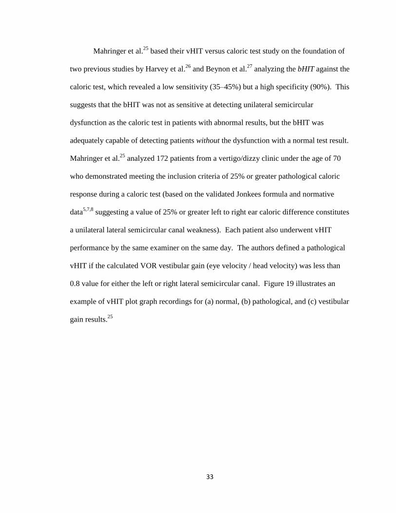
Mahringer et al.25
based their vHIT versus caloric test study on the foundation of
two previous studies by Harvey et al.26
and Beynon et al.27
analyzing the bHIT against the
caloric test, which revealed a low sensitivity (35–45%) but a high specificity (90%). This
suggests that the bHIT was not as sensitive at detecting unilateral semicircular
dysfunction as the caloric test in patients with abnormal results, but the bHIT was
adequately capable of detecting patients without the dysfunction with a normal test result.
Mahringer et al.25
analyzed 172 patients from a vertigo/dizzy clinic under the age of 70
who demonstrated meeting the inclusion criteria of 25% or greater pathological caloric
response during a caloric test (based on the validated Jonkees formula and normative
data5,7,8
suggesting a value of 25% or greater left to right ear caloric difference constitutes
a unilateral lateral semicircular canal weakness). Each patient also underwent vHIT
performance by the same examiner on the same day. The authors defined a pathological
vHIT if the calculated VOR vestibular gain (eye velocity / head velocity) was less than
0.8 value for either the left or right lateral semicircular canal. Figure 19 illustrates an
example of vHIT plot graph recordings for (a) normal, (b) pathological, and (c) vestibular
gain results.25
34
_____________________________________________________________________________ Figure 19 vHIT plot graph. (a) normal, (b) pathological, and (c) vestibular gain
For (a) and (b) black line is eye velocity, gray line is head velocity. For (c) open circle is single gain value of normal, black circle is mean gain value of normal. Gray square is singe gain value of pathological, and black square is mean value of pathological. From: Mahringer et al., 2014
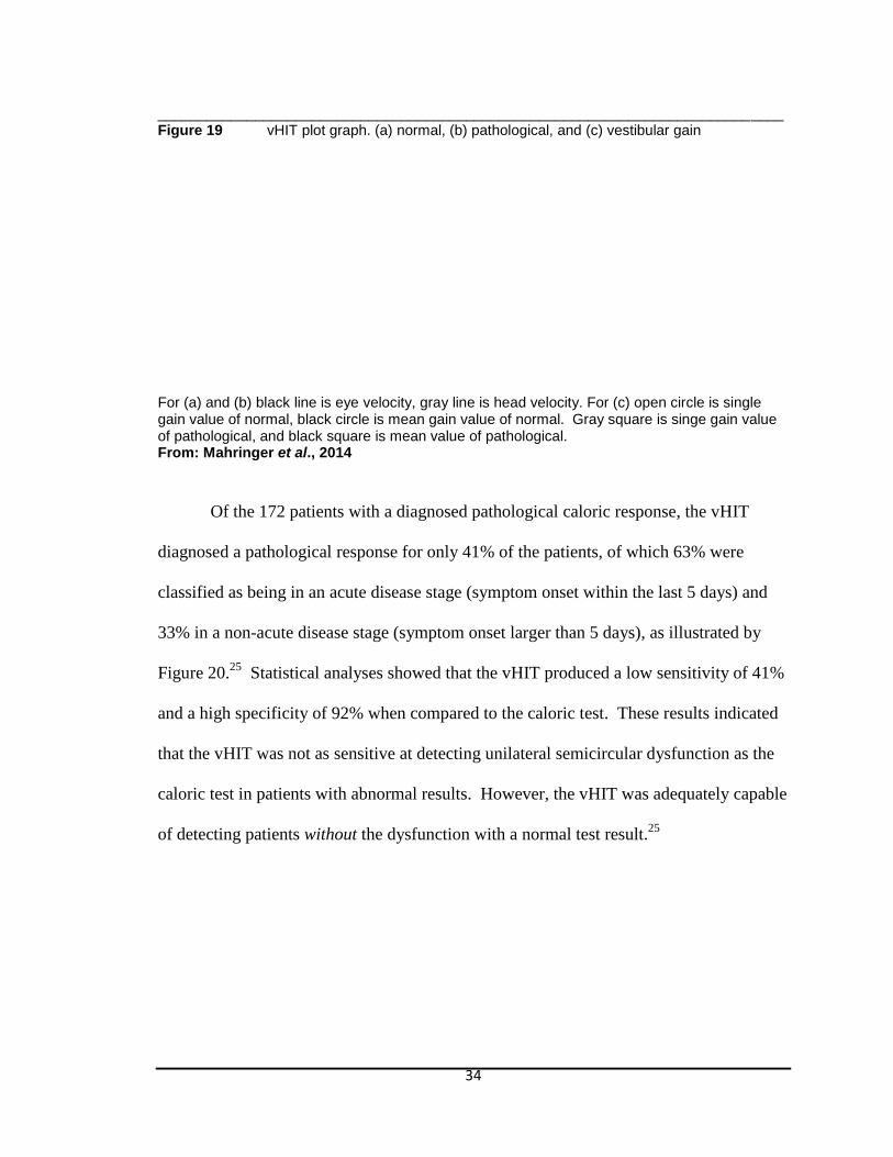
Of the 172 patients with a diagnosed pathological caloric response, the vHIT
diagnosed a pathological response for only 41% of the patients, of which 63% were
classified as being in an acute disease stage (symptom onset within the last 5 days) and
33% in a non-acute disease stage (symptom onset larger than 5 days), as illustrated by
Figure 20.25
Statistical analyses showed that the vHIT produced a low sensitivity of 41%
and a high specificity of 92% when compared to the caloric test. These results indicated
that the vHIT was not as sensitive at detecting unilateral semicircular dysfunction as the
caloric test in patients with abnormal results. However, the vHIT was adequately capable
of detecting patients without the dysfunction with a normal test result.25
35
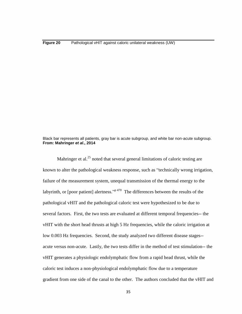
Figure 20 Pathological vHIT against caloric unilateral weakness (UW)
Black bar represents all patients, gray bar is acute subgroup, and white bar non-acute subgroup. From: Mahringer et al., 2014
Mahringer et al.25
noted that several general limitations of caloric testing are
known to alter the pathological weakness response, such as “technically wrong irrigation,
failure of the measurement system, unequal transmission of the thermal energy to the
labyrinth, or [poor patient] alertness.”p 470
The differences between the results of the
pathological vHIT and the pathological caloric test were hypothesized to be due to
several factors. First, the two tests are evaluated at different temporal frequencies-- the
vHIT with the short head thrusts at high 5 Hz frequencies, while the caloric irrigation at
low 0.003 Hz frequencies. Second, the study analyzed two different disease stages--
acute versus non-acute. Lastly, the two tests differ in the method of test stimulation-- the
vHIT generates a physiologic endolymphatic flow from a rapid head thrust, while the
caloric test induces a non-physiological endolymphatic flow due to a temperature
gradient from one side of the canal to the other. The authors concluded that the vHIT and
36
caloric test assessment tools complemented each other in identifying vestibular
hypofunction of the lateral semicircular canals; the vHIT identifies dysfunction at high
frequencies, while caloric test identifies dysfunction at low frequencies. They postulated
that to save time clinically, the vHIT should be performed first and, if unremarkable, a
caloric test should then be undertaken.
Another study by McCaslin et al.9 also evaluated the sensitivity and specificity of
the vHIT against what they considered the “gold standard” caloric test for detecting
peripheral vestibular dysfunction of the lateral semicircular canals, with an added
component of a self-reported dizziness handicap outcome measure (Dizziness Handicap
Inventory) which is outside the scope of this paper. 115 patients under the age of 65 with
symptoms of dizziness and negative MRI findings were enrolled and underwent both
caloric testing and vHIT assessment at the same appointment. For statistical analysis,
researchers were blinded of the results of the caloric test during interpretation of vHIT
data. Patients were placed into four groups based on calculated caloric asymmetry
between the left and right ears, as Group 1: 0–25%, Group 2: 26–50%, Group 3: 51–75%,
and Group 4: 76–100%. The vHIT test was considered abnormal if VOR vestibular gain
dropped below 0.7, and if covert and overt saccades were present for >50% of the head
thrust trials.
Findings revealed that the more severe the caloric asymmetry from the four
groups the further the VOR vestibular gain reduced, and the more the presence of overt
and/or covert corrective saccades increased. These results suggested impaired peripheral
vestibular function of the lateral semicircular canals as depicted in Figure 21.9 Statistical
analyses showed the vHIT produced a high sensitivity of 78% and a higher specificity of
37
95% when compared to the caloric test at cutoff point of 39.50% caloric asymmetry. The
high sensitivity indicates the vHIT can adequately detect unilateral semicircular
dysfunction as the caloric test can in patients with at least 39.50% caloric asymmetry.
The high specificity suggests the vHIT is also adequately capable of detecting patients
without the dysfunction with a normal test result. Additionally, no significant
correlations were found between the two test results and the self-reported dizziness
handicap outcome measure, as hypothesized.9
Figure 21 Normal versus abnormal vHIT against 4 groups of caloric asymmetry
From: McCaslin et al., 2014
McCaslin et al.9 questioned the discrepancy in the findings which deemed the
caloric test as abnormal while the vHIT as normal, specifically at caloric asymmetries
between 25 and 40%. Long historical use of caloric testing has established the value of
25% or greater ear differential as being a valid indicator of pathological unilateral lateral
38
semicircular canal impairment. The authors acknowledged the commonly known
limitations of the caloric test (such as non-physiological and low frequencies, time-
intensiveness, and patient discomfort) but also highlighted some issues with vHIT testing.
Some of these issues included difficulty relaxing neck musculature and adhering to
instructions of fixating gaze on the target, which can both negatively affect head thrust
performance. They concluded that vHIT and caloric data are not redundant such that the
vHIT should replace the caloric test for the “gold standard” distinction, but instead, the
tests are complementary. They did, however, offer the many evolved advantages the
vHIT has over caloric testing, such as high specificity in pathological peripheral
vestibular cases, quick test time, non-invasive procedures, and the ability to test vertical
semicircular canals which caloric testing cannot do.
vHIT versus Rotary Chair Test
A literature search reveals few comparisons between the vHIT (or even the bHIT)
and the rotary chair test. However, many studies comparing the rotary chair test with the
caloric test exist. Generally, these two tests are performed in a standardized protocol that
includes a list of several subset tests (such as ocular saccades, smooth pursuit, tracking,
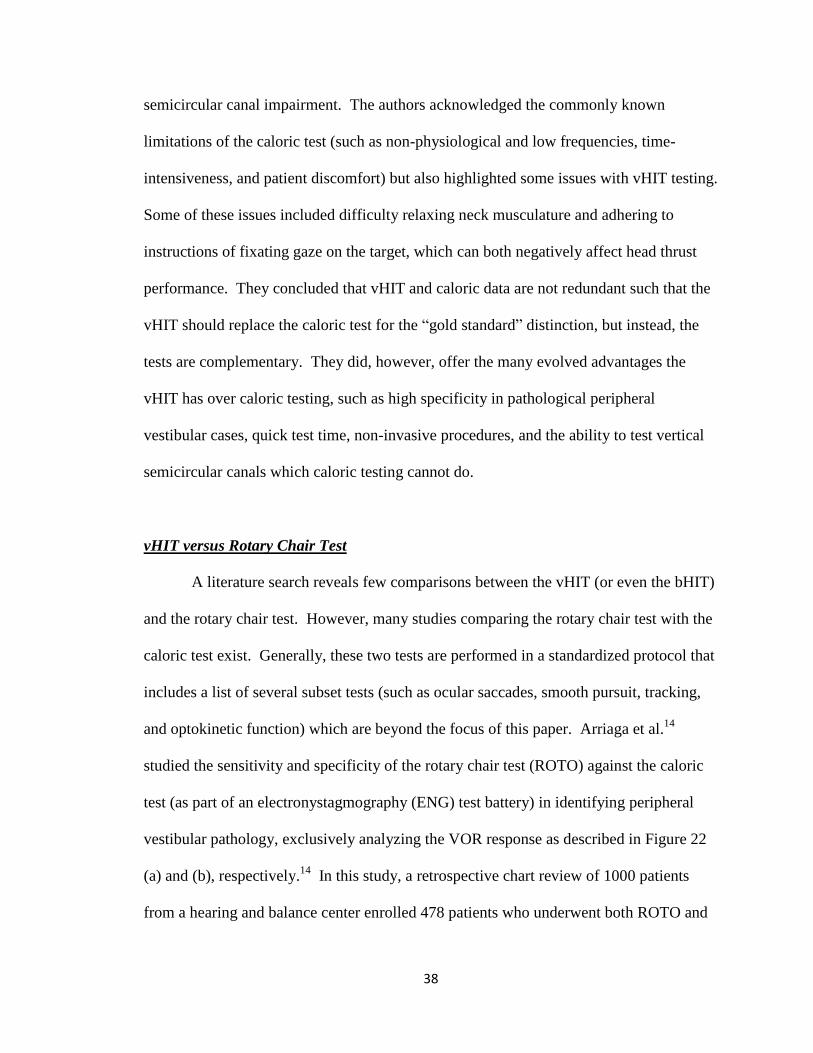
and optokinetic function) which are beyond the focus of this paper. Arriaga et al.14
studied the sensitivity and specificity of the rotary chair test (ROTO) against the caloric
test (as part of an electronystagmography (ENG) test battery) in identifying peripheral
vestibular pathology, exclusively analyzing the VOR response as described in Figure 22
(a) and (b), respectively.14
In this study, a retrospective chart review of 1000 patients
from a hearing and balance center enrolled 478 patients who underwent both ROTO and
39
ENG testing. The ROTO test was defined as abnormal if there were two frequencies with
abnormal gain, phase, or symmetry on VOR testing. The ENG test was defined as
abnormal if the caloric left versus right ear differential was greater than 25%.
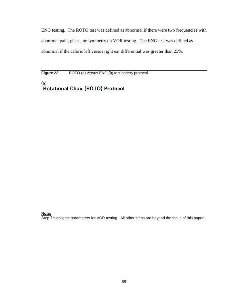
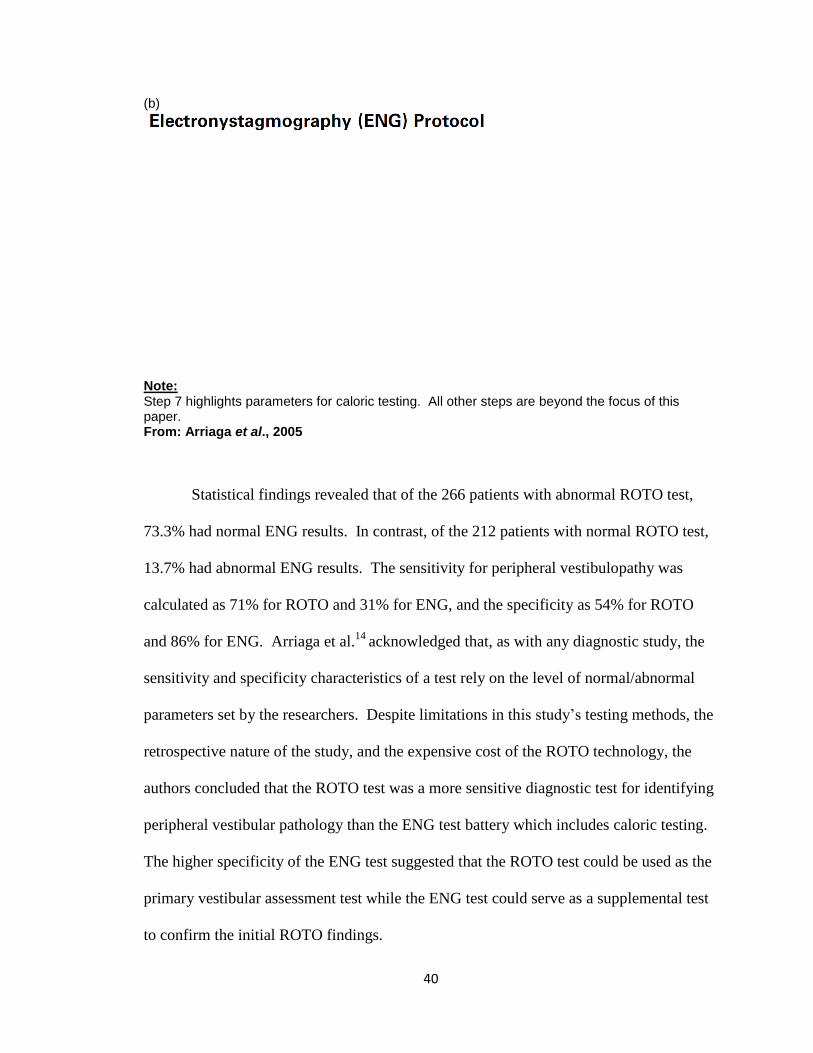
Figure 22 ROTO (a) versus ENG (b) test battery protocol
(a)
Note: Step 7 highlights parameters for VOR testing. All other steps are beyond the focus of this paper.
40
(b)
Note: Step 7 highlights parameters for caloric testing. All other steps are beyond the focus of this paper. From: Arriaga et al., 2005
Statistical findings revealed that of the 266 patients with abnormal ROTO test,
73.3% had normal ENG results. In contrast, of the 212 patients with normal ROTO test,
13.7% had abnormal ENG results. The sensitivity for peripheral vestibulopathy was
calculated as 71% for ROTO and 31% for ENG, and the specificity as 54% for ROTO
and 86% for ENG. Arriaga et al.14
acknowledged that, as with any diagnostic study, the
sensitivity and specificity characteristics of a test rely on the level of normal/abnormal
parameters set by the researchers. Despite limitations in this study’s testing methods, the
retrospective nature of the study, and the expensive cost of the ROTO technology, the
authors concluded that the ROTO test was a more sensitive diagnostic test for identifying
peripheral vestibular pathology than the ENG test battery which includes caloric testing.
The higher specificity of the ENG test suggested that the ROTO test could be used as the
primary vestibular assessment test while the ENG test could serve as a supplemental test
to confirm the initial ROTO findings.
41
Despite the lack of literature comparing the vHIT to the rotary chair test, the
rotary chair test in itself presents with many limitations as previously mentioned (such as
being very costly to own and operate, the large amount of space the chair requires,
restrictions within the available range of head rotation frequencies, set-up barriers, and
limited clinical applicability).13
Such substantial barriers restrict the rotary chair test
from being an ideal and practical assessment tool in today’s fast-paced, evidence-based,
technologically advanced, and patient-driven medical field.5,12,13
vHIT versus bHIT
The bHIT has been widely used as a highly specific clinical assessment tool for
the VOR response in detecting peripheral vestibular dysfunction of the semicircular
canals since its inception in 1988.3,16,20
However, as a result of low sensitivity and lack
of objective and quantifiable data, the limitations in the bHIT fueled the evolution of the
vHIT.3,16,18
Perez-Fernandez et al.20
performed a comparative study of the bHIT against the
vHIT in 179 patients with various types of balance and dizziness peripheral vestibular
disorders. The bHIT method was performed on all patients with three manual,
randomized head thrusts in the left and right horizontal directions, with visual
examination by the experimenter to determine whether the test was normal (absence of
ocular overt saccades at end of the head movement in at least two thrusts) or abnormal
(presence of overt saccades). The vHIT method was then performed on the same patients
using a pair of video goggles with built-in, high-definition, high-speed camera which
recorded eye and head movements during 20 quick, experimenter-delivered, random,
42
manual head thrusts in the same left and right horizontal directions as the bHIT method.
VOR vestibular gains below 0.6 were considered abnormal, and corrective saccadic eye
movements, if present, were classified as either overt or covert.
The results of this study by Perez-Fernandez et al.20
showed variances in normal
and abnormal bHIT and vHIT findings with an overlap of only 67.9% of head thrusts
performed. The majority of the findings showed the bHIT as normal while the vHIT as
abnormal. The authors raised several reasons for this discrepancy. First, only three
thrusts were performed to each side in the bHIT method versus 20 in the vHIT method.
Second, there was no quantifiable measure of the head movement velocity in the bHIT
method as there was with the recorded thrusts delivered during the vHIT method. This
then eliminates the ability of the bHIT method to calculate the crucial VOR gain equation
that is vital in studying the functionality of the peripheral vestibular system. Third, and
most importantly, the bHIT lacks the ability to detect covert saccades by simple visual
observation by an experimenter, which can lead to a false negative diagnosis of “normal”
peripheral vestibular function. These covert saccades, however, are easily recordable
with the vHIT method. The authors concluded that the methodology of the vHIT device
provided the necessary objective data in a simplistic, time-sensitive, and versatile manner
while still allowing it to be as clinically applicable as the bHIT method.
Another study by Blodow et al.18
also evaluated the accuracy of the vHIT
assessment tool on 117 patients with diagnosed peripheral vestibular dysfunction (based
on previously performed caloric test, cranial MRI, and bHIT results) and 20 healthy
subjects. All participants underwent a vHIT assessment which included wearing a
lightweight video goggle with an attached video-oculography camera. A minimum of 10
43
manual and unpredictable head thrusts in the left and right horizontal plane were
delivered as the patient fixated gaze on a dot located on a wall 1.2 meters ahead.
Statistical data defined a VOR vestibular gain of less than 0.79 and the presence of covert
and/or overt saccades as an abnormal VOR response. The results found that the healthy
subjects had a high VOR vestibular gain of 0.96 for left and right lateral semicircular
canals. For the patients with varying types of peripheral vestibular dysfunction, the VOR
vestibular gain was found to have a low overall mean of 0.44. The authors concluded
that the vHIT assessment tool can accurately detect abnormal VOR responses and record
the covert saccades that are missed by exclusive visual examination of the bHIT tool,
making diagnosis of peripheral vestibular dysfunction more definitive.
vHIT versus Scleral Search Coil Technique
Two highly acclaimed and widely referenced research studies compared the
effectiveness of the vHIT assessment tool to the current gold standard scleral search coil
technique.16,23
The MacDougall et al.16
study was a prospective, cross-sectional
comparison study that enrolled 16 participants ranging from 29 to 66 years of age, of
which eight were patients with confirmed peripheral vestibular dysfunction (ranging from
five months to 27 years of symptoms), and eight were healthy subjects that served as the
control group. Both groups wore the video-oculographic goggles with the built-in, high-
speed camera, as well as the scleral search coil contact lens in the right eye to allow for
simultaneous recording of both tests, the vHIT and the scleral search coil technique. All
participants were instructed to fixate on a laser dot on a screen 91 cm in front of them in
dim light while approximately 50 horizontal manual head thrusts at random velocity,
44
amplitude, and frequency were delivered to them by the same experimenter. Two data
sets were obtained for each recording session to show the reliability of the calculated
gains of the video-oculography and the search coil methods. Criterion for abnormal VOR
vestibular gain was 0.68 or less.
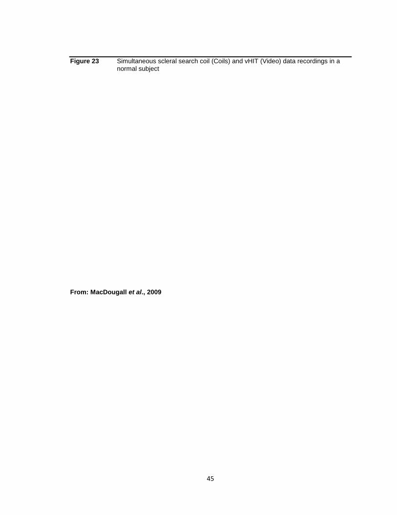
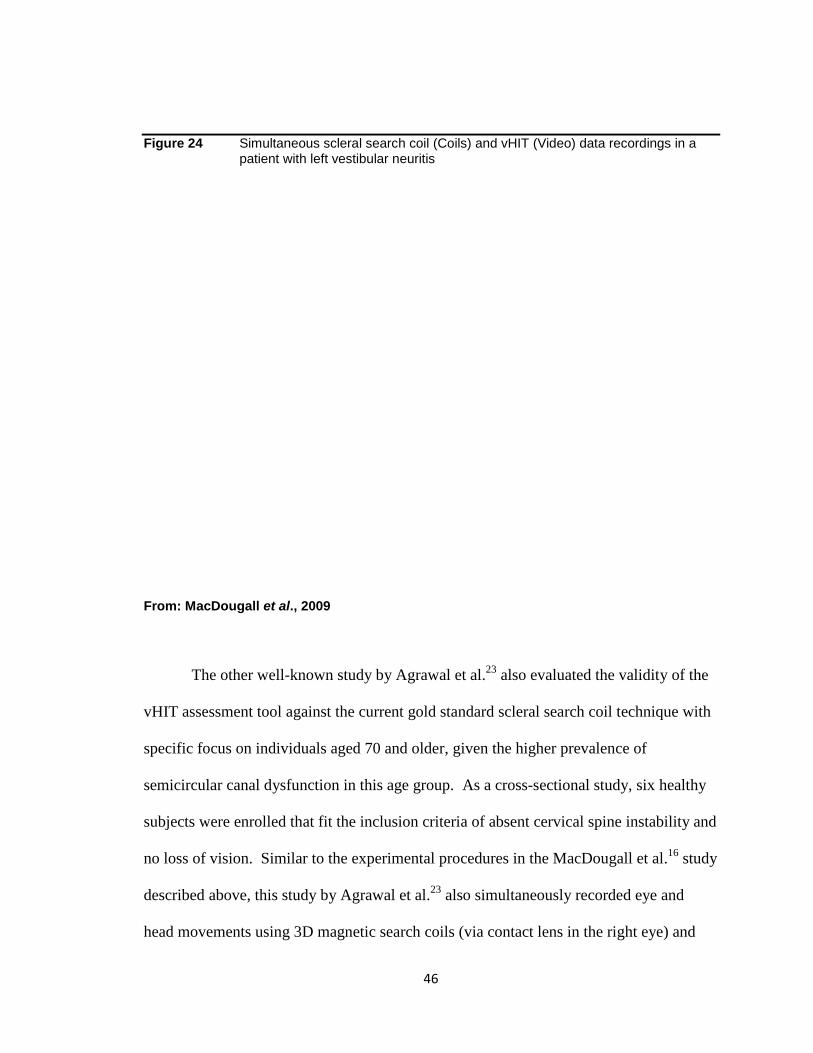
Findings from this MacDougall et al.16
study revealed that the simultaneous
recordings of VOR response and presence of saccadic eye movements from the vHIT and
scleral search coil technique were closely comparable, and without any significant
differences between the control and patient groups (as shown in Figures 23 and 24,
respectively16
). The sensitivity and specificity of both these tests were 1.0 (95%
confidence interval 0.69–1.0). The authors concluded that if the vHIT and the scleral
search coil technique produced equal results in both healthy subjects and patients with
pre-diagnosed impaired peripheral vestibular function, then the vHIT could be considered
an accurate and valid assessment tool.
45
Figure 23 Simultaneous scleral search coil (Coils) and vHIT (Video) data recordings in a normal subject
From: MacDougall et al., 2009
46
Figure 24 Simultaneous scleral search coil (Coils) and vHIT (Video) data recordings in a patient with left vestibular neuritis
From: MacDougall et al., 2009
The other well-known study by Agrawal et al.23
also evaluated the validity of the
vHIT assessment tool against the current gold standard scleral search coil technique with
specific focus on individuals aged 70 and older, given the higher prevalence of
semicircular canal dysfunction in this age group. As a cross-sectional study, six healthy
subjects were enrolled that fit the inclusion criteria of absent cervical spine instability and
no loss of vision. Similar to the experimental procedures in the MacDougall et al.16
study
described above, this study by Agrawal et al.23
also simultaneously recorded eye and
head movements using 3D magnetic search coils (via contact lens in the right eye) and
47
2D video-oculography (via video goggles with built-in, high-speed camera over the left
eye). Subjects were instructed to fixate gaze at a dot located 124 cm directly in front of
them at eye level. Recordings were performed twice to measure test-retest reliability for
both of the tests. Comparison measurement criterion was the ‘best value’ angular VOR
vestibular gain (AVOR gain) for each head thrust.
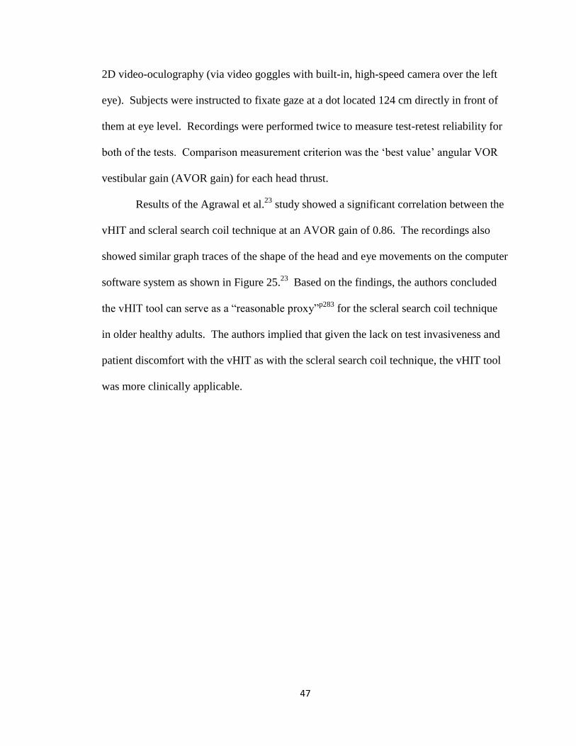
Results of the Agrawal et al.23
study showed a significant correlation between the
vHIT and scleral search coil technique at an AVOR gain of 0.86. The recordings also
showed similar graph traces of the shape of the head and eye movements on the computer
software system as shown in Figure 25.23
Based on the findings, the authors concluded
the vHIT tool can serve as a “reasonable proxy”p283
for the scleral search coil technique
in older healthy adults. The authors implied that given the lack on test invasiveness and
patient discomfort with the vHIT as with the scleral search coil technique, the vHIT tool
was more clinically applicable.
48
Figure 25 Individual head velocity and eye velocity traces during simultaneous scleral search coil (Search Coil) and vHIT (VOG for video oculography) data recordings of a normal subject. Yaw, Pitch, Roll represent head angular velocity positions.
From: Agrawal et al., 2014
49
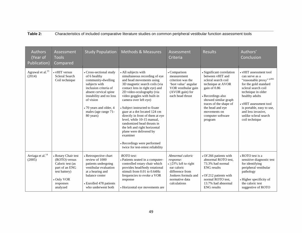
____________________________________________________________________________________________________________________Table 2: Characteristics of included comparative literature studies on common peripheral vestibular function assessment tools
Authors (Year of
Publication)
Assessment Tools Compared
Study Population Methods & Measures Assessment Criteria
Results Authors’ Conclusion
Agrawal et al.23
(2014)
vHIT versus
Scleral Search
Coil technique
Cross-sectional study
of 6 healthy
community-dwelling
subjects with
inclusion criteria of
absent cervical spine
instability and no loss
of vision
70 years and older, 4
males (age range 71-
80 years)
All subjects with
simultaneous recording of eye
and head movements using
3D magnetic search coils (via
contact lens in right eye) and
2D video-oculography (via
video goggles with built-in
camera over left eye)
Subject instructed to fixate
gaze at a dot located 124 cm
directly in front of them at eye
level, while 10-15 manual,
randomized head thrusts in
the left and right horizontal
plane were delivered by
examiner
Recordings were performed
twice for test-retest reliability
Comparison
measurement
criterion was the
‘best value’ angular
VOR vestibular gain
(AVOR gain) for
each head thrust
Significant correlation
between vHIT and
scleral search coil
technique at AVOR
gain of 0.86
Recordings also
showed similar graph
traces of the shape of
the head and eye
movements on
computer software
program
vHIT assessment tool
can serve as a
“reasonable proxy” p283
for the gold standard
scleral search coil
technique in older
healthy adults
vHIT assessment tool
is portable, easy to use,
and less invasive,
unlike scleral search
coil technique
Arriaga et al.14
(2005)
Rotary Chair test
(ROTO) versus
Caloric test (as
part of an ENG
test battery)
Only VOR
responses
analyzed
Retrospective chart
review of 1000
patients undergoing
vestibular evaluation
at a hearing and
balance center
Enrolled 478 patients
who underwent both
ROTO test:
Patients seated in a computer-
controlled rotary chair which
provides head/body rotational
stimuli from 0.01 to 0.64Hz
frequencies to evoke a VOR
response
Horizontal eye movements are
Abnormal caloric
response:
≥25% left to right
ear caloric
difference from
Jonkees formula and
normative data
calculations
Of 266 patients with
abnormal ROTO test,
73.3% had normal
ENG results
Of 212 patients with
normal ROTO test,
13.7% had abnormal
ENG results
ROTO test is a
sensitive diagnostic test
for identifying
peripheral vestibular
pathology
Higher specificity of
the caloric test
suggestive of ROTO
50
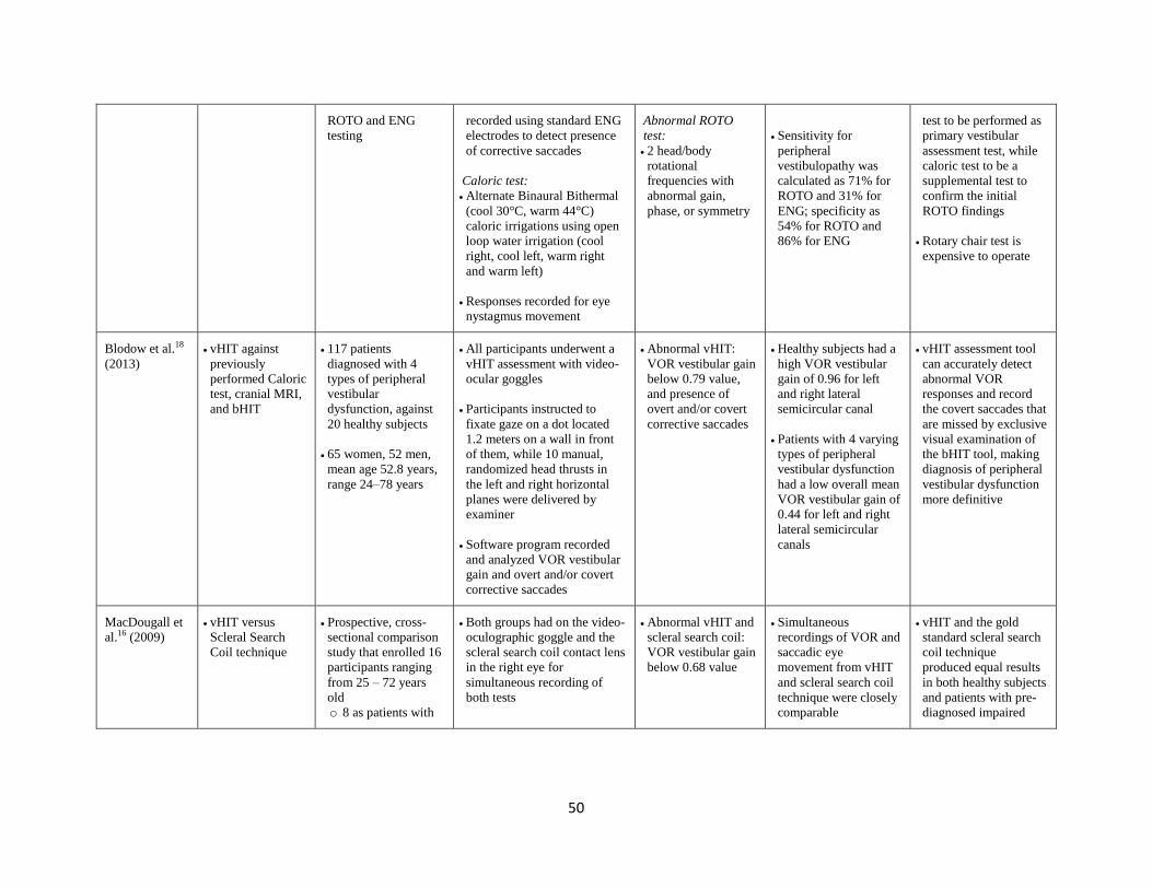
ROTO and ENG
testing
recorded using standard ENG
electrodes to detect presence
of corrective saccades
Caloric test:
Alternate Binaural Bithermal
(cool 30°C, warm 44°C)
caloric irrigations using open
loop water irrigation (cool
right, cool left, warm right
and warm left)
Responses recorded for eye
nystagmus movement
Abnormal ROTO
test:
2 head/body
rotational
frequencies with
abnormal gain,
phase, or symmetry
Sensitivity for
peripheral
vestibulopathy was
calculated as 71% for
ROTO and 31% for
ENG; specificity as
54% for ROTO and
86% for ENG
test to be performed as
primary vestibular
assessment test, while
caloric test to be a
supplemental test to
confirm the initial
ROTO findings
Rotary chair test is
expensive to operate
Blodow et al.18
(2013)
vHIT against
previously
performed Caloric
test, cranial MRI,
and bHIT
117 patients
diagnosed with 4
types of peripheral
vestibular
dysfunction, against
20 healthy subjects
65 women, 52 men,
mean age 52.8 years,
range 24–78 years
All participants underwent a
vHIT assessment with video-
ocular goggles
Participants instructed to
fixate gaze on a dot located
1.2 meters on a wall in front
of them, while 10 manual,
randomized head thrusts in
the left and right horizontal
planes were delivered by
examiner
Software program recorded
and analyzed VOR vestibular
gain and overt and/or covert
corrective saccades
Abnormal vHIT:
VOR vestibular gain
below 0.79 value,
and presence of
overt and/or covert
corrective saccades
Healthy subjects had a
high VOR vestibular
gain of 0.96 for left
and right lateral
semicircular canal
Patients with 4 varying
types of peripheral
vestibular dysfunction
had a low overall mean
VOR vestibular gain of
0.44 for left and right
lateral semicircular
canals
vHIT assessment tool
can accurately detect
abnormal VOR
responses and record
the covert saccades that
are missed by exclusive
visual examination of
the bHIT tool, making
diagnosis of peripheral
vestibular dysfunction
more definitive
MacDougall et
al.16 (2009)
vHIT versus
Scleral Search
Coil technique
Prospective, cross-
sectional comparison
study that enrolled 16
participants ranging
from 25 – 72 years
old
o 8 as patients with
Both groups had on the video-
oculographic goggle and the
scleral search coil contact lens
in the right eye for
simultaneous recording of
both tests
Abnormal vHIT and
scleral search coil:
VOR vestibular gain
below 0.68 value
Simultaneous
recordings of VOR and
saccadic eye
movement from vHIT
and scleral search coil
technique were closely
comparable
vHIT and the gold
standard scleral search
coil technique
produced equal results
in both healthy subjects
and patients with pre-
diagnosed impaired
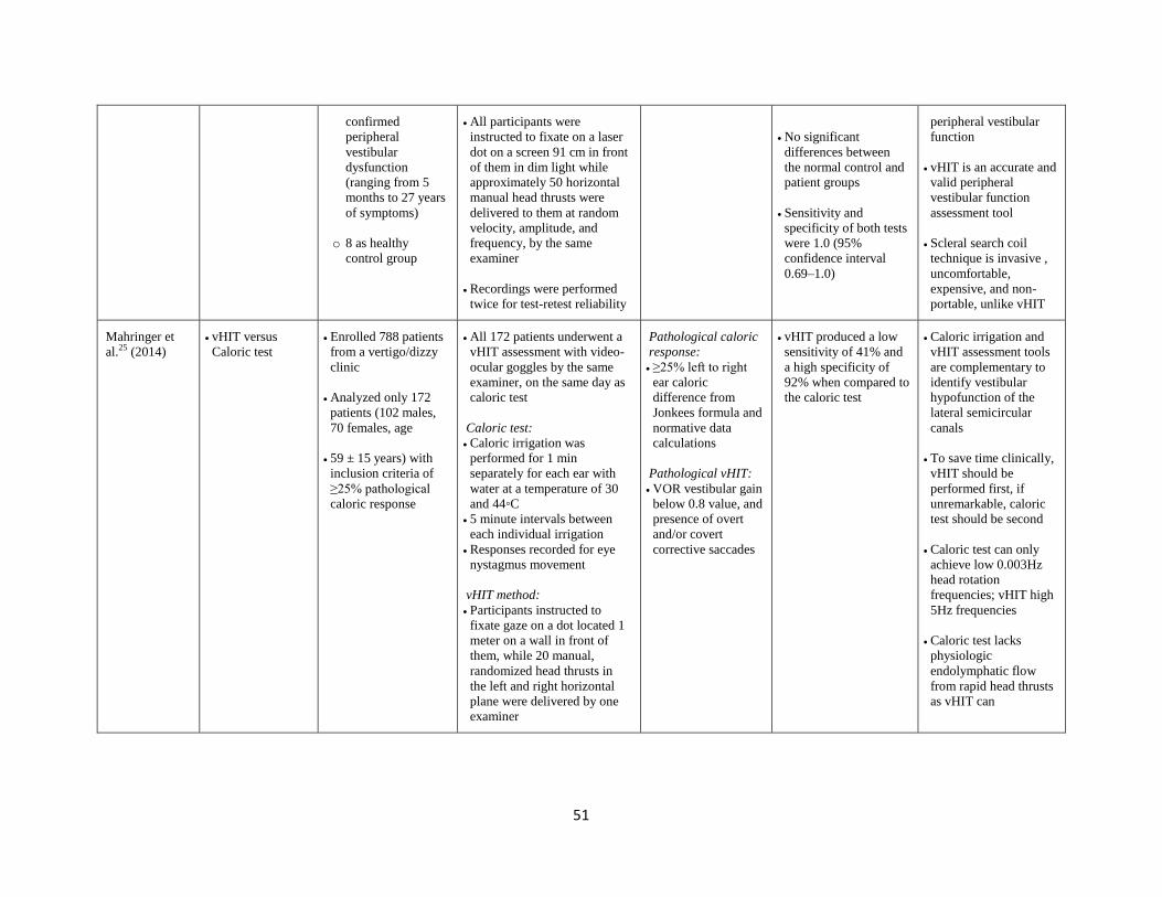
51
confirmed
peripheral
vestibular
dysfunction
(ranging from 5
months to 27 years
of symptoms)
o 8 as healthy
control group
All participants were
instructed to fixate on a laser
dot on a screen 91 cm in front
of them in dim light while
approximately 50 horizontal
manual head thrusts were
delivered to them at random
velocity, amplitude, and
frequency, by the same
examiner
Recordings were performed
twice for test-retest reliability
No significant
differences between
the normal control and
patient groups
Sensitivity and
specificity of both tests
were 1.0 (95%
confidence interval
0.69–1.0)
peripheral vestibular
function
vHIT is an accurate and
valid peripheral
vestibular function
assessment tool
Scleral search coil
technique is invasive ,
uncomfortable,
expensive, and non-
portable, unlike vHIT
Mahringer et
al.25 (2014)
vHIT versus
Caloric test
Enrolled 788 patients
from a vertigo/dizzy
clinic
Analyzed only 172
patients (102 males,
70 females, age
59 ± 15 years) with
inclusion criteria of
≥25% pathological
caloric response
All 172 patients underwent a
vHIT assessment with video-
ocular goggles by the same
examiner, on the same day as
caloric test
Caloric test:
Caloric irrigation was
performed for 1 min
separately for each ear with
water at a temperature of 30
and 44◦C
5 minute intervals between
each individual irrigation
Responses recorded for eye
nystagmus movement
vHIT method:
Participants instructed to
fixate gaze on a dot located 1
meter on a wall in front of
them, while 20 manual,
randomized head thrusts in
the left and right horizontal
plane were delivered by one
examiner
Pathological caloric
response:
≥25% left to right
ear caloric
difference from
Jonkees formula and
normative data
calculations
Pathological vHIT:
VOR vestibular gain
below 0.8 value, and
presence of overt
and/or covert
corrective saccades
vHIT produced a low
sensitivity of 41% and
a high specificity of
92% when compared to
the caloric test
Caloric irrigation and
vHIT assessment tools
are complementary to
identify vestibular
hypofunction of the
lateral semicircular
canals
To save time clinically,
vHIT should be
performed first, if
unremarkable, caloric
test should be second
Caloric test can only
achieve low 0.003Hz
head rotation
frequencies; vHIT high
5Hz frequencies
Caloric test lacks
physiologic
endolymphatic flow
from rapid head thrusts
as vHIT can
52
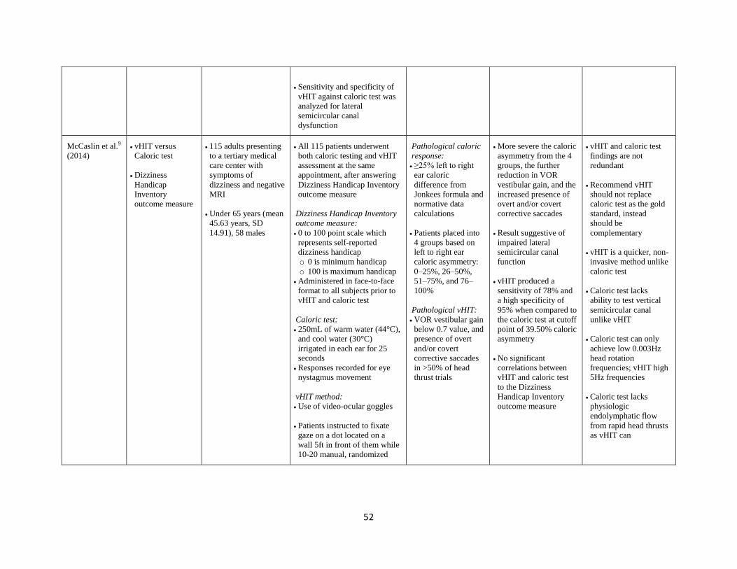
Sensitivity and specificity of
vHIT against caloric test was
analyzed for lateral
semicircular canal
dysfunction
McCaslin et al.9
(2014)
vHIT versus
Caloric test
Dizziness
Handicap
Inventory
outcome measure
115 adults presenting
to a tertiary medical
care center with
symptoms of
dizziness and negative
MRI
Under 65 years (mean
45.63 years, SD
14.91), 58 males
All 115 patients underwent
both caloric testing and vHIT
assessment at the same
appointment, after answering
Dizziness Handicap Inventory
outcome measure
Dizziness Handicap Inventory
outcome measure:
0 to 100 point scale which
represents self-reported
dizziness handicap
o 0 is minimum handicap
o 100 is maximum handicap
Administered in face-to-face
format to all subjects prior to
vHIT and caloric test
Caloric test:
250mL of warm water (44°C),
and cool water (30°C)
irrigated in each ear for 25
seconds
Responses recorded for eye
nystagmus movement
vHIT method:
Use of video-ocular goggles
Patients instructed to fixate
gaze on a dot located on a
wall 5ft in front of them while
10-20 manual, randomized
Pathological caloric
response:
≥25% left to right
ear caloric
difference from
Jonkees formula and
normative data
calculations
Patients placed into
4 groups based on
left to right ear
caloric asymmetry:
0–25%, 26–50%,
51–75%, and 76–
100%
Pathological vHIT:
VOR vestibular gain
below 0.7 value, and
presence of overt
and/or covert
corrective saccades
in >50% of head
thrust trials
More severe the caloric
asymmetry from the 4
groups, the further
reduction in VOR
vestibular gain, and the
increased presence of
overt and/or covert
corrective saccades
Result suggestive of
impaired lateral
semicircular canal
function
vHIT produced a
sensitivity of 78% and
a high specificity of
95% when compared to
the caloric test at cutoff
point of 39.50% caloric
asymmetry
No significant
correlations between
vHIT and caloric test
to the Dizziness
Handicap Inventory
outcome measure
vHIT and caloric test
findings are not
redundant
Recommend vHIT
should not replace
caloric test as the gold
standard, instead
should be
complementary
vHIT is a quicker, non-
invasive method unlike
caloric test
Caloric test lacks
ability to test vertical
semicircular canal
unlike vHIT
Caloric test can only
achieve low 0.003Hz
head rotation
frequencies; vHIT high
5Hz frequencies
Caloric test lacks
physiologic
endolymphatic flow
from rapid head thrusts
as vHIT can
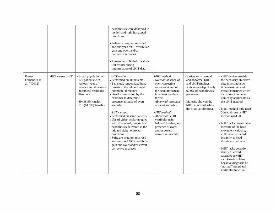
53
head thrusts were delivered in
the left and right horizontal
directions
Software program recorded
and analyzed VOR vestibular
gain and overt and/or
corrective saccades
Researchers blinded of caloric
test results during
interpretation of vHIT data
Perez-
Fernandez et
al.20 (2012)
vHIT versus bHIT Broad population of
179 patients with
various types of
balance and dizziness
peripheral vestibular
disorders
69 (38.5%) males,
110 (61.5%) females
bHIT method:
Performed on all patients
3 manual, randomized head
thrusts in the left and right
horizontal directions
visual examination by the
examiner to determine
presence/absence of overt
saccades
vHIT method:
Performed on same patients
Use of video-ocular goggles
with 20 manual, randomized
head thrusts delivered in the
left and right horizontal
directions
Software program recorded
and analyzed VOR vestibular
gain and overt and/or covert
corrective saccades
bHIT method:
Normal: absence of
overt corrective
saccades at end of
the head movement
in at least two head
thrusts
Abnormal: presence
of overt saccades
vHIT method:
Abnormal: VOR
vestibular gain
below 0.6 value, and
presence of overt
and/or covert
corrective saccades
Variances in normal
and abnormal bHIT
and vHIT findings,
with an overlap of only
67.9% of head thrusts
performed
Majority showed the
bHIT as normal while
the vHIT as abnormal
vHIT device provide
the necessary objective
data in a simplistic,
time-sensitive, and
versatile manner which
can allow it to be as
clinically applicable as
the bHIT method
bHIT method only used
3 head thrusts; vHIT
method used 20
bHIT lacks quantifiable
measure of the head
movement velocity;
vHIT able to record
instantly as head
thrusts are delivered
bHIT lacks detection
ability of covert
saccades as vHIT
canleads to false
negative diagnosis of
“normal” peripheral
vestibular function
54
SECTION 4: Discussion & Conclusion
Discussion
The vestibulo-ocular reflex (VOR) response is a coaction between the six
semicircular canals and the twelve extraocular muscles to stabilize gaze on a target, such
that head movements in one direction trigger the eyes to move with equal speed and
distance to the opposite direction.1-3,24
The ability to maintain a clear vision of a target
during natural rapid head movements is a vital automatic reflex that allows humans to be
mobile without blurred vision, dizziness, nausea, loss of postural control, and spatial
disorientation.1
In contrast, a deficient VOR response lacks signals from head movements to
stimulate eye movements, such that the eyes have to perform a quick, compensatory,
catch-up movement, known as a corrective saccade, to relocate the target back onto the
visual field.1-3
These saccades occur either during the head movements (covert saccades),
or at the end of the head movements (overt saccades).1-3
The presence of any saccades
indicates dysfunction of the semicircular canal.1,4
Thus, the integrity of the VOR
response and saccadic eye movement is not only crucial in understanding but central to
determining normal versus abnormal semicircular canal functionality of the peripheral
vestibular system. Determining this specific functionality requires appropriate
assessment tools which provide accurate, objective, and reliable measurements of head
rotations and eye movements.
Clinicians and researchers since early 20th century have designed several
functional assessment tools to specifically measure the VOR response and saccadic eye
55
movement, five of which are currently still widely used in practice: the caloric test, rotary
chair test, bedside head impulse test (bHIT), scleral search coil technique, and video head
impulse test (vHIT). With the advent of new technology, evidence-based medicine, and
patient-driven and cost-effective clinical care, some of these traditionally used functional
assessment tools lack validity and reliability.14
The newest assessment tool, the vHIT,
has been purported by many clinicians and researchers to have potential to be the next
“gold standard.”9,14,20,23
It should be noted that these functional assessment tools are not to be considered
diagnostic, but rather demonstrative in quantifying peripheral vestibular function, as
neurotologists would argue “vestibular function represents a complex interaction between
peripheral and central vestibular physiology with changes in each of the system’s
components during disease.”14, p332
Arriaga et al.14
convey the challenges in medicine of
establishing a test as a “gold standard” since the specificity and sensitivity characteristics
of any assessment tool rely on the parameters set by researchers for “normal” and
“abnormal.” Thus they suggest utilizing the “best available”14, p332
standard instead, in
which the clinician decides on which assessment tool to perform on patients to provide
the most valid and reliable results.
In the current literature, the vHIT was consistently found to provide accurate
objective data in identifying peripheral vestibular dysfunction of the semicircular
canals.3,9,14,16,18,20,23,25
When the vHIT tool is compared against the age-tested caloric test,
the high sensitivity and specificity characteristics suggest the vHIT is adequately capable
of detecting unilateral semicircular dysfunction in patients with abnormal results, as well
as detecting patients without the dysfunction with normal test results, respectively.9,25
56
Despite the lack of evidence for a comparison to the vHIT tool, the rotary chair
test in itself has been found to be an ineffective tool clinically and has questionable
reliability and validity.12,13
The rotary chair test thus falls short of what the vHIT tool has
developed into clinically and objectively.
Comparison of the objective vHIT tool against the subjective bHIT method (from
which the basis of the vHIT tool derives from), the vHIT surpasses the known limitations
of the bHIT method by being quantitative and more accurate at identifying peripheral
vestibular dysfunction of the semicircular canals.18,20
Finally, when the vHIT tool is compared against the current gold standard scleral
search coil technique, the vHIT stands to be as accurate and sensitive in identifying
peripheral vestibular dysfunction of the semicircular canals as the scleral search coil
technique in both middle-aged and older healthy controls as well as in patients in an acute
and non-acute peripheral vestibular disease stage.16,23
Two highly acclaimed studies
performed simultaneous vHIT and scleral search coil testing of the VOR response and
saccadic eye movement. A high and equal sensitivity and specificity of 1.0 was produced
in both assessment tools, concluding that the vHIT can provide the same reliability and
validity as the gold standard scleral search coil technique.16,23
The major limitation shared by the vHIT, the bHIT method, and the scleral search
coil technique is the general clinical precaution or contraindication for a patient with a
neck injury or limitations in cervical range of motion.10
In such cases, the clinician can
refer to the caloric test, rotary chair test, or other functional assessment tests which limit
or avoid head and neck movements. A minor limitation of the vHIT is the minimal
57
slippage of the goggles usually from lack of a snug fit of the video-ocular goggles which
can be easily fixed by readjustment.3,10,16,21,23
It is worthy to note that given the recent development of the vHIT tool, a standard
protocol has not yet been established of how many head thrusts to perform, or the
distance of the patient from the fixation point, as there are only a few manufacturers that
have developed the vHIT device to date. However, most recent studies testing the vHIT
tool have taken the methodology from studies on the gold standard scleral search coil
technique and incorporated into their vHIT research design.
Considering the consistent favorable results from the studies on the benefits of the
vHIT tool, and the added value of its portability, simplicity, affordability, quickness, non-
invasiveness, and widespread clinical applicability over its counterpart assessment
tools,9,16,18,20,23,25
the implications of this new assessment tool solidify the message of
vHIT being the “best available” reference standard.14, p332
The vHIT has enhanced the
clinical ease of detecting VOR response and saccadic eye movement by eliminating
various limitations from traditional assessment tools, yet remaining accurate, reliable, and
valid at identifying peripheral vestibular dysfunction of the semicircular canals.
Conclusion:
“Eyes provide [the] most accessible window for exploring vestibular function”
(Goebel et al.11, p401
). The findings of this comparative literature search suggest the
newest assessment tool, the vHIT, can be considered objective, valid, complementary,
and statistically equivalent to the traditional assessment tools: the caloric test, rotary chair
test, bHIT, and scleral search coil technique. To save time, be more cost-effective,
58
prevent patient discomfort, and deliver ease of clinical use, the vHIT stands to provide
the “best available” reference standard for an assessment tool in identifying peripheral
vestibular dysfunction of the semicircular canals. Given the strong validity and clinical
applicability already available, the vHIT can provide even more impact on further clinical
research for the functionality of the entire vestibular system. There are widespread
clinical research opportunities to assess the theoretical effects of vHIT on broader topics
such as imaging, pharmacological agents, or vestibular rehabilitation.
Acknowledgements:
The author thanks Dr. Roberta O’Shea for advising and editing this project, and
Dr. Miriam Redleaf and Dr. Renee Theiss for their support in being a part of the
dissertation committee.
59
SECTION 5: References
1. O’Sullivan S, Schmitz T. eds. Physical Rehabilitation: Assessment and Treatment. 4
th
ed. Philadelphia, PA: F.A.Davis Company; 2000.
2. Colagiorgio P, Colnaghi S, Versino M, Ramat S. A new tool for investigating the
functional testing of the VOR. Frontiers in neurology. 2013;4:1-9.
3. Curthoys I, MacDougall H, Manzari L, Burgess A, Bradshaw A, McGarvie L, et al.
Clinical application of a new objective test of semicircular canal dynamic function–the
video head impulse test (vHIT). A safe, simple, and fast clinical vestibular test. In
Vertigo–Kontroverses und Bewährtes 8. Hennig Symposium, New York: SpringerWien.
2011: 53-64.
4. Heuberger M, Sağlam M, Todd N, Jahn K, Schneider E, Lehnen N. Covert anti-
compensatory quick eye movements during head impulses. PloS one, 9(4), e93086.5.
doi:10.1371/journal.pone.0093086.
5. Jacobson G, Newman C. Background and technique of caloric testing. In: Handbook of
Balance Function Testing. St. Louis, MO: Mosby Year Book; 1993:156-92.
6. Caloric stimulation. Medline Plus Web site.
http://www.nlm.nih.gov/medlineplus/ency/article/003429.htm. Published 2013. Accessed
April 9, 2015.
7. Weerts A, De Meyer G, Pauwels G et al. Pharmaceutical countermeasures have
opposite effects on the utricles and semicircular canals in man. Audiology & neuro-
otology. 2011;17(4):235-242.
8. Perez N, Rama-Lopez J. Head-impulse and caloric tests in patients with dizziness. Otol
Neurotol. 2003;24(6):913–917.
9. McCaslin, Devin L, et al. Predictive properties of the video head impulse test:
Measures of caloric symmetry and self-report dizziness handicap." Ear and hearing.
2014;35(5):185-191.
10. Craig J, Crumley-Welsh W, Kattah J et al. Assessing the patient with vestibular
symptoms. Otometrics Web site. Accesses March 10, 2015.
11. Goebel JA, Hanson JM, Fishel DG. Interlaboratory variability of rotational chair test
results. Otolaryngol Head Neck Surg. 1994;110:400-405.
12. Furman JM, Goebel JA, Hamid MA, et al. Interlaboratory variability of rotational
chair test results II: analysis of simulated data. Otolaryngol Head Neck Surg.
2001;125(5):23-30.
60
13. Phillips J. Rotary chair testing. In: Manual of Pediatric Balance Disorders. San Diego,
CA: Plural Publishing, Inc.; 2013:59-74.
14. Arriaga MA, Chen DA, Cenci KA. Rotational chair (ROTO) instead of
electronystagmography (ENG) as the primary vestibular test. Otolaryngol Head Neck
Surg. 2005;133(3):329-33.
15. Davalos-Bichara M, Agrawal Y. Normative results of healthy older adults on standard
clinical vestibular tests. Otology & Neurotology. 2014;35(2):297.
16. MacDougall HG, Weber KP, McGarvie LA, Halmagyi GM, Curthoys IS. The video
head impulse test diagnostic accuracy in peripheral vestibulopathy. Neurology.
2009;73(14):1134-1141.
17. Jorns-Haderli M, Straumann D, Palla A. Accuracy of the bedside head impulse test in
detecting vestibular hypofunction. J Neurol Neurosurg Psychiatry. 2007;78(10):1113–
1118. doi:10.1136/jnnp.2006.109512
18. Blödow A, Pannasch S, Walther LE. Detection of isolated covert saccades with the
video head impulse test in peripheral vestibular disorders. Auris Nasus Larynx.
2013:40(4):348-351.
19. Weber KP, MacDougall HG, Halmagyi GM, Curthoys IS. Impulsive Testing of
Semicircular‐Canal Function Using Video‐oculography. Annals of the New York
Academy of Sciences. 2009;1164(1):486-491.
20. Pérez-Fernández N, Gallegos-Constantino V, Barona-Lleo L, Manrique-Huarte R.
Clinical and video-assisted examination of the vestibulo-ocular reflex: A comparative
study. Acta Otorrinolaringologica (English Edition). 2012;63(6):429-435.
21. Migliaccio AA, Cremer PD. The 2D modified head impulse test: A 2D technique for
measuring function in all six semi-circular canals. Journal of Vestibular Research.
2011;21(4):227-234.
22. Chen L, Todd M, Halmagyi GM, Aw S. Head impulse gain and saccade analysis in
pontine-cerebellar stroke and vestibular neuritis. Neurology. 2014;83(17):1513-1522.
23. Agrawal Y, Schubert MC, Migliaccio AA, Zee DS, Schneider E, et al. Evaluation of
quantitative head impulse testing using search coils versus videooculography in older
individuals. Otol Neurotol. 2014;35:283–288.
24. Robinson DA. A method of measuring eye movement using a scleral search coil in a
magnetic field. IEEE Trans Biomed Eng. 1963;10:137–145.
61
25. Mahringer A, Rambold HA. Caloric test and video-head-impulse: a study of
vertigo/dizziness patients in a community hospital. European Archives of Oto-Rhino-
Laryngology. 2014;271(3):463-472.
26. Harvey SA, Wood DJ, Feroah TR. Relationship of the head impulse test and head-
shake nystagmus in reference to caloric testing. Am J Otol. 1997;18(1):207-213.
27. Beynon GJ, Jani P, Baguley DM. A clinical evaluation of head impulse testing. Clin
Otolaryngol Allied Sci. 1998;23(2):117–122.


![Electrical Vestibular Stimulation after Vestibular ......electrical stimulation of the vestibular system to one ear [4,5,9]. However studies have also reported vestibular responses](https://img.pdfslide.us/doc/110x75/60f6b0762ca1b41e91018b73/electrical-vestibular-stimulation-after-vestibular-electrical-stimulation.jpg)