Embed Size (px)
Citation preview
Open Access2008Desjardinset al.Volume 9, Issue 12, Article R183ResearchComparative genomics of mutualistic viruses of Glyptapanteles parasitic waspsChristopher A Desjardins*¥, Dawn E Gundersen-Rindal†, Jessica B Hostetler*, Luke J Tallon*#, Douglas W Fadrosh***, Roger W Fuester‡, Monica J Pedroni†††, Brian J Haas*‡‡, Michael C Schatz§, Kristine M Jones*#, Jonathan Crabtree*#, Heather Forberger* and Vishvanath Nene*¶
Addresses: *J Craig Venter Institute, Rockville, MD 20850, USA. †USDA-ARS Invasive Insect Biocontrol and Behavior Laboratory, Beltsville, MD 20705, USA. ‡USDA-ARS Beneficial Insect Introduction Research Laboratory, Newark, DE 19713, USA. §Center for Bioinformatics and Computational Biology, University of Maryland, College Park, MD 20742, USA. ¶Institute for Genome Sciences and Department of Microbiology and Immunology, University of Maryland School of Medicine, Baltimore, MD 21201, USA. ¥Current address: Department of Biology, University of Rochester, Rochester, NY 14627, USA. #Current address: Institute for Genome Sciences, University of Maryland School of Medicine, Baltimore, MD 21201, USA. **Current address: J Craig Venter Institute, La Jolla, CA 92121, USA. ††Current address: Department of Veterinary Microbiology and Pathology, Washington State University, WA 99164, USA. ‡‡Current address: The Broad Institute, Cambridge, MA 02142, USA.
Correspondence: Christopher A Desjardins. Email: [email protected]
© 2008 Desjardins et al.; licensee BioMed Central Ltd. This is an open access article distributed under the terms of the Creative Commons Attribution License (http://creativecommons.org/licenses/by/2.0), which permits unrestricted use, distribution, and reproduction in any medium, provided the original work is properly cited.Glyptapanteles polydnavirus genome sequences<p>Comparative genome analysis of two endosymbiotic polydnaviruses from Glyptapanteles parasitic wasps reveals new insights into the evolutionary arms race between host and parasite.</p>
Abstract
Background: Polydnaviruses, double-stranded DNA viruses with segmented genomes, have evolved as obligateendosymbionts of parasitoid wasps. Virus particles are replication deficient and produced by female wasps from proviralsequences integrated into the wasp genome. These particles are co-injected with eggs into caterpillar hosts, where viralgene expression facilitates parasitoid survival and, thereby, survival of proviral DNA. Here we characterize and comparethe encapsidated viral genome sequences of bracoviruses in the family Polydnaviridae associated with Glyptapanteles gypsymoth parasitoids, along with near complete proviral sequences from which both viral genomes are derived.
Results: The encapsidated Glyptapanteles indiensis and Glyptapanteles flavicoxis bracoviral genomes, each composed of 29different size segments, total approximately 517 and 594 kbp, respectively. They are generated from a minimum of sevendistinct loci in the wasp genome. Annotation of these sequences revealed numerous novel features for polydnaviruses,including insect-like sugar transporter genes and transposable elements. Evolutionary analyses suggest that positiveselection is widespread among bracoviral genes.
Conclusions: The structure and organization of G. indiensis and G. flavicoxis bracovirus proviral segments as multiple locicontaining one to many viral segments, flanked and separated by wasp gene-encoding DNA, is confirmed. Rapid evolutionof bracovirus genes supports the hypothesis of bracovirus genes in an 'arms race' between bracovirus and caterpillar.Phylogenetic analyses of the bracoviral genes encoding sugar transporters provides the first robust evidence of a wasporigin for some polydnavirus genes. We hypothesize transposable elements, such as those described here, could facilitatetransfer of genes between proviral segments and host DNA.
Published: 30 December 2008
Genome Biology 2008, 9:R183 (doi:10.1186/gb-2008-9-12-r183)
Received: 14 July 2008Accepted: 30 December 2008
The electronic version of this article is the complete one and can be found online at http://genomebiology.com/2008/9/12/R183
Genome Biology 2008, 9:R183

http://genomebiology.com/2008/9/12/R183 Genome Biology 2008, Volume 9, Issue 12, Article R183 Desjardins et al. R183.2
BackgroundThe capacity to sequence and analyze complex genomes hasenabled rapid progress toward understanding atypical bio-logical systems, such as the obligate mutualistic association ofpolydnaviruses (PDVs) with certain parasitic wasps [1]. PDVshave evolved a distinctive life strategy as they exist in two dis-tinct forms, as a proviral form integrated into the genome ofmale and female parasitoids [2,3], and in a virus form that isreplication deficient. PDV encapsidated genomes are unlikeany other viral genomes as they consist of multiple circulardouble-stranded DNA molecules, referred to as segments.Proviral DNA is amplified from the parasitoid genome, fol-lowed by excision, circularization, and encapsidation of seg-ments into virus particles, and occurs only within femaleovarian calyx epithelial cells [4-6]. PDVs are also distinctivein that they require two separate hosts to maintain their lifecycle. PDV virions are released into the oviduct lumen with noobvious pathology to the primary host. During oviposition,virions, wasp eggs and other parasitism-associated factors aredelivered into a secondary host, usually a lepidopteran, wherePDV gene expression disrupts immune functions, physiology,and development [7-9]. Virion-induced pathology within thesecondary host ensures survival of the PDV proviral formwithin the parasitoid life cycle.
Genome sequencing and analyses undertaken to elucidate thegenetic complement of PDVs relating to their unusual biologyhave focused mainly on encapsidated viral genomes as iso-lated from female parasitoid reproductive tracts. Comprehen-sive genome studies have been conducted for representativemembers of the two genera of PDVs, bracoviruses (BVs) andichnoviruses (IVs), described as obligate endosymbionts ofbraconid and ichneumonid parasitoids, respectively. Braco-nid parasitoids harboring BVs are monophyletic and com-prise the microgastroid complex of seven subfamilies(Cheloninae, Dirrhoponae, Mendesellinae, Khoikhoiinae,Cardiochilinae, Miracinae and Microgastrinae) with greaterthan 17,500 species [10]. BV genomes sequenced to date areexclusive to subfamily Microgastrinae, Cotesia congregataBV (CcBV) [11], and Microplitis demolitor BV (MdBV) [12];IV genomes sequenced include members from the ichneumo-nid subfamily Campopleginae, Campolitis sonorensis IV(CsIV) [12], Hyposoter fugitivus IV and Tranosema rostraleIV [13], and from subfamily Banchinae, Glypta fumiferanaeIV [14]. General characteristics that differ in sequencedencapsidated BV and IV viral genomes include segmentnumber, size and abundance, while similar characteristicsinclude a low gene coding density, multiple intron-containingprotein coding genes and the presence of multi-gene families[11-16].
Conventional concepts in virology do not adequately describethe viral and proviral forms of PDV genomes. EncapsidatedPDV genomes sequenced to date lack known viral structuralproteins and DNA replication-associated enzymes, and thereis evidence that in CsIV, a gene coding for one structural pro-
tein is not encoded by the encapsidated genome [17]. It hasbeen hypothesized that PDVs have a viral ancestry and haveundergone reductive genome evolution, where replicationand coat protein genes have been transferred to other regionsof the wasp genome [18]. Although PDVs are classified asviruses [1], a contrasting hypothesis is that PDVs haveevolved from genetic elements that have captured parasitoidgenes and a virion production system [19]. Here, we use theterm viral genome to represent segment sequences encapsi-dated in virus particles and proviral segment to represent thelinear integrated form of a circular viral genome segment. Weconservatively describe the proviral genome to encompass allproviral segments and their excision motifs.
PDV viral genome sequences have advanced our understand-ing of PDV-mediated pathology in parasitism and have begunto unravel evolutionary relationships of this unusual group ofviruses. However, very little is known about the compositionand sequence of PDV proviral genomes beyond the predictionof proviral segment sequences. Early studies based on CsIVshowed that several proviral segments were flanked bygenomic DNA that was not encapsidated, suggesting CsIVproviral segment sequences are dispersed in the waspgenome [20-22]. In contrast, BV proviral segment sequencesare thought to be located at a single locus in a tandem array[23-25]. This hypothesis is based on both in situ hybridizationevidence where probes from 3 of 30 different CcBV viral seg-ments hybridized to the same region of a single wasp chromo-some [25] and studies of CcBV and Chelonus inanitus BV(CiBV) in which proviral segments were flanked on one orboth sides by a different proviral segment [23,24]. A directDNA sequence repeat was seen at the boundaries of the fewproviral segments examined [23], and it appears to mark thesite of proviral segment excision, possibly via conservativesite-specific recombination, as a single copy of the repeat wasnoted within the corresponding circularized viral segment[23]. A working model for BV viral segment production is thatthey are derived from a large precursor molecule encompass-ing multiple proviral segments, which is amplified after exci-sion of the precursor from the wasp chromosome [26,27].Studies of CcBV and CiBV show that there is no replication ofmature viral segments [23,26-28].
Recently, we presented a global examination and descriptionof proviral segment sequences of a BV associated with Glyp-tapanteles indiensis (GiBV) [15], a parasitoid of gypsy moth.In contrast to earlier concepts, our data showed that some butnot all of approximately 24 GiBV proviral segment sequencesare tandemly arrayed. We provided the first detailed analysisof a major part of a PDV proviral genome, a 223 kbp locuslabeled proviral locus 1, which encodes a tandem array of 8GiBV proviral segments separated by inter-segmental regionsthat varies in length from 117 to 8,369 bp [15]. We also pro-posed that it was reasonable to consider inter-segmental DNAseparating tandemly arrayed proviral segments as compo-nents of the proviral genome. Structural and compositional
Genome Biology 2008, 9:R183

http://genomebiology.com/2008/9/12/R183 Genome Biology 2008, Volume 9, Issue 12, Article R183 Desjardins et al. R183.3
analyses revealed that this array of proviral segments wasflanked by 6-7 kbp sequence repeats and that proviral seg-ment sequences had a distinct nucleotide composition frominter-segmental and flanking non-segment DNA. Compara-tive sequence analysis revealed conserved motifs at the sitesof excision of segments from proviral DNA, which suggestedthat there is directionality to the mechanism of segment exci-sion, a conclusion also supported in CiBV [29]. The motifs weidentified are also found in other BV genomes, suggesting ahighly conserved mechanism of BV proviral segment exci-sion. Analyses of DNA polymorphisms in the eight GiBV viralsegment sequences gave evidence for selection acting on bothprotein-coding and non-coding DNA, indicating non-codingsegmental DNA may serve functional purposes [15].
Building on our previous work, we have expanded the knowl-edge of the BV proviral form through detailed analysis of anadditional five loci encoding GiBV proviral segments. Herewe revise our estimate of the GiBV viral segment number to29, encoded by a minimum of 7 proviral loci, providing anearly complete description of the GiBV viral genome. Inaddition, we provide equally extensive data showing similarcharacteristics of proviral segment sequences of GfBV, a BVassociated with another wasp in the same genus as GiBV, G.flavicoxis. Comparative analyses reveal a high degree of syn-teny between loci that code for GiBV and GfBV proviral seg-ments as well as flanking DNA. Not all loci are flanked bysequence repeat elements, but segment excision motifs havebeen conserved. Proviral segment sequences exhibit clear dif-ferences in their nucleotide composition relative to flankingDNA. GiBV and GfBV genes appear to be evolving rapidly,and GiBV and GfBV contain a novel multi-gene family thatcodes for sugar transporter proteins. We also identify a recentinsertion of a transposable element (TE) within a populationof GiBV and more ancient TEs associated with BV genomes.This work represents the most comprehensive characteriza-tion of PDV proviral genomes and of the structural organiza-tion of proviral segments to date.
Results and discussionSequence characterization of GiBV and GfBV viral genomesIn an early study, field inversion gel analysis of the GiBV viralgenome estimated the presence of 13 segments and a cumula-tive viral genome size of approximately 250 kbp [30]. At thestart of our project the viral genome characteristics of GfBVwere unknown, but presumed to be similar to those of GiBV.We undertook a whole genome shotgun (WGS) sequencingapproach to sequence the GiBV and GfBV viral genomes.Viral DNA was sequenced to approximately 8× coverageusing purified virions pooled from the calyx fluid of approxi-mately 400 and 50 female wasps from G. indiensis and G. fla-vicoxis, respectively (see Materials and methods). Followinga manual effort to close sequence and physical gaps, we wereable to derive a complete consensus viral segment sequence
for 21 GiBV viral segments (8 of which were described previ-ously [15]); 4 segments (numbers 17, 19, 21 and 29) remainedas partial sequences (Figure 1) due to technical difficultiesprimarily associated with sequence repeats. GiBV viral seg-ment 25 corresponded to the GiBV genome 'segment F' previ-ously sequenced and characterized in detail [31,32]. ForGfBV, we were able to complete sequence analysis of 27 viralsegments and 1 (number 13) remained a partial sequence(Figure 1). Nucleotide sequence polymorphisms occurredmore frequently in GiBV than in GfBV, and we presume thehigher sequence success rate for GfBV was due to sampling amore homogeneous viral population relative to GiBV. Thediscrepancy in GiBV viral genome statistics with earlier esti-mates [30] is due to finding multiple GiBV viral segments ofsimilar size, which would have co-migrated on the agarosegel. The 25 GiBV and 28 GfBV viral segment sequencestotaled 489 kbp and 581 kbp, respectively. These aggregategenome sizes and viral segment numbers have been furtherrevised based on proviral sequence data (described below).Individual WGS sequence reads were deposited in the NCBITrace Archive [1643848625-1643870960, 1813616562-1813617310]. Consensus sequence for viral segments, whichranged in length from 9.7 to 39.0 kbp for GiBV and 3.8 to 50.7kbp for GfBV have been deposited in GenBank [Gen-Bank:EU001243-EU001285].
Sequence characterization of loci encoding GiBV and GfBV proviral segmentsWe previously reported that a radioactive probe from totalGiBV viral DNA hybridized at varying intensity to 127 clonesfrom a bacterial artificial chromosome (BAC) library of 9,216clones derived from G. indiensis larvae; 17 GiBV viral seg-ment sequences for which probes were available mapped toBAC clones that segregated into 7 distinct non-overlappinggroups [15]. A similar size BAC library from G. flavicoxisyielded 154 clones that hybridized with total GfBV viral DNA.A combination of BAC end sequencing of 60 BACs and 10GfBV segment-specific PCRs also segregated these BACclones into 7 groups. GiBV and GfBV proviral segmentsequences not present in the selected BAC clones were iso-lated by rescreening the arrayed BAC libraries with radioac-tive viral segment-specific PCR products. Probes for GiBVviral segment number 29 and 30 and GfBV viral segmentnumber 29 and 31 did not hybridize to any BAC clones, whilehybridizations of a probe for GiBV segment 28 proved to befalse positives. The former may be due to representationalbias in the BAC libraries that were screened.
A total of 11 BAC clones from G. indiensis (2 BAC clones werefrom a prior study [15]) and 9 BAC clones from G. flavicoxishave been sequenced. Clone selection was based on a combi-nation of proviral segment sequence composition, BAC fin-gerprinting, and BAC end sequencing to try to ensure that theBAC clones chosen for sequencing contained the greatest pos-sible coverage of proviral segment sequences. The G. indien-sis and G. flavicoxis BAC sequence data totaled 1.21 and 1.16
Genome Biology 2008, 9:R183
http://genomebiology.com/2008/9/12/R183 Genome Biology 2008, Volume 9, Issue 12, Article R183 Desjardins et al. R183.4
Mbp, respectively. The location of proviral segments withinthe BAC clones was determined by sequence alignment ofviral segment sequences, as well as by searching for individ-ual segment excision motifs (see below). Overlapping BACsequences were fused, if possible within an inter-segmentalregion, to create pseudo-molecule sequences reducing uniquesequence data for G. indiensis and G. flavicoxis to 1.08 Mbpand 1.00 Mbp, respectively. The length of contiguoussequences ranged in size from 48.8 to 233.6 kbp for G. indi-ensis and 91.7 to 279.3 kbp for G. flavicoxis. Sequences wereannotated as described in Materials and methods. These data
have been deposited in GenBank [GenBank:EF710644-EF710659; AC191960] was previously submitted [15].
Identification of novel GiBV and GfBV viral segmentsPreviously, using MEME [33], we discovered a highly con-served 6 bp DNA sequence embedded within an approxi-mately 60 bp motif that appears to function as the site ofexcision and circularization of 8 GiBV proviral segmentsequences [15]. The analysis of such motifs was expanded toencompass all available viral and proviral sequence data forGiBV and GfBV. Viral, 5' proviral and 3' proviral excision
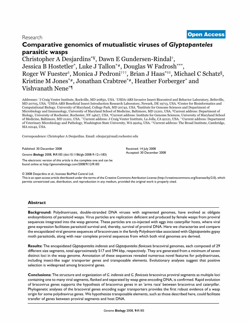
Summary of GiBV and GfBV sequence dataFigure 1Summary of GiBV and GfBV sequence data. Homologous viral segments between GiBV and GfBV are depicted in the same row and have been assigned the same number; blank spaces represent the absence of a homologous segment in one of the genomes. To the right, columns indicate the locus containing the segment and whether the segment is complete (C), partial (P), or absent (-) in the viral and proviral sequences from each genome. Incomplete sequence indicators (<>) are shown only for segments for which both viral and proviral sequence are absent or incomplete.
>
>
>29 <
hypothetical protein
protein tyrosine phosphatase
viral ankyrin
sugar transporter
c-type lectin
cystatin
GiBV
tRNA
Kbp0 10 20 30 40 50
Kbp0 10 20 30 40 50
GfBV
13 < >
<> incomplete sequence
locus
?
?
?
7
7
6
5
4
3
3
2
2
2
2
2
2
2
2
2
2
2
2
2
1
1
1
1
1
1
1
1
viral
C
P
C
C
C
C
C
C
P
C
P
-
P
-
C
C
C
-
C
C
-
C
C
C
C
C
C
C
C
proviral
-
-
-
C
C
C
C
C
C
C
C
P
P
C
C
C
C
C
C
C
C
C
C
C
C
C
C
C
C
viral
C
C
C
C
C
C
C
C
C
C
C
C
C
C
C
C
P
C
C
C
C
-
C
C
C
C
C
C
C
proviral
-
-
C
P
-
C
C
C
C
C
C
C
C
C
C
P
P
C
C
C
C
C
C
C
C
C
C
C
C
GiBV GfBV
30
28
26
25
24
23
22
21
20
19
18
17
16
15
14
13
12
11
10
9
8
7
6
5
4
3
2
1
31
29
28
27
26
25
24
23
22
21
20
19
18
17
15
14
12
11
10
9
8
7
6
5
4
3
2
1
Genome Biology 2008, 9:R183
http://genomebiology.com/2008/9/12/R183 Genome Biology 2008, Volume 9, Issue 12, Article R183 Desjardins et al. R183.5
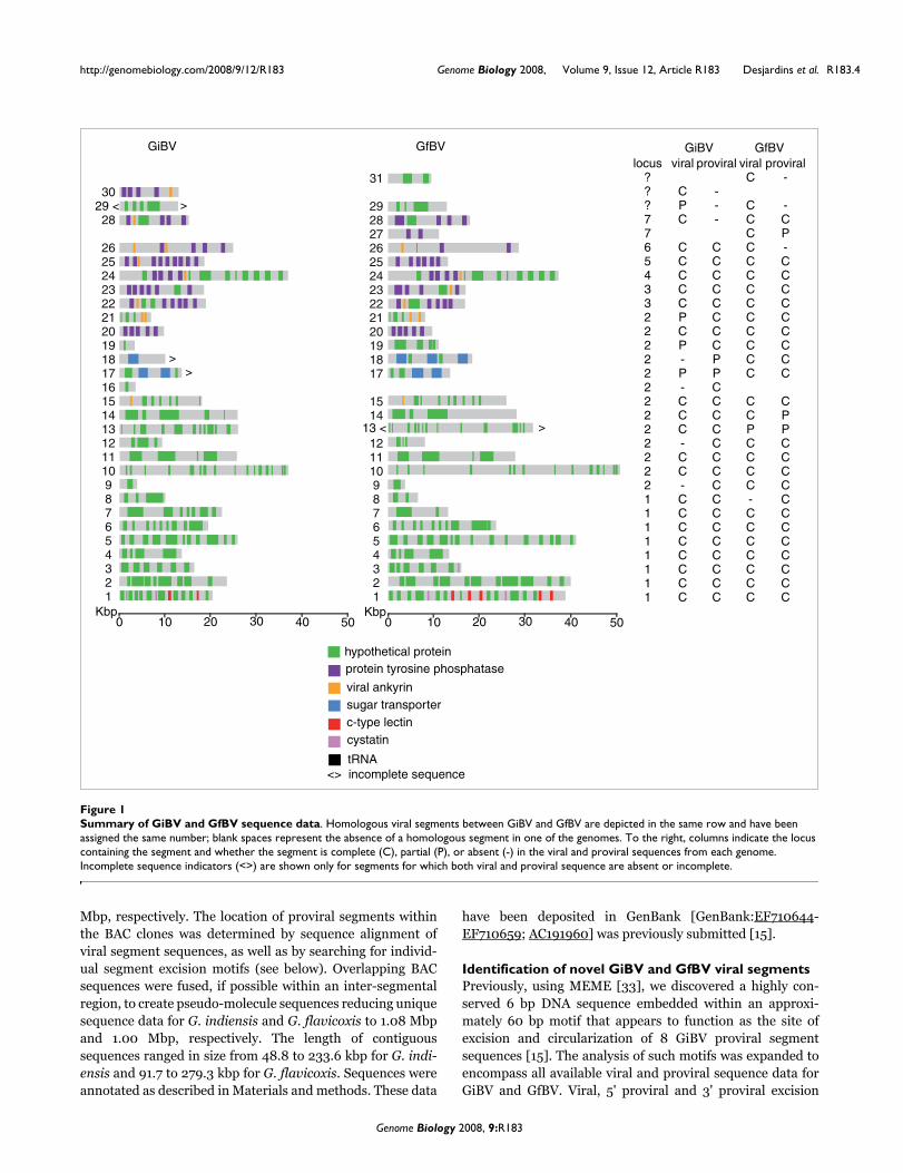
motifs are shown in Figure 2. There are some differences, pri-marily outside the hexamer repeat, between the new consen-sus excision motifs for GiBV and those described previously[15]. Additionally, the GfBV excision motif is highly similar tothat of GiBV. As we demonstrated before for GiBV, differ-ences between the extended sequence motifs at each end of aGfBV proviral segment indicate there is directionality to themechanism of segment excision from the site of integration.
Using the extended motifs and MAST [34] we searched BACsequences in order to detect the boundaries of proviral seg-ment sequences for which we had partial viral segment
sequence data, and we extended this search to all BACs. Inaddition to detecting some of the missing viral segmentsequences, MAST predicted the presence of four novel provi-ral segments for GiBV (numbers 9, 12, 16 and 18) and one(number 8) for GfBV (Figure 1). All these potential proviralsegments had exact sequence matches to unassembledsequences in the viral WGS sequence data. Sequence cover-age in all cases was below one-fold and viral segmentsequence versions of the novel proviral segments could not beassembled from the WGS sequence data alone. To determinewhether these sequences represented potential false posi-tives, we searched all inter-segmental and flanking non-seg-
Nucleotide conservation extends around the proviral segment excision siteFigure 2Nucleotide conservation extends around the proviral segment excision site. Viral, 5' proviral, and 3' proviral motifs are shown for the GiBV and GfBV genomes, with the excision site highlighted. For each proviral motif, underlined sequence represents non-encapsidated sequence while unmarked sequence represents proviral segment sequence. Following excision, DNA is circularized at the excision site forming the viral motif.
(a) GiBV 5' proviral motif
(b) GiBV 3' proviral motif
(c) GiBV viral motif
(d) GfBV 5' proviral motif
(e) GfBV 3' proviral motif
(f) GfBV viral motif
excision site
Genome Biology 2008, 9:R183
http://genomebiology.com/2008/9/12/R183 Genome Biology 2008, Volume 9, Issue 12, Article R183 Desjardins et al. R183.6
mental regions against the unassembled viral WGS sequencedata and found no evidence for them in the WGS data. Thus,a combination of the viral and proviral sequence data predictsthat GiBV and GfBV viral genomes each contain 29 segments,although 3 GiBV and 1 GfBV viral segments remain partiallysequenced (Figure 1).
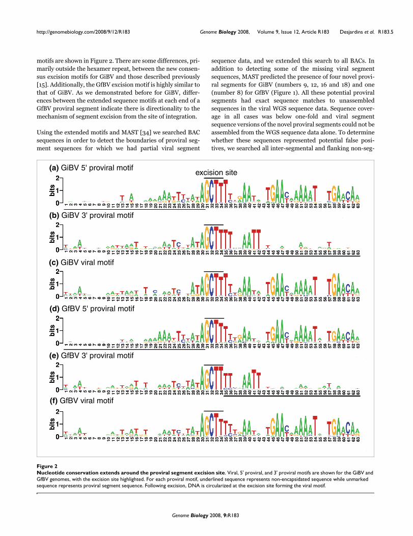
Loci encoding GiBV and GfBV proviral segments exhibit a high degree of syntenyBased on GiBV and GfBV proviral segment location and con-servation of gene order and sequence similarity within provi-ral segments, 27 of the 29 proviral segments were classifiedinto pairs of segment homologs (Figures 1 and 3). Each mem-ber of a segment pair was assigned the same number. Seg-ments 16 and 30 appear to be unique to GiBV, while segments27 and 31 appear to be unique to GfBV (Figure 1). In additionto synteny between GiBV and GfBV proviral segments, thereis clear evidence for conservation of gene order and identityof genes in wasp DNA flanking proviral segment sequences(Figure 3). These data depict the near entirety of the GiBV andGfBV proviral segments; loci encoding three GiBV and threeGfBV proviral segments remain to be identified (Figure 1).
The largest region encoding proviral DNA in both species isrepresented by two linked proviral loci, 1 and 2. GiBV provirallocus 1 [15] and GfBV locus 1 consist of 8 proviral segmentseach, while GiBV and GfBV proviral locus 2 consists of 12 and13 proviral segments, respectively (Table 1). Although theBAC clones we sequenced did not overlap at these loci, con-sideration of synteny suggests that they are linked (Figure 3).Synthetic oligonucleotide primers were designed near theends of GiBV proviral segment 17 and 18 to close the existinggap between BAC clones within GiBV locus 2. However, PCRanalysis of pupal stage G. indiensis parasitoid DNA failed,possibly due the large size of the estimated gap of approxi-
mately 20 kbp. Additional primers were designed based on aninter-segmental parasitoid sodium:neurotransmitter sym-porter gene present in the homologous GfBV region (Figure3), which were then used in appropriate combination withGiBV proviral segment 17 and 18 primers. An amplicon ofapproximately 3 kbp (symporter to GiBV proviral segment18) was obtained and end-sequenced (data not shown), veri-fying the predicted linkage. We were not able to link the largegap from GiBV proviral segment 17 to the symporter, but thedata strongly support linkage of GiBV proviral segment 17and 18 as occurs in GfBV. Synthetic oligonucleotides designedto either side of the gap separating GiBV proviral locus 1amplified an approximately 5 kbp product and end-sequencedata verified physical linkage of the two loci (data not shown).The strong syntenic relationships between GiBV and GfBVproviral genomes argue for the correct assembly and repre-sentation of GiBV proviral locus 2 and its linkage to provirallocus 1 (Figure 3).
The remaining GiBV and GfBV proviral loci (Table 1, Figure3) contain either a single proviral segment (loci 4-6) or twoproviral segments in tandem array (loci 3 and 7). Locus 6 andlocus 7 sequences are only available for GiBV and GfBV,respectively, but must exist for both genomes based on viralsegment sequence data (Figure 1). BAC clones containing loci3-7 do not overlap with each other or with those encoding loci1 and 2. The genomic context surrounding three proviral seg-ment sequences from each virus remains unknown, and theycould occur as single or tandemly linked sequences. Unlessthese proviral segments are linked to the loci we have alreadydefined (in which case there would be seven loci), our currentdata indicate that GiBV and GfBV proviral segment sequenceswill occupy eight or nine loci. It remains possible thatsequence analysis of the missing proviral loci may identifyadditional viral segment sequences.
Structural organization and synteny of proviral segments of GfBV and GiBVFigure 3Structural organization and synteny of proviral segments of GfBV and GiBV. Detailed diagrams of proviral loci for GfBV and GiBV are depicted. For each, the corresponding segment number of the encapsidated viral segment is given for each segment, with the > or < symbol depicting the directionality of segment excision. Black boxes represent long tandem repeats, and three repeat classes are listed as A, B, and C. Regions of synteny between proviral segment and flanking DNA are shaded in gray. Strand-specific protein coding genes within proviral segments are depicted by green boxes, while genes encoded in flanking DNA at each locus are depicted by purple boxes. The length for each locus is shown in kbp.
22> <23
22> <23
0 Kbp 50 Kbp
Gf
Gi
Locus 3
Gi
0 Kbp 50 Kbp
26>
Locus 6
0 Kbp 50 Kbp 100 Kbp
<24
<24Locus 4 Locus 5
0 Kbp 50 Kbp 100 Kbp
25>
25>
Locus 7
Gf
0 Kbp 50 Kbp 100 Kbp
<27
Locus 1
Gf
Gi
0 Kbp 50 Kbp 100 Kbp 150 Kbp 200 Kbp 250 Kbp 300 Kbp 350 Kbp 400 Kbp 450 Kbp 500 Kbp 550 Kbp
<1 2> <3 4> <5 6> 7> 8> 9> <10 <11 12> <13 <14 15> <16 17>18>19>20><21
<1 4> <5 9> <138> <14 15> 18> 19>17> <2120><10 <11 12>6> 7>2> <3
Locus 2
A B A
A
B
A
<28
C
Genome Biology 2008, 9:R183
http://genomebiology.com/2008/9/12/R183 Genome Biology 2008, Volume 9, Issue 12, Article R183 Desjardins et al. R183.7
We previously described two different long (6-7 kbp) tandemrepeats flanking GiBV proviral locus 1 (named L1R1 andL1R2) [15]. These repeats were not found to universally flankthe remaining GiBV proviral loci or to flank GfBV provirallocus 1. A long (7.5-8 kbp) tandem repeat similar to L1R1 wasfound in DNA adjacent to GiBV proviral loci 4 and 5 (Figure3, A repeats) but these repeats were not found in the homolo-gous GfBV proviral loci. An additional smaller (1 kbp) repeatsimilar to L1R1 was found on the 3' side of GiBV proviral locus2. The L1R2 repeat was not found at any other GiBV proviralloci, although L1R2-like repeats (approximately 1.5 kbp) werefound near GfBV proviral loci 4 and 7 (Figure 3, B repeats).GiBV proviral locus 6 does have a long (approximately 6 kbp)tandem repeat on the 3' side (Figure 3, C repeat), although itdoes not share sequence similarity to either L1R1 or L2R2.
As found in GiBV proviral locus 1 [15], inter-segmentalregions separating tandemly arrayed proviral segments inboth the GiBV and GfBV proviral genomes are generally small(<1 kbp) and do not code for proteins. One exception is theapproximately 9 kbp region separating GfBV proviral seg-ments 17 and 18, which codes for a parasitoid sodium:neuro-transmitter symporter. As described above, a similar hostgene is likely to exist in the homologous region in G. indiensis.
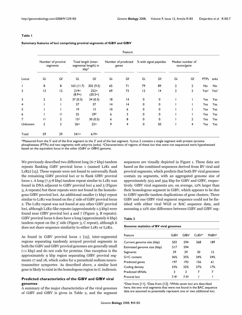
Predicted characteristics of the GiBV and GfBV viral genomesA summary of the major characteristics of the viral genomesof GiBV and GfBV is given in Table 2, and the segment
sequences are visually depicted in Figure 1. These data arebased on the combined sequences derived from BV viral andproviral segments, which predicts that both BV viral genomescontain 29 segments, with an aggregated genome size ofapproximately 503 and 594 kbp for GiBV and GfBV, respec-tively. GfBV viral segments are, on average, 12% larger thantheir homologous segment in GiBV, which appears to be dueto GfBV-specific tandem duplications of gene clusters. ThreeGiBV and one GfBV viral segment sequence could not be fin-ished with either viral WGS or BAC sequence data, andassuming a 12% size difference between GiBV and GfBV seg-
Table 1
Summary features of loci comprising proviral segments of GiBV and GfBV
Feature
Number of proviral segments
Total length (inter-segmental length) in
kbp*
Number of predicted genes
% with signal peptides Median number of exons/gene
Locus Gi Gf Gi Gf Gi Gf Gi Gf Gi Gf PTPs anks
1 8 8 163 (11.7) 202 (9.5) 65 71 79 89 2 2 No No
2 13 12 219+ (8.9+)
252+ (20.5+)
69 73 12 14 2 2 Yes† Yes†
3 2 2 37 (0.3) 34 (0.3) 18 14 0 0 1 1 Yes Yes
4 1 1 37 37 14 14 0 0 1 1 Yes Yes
5 1 1 19 13 10 6 0 0 1 1 Yes Yes
6 1 1‡ 25 292 6 3 0 0 1 1 Yes Yes
7 1‡ 2 152 30 (0.5) 6 8 0 0 1 2 Yes Yes
Unknown 2 2 26+ 22+ 9 4 0 50 1 4 Yes Yes
Total 29 29 541+ 619+
*Measured from the 5' end of the first segment to the 3' end of the last segment. †Locus 2 contains a single segment with protein tyrosine phosphatases (PTPs) and two segments with ankyrins (anks). ‡Characteristics of regions of these loci that were not sequenced were hypothesized based on the equivalent locus in the other (GiBV or GfBV) genome.
Table 2
Genome statistics of BV viral genomes
Feature GiBV GfBV CcBV* MdBV†
Current genome size (kbp) 503 594 568 189
Estimated genome size (kbp) 517 594
Segments 29 29 30 15
G+C content 36% 35% 34% 34%
Predicted genes 197 193 156 61
Coding density 33% 32% 27% 17%
Predicted tRNAs 3 3 7 7
Proviral loci 7-9‡ 7-9‡ ? ?
*Data from [11]. †Data from [12]. ‡While seven loci are described here, the two viral segments that were not found in the BAC sequence data are assumed to potentially represent one or two additional loci.
Genome Biology 2008, 9:R183

http://genomebiology.com/2008/9/12/R183 Genome Biology 2008, Volume 9, Issue 12, Article R183 Desjardins et al. R183.8
ment homologs, we estimate that we are missing approxi-mately 14 kbp of GiBV and <1 kbp GfBV viral genomesequence data. We thus estimate a cumulative viral genomesize to be 517 kbp for GiBV and 594 kbp for GfBV.
The GiBV and GfBV viral genomes are predicted to encodesimilar numbers of proteins, 197 versus 193, respectively, and58% and 63%, respectively, of the genes are predicted to con-tain introns. Both genomes have a similar average G+C con-tent (35-36%) and protein coding density (32-33%). Thegenomes contain many gene families found in other bracovi-ruses, including protein tyrosine phosphatases (PTPs), viralankyrins, C-type lectins, and cystatins; features of these genefamilies are listed in Table 3. Encoded PTPs, ankyrins, andcystatins are mostly predicted to be single exon genes while C-type lectins are always predicted to be two-exon genes. PTPsand ankyrins are not predicted to encode signal peptideswhile C-type lectins and cystatins are often predicted toencode them. Additionally, as reported for CcBV and MdBV,the GfBV and GiBV genomes are predicted to encode a smallnumber of tRNAs (Table 2).
A high degree of partitioning is seen between viral segmentgene content when the segments are grouped by provirallocus membership (Table 1). The majority of genes in locus 1are predicted to encode signal peptides, while few genes inany other loci are. Loci 1 and 2 both contain many proviralsegments, most of which encode hypothetical proteins ofunknown function and contain one intron. In contrast, theremaining loci contain only one or two proviral segments andpredominantly encode PTPs and viral ankyrins that are notpredicted to contain introns. A recent study of CiBV showedthat genes on the same viral segment were generallyexpressed in the secondary host in the same time period [35].Since BV viral segments are present in virion DNA prepara-tions in different abundances, the locus-specific partitioningof GiBV and GfBV proviral segments may represent someadditional form of control over BV viral gene delivery.
Proviral segments and flanking regions show fundamental sequence differencesAnnotation of non-encapsidated DNA sequences flankingGiBV and GfBV proviral segment sequences predicted thepresence of 78 and 71 genes, respectively. Nine genes thatflank GiBV proviral locus 1 were described previously [15].Very few flanking genes showed similarity to known virus(including PDV) genes; those that did are discussed below.However, many had a high sequence similarity with insectgenes (78% for flanking genes and 29% for segment genes;BLASTP, E < e-10). Additionally, flanking genes tended tohave more exons per gene than proviral segment genes,although the difference was not statistically significant (2 ± 1for segment genes and 3 ± 2 for flanking genes).
Proviral segment and flanking DNA are distinct not only atthe gene level, but on a nucleotide composition level as well.Previously, we demonstrated that for GiBV proviral locus 1,proviral segment sequences had nucleotide compositionssimilar to each other and distinct from flanking sequences[15]. We extended the analysis of trinucleotide frequencies toencompass all available GiBV and GfBV proviral segment,flanking, and inter-segmental sequence data. The results areshown in Figure 4. The majority of proviral segment andflanking sequences cluster into distinct groups, and the shortterminal-branch lengths indicate highly similar compositionwithin these groups. Inter-segmental regions also tend tocluster together, although with a higher degree of variation,indicated by the longer terminal-branch lengths. This wasexpected as the generally shorter lengths of inter-segmentalsequences make calculating trinucleotide frequencies lessaccurate. These results were consistent across both the GiBVand GfBV sequence data.
GiBV and GfBV proviral segment genes are evolving rapidlyAs koinobiont endoparasitoids, PDV-carrying wasps developas larvae within caterpillars that are still undergoing develop-ment. Therefore, they are in an arms race with the caterpillars
Table 3
Features of gene families of GiBV and GfBV
Feature
Number of copies Number with signal peptide Number of exons Average M-K test
Gene family GiBV GfBV Loci Gi Gf Gi Gf dN/dS (n) p-value
PTPs 42 31 2-7 0 0 1 1-2 0.80 (16) <0.001
Ankyrins 9 8 2-7 0 0 1 1 0.59 (3) NA
C-type lectins 2 5 1 1 5 2 2 1.09 (1) NA
Cystatins 1 2 1 1 2 1 1 2.23 (1) NA
Sugar transporters 3 5 2 2 0 6-7 7-8 0.23 (2) NA
NA, not enough data for analysis.
Genome Biology 2008, 9:R183

http://genomebiology.com/2008/9/12/R183 Genome Biology 2008, Volume 9, Issue 12, Article R183 Desjardins et al. R183.9
and PDVs contribute to survival against attacks by theimmune system of a living caterpillar. Given this situation, itis plausible to hypothesize that PDV genes on a whole mustevolve rapidly, particularly since conserved genes that typi-cally control viral replication and particle formation appear tobe absent from the viral genome. We utilized sequence diver-gence between GiBV and GfBV genes to test this hypothesis.All genes described here were divided into two sets: oneencompassing genes encoded by proviral segments, and oneincluding flanking genes. Genes were considered orthologs ifthey had reciprocal best BLAST hits to each other andappeared in syntenic positions. Using these criteria, we iden-tified 72 orthologous gene pairs in the proviral segment geneset and 41 in the flanking gene set.
In order to examine the strength and direction of selectionacting on these genes, the ratio of non-synonymous to synon-ymous substitutions (dN/dS) was calculated and a histogramsummarizing the results is shown in Figure 5. Detailed resultsfor each gene pair are shown in Additional data file 1. Flank-ing wasp genes predominantly have dN/dS values near 0(median = 0.23, average = 0.38), indicating that they areunder strong negative selection, which is expected for genesinvolved in essential cellular processes. On the other hand,
proviral segment genes have dN/dS values centered near 1(median = 0.92, average = 0.96), suggesting that many ofthese genes are under neutral or positive selection. Analysiswith the Mann-Whitney U test shows that, overall, segment
Clustering diagram of proviral segments, flanking sequences, and inter-segmental sequences by nucleotide compositionFigure 4Clustering diagram of proviral segments, flanking sequences, and inter-segmental sequences by nucleotide composition. The clustering was generated using the neighbor-joining algorithm on relative trinucleotide frequences for each sequence region. Inter-segmental sequences are shown in black (unlabeled), proviral segment sequences in red, flanking sequences in green, and the GiBV TE in blue.
gibv gfbv
Proviral genomesegments
Flanking regions
Flanking regionsProviral genome segments
Flanking regionGiBV TE
Proviral genomesegments
Proviral genome segment
Proviral genome segmentflanking region
Flanking regions
Histogram of dN/dS values for viral and flanking genes in GiBV and GfBV proviral sequencesFigure 5Histogram of dN/dS values for viral and flanking genes in GiBV and GfBV proviral sequences. dN/dS values were calculated for 72 and 41 homologous pairs of viral genes in flanking DNA, respectively. Genes in flanking DNA have dN/dS values centered near 0 while viral genes have dN/dS values centered near 1.
0
5
10
15
20
25
0.2 0.4 0.6 0.8 1 1.2 1.4 1.6 1.8 2 2.2 2.4
dN/dS
# o
f genes
Flanking (n=40)
Viral (n=72)
Genome Biology 2008, 9:R183
http://genomebiology.com/2008/9/12/R183 Genome Biology 2008, Volume 9, Issue 12, Article R183 Desjardins et al. R183.10
genes are evolving at significantly different rates than flank-ing genes (p < 0.001).
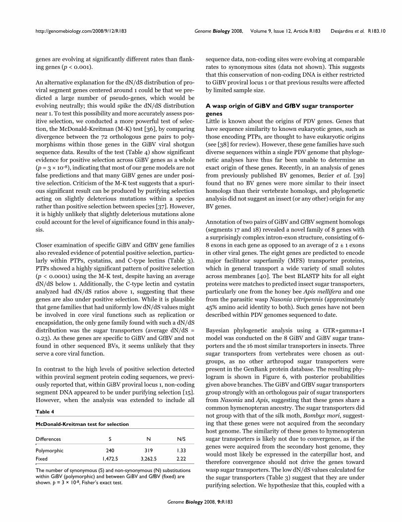
An alternative explanation for the dN/dS distribution of pro-viral segment genes centered around 1 could be that we pre-dicted a large number of pseudo-genes, which would beevolving neutrally; this would spike the dN/dS distributionnear 1. To test this possibility and more accurately assess pos-itive selection, we conducted a more powerful test of selec-tion, the McDonald-Kreitman (M-K) test [36], by comparingdivergence between the 72 orthologous gene pairs to poly-morphisms within those genes in the GiBV viral shotgunsequence data. Results of the test (Table 4) show significantevidence for positive selection across GiBV genes as a whole(p = 3 × 10⁻⁸), indicating that most of our gene models are notfalse predictions and that many GiBV genes are under posi-tive selection. Criticism of the M-K test suggests that a spuri-ous significant result can be produced by purifying selectionacting on slightly deleterious mutations within a speciesrather than positive selection between species [37]. However,it is highly unlikely that slightly deleterious mutations alonecould account for the level of significance found in this analy-sis.
Closer examination of specific GiBV and GfBV gene familiesalso revealed evidence of potential positive selection, particu-larly within PTPs, cystatins, and C-type lectins (Table 3).PTPs showed a highly significant pattern of positive selection(p < 0.0001) using the M-K test, despite having an averagedN/dS below 1. Additionally, the C-type lectin and cystatinanalyzed had dN/dS ratios above 1, suggesting that thesegenes are also under positive selection. While it is plausiblethat gene families that had uniformly low dN/dS values mightbe involved in core viral functions such as replication orencapsidation, the only gene family found with such a dN/dSdistribution was the sugar transporters (average dN/dS =0.23). As these genes are specific to GiBV and GfBV and notfound in other sequenced BVs, it seems unlikely that theyserve a core viral function.
In contrast to the high levels of positive selection detectedwithin proviral segment protein coding sequences, we previ-ously reported that, within GiBV proviral locus 1, non-codingsegment DNA appeared to be under purifying selection [15].However, when the analysis was extended to include all
sequence data, non-coding sites were evolving at comparablerates to synonymous sites (data not shown). This suggeststhat this conservation of non-coding DNA is either restrictedto GiBV proviral locus 1 or that previous results were affectedby limited sample size.
A wasp origin of GiBV and GfBV sugar transporter genesLittle is known about the origins of PDV genes. Genes thathave sequence similarity to known eukaryotic genes, such asthose encoding PTPs, are thought to have eukaryotic origins(see [38] for review). However, these gene families have suchdiverse sequences within a single PDV genome that phyloge-netic analyses have thus far been unable to determine anexact origin of these genes. Recently, in an analysis of genesfrom previously published BV genomes, Bezier et al. [39]found that no BV genes were more similar to their insecthomologs than their vertebrate homologs, and phylogeneticanalysis did not suggest an insect (or any other) origin for anyBV genes.
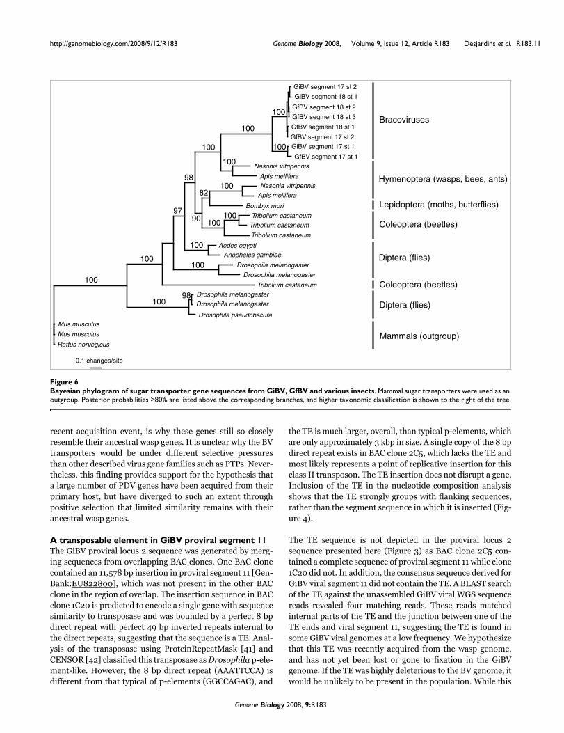
Annotation of two pairs of GiBV and GfBV segment homologs(segments 17 and 18) revealed a novel family of 8 genes witha surprisingly complex intron-exon structure, consisting of 6-8 exons in each gene as opposed to an average of 2 ± 1 exonsin other viral genes. The eight genes are predicted to encodemajor facilitator superfamily (MFS) transporter proteins,which in general transport a wide variety of small solutesacross membranes [40]. The best BLASTP hits for all eightproteins were matches to predicted insect sugar transporters,particularly one from the honey bee Apis mellifera and onefrom the parasitic wasp Nasonia vitripennis (approximately45% amino acid identity to both). Such genes have not beendescribed within PDV genomes sequenced to date.
Bayesian phylogenetic analysis using a GTR+gamma+Imodel was conducted on the 8 GiBV and GiBV sugar trans-porters and the 16 most similar transporters in insects. Threesugar transporters from vertebrates were chosen as out-groups, as no other arthropod sugar transporters werepresent in the GenBank protein database. The resulting phy-logram is shown in Figure 6, with posterior probabilitiesgiven above branches. The GiBV and GfBV sugar transportersgroup strongly with an orthologous pair of sugar transportersfrom Nasonia and Apis, suggesting that these genes share acommon hymenopteran ancestry. The sugar transporters didnot group with that of the silk moth, Bombyx mori, suggest-ing that these genes were not acquired from the secondaryhost genome. The similarity of these genes to hymenopteransugar transporters is likely not due to convergence, as if thegenes were acquired from the secondary host genome, theywould most likely be expressed in the caterpillar host, andtherefore convergence should not drive the genes towardwasp sugar transporters. The low dN/dS values calculated forthe sugar transporters (Table 3) suggest that they are underpurifying selection. We hypothesize that this, coupled with a
Table 4
McDonald-Kreitman test for selection
Differences S N N/S
Polymorphic 240 319 1.33
Fixed 1,472.5 3,262.5 2.22
The number of synonymous (S) and non-synonymous (N) substitutions within GiBV (polymorphic) and between GiBV and GfBV (fixed) are shown. p = 3 × 10⁻⁸, Fisher's exact test.
Genome Biology 2008, 9:R183
http://genomebiology.com/2008/9/12/R183 Genome Biology 2008, Volume 9, Issue 12, Article R183 Desjardins et al. R183.11
recent acquisition event, is why these genes still so closelyresemble their ancestral wasp genes. It is unclear why the BVtransporters would be under different selective pressuresthan other described virus gene families such as PTPs. Never-theless, this finding provides support for the hypothesis thata large number of PDV genes have been acquired from theirprimary host, but have diverged to such an extent throughpositive selection that limited similarity remains with theirancestral wasp genes.
A transposable element in GiBV proviral segment 11The GiBV proviral locus 2 sequence was generated by merg-ing sequences from overlapping BAC clones. One BAC clonecontained an 11,578 bp insertion in proviral segment 11 [Gen-Bank:EU822800], which was not present in the other BACclone in the region of overlap. The insertion sequence in BACclone 1C20 is predicted to encode a single gene with sequencesimilarity to transposase and was bounded by a perfect 8 bpdirect repeat with perfect 49 bp inverted repeats internal tothe direct repeats, suggesting that the sequence is a TE. Anal-ysis of the transposase using ProteinRepeatMask [41] andCENSOR [42] classified this transposase as Drosophila p-ele-ment-like. However, the 8 bp direct repeat (AAATTCCA) isdifferent from that typical of p-elements (GGCCAGAC), and
the TE is much larger, overall, than typical p-elements, whichare only approximately 3 kbp in size. A single copy of the 8 bpdirect repeat exists in BAC clone 2C5, which lacks the TE andmost likely represents a point of replicative insertion for thisclass II transposon. The TE insertion does not disrupt a gene.Inclusion of the TE in the nucleotide composition analysisshows that the TE strongly groups with flanking sequences,rather than the segment sequence in which it is inserted (Fig-ure 4).
The TE sequence is not depicted in the proviral locus 2sequence presented here (Figure 3) as BAC clone 2C5 con-tained a complete sequence of proviral segment 11 while clone1C20 did not. In addition, the consensus sequence derived forGiBV viral segment 11 did not contain the TE. A BLAST searchof the TE against the unassembled GiBV viral WGS sequencereads revealed four matching reads. These reads matchedinternal parts of the TE and the junction between one of theTE ends and viral segment 11, suggesting the TE is found insome GiBV viral genomes at a low frequency. We hypothesizethat this TE was recently acquired from the wasp genome,and has not yet been lost or gone to fixation in the GiBVgenome. If the TE was highly deleterious to the BV genome, itwould be unlikely to be present in the population. While this
Bayesian phylogram of sugar transporter gene sequences from GiBV, GfBV and various insectsFigure 6Bayesian phylogram of sugar transporter gene sequences from GiBV, GfBV and various insects. Mammal sugar transporters were used as an outgroup. Posterior probabilities >80% are listed above the corresponding branches, and higher taxonomic classification is shown to the right of the tree.
Apis mellifera
Apis mellifera
Nasonia vitripennis
Nasonia vitripennis
Bombyx mori
Tribolium castaneum
Tribolium castaneum
Tribolium castaneum
Anopheles gambiae
Drosophila melanogaster
Aedes egypti
Drosophila melanogaster
Tribolium castaneum
Drosophila melanogaster
Drosophila melanogaster
Drosophila pseudobscura
Mus musculus
Rattus norvegicus
Mus musculus
0.1 changes/site
GfBV segment 17 st 1
GiBV segment 17 st 1
GiBV segment 18 st 1
GiBV segment 17 st 2
GfBV segment 18 st 3
GfBV segment 18 st 2
GfBV segment 17 st 2
GfBV segment 18 st 1
Hymenoptera (wasps, bees, ants)
Bracoviruses
Lepidoptera (moths, butterflies)
Coleoptera (beetles)
Diptera (flies)
Coleoptera (beetles)
Diptera (flies)
Mammals (outgroup)
100
100
100
100
100
100
82
100
10098
100100
100100
90
100
98
97
Genome Biology 2008, 9:R183
http://genomebiology.com/2008/9/12/R183 Genome Biology 2008, Volume 9, Issue 12, Article R183 Desjardins et al. R183.12
TE does not encode any genes other than a transposase, itdoes suggest that TEs can enter a BV genome and becomepackaged successfully. No evidence of this TE was found ineither viral or proviral GfBV sequence data.
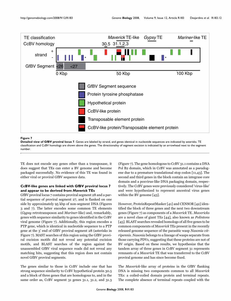
CcBV-like genes are linked with GfBV proviral locus 7 and appear to be derived from Maverick TEsGfBV proviral locus 7 contains proviral segment 28 and a par-tial sequence of proviral segment 27, and is flanked on oneside by approximately 95 kbp of non-segment DNA (Figures3 and 7). The latter encodes some common TE elements(Gypsy retrotransposon and Mariner-like) and, remarkably,genes with sequence similarity to genes identified in the CcBVviral genome (Figure 7). Additionally, this region encodes aPTP gene, which is identical in nucleotide sequence to a PTPgene at the 3' end of GfBV proviral segment 28 (asterisks inFigure 7). MAST searches of this region using the GfBV provi-ral excision motifs did not reveal any potential excisionmotifs, and BLAST searches of the region against theunassembled GfBV viral sequence reads did not reveal anymatching hits, suggesting that this region does not containnovel GfBV proviral segments.
The genes similar to those in CcBV include one that hasstrong sequence similarity to CcBV hypothetical protein 30.5and a block of three genes that are homologous to, and in thesame order as, CcBV segment 31 genes 31.1, 31.2, and 31.3
(Figure 7). The gene homologous to CcBV 31.1 contains a DNAPol B2 domain, which in CcBV was annotated as a pseudog-ene due to a premature translational stop codon [11,43]. Thesecond and third genes in the block contain an integrase coredomain and a poxvirus-like DNA packaging domain, respec-tively. The CcBV genes were previously considered 'virus-like'and were hypothesized to represent ancestral virus geneswithin the BV genome [43].
However, ProteinRepeatMasker [41] and CENSOR [42] iden-tified the block of three genes and the next two downstreamgenes (Figure 7) as components of a Maverick TE. Mavericksare a novel class of giant TEs [44], also known as Polintons[45]. BLAST searches revealed homologs of all five genes to becommon components of Maverick TEs present in the recentlyreleased genome sequence of the parasitic wasp Nasonia vit-ripennis. Nasonia belongs to a lineage of wasps separate fromthose carrying PDVs, suggesting that these proteins are not ofBV origin. Based on these results, we hypothesize that thetandem array of three genes in CcBV segment 31 representsremnants of a Maverick TE that was transferred to the CcBVproviral genome and has since become fixed.
The Maverick-like array of proteins in the GfBV flankingDNA is missing two components common to all MaverickTEs: a coiled-coiled domain protein and terminal repeats.The complete absence of terminal repeats coupled with the
Detailed view of GfBV proviral locus 7Figure 7Detailed view of GfBV proviral locus 7. Genes are labeled by strand, and genes identical in nucleotide sequences are indicated by asterisks. TE classification and CcBV homologs are shown above the genes. The directionality of segment excision is indicated by an arrowhead next to the segment number.
GfBV Segment sequence
Protein tyrosine phosphatase
Hypothetical protein
CcBV-like protein
Transposable element protein
CcBV-like protein/Transposable element protein
**
Gypsy TE Mariner-like TEMaverick TE-like
CcBV homology
TE classification
30.5 31.1,2,3
50 Kbp0 Kbp 100 Kbp
<27<26
strand+
-
GfBV Segment
Genome Biology 2008, 9:R183

http://genomebiology.com/2008/9/12/R183 Genome Biology 2008, Volume 9, Issue 12, Article R183 Desjardins et al. R183.13
maintenance of open reading frames is particularly unusual.Since TEs cannot function without terminal repeats, TE openreading frames typically degrade rapidly after their terminalrepeats are lost. This suggests that selection on the waspgenome is maintaining these open reading frames, althoughtheir potential function is unknown. The abundance of bothTEs and BV-like genes encoded in DNA flanking GfBV provi-ral locus 7 may indicate that this region is a 'hotspot' for themovement of DNA between proviral and flanking sequences.It will be of interest to determine if similar sequences arepresent in the homologous GiBV locus.
ConclusionHere we provide a comprehensive sequence and computa-tional analysis of the viral genome of BVs isolated from twodifferent Glyptapanteles species, G. indiensis and G. flavi-coxis, and an analysis of proviral sequences from whichencapsidated viral genomes are derived. The sequencing ofboth BV genomic forms, that is, viral and proviral segments,was critical in developing a more complete definition of boththe viral genome as well as the structural organization of alarge component of the proviral genome, given that the latterunexpectedly led to the discovery of four GiBV and one GfBVviral segment not evident from sequencing the viral genome.
Although GiBV and GfBV proviral segments are not all tan-demly arrayed, synteny between the two sets of sequencesand preliminary linkage data for GiBV suggest that approxi-mately 70% of the 29 proviral segments in each genome areclustered in a single genomic region consisting of two proviralloci that span a region of approximately 580 kbp. Each locuscontains tandemly arrayed proviral segments separated fromeach other by a short stretch of non-segment DNA (Figure 3).The remaining GiBV and GfBV proviral loci identified containeither one or two proviral segments. Most of the seven char-acterized loci are flanked by long stretches of non-segmentDNA that encode insect proteins. Not every proviral locus isflanked by long tandem repeats, as was reported for provirallocus 1 [15]. So it is presently unclear how proviral loci aredemarcated within the wasp genome sequence. Qualitativeand quantitative studies of proviral sequences at the time ofproduction of viral segments will be needed to accuratelydelineate the boundaries of amplified proviral sequences. Oursequence data are not incompatible with the hypothesis thatall proviral loci are linked in a macrolocus, as suggested forCcBV proviral segments [25,27]. Additional mapping experi-ments, for example, fluorescence in situ hybridization analy-sis using segment-specific probes or generation of a BACtiling path among loci should resolve the spatial relationshipsamong the GiBV and GfBV proviral loci. If present, a macrolo-cus encoding proviral segments would exceed 1 Mbp in size.
BVs are thought to have a monophyletic origin about 100 mil-lion years ago [46], a hypothesis supported by the high degreeof synteny between GiBV and GfBV proviral loci and flanking
DNA, as well as the highly conserved proviral segment exci-sion motif among BVs [15]. The compositional difference insequences at proviral loci could be indicative of independentorigins of BVs, but they could also be the result of selectivepressures from the primary and or secondary host environ-ment. It will be of interest to compare proviral loci from basalmicrograstrines, such as Microplitis, to those of more derivedmiscogastrines such as Glyptapanteles. Such comparisonscould determine if there was an ancient site for BV integrationand, if such a site exists, the extent to which it has been con-served. In addition, whole host genome sequence data arelikely to contribute to a more complete understanding of theevolution of BVs.
The functions and origins of PDV genes are often difficult todefine. Comparative sequence analyses revealed widespreadpositive selection across a large number of GiBV and GfBVgenes, which supports their role in an 'arms race' betweenvirus and caterpillar. This rapid evolution may help toobscure the origins of many PDV genes. However, GiBV andGfBV both encode a gene family of sugar transporters notfound in other sequenced PDV genomes. Phylogenetic analy-ses suggest that these were wasp genes that were moved intothe proviral genome, providing evidence for an insect originfor these BV genes. This suggests that the proviral BV genomemay be at least partially derived from the wasp genome, andpossibly represents a mosaic of an ancestral virus and morerecently inserted host genes.
Transposable elements represent a plausible mechanism foracquiring virulence or other desirable genes from the host[27,47]. Our evidence shows a p-element like and a Maverick-like element associated with BV genomes. Whether the latterplays a role in BV biology is unknown, although it is interest-ing to note the similarities between the proposed methods ofreplication in Mavericks [45] and PDVs, as both are hypoth-esized to replicate extrachromosomally from a circular orstem-loop molecule. Both described TEs, the former recentlyacquired and the latter ancient, could have served to movecore viral genes out of the ancestral BV genome to the hostgenome and virulence genes into it. Alternatively, genes couldhave moved out of the proviral genome by the degradation ofexcision motifs of proviral segments, thereby creating'pseudo-segments' that are no longer encapsidated.
Materials and methodsRearing of parasitoid waspsOutbred populations of G. indiensis and G. flavicoxis, solitaryand gregarious endoparasitoids of gypsy moths (Lymantriadispar), respectively, were maintained at the USDA-ARS-Beneficial Insects Introduction Research Unit, Newark, Dela-ware, until they were moved to Beltsville, Maryland in 2007.The maintenance protocol and history of the G. indiensis col-ony was described previously [15]. G. flavicoxis was rearedunder the same protocol with the exception that G. flavicoxis
Genome Biology 2008, 9:R183
http://genomebiology.com/2008/9/12/R183 Genome Biology 2008, Volume 9, Issue 12, Article R183 Desjardins et al. R183.14
parasitizes late third instar gypsy moth larvae. Cocoonsformed from parasitized hosts were stored at 24°C until adultparasitoid emergence and then separated by sex. For BAClibrary material, G. flavicoxis larvae were dissected from par-asitized gypsy moth larvae 10 days post-parasitization, brieflyrinsed in phosphate buffered saline, flash frozen in liquidnitrogen and stored frozen at -80°C.
Virion purification and DNA extractionVirions were purified from G. indiensis and G. flavicoxisfemales using established protocols [48]. Briefly, femalewasps were anaesthetized in 75% ethanol and rinsed in phos-phate buffered saline. Ovaries were dissected from thefemales in a drop of phosphate buffered saline and ruptured,draining the calyx fluid. Pooled calyx fluid was subsequentlyfiltered through a 0.45 μm filter to remove eggs and cellulardebris [49]. Viral DNA was extracted according to establishedprotocols [30].
Identification of BAC clones containing proviral segment DNABAC libraries of G. indiensis and G. flavicoxis with a 120 kbaverage insert size were constructed by Amplicon Express(Pullman, WA, USA), using a partial BamHI digest insertedinto an MboI site of a pECBAC1 vector. A nylon filter arrayedwith 9,216 BAC clones was created from each library. In orderto identify BAC clones containing proviral segment DNA, BVencapsidated viral DNA from each was radioactively labeledwith 32P-labeled α-dCTP (NEN/Perkin-Elmer, Waltham, MA,USA) using the Redi-prime II DNA labeling kit (AmershamBiosciences, Piscataway, NJ, USA). Labeled DNA was thenpurified using a QIAquick PCR purification kit (Qiagen,Valencia, CA, USA). The filter was pre-hybridized at 65°C forat least 3 hours with Rapid-hyb Buffer (Amersham Bio-sciences) and 500 μg of salmon testes DNA (denatured at100°C; Sigma-Aldritch, St. Louis, MO, USA). The probe wasadded and allowed to hybridize overnight at 65°C. The filterwas then washed 2 times for 60 minutes each at 65°C with a0.1 × SSC/0.1% SDS solution, wrapped in plastic wrap, andautoradiographed using Kodac BioMax MS film.
Viral and BAC clone sequencingApproximately 7.5 μg of BV encapsidated DNA was shearedand DNA fragments in the size range 3.5-4.5 kbp purifiedafter separation by agarose gel electrophoresis. The frag-ments were blunt ended and, after addition of BstXI adaptors,cloned into the BstXI site of pHOS2. Shotgun libraries weresimilarly made for each BAC clone. Celera Assembler [50]and TIGR Assembler [51] were used to assemble randomsequence data for BV and BAC clones. Gap closure wasassisted by a closure editor tool called Cloe that also permitsthe manual inspection and editing of sequence data. A varietyof methods were used to close gaps, including re-sequencingthe ends of random clones, transposon assisted sequencing(GPS, New England Biolabs™, Ipswitch, MA, USA) or 'micro-library' construction of single or pooled templates, and con-
version of physical gaps to sequence gaps using 'POMP'(pipette optimal multiplex PCR) [52] and or/a 'GenomeWalker' kit (Invitrogen™).
Viral segment-specific PCRs and hybridizationsPrimers were developed specific to individual identified GiBVand GfBV viral segment sequences as described in [15]. PCRwas performed in a 10 μl solution that included 0.1 μl tem-plate DNA, 0.3 μl 50 mM MgCl2, 1 μl 10× PCR buffer, 0.2 μl10 mM dNTPs, 7.9 μl H2O, 0.1 μl Platinum Taq (Invitrogen),0.2 μl F primer (20 pm/μl), and 0.2 μl RC primer (20 pm/μl).The PCR protocol was 94° for 2 minutes; 35 cycles of 94° for30 s, 58° for 30 s, 72° for 45 s; followed by 72° for 7 minutes.PCR products to be used for hybridizations were purifiedusing a QIAquick PCR purification kit (Qiagen). Segment-specific hybridizations were done as described above for totalviral DNA hybridizations.
Derivation of consensus segment sequences for GiBV and GfBVBecause individual sequence reads could not be associatedwith individual wasps, a conical consensus sequence was gen-erated for each BV segment using the SliceTools package [53].At a given position in a conical consensus, all bases with acumulative quality value within 50% of the highest cumula-tive quality value were assigned to that position.
AnnotationA combination of SoftBerry's FGENESH [54] using the honeybee (A. mellifera) training set, and the Beijing Genome Insti-tute's BGF [55] trained on the silkmoth (B. mori) were usedfor gene prediction, in addition to the AAT package [56],which allows spliced alignment of proteins to genomic DNA,thereby revealing potential exon-intron boundaries. Genemodels from FGENESH were generally accepted except whenmultiple other sources of information contradicted thosemodels. SignalP [57,58] and tRNAScan-SE [59] were used topredict signal peptides and tRNAs, respectively. Transposa-ble elements were annotated using ProteinRepeatMasker [41]and CENSOR [42].
Motif analysesExcision motifs were generated by cutting out a sequenceextending 30 bp upstream and downstream from the GCTexcision site at the 5' and 3' boundaries of proviral segmentsand at the GCT circularization site of viral segments for bothGiBV and GfBV. No additional alignment was conducted onthe sequences. All motifs were visualized using WebLogo[60,61]. Proviral excision motifs were also generated withthese sequences using MEME [33], and the resulting motifswere used to search BAC sequences for potential additionalproviral segments using MAST [34].
Comparative genomic analysesJaccard orthologous gene clusters between GiBV and GfBVwere calculated using Sybil [62]. Syntenic blocks were
Genome Biology 2008, 9:R183

http://genomebiology.com/2008/9/12/R183 Genome Biology 2008, Volume 9, Issue 12, Article R183 Desjardins et al. R183.15
defined as two or more adjacent orthologous gene clusters,and the results were visualized using Sybil [62]. This informa-tion, in addition to conserved location (locus and positionwithin that locus) of proviral segments, was used to definehomologous segments between GiBV and GfBV.
Phylogenetic and compositional analysesFor the phylogenetic analysis, GiBV and GfBV MFS trans-porter genes were searched against the GenBank non-redun-dant protein database using BLASTP. The top 18 hits fromunique organisms were downloaded from GenBank (N. vit-ripennis, [GenBank:XP_001607065; XP_001602960]; A.mellifera, [GenBank:XP_001120868; XP_395522]; Tribo-lium castaneum, [GenBank:XP_973694; XP_973659], [Gen-Bank:XP_966705; XP_966524]; Anopheles gambiae,[GenBank:XP_311836]; Aedes egypti, [Gen-Bank:XP_001649205]; Drosophila melanogaster, [Gen-Bank:NP_611451; NP_524479; CAA73031; XP_001361445];Drosophila pseudoobscura, [GenBank:XP_001358762];Mus musculus, [GenBank:NP_035525; CAC36405]; andRattus norvegicus, [GenBank:NP_062103]). Additionally,the single GfBV MFS transporter gene was searched againstB. mori expressed sequence tags in GenBank, and the singlestrong hit (E = e-61; [Genbank:BJ985900]) was downloadedfrom GenBank and translated. These sequences, in additionto the eight MFS transporters predicted for GiBV and GfBV,were aligned using ClustalW [63], and regions of ambiguousalignment were removed using Seaview [64], resulting in analignment of 448 amino acids. The phylogenetic analysis wasconducted using MrBayes 3.1.2 [65,66], sampling every 1,000generations for 1 × 10⁷ generations. The first 50% of genera-tions were discarded as burn-in, and posterior probabilitieswere calculated from the remaining 501 sampled generations.
For the nucleotide composition analysis, relative trinucle-otide frequencies [67] were calculated for all segment, inter-segmental, and flanking sequences. A Euclidean distancematrix was then constructed from those frequencies. Thesequences were then clustered using the neighbor-joiningalgorithm in PAUP* [68] and the resulting tree was visualizedwith Treeview [69].
Molecular evolution analysesAll proteins described here for each genome were divided intotwo sets: those encoded by proviral segments and flankinggenes. For each set, BLASTP in WU-BLAST [70] was used tosearch the GiBV (or Gi) proteins against the GfBV (or Gf) pro-teins and vice versa. Nucleotide sequences of reciprocal besthit pairs that appeared in syntenic regions (in the same seg-ment for proviral genes and in the same region for flankinggenes) were aligned using the cdna_fast_pair method in T-Coffee [71], and pairs with ambiguous or frameshifted align-ments were removed. The codeml program in PAML [72,73]was used to calculate dN/dS (Ka/Ks) for the remaining genepair alignments. Pairs of proviral genes were further analyzedby using codeml in PAML [72,73] to calculate the number of
silent and replacement substitutions. The number of silentand replacement polymorphisms within the GiBV shotgunsequence data for these genes with at least 3× coverage werethen calculated using previously described methods [15]. TheM-K test [36] was then utilized to test gene pairs for evidenceof positive or negative selection.
AbbreviationsBAC: bacterial artificial chromosome; BV: bracovirus; CcBV:Cotesia congregata bracovirus; CiBV: Chelonus inanitus bra-covirus; CsIV: Campoletis sonorensis ichnovirus; dN/dS:ratio of non-synonymous to synonymous substitutions;GfBV: Glyptapanteles flavicoxis bracovirus; GiBV: Glyptap-anteles indiensis bracovirus; IV: ichnovirus; MdBV: Microp-litis demolitor bracovirus; MFS: major facilitatorsuperfamily; M-K: McDonald-Kreitman; PDV: polydnavirus;PTP: protein tyrosine phosphatase; TE: transposable ele-ment; WGS: whole genome shotgun.
Authors' contributionsVN and DEGR conceived the project. VN, CAD, and DEGRcoordinated the project. CAD, DEGR, VN, and MJP designedand performed laboratory procedures and experiments. CAD,MCS, JC, and VN designed and performed computationalanalyses. CAD, VN, and DEGR wrote the manuscript. CADand BJH conducted genome annotation. JBH, LJT, and KMJconducted genome closure. RWF reared parasitoids. DWFand HF conducted library construction. All authors read andapproved this manuscript.
Additional data filesThe following additional data are available with the onlineversion of this article. Additional data file 1 is a table showingthe molecular evolutionary analyses of GiBV and GfBV.Additional data file 1Molecular evolutionary analyses of GiBV and GfBVMolecular evolutionary analyses of GiBV and GfBV.Click here for file
AcknowledgementsWe would like to thank Teresa Utterback, Tamara Feldblyum, and the staffat J Craig Venter Institute's Joint Technology Center for sequencing andviral library construction, and the JCVI IT department for general support.We would also like to thank Jessica Vamathevan and Mihai Pop for initialwork on viral genome closure and analysis, Hean Koo and Jessica Chen forhandling sequence submissions, Linda Hannick for help with gene familycomputation, and Daven Presgraves for advice on SNP analysis. Funding forthis study was provided by the National Science Foundation (0413618) andUnited States Department of Agriculture (2004-35600-15032).
References1. Stoltz DB, Beckage NE, Blissard GW, Fleming JGW, Krell PJ, Theil-
mann DA, Summers MD, Webb BA: Polydnaviridae. In Virus Tax-onomy Sixth Report of the International Committee on Taxonomy of VirusesEdited by: Murphy FA, Fauquet CM, Bishop DHL, Ghabrial SA, JarvisAW, Martelli GP, Mayo MA, Summers MD. Vienna: Springer Verlag;1995:143-147.
2. Fleming JA: The integration of polydnavirus genomes in para-sitoid genomes: implications for biocontrol and genetic anal-yses of parasitoid wasps. Biol Control 1991, 1:127-135.
Genome Biology 2008, 9:R183

http://genomebiology.com/2008/9/12/R183 Genome Biology 2008, Volume 9, Issue 12, Article R183 Desjardins et al. R183.16
3. Gruber A, Stettler P, Heiniger P, Schumperli D, Lanzrein B: Polydna-virus DNA of the braconid wasp Chelonus inanitus is inte-grated in the wasp's genome and excised only in later pupaland adult stages of the female. J Gen Virol 1996, 77:2873-2879.
4. Theilmann DA, Summers MD: Molecular analysis of Campoletissonorensis virus DNA in the lepidopteran host Heliothis vires-cens. J Gen Virol 1986, 67:1961-1969.
5. Norton WN, Vinson SB: Correlating the initiation of virus rep-lication with a specific phase of pupal development in an ich-neumonid parasitoid. Cell Tissue Res 1983, 231:387-398.
6. Wyder S, Blank F, Lanzrein B: Fate of polydnavirus DNA of theegg-larval parasitoid Chelonus inanitus in the host Spodopteralittoralis. J Insect Physiol 2003, 49:491-500.
7. Lavine MD, Beckage NE: Polydnaviruses: potent mediators ofhost insect immune dysfunction. Parasitol Today 1995,11:368-378.
8. Lawrence PO, Lanzrein B: Hormonal interactions betweeninsect endoparasites and their host insects. In Parasites andPathogens of Insects. Parasites Volume 1. Edited by: Beckage NE,Thompson SN, Federici BA. New York: Academic Press; 1993:59-86.
9. Webb BA, Strand MR: The biology and genomics of polydnavi-ruses. In Comprehensive Molecular Insect Science Volume 6. Edited by:Gilbert LI, I I, Gill S. San Diego: Elsevier; 2005:323-360.
10. Whitfield JB: Estimating the age of the polydnavirus/braconidwasp symbiosis. Proc Natl Acad Sci USA 2002, 99:7508-7513.
11. Espagne E, Dupuy C, Huguet E, Cattolico L, Provost B, Martins N,Poirie M, Periquet G, Drezen JM: Genome sequence of a polyd-navirus: insights into symbiotic virus evolution. Science 2004,306:286-289.
12. Webb BA, Strand MR, Dickey SE, Beck MH, Hilgarth RS, Barney WE,Kadash K, Kroemer JA, Lindstrom KG, Rattanadechakul W, ShelbyKS, Thoetkiattikul H, Turnbull MW, Witherell RA: Polydnavirusgenomes reflect their dual roles as mutualists and patho-gens. Virology 2006, 347:160-174.
13. Tanaka K, Lapointe R, Barney WE, Makkay AM, Stoltz D, Cusson M,Webb BA: Shared and species-specific features among ichno-virus genomes. Virology 2007, 363:26-35.
14. Lapointe R, Tanaka K, Barney WE, Whitfield JB, Banks JC, Beliveau C,Stoltz D, Webb BA, Cusson M: Genomic and morphological fea-tures of a banchine polydnavirus: comparison with bracovi-ruses and ichnoviruses. J Virol 2007, 81:6491-6501.
15. Desjardins CA, Gundersen-Rindal DE, Hostetler JB, Tallon LJ, FuesterRW, Schatz MC, Pedroni MJ, Fadrosh DW, Haas BJ, Toms BS, ChenD, Nene V: Structure and evolution of a proviral locus of Glyp-tapanteles indiensis bracovirus. BMC Microbiol 2007, 7:61.
16. Kroemer JA, Webb BA: Polydnavirus genes and genomes:emerging gene families and new insights into polydnavirusreplication. Annu Rev Entomol 2004, 49:431-456.
17. Deng L, Stoltz DB, Webb BA: A gene encoding a polydnavirusstructural polypeptide is not encapsidated. Virology 2000,269:440-450.
18. Federici BA, Bigot Y: Origin and evolution of polydnaviruses bysymbiogenesis of insect DNA viruses in endoparasitic wasps.J Insect Physiol 2003, 49:419-432.
19. Whitfield JB, Asgari S: Virus or not? Phylogenetics of polydnavi-ruses and their wasp carriers. J Insect Physiol 2003, 49:397-405.
20. Fleming JG, Summers MD: Polydnavirus DNA is integrated inthe DNA of its parasitoid wasp host. Proc Natl Acad Sci USA 1991,88:9770-9774.
21. Cui L, Webb BA: Homologous sequences in the Campoletissonorensis polydnavirus genome are implicated in replicationand nesting of the W segment family. J Virol 1997,71:8504-8513.
22. Rattanadechakul W, Webb BA: Characterization of Campoletissonorensis ichnovirus unique segment B and excision locusstructure. J Insect Physiol 2003, 49:523-532.
23. Savary S, Beckage N, Tan F, Periquet G, Drezen JM: Excision of thepolydnavirus chromosomal integrated EP1 sequence of theparasitoid wasp Cotesia congregata (Braconidae, Microgasti-nae) at potential recombinase binding sites. J Gen Virol 1997,78:3125-3134.
24. Wyder S, Tschannen A, Hochuli A, Gruber A, Saladin V, Zumbach S,Lanzrein B: Characterization of Chelonus inanitus polydnavirussegments: sequences and analysis, excision site and demon-stration of clustering. J Gen Virol 2002, 83:247-256.
25. Belle E, Beckage NE, Rousselet J, Poirie M, Lemeunier F, Drezen JM:Visualization of polydnavirus sequences in a parasitoid waspchromosome. J Virol 2002, 76:5793-5796.
26. Pasquier-Barre F, Dupuy C, Huguet E, Monteiro F, Moreau A, PoirieM, Drezen JM: Polydnavirus replication: the EP1 segment ofthe parasitoid wasp Cotesia congregata is amplified within alarger precursor molecule. J Gen Virol 2002, 83:2035-2045.
27. Drezen JM, Provost B, Espagne E, Cattolico L, Dupuy C, Poirie M, Per-iquet G, Huguet E: Polydnavirus genome: integrated vs. freevirus. J Insect Physiol 2003, 49:407-417.
28. Marti D, Grossniklaus-Burgin C, Wyder S, Wyler T, Lanzrein B:Ovary development and polydnavirus morphogenesis in theparasitic wasp Chelonus inanitus. I. Ovary morphogenesis,amplification of viral DNA and ecdysteroid titres. J Gen Virol2003, 84:1141-1150.
29. Annaheim M, Lanzrein B: Genome organization of the Chelonusinanitus polydnavirus: excision sites, spacers and abundanceof proviral and excised segments. J Gen Virol 2007, 88:450-457.
30. Chen YP, Gundersen-Rindal DE: Morphological and genomiccharacterization of the polydnavirus associated with the par-asitoid wasp Glyptapanteles indiensis (Hymenoptera: Braco-nidae). J Gen Virol 2003, 84:2051-2060.
31. Gundersen-Rindal D, Dougherty EM: Evidence for integration ofGlyptapanteles indiensis polydnavirus DNA into the chromo-some of Lymantria dispar in vitro. Virus Res 2000, 66:27-37.
32. Gundersen-Rindal DE, Pedroni MJ: Characterization and tran-scriptional analysis of protein tyrosine phosphatase genesand an ankyrin repeat gene of the parasitoid Glyptapantelesindiensis polydnavirus in the parasitized host. J Gen Virol 2006,87:311-322.
33. Bailey TL, Elkan C: Fitting a mixture model by expectationmaximization to discover motifs in biopolymers. In Proceed-ings of the Second International Conference on Intelligent Systems forMolecular Biology Menlo Park, California: AAAI Press; 1994:28-36.
34. Bailey TL, Gribskov M: Combining evidence using p-values:application to sequence homology searches. Bioinformatics1998, 14:48-54.
35. Weber B, Annaheim M, Lanzrein B: Transcriptional analysis ofpolydnaviral genes in the course of parasitization revealssegment-specific patterns. Arch Insect Biochem Physiol 2007,66:9-22.
36. McDonald JH, Kreitman M: Adaptive protein evolution at theAdh locus in Drosophila. Nature 1991, 351:652-654.
37. Hughes AL: Looking for Darwin in all the wrong places: themisguided quest for positive selection at the nucleotidesequence level. Heredity 2007, 99:364-373.
38. Dupuy C, Huguet E, Drezen JM: Unfolding the evolutionary storyof polydnaviruses. Virus Res 2006, 117:81-89.
39. Bezier A, Herbiniere J, Serbielle C, Lesobre J, Wincker P, Huguet E,Drezen JM: Bracovirus gene products are highly divergentfrom insect proteins. Arch Insect Biochem Physiol 2008, 67:172-187.
40. Pao SS, Paulsen IT, Saier MH Jr: Major facilitator superfamily.Microbiol Mol Biol Rev 1998, 62:1-34.
41. RepeatMasker Open-3.0 [http://www.repeatmasker.org]42. Kohany O, Gentles AJ, Hankus L, Jurka J: Annotation, submission
and screening of repetitive elements in Repbase: Rep-baseSubmitter and Censor. BMC Bioinformatics 2006, 7:474.
43. Drezen JM, Bezier A, Lesobre J, Huguet E, Cattolico L, Periquet G,Dupuy C: The few virus-like genes of Cotesia congregata bra-covirus. Arch Insect Biochem Physiol 2006, 61:110-122.
44. Pritham EJ, Putliwala T, Feschotte C: Mavericks, a novel class ofgiant transposable elements widespread in eukaryotes andrelated to DNA viruses. Gene 2007, 390:3-17.
45. Kapitonov VV, Jurka J: Self-synthesizing DNA transposons ineukaryotes. Proc Natl Acad Sci USA 2006, 103:4540-4545.
46. Murphy N, Banks JC, Whitfield JB, Austin AD: Phylogeny of theparasitic microgastroid subfamilies (Hymenoptera: Braconi-dae) based on sequence data from seven genes, with animproved time estimate of the origin of the lineage. Mol Phy-logenet Evol 2008, 47:378-395.
47. Webb BA: Polydnavirus biology, genome structure, and evo-lution. In The Insect Viruses Edited by: Miller LK, Ball LA. New York:Plenum Press; 1998:105-139.
48. Krell PJ, Summers MD, Vinson SB: Virus with a multipartitesuperhelical DNA genome from the Ichneumonid parasitoidCampoletis sonorensis. J Virol 1982, 43:859-870.
49. Beckage NE, Tan FF, Schleifer KW, Lane RD, Cherubin LL: Charac-terization and biological effects of Cotesia congregata polyd-navirus on host larvae of the tobacco hornworm, Manducasexta. Arch Insect Biochem Physiol 1994, 26:165-195.
50. Myers EW, Sutton GG, Delcher AL, Dew IM, Fasulo DP, Flanigan MJ,
Genome Biology 2008, 9:R183
http://genomebiology.com/2008/9/12/R183 Genome Biology 2008, Volume 9, Issue 12, Article R183 Desjardins et al. R183.17
Kravitz SA, Mobarry CM, Reinert KH, Remington KA, Anson EL,Bolanos RA, Chou HH, Jordan CM, Halpern AL, Lonardi S, BeasleyEM, Brandon RC, Chen L, Dunn PJ, Lai Z, Liang Y, Nusskern DR, ZhanM, Zhang Q, Zheng X, Rubin GM, Adams MD, Venter JC: A whole-genome assembly of Drosophila. Science 2000, 287:2196-2204.
51. Sutton G, White O, Adams M, Kerlavage A: TIGR Assembler: Anew tool for assembling large shotgun sequencing projects.Genome Sci Technol 1995, 1:9-19.
52. Tettelin H, Radune D, Kasif S, Khouri H, Salzberg SL: Optimizedmultiplex PCR: efficiently closing a whole-genome shotgunsequencing project. Genomics 1999, 62:500-507.
53. Slice Tools [http://slicetools.sourceforge.net]54. SoftBerry - FGENESH [http://www.softberry.com]55. Beijing Gene Finder [http://bgf.genomics.org.cn]56. Huang X, Adams MD, Zhou H, Kerlavage AR: A tool for analyzing
and annotating genomic sequences. Genomics 1997, 46:37-45.57. Nielsen H, Engelbrecht J, Brunak S, von Heijne G: Identification of
prokaryotic and eukaryotic signal peptides and prediction oftheir cleavage sites. Protein Eng 1997, 10:1-6.
58. Bendtsen JD, Nielsen H, von Heijne G, Brunak S: Improved predic-tion of signal peptides: SignalP 3.0. J Mol Biol 2004, 340:783-795.
59. Lowe TM, Eddy SR: tRNAscan-SE: a program for improveddetection of transfer RNA genes in genomic sequence.Nucleic Acids Res 1997, 25:955-964.
60. Crooks GE, Hon G, Chandonia JM, Brenner SE: WebLogo: asequence logo generator. Genome Res 2004, 14:1188-1190.
61. Schneider TD, Stephens RM: Sequence logos: a new way to dis-play consensus sequences. Nucleic Acids Res 1990, 18:6097-6100.
62. Crabtree J, Angiuoli SV, Wortman JR, White OR: Sybil: methodsand software for multiple genome comparison and visualiza-tion. Methods Mol Biol 2007, 408:93-108.
63. Chenna R, Sugawara H, Koike T, Lopez R, Gibson TJ, Higgins DG,Thompson JD: Multiple sequence alignment with the Clustalseries of programs. Nucleic Acids Res 2003, 31:3497-3500.
64. Galtier N, Gouy M, Gautier C: SEAVIEW and PHYLO_WIN:two graphic tools for sequence alignment and molecularphylogeny. Comput Appl Biosci 1996, 12:543-548.
65. Ronquist F, Huelsenbeck JP: MrBayes 3: Bayesian phylogeneticinference under mixed models. Bioinformatics 2003,19:1572-1574.
66. Huelsenbeck JP, Ronquist F: MRBAYES: Bayesian inference ofphylogenetic trees. Bioinformatics 2001, 17:754-755.
67. Karlin S, Ladunga I: Comparisons of eukaryotic genomicsequences. Proc Natl Acad Sci USA 1994, 91:12832-12836.
68. Swofford DL: Phylogenetic Analysis Using Parsimony (*and Other Meth-ods). Version 4th edition. Sunderland, Massachusetts: Sinauer Associ-ates; 2003.
69. Page RD: TreeView: an application to display phylogenetictrees on personal computers. Comput Appl Biosci 1996,12:357-358.
70. WU-BLAST [http://blast.wustl.edu]71. Notredame C, Higgins DG, Heringa J: T-Coffee: A novel method
for fast and accurate multiple sequence alignment. J Mol Biol2000, 302:205-217.
72. Yang Z: PAML 4: phylogenetic analysis by maximum likeli-hood. Mol Biol Evol 2007, 24:1586-1591.
73. Yang Z: PAML: a program package for phylogenetic analysisby maximum likelihood. Comput Appl Biosci 1997, 13:555-556.
Genome Biology 2008, 9:R183


![Mutualistic Growth of the SulfateReducer Desulfovibrio ...digital.csic.es/bitstream/10261/79980/1/Mutualistic growth of the... · Meyerhof–Parnas pathway [9, 10], the degradation](https://img.pdfslide.us/doc/110x75/5e68148dbef0cd325b1073c5/mutualistic-growth-of-the-sulfatereducer-desulfovibrio-growth-of-the-meyerhofaparnas.jpg)