Embed Size (px)
Citation preview
Emine S Kursun-Çakmak et al
150
Comparative Evaluation of the Radiopacity of Bone Graft Materials used in Dentistry1Emine S Kursun-Çakmak, 2Nihat Akbulut, 3Dogan D Öztas
ABSTRACTIntroduction: Ample radiopacity in order to distinguish from the surrounding tissues is a desirable property of dental graft materials. A total of 15 bone graft materials’ (BGMs) opacities were analyzed in this study.
Materials and methods: Graft materials were placed in the implant cavity (5 × 10 mm) in cadaver’s mandible respectively. Cavity was exposed by using periapical film and a dental X-ray machine at 70 kVp and 8 mA. The optical density of the radio-graphic images was measured with a transmission densitom-eter. One-way analysis of variance (ANOVA) was conducted for statistical analysis.
Results: Among the materials tested, the most radiolucent bone grafts were Grafton and Allogenix with a statistical signifi-cance of p ≥ 0.05. 4Bone and Bego Oss exhibited the highest radiopacity with a statistical significance of p ≥ 0.05. Inadequate radiopacity of the dental graft materials may lead to confusion among clinicians in the radiographical follow-up. Among 15 BGMs tested, only three had higher density than bone tissue.
Conclusion: The radiopacity of the BGM was found to be higher than bone at only three of them.
Keywords: Bone graft, Cadaver, Densitometry, Radiopacity.
How to cite this article: Kursun-Çakmak ES, Akbulut N, Öztas DD. Comparative Evaluation of the Radiopacity of Bone Graft Materials used in Dentistry. J Contemp Dent 2017;7(3):150-155.
Source of support: Nil
Conflict of interest: None
INTRODUCTION
Bone graft materials are frequently used in orthopedics, periodontics, and in oral and maxillofacial surgery with effective clinical outcomes.1 The BGMs that are presently
1,2Associate Professor, 3Professor1Department of Oral and Maxillofacial Radiology, Ministry of Health, Public Hospitals Agency of Turkey, Ankara, Turkey2Department of Oral and Maxillofacial Surgery, Faculty of Dentistry, Gaziosmanpasa University, Tokat, Turkey3Department of Prosthodontic Dentistry, Faculty of Dentistry Ankara University, Ankara, Turkey
Corresponding Author: Emine S Kursun-Çakmak, Associate Professor, Department of Oral and Maxillofacial Radiology Ministry of Health, Public Hospitals Agency of Turkey, Ankara Turkey, Phone: +905364563921, e-mail: [email protected]
JCD
Original rESEarCH10.5005/jp-journals-10031-1204
used in dental clinics are autogenous bones, allogeneic bones, xenogeneic bones, and alloplastic materials.2 Although autogenous bone grafts are thought to be the gold standard for bone grafting, it has some disadvan-tages, such as the formation of a second surgical region, causing morbidity in the donor region, and only being able to take a limited amount.3 The need for an allogeneic source of bone arose from the need for increased donor material and the problems associated with autogenous bone procurement mentioned above.4 Allogenic bone is usually processed as a freeze-dried graft or as a demin-eralized bone matrix (DBM). The former is usually placed with autogenous grafts due to the lack of osteogenic and osteoinductive capabilities.1 Xenografts are more available in greater supply than allografts and have larger sizes. Most of the xenografts that are currently used have porcine and bovine origins, because of their similarity to the human bone regarding chemical com-position (mainly carbonated hydroxyapatite and Type I collagen) and structure. Also, the interest in natural coral exoskeletons has been increasing.1 Synthetic grafts are the other alternatives to the BGMs. The advantages of these materials include reduced morbidity of harvesting autogenous and/or allograft bone, increased availability, and decreased anesthetic/operative time and associated costs. Commercial materials differ in the tailoring of their size, form, osteoconductivity, osteoinductivity, and resorption kinetics.5 Despite the increase in the number of procedures that require bone grafts, there has not been an ideal bone graft substitute.6
Due to the radiopacity of graft materials, it is possible to radiologically detect the form and voids within the material. Enough radiopacity in order to be distinguished from the surrounding anatomic structures is a desirable property for dental graft materials as well as all bioma-terials. A number of studies focusing on the radiopacity of dental materials including direct restorative materials, cavity liners, denture base materials, elastomeric impres-sion materials, endodontic sealers, posts and retrograde materials, adhesive systems, etc., have been reported.7
As a general rule, densitometers are used for reading optical densities on radiographic films, in accordance with the recommendations of the American Dental Asso-ciation.8 In the transmission densitometer, the obtained optical density is a logarithmic measure of the ratio of
Comparative Evaluation of the Radiopacity of Bone Graft Materials
Journal of Contemporary Dentistry, September-December 2017;7(3):150-155 151
JCD
transmitted to incident light through the film image.9 Radiopacity is usually expressed in terms of aluminum thickness and many researchers use aluminum step-wedges to compare the radiopacity of restorative materi-als under typical radiographic conditions.7,10
The aim of the present study is to detect the radi-opacity of commercially available BGMs in cadavers’ mandibles to mimic the in vivo conditions and compare them with each other and bone tissue.
MATERIALS AND METHODS
This study evaluated the radiopacity of 15 BGMs that are commercially available. Their specifications are given in Table 1. Of the BGMs tested, six materials were allografts, three materials were xenografts, and the remaining six were synthetic grafts. Eight BGMs’ grain size was higher than 0.6 mm and 4 BGMs’ grain size was lower than 0.6 mm.
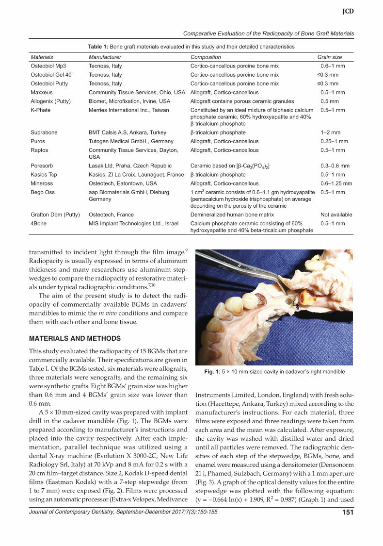
A 5 × 10 mm-sized cavity was prepared with implant drill in the cadaver mandible (Fig. 1). The BGMs were prepared according to manufacturer’s instructions and placed into the cavity respectively. After each imple-mentation, parallel technique was utilized using a dental X-ray machine (Evolution X 3000-2C, New Life Radio logy Srl, Italy) at 70 kVp and 8 mA for 0.2 s with a 20 cm film–target distance. Size 2, Kodak D-speed dental films (Eastman Kodak) with a 7-step stepwedge (from 1 to 7 mm) were exposed (Fig. 2). Films were processed using an automatic processor (Extra-x Velopex, Medivance
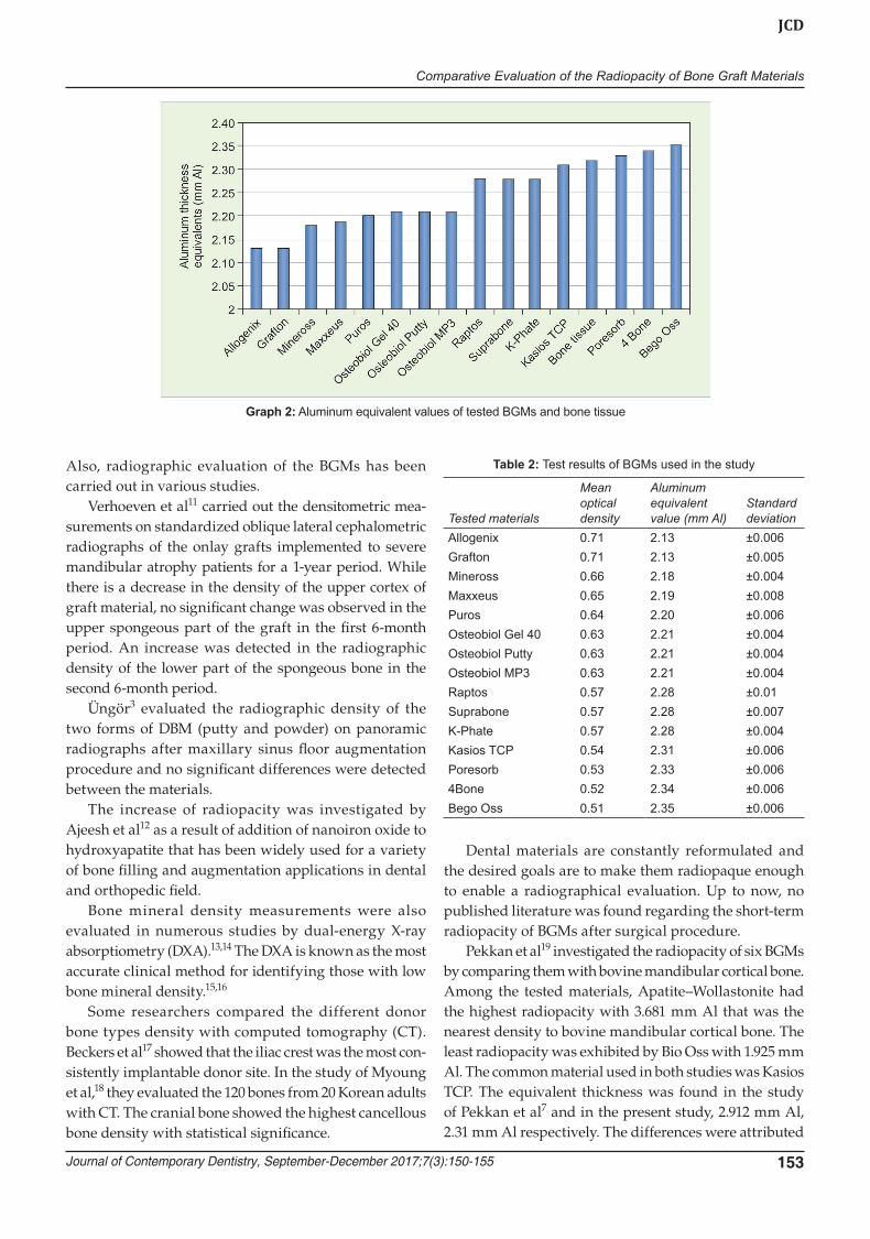
Instruments Limited, London, England) with fresh solu-tion (Hacettepe, Ankara, Turkey) mixed according to the manufacturer’s instructions. For each material, three films were exposed and three readings were taken from each area and the mean was calculated. After exposure, the cavity was washed with distilled water and dried until all particles were removed. The radiographic den-sities of each step of the stepwedge, BGMs, bone, and enamel were measured using a densitometer (Densonorm 21 i, Phamed, Sulzbach, Germany) with a 1 mm aperture (Fig. 3). A graph of the optical density values for the entire stepwedge was plotted with the following equation: (y = −0.664 ln(x) + 1.909, R2 = 0.987) (Graph 1) and used
Table 1: Bone graft materials evaluated in this study and their detailed characteristics
Materials Manufacturer Composition Grain sizeOsteobiol Mp3 Tecnoss, Italy Cortico-cancellous porcine bone mix 0.6–1 mmOsteobiol Gel 40 Tecnoss, Italy Cortico-cancellous porcine bone mix ≤0.3 mmOsteobiol Putty Tecnoss, Italy Cortico-cancellous porcine bone mix ≤0.3 mmMaxxeus Community Tissue Services, Ohio, USA Allograft, Cortico-cancellous 0.5–1 mmAllogenix (Putty) Biomet, Microfixation, Irvine, USA Allograft contains porous ceramic granules 0.5 mmK-Phate Merries International Inc., Taiwan Constituted by an ideal mixture of biphasic calcium
phosphate ceramic, 60% hydroxyapatite and 40% β-tricalcium phosphate
0.5–1 mm
Suprabone BMT Calsis A.S, Ankara, Turkey β-tricalcium phosphate 1–2 mmPuros Tutogen Medical GmbH , Germany Allograft, Cortico-cancellous 0.25–1 mmRaptos Community Tissue Services, Dayton,
USAAllograft, Cortico-cancellous 0.5–1 mm
Poresorb Lasak Ltd, Praha, Czech Republic Ceramic based on [β-Ca3(PO4)2] 0.3–0.6 mmKasios Tcp Kasios, ZI La Croix, Launaguet, France β-tricalcium phosphate 0.5–1 mmMineross Osteotech, Eatontown, USA Allograft, Cortico-cancellous 0.6–1.25 mmBego Oss aap Biomaterials GmbH, Dieburg,
Germany1 cm3 ceramic consists of 0.6–1.1 gm hydroxyapatite (pentacalcium hydroxide trisphosphate) on average depending on the porosity of the ceramic
0.5–1 mm
Grafton Dbm (Putty) Osteotech, France Demineralized human bone matrix Not available4Bone MIS Implant Technologies Ltd., Israel Calcium phosphate ceramic consisting of 60%
hydroxyapatite and 40% beta-tricalcium phosphate 0.5–1 mm
Fig. 1: 5 × 10 mm-sized cavity in cadaver’s right mandible
Emine S Kursun-Çakmak et al
152
to determine the aluminum thickness equivalent values of the materials. The mean radiopacity values for each of the materials tested were compared using ANOVA and post hoc Tukey honest significant difference tests (p < 0.05).
RESULTS
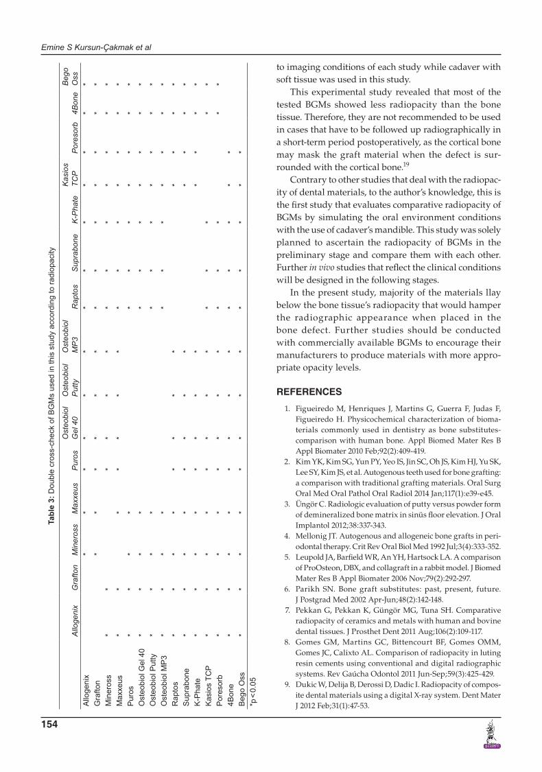
The transmission densitometry optical density values of BGMs varied from 0.51 (the most radiopaque) to 0.71 (the most radiolucent). The aluminum equivalent thicknesses of the BGMs ranged from 2.13 to 2.35 mm Al (Graph 2). Bego Oss (aap Biomaterials GmbH, Germany) exhibited the highest radiopacity of the materials tested, and Allogenix putty (Biomet, USA) exhibited the lowest (Table 2). The mandibular bone density was detected
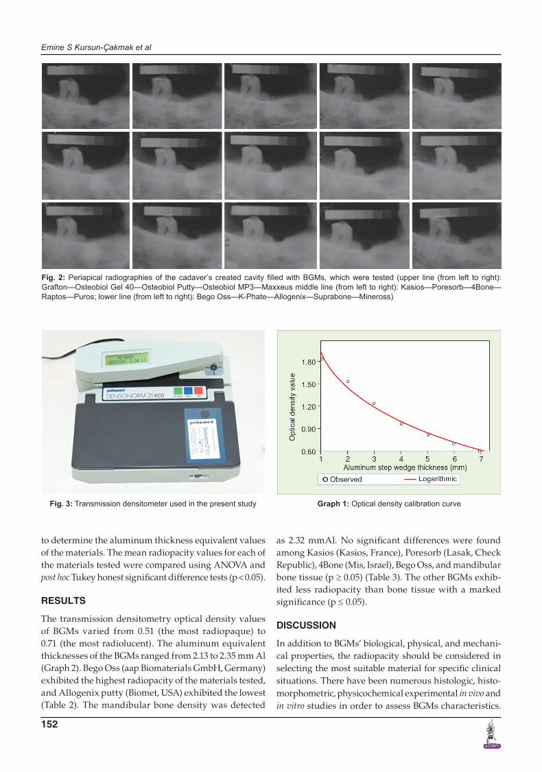
Fig. 2: Periapical radiographies of the cadaver’s created cavity filled with BGMs, which were tested (upper line (from left to right): Grafton—Osteobiol Gel 40—Osteobiol Putty—Osteobiol MP3—Maxxeus middle line (from left to right): Kasios—Poresorb—4Bone—Raptos—Puros; lower line (from left to right): Bego Oss—K-Phate—Allogenix—Suprabone—Mineross)
Fig. 3: Transmission densitometer used in the present study Graph 1: Optical density calibration curve
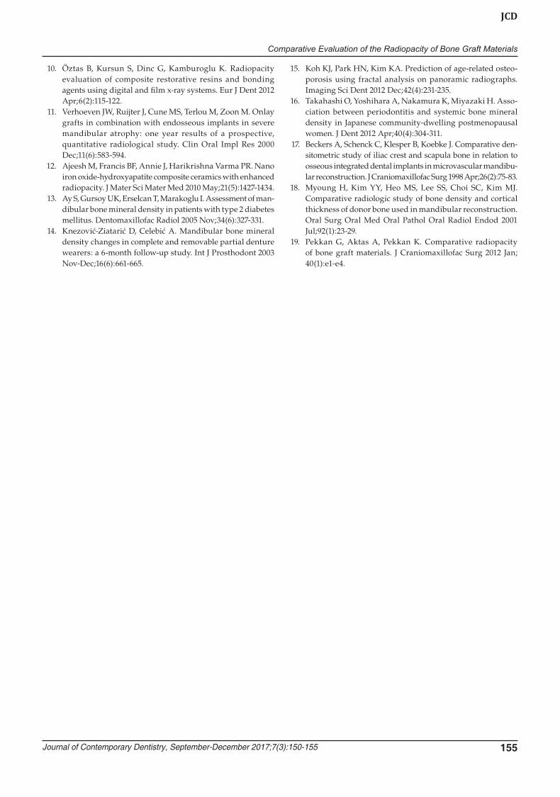
as 2.32 mmAl. No significant differences were found among Kasios (Kasios, France), Poresorb (Lasak, Check Republic), 4Bone (Mis, Israel), Bego Oss, and mandibular bone tissue (p ≥ 0.05) (Table 3). The other BGMs exhib-ited less radiopacity than bone tissue with a marked significance (p ≤ 0.05).
DISCUSSION
In addition to BGMs’ biological, physical, and mechani-cal properties, the radiopacity should be considered in selecting the most suitable material for specific clinical situations. There have been numerous histologic, histo-morphometric, physicochemical experimental in vivo and in vitro studies in order to assess BGMs characteristics.
Comparative Evaluation of the Radiopacity of Bone Graft Materials
Journal of Contemporary Dentistry, September-December 2017;7(3):150-155 153
JCD
Also, radiographic evaluation of the BGMs has been carried out in various studies.
Verhoeven et al11 carried out the densitometric mea-surements on standardized oblique lateral cephalometric radiographs of the onlay grafts implemented to severe mandibular atrophy patients for a 1-year period. While there is a decrease in the density of the upper cortex of graft material, no significant change was observed in the upper spongeous part of the graft in the first 6-month period. An increase was detected in the radiographic density of the lower part of the spongeous bone in the second 6-month period.
Üngör3 evaluated the radiographic density of the two forms of DBM (putty and powder) on panoramic radiographs after maxillary sinus floor augmentation procedure and no significant differences were detected between the materials.
The increase of radiopacity was investigated by Ajeesh et al12 as a result of addition of nanoiron oxide to hydroxyapatite that has been widely used for a variety of bone filling and augmentation applications in dental and orthopedic field.
Bone mineral density measurements were also evaluated in numerous studies by dual-energy X-ray absorptiometry (DXA).13,14 The DXA is known as the most accurate clinical method for identifying those with low bone mineral density.15,16
Some researchers compared the different donor bone types density with computed tomography (CT). Beckers et al17 showed that the iliac crest was the most con-sistently implantable donor site. In the study of Myoung et al,18 they evaluated the 120 bones from 20 Korean adults with CT. The cranial bone showed the highest cancellous bone density with statistical significance.
Table 2: Test results of BGMs used in the study
Tested materials
Mean optical density
Aluminum equivalent value (mm Al)
Standard deviation
Allogenix 0.71 2.13 ±0.006Grafton 0.71 2.13 ±0.005Mineross 0.66 2.18 ±0.004Maxxeus 0.65 2.19 ±0.008Puros 0.64 2.20 ±0.006Osteobiol Gel 40 0.63 2.21 ±0.004Osteobiol Putty 0.63 2.21 ±0.004Osteobiol MP3 0.63 2.21 ±0.004Raptos 0.57 2.28 ±0.01Suprabone 0.57 2.28 ±0.007K-Phate 0.57 2.28 ±0.004Kasios TCP 0.54 2.31 ±0.006Poresorb 0.53 2.33 ±0.0064Bone 0.52 2.34 ±0.006Bego Oss 0.51 2.35 ±0.006
Graph 2: Aluminum equivalent values of tested BGMs and bone tissue
Dental materials are constantly reformulated and the desired goals are to make them radiopaque enough to enable a radiographical evaluation. Up to now, no published literature was found regarding the short-term radiopacity of BGMs after surgical procedure.
Pekkan et al19 investigated the radiopacity of six BGMs by comparing them with bovine mandibular cortical bone. Among the tested materials, Apatite–Wollastonite had the highest radiopacity with 3.681 mm Al that was the nearest density to bovine mandibular cortical bone. The least radiopacity was exhibited by Bio Oss with 1.925 mm Al. The common material used in both studies was Kasios TCP. The equivalent thickness was found in the study of Pekkan et al7 and in the present study, 2.912 mm Al, 2.31 mm Al respectively. The differences were attributed
Emine S Kursun-Çakmak et al
154
Tabl
e 3:
Dou
ble
cros
s-ch
eck
of B
GM
s us
ed in
this
stu
dy a
ccor
ding
to ra
diop
acity
Allo
geni
xG
rafto
nM
iner
oss
Max
xeus
Pur
osO
steo
biol
G
el 4
0O
steo
biol
P
utty
Ost
eobi
ol
MP
3R
apto
sS
upra
bone
K-P
hate
Kas
ios
TCP
Por
esor
b4B
one
Beg
o O
ssA
lloge
nix
**
**
**
**
**
**
*G
rafto
n*
**
**
**
**
**
**
Min
eros
s*
**
**
**
**
**
**
Max
xeus
**
**
**
**
**
**
**
Pur
os*
**
**
**
**
**
Ost
eobi
ol G
el 4
0*
**
**
**
**
**
Ost
eobi
ol P
utty
**
**
**
**
**
*O
steo
biol
MP
3*
**
**
**
**
**
Rap
tos
**
**
**
**
**
**
Sup
rabo
ne*
**
**
**
**
**
*K
-Pha
te*
**
**
**
**
**
*K
asio
s TC
P*
**
**
**
**
**
**
Por
esor
b*
**
**
**
**
**
**
4Bon
e*
**
**
**
**
**
**
Beg
o O
ss*
**
**
**
**
**
**
*p <
0.0
5
to imaging conditions of each study while cadaver with soft tissue was used in this study.
This experimental study revealed that most of the tested BGMs showed less radiopacity than the bone tissue. Therefore, they are not recommended to be used in cases that have to be followed up radiographically in a short-term period postoperatively, as the cortical bone may mask the graft material when the defect is sur-rounded with the cortical bone.19
Contrary to other studies that deal with the radiopac-ity of dental materials, to the author’s knowledge, this is the first study that evaluates comparative radiopacity of BGMs by simulating the oral environment conditions with the use of cadaver’s mandible. This study was solely planned to ascertain the radiopacity of BGMs in the preliminary stage and compare them with each other. Further in vivo studies that reflect the clinical conditions will be designed in the following stages.
In the present study, majority of the materials llay below the bone tissue’s radiopacity that would hamper the radiographic appearance when placed in the bone defect. Further studies should be conducted with commercially available BGMs to encourage their manufacturers to produce materials with more appro-priate opacity levels.
REFERENCES
1. Figueiredo M, Henriques J, Martins G, Guerra F, Judas F, Figueiredo H. Physicochemical characterization of bioma-terials commonly used in dentistry as bone substitutes-comparison with human bone. Appl Biomed Mater Res B Appl Biomater 2010 Feb;92(2):409-419.
2. Kim YK, Kim SG, Yun PY, Yeo IS, Jin SC, Oh JS, Kim HJ, Yu SK, Lee SY, Kim JS, et al. Autogenous teeth used for bone grafting: a comparison with traditional grafting materials. Oral Surg Oral Med Oral Pathol Oral Radiol 2014 Jan;117(1):e39-e45.
3. Üngör C. Radiologic evaluation of putty versus powder form of demineralized bone matrix in sinüs floor elevation. J Oral Implantol 2012;38:337-343.
4. Mellonig JT. Autogenous and allogeneic bone grafts in peri-odontal therapy. Crit Rev Oral Biol Med 1992 Jul;3(4):333-352.
5. Leupold JA, Barfield WR, An YH, Hartsock LA. A comparison of ProOsteon, DBX, and collagraft in a rabbit model. J Biomed Mater Res B Appl Biomater 2006 Nov;79(2):292-297.
6. Parikh SN. Bone graft substitutes: past, present, future. J Postgrad Med 2002 Apr-Jun;48(2):142-148.
7. Pekkan G, Pekkan K, Güngör MG, Tuna SH. Comparative radiopacity of ceramics and metals with human and bovine dental tissues. J Prosthet Dent 2011 Aug;106(2):109-117.
8. Gomes GM, Martins GC, Bittencourt BF, Gomes OMM, Gomes JC, Calixto AL. Comparison of radiopacity in luting resin cements using conventional and digital radiographic systems. Rev Gaúcha Odontol 2011 Jun-Sep;59(3):425-429.
9. Dukic W, Delija B, Derossi D, Dadic I. Radiopacity of compos-ite dental materials using a digital X-ray system. Dent Mater J 2012 Feb;31(1):47-53.
Comparative Evaluation of the Radiopacity of Bone Graft Materials
Journal of Contemporary Dentistry, September-December 2017;7(3):150-155 155
JCD
10. Öztas B, Kursun S, Dinc G, Kamburoglu K. Radiopacity evaluation of composite restorative resins and bonding agents using digital and film x-ray systems. Eur J Dent 2012 Apr;6(2):115-122.
11. Verhoeven JW, Ruijter J, Cune MS, Terlou M, Zoon M. Onlay grafts in combination with endosseous implants in severe mandibular atrophy: one year results of a prospective, quantitative radiological study. Clin Oral Impl Res 2000 Dec;11(6):583-594.
12. Ajeesh M, Francis BF, Annie J, Harikrishna Varma PR. Nano iron oxide-hydroxyapatite composite ceramics with enhanced radiopacity. J Mater Sci Mater Med 2010 May;21(5):1427-1434.
13. Ay S, Gursoy UK, Erselcan T, Marakoglu I. Assessment of man-dibular bone mineral density in patients with type 2 diabetes mellitus. Dentomaxillofac Radiol 2005 Nov;34(6):327-331.
14. Knezović-Ziatarić D, Celebić A. Mandibular bone mineral density changes in complete and removable partial denture wearers: a 6-month follow-up study. Int J Prosthodont 2003 Nov-Dec;16(6):661-665.
15. Koh KJ, Park HN, Kim KA. Prediction of age-related osteo-porosis using fractal analysis on panoramic radiographs. Imaging Sci Dent 2012 Dec;42(4):231-235.
16. Takahashi O, Yoshihara A, Nakamura K, Miyazaki H. Asso-ciation between periodontitis and systemic bone mineral density in Japanese community-dwelling postmenopausal women. J Dent 2012 Apr;40(4):304-311.
17. Beckers A, Schenck C, Klesper B, Koebke J. Comparative den-sitometric study of iliac crest and scapula bone in relation to osseous integrated dental implants in microvascular mandibu-lar reconstruction. J Craniomaxillofac Surg 1998 Apr;26(2):75-83.
18. Myoung H, Kim YY, Heo MS, Lee SS, Choi SC, Kim MJ. Comparative radiologic study of bone density and cortical thickness of donor bone used in mandibular reconstruction. Oral Surg Oral Med Oral Pathol Oral Radiol Endod 2001 Jul;92(1):23-29.
19. Pekkan G, Aktas A, Pekkan K. Comparative radiopacity of bone graft materials. J Craniomaxillofac Surg 2012 Jan; 40(1):e1-e4.