Embed Size (px)
Citation preview
Brit. J. Ophthal. (1967) 51, 361
COMMUNICATIONS
RETINAL VASCULrrIS ASSOCIATED WITHANTERIOR UVEITIS*t
BY
J. NOLAN AND J. F. CULLENDepartment of Ophthalmology, Royal Infirmary, Edinburgh
INFLAMMATION of the anterior uvea is often accompanied by inflammatory changesin the posterior uvea, because the iris, ciliary body, and choroid are parts of a singleentity. Anterior uveitis accompanied by a retinitis or retinal vasculitis presents amuch rarer combination in spite of the functional dependence which the retina comesto have upon the choroid. This is no doubt because the retina and the posterioruvea are morphologically different structures.
Nonetheless, retinal vasculitis associated with an anterior uveitis does occur, andwe have seen five such cases in the last 4 years. All the patients belonged tothe younger age group (16 to 39 years) and, despite full investigations in every case,no definite aetiology has been established in any, although one (Case 5) had beendiagnosed in the past as an example of Reiter's syndrome.
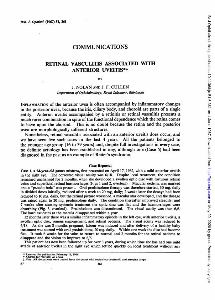
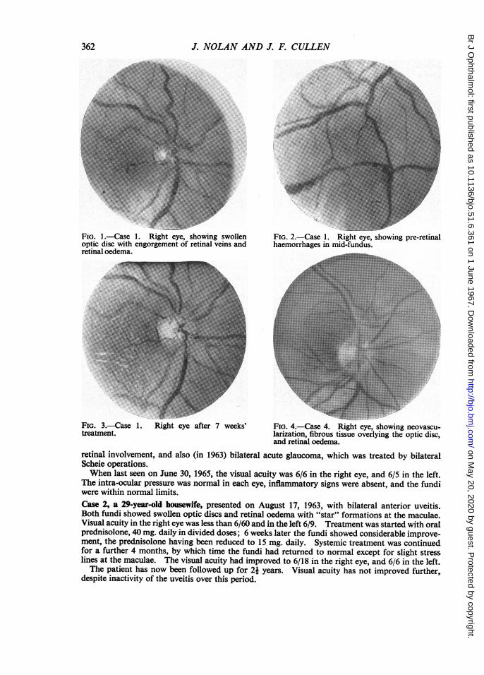
Cas ReportsCase 1, a 24-year-old games mistress, first presented on April 17, 1962, with a mild anterior uveitisin the right eye. The corrected visual acuity was 6/18. Despite local treatment, the conditionremained unchanged for 2 months, when she developed a swollen optic disc with tortuous retinalveins and superficial retinal haemorrhages (Figs 1 and 2, overleaf). Macular oedema was markedand a "pseudo-hole" was present. Oral prednisolone therapy was therefore started, 30 mg. dailyin divided doses initially, reduced after a week to 20 mg, daily; 2 weeks later the dosage had beenreduced to 10 mg. daily, but the retinal picture worsened, a macular star developed, and the dosagewas raised again to 20 mg. prednisolone daily. The condition thereafter improved steadily, and7 weeks after starting systemic treatment the optic disc was flat and the haemorrhages wereabsorbing (Fig. 3, overleaf). Prednisolone was discontinued. The visual acuity was then 6/6.The hard exudates at the macula disappeared within a year.
12 months later there was a similar inflammatory episode in the left eye, with anterior uveitis, aswollen optic disc, venous engorgement, and retinal oedema. The visual acuity was reduced to6/18. As she was 8 months pregnant, labour was induced and after delivery of a healthy babytreatment was started with oral prednisolone, 20 mg. daily. Within one week the disc had becomeflat. It took 6 weeks for the veins to return to normal and 2 months for the retinal oedema todisappear and the vision to improve to 6/6.
This patient has now been followed up for over 3 years, during which time she has had one mildattack of anterior uveitis in the right eye which settled quickly on local treatment without any
* Received for publication February 16, 1966.t Address for reprints: As abovet Note: All the patients were treated from the outset with topical corticosteroid and atropine drops.
27 361
on May 20, 2020 by guest. P
rotected by copyright.http://bjo.bm
j.com/
Br J O
phthalmol: first published as 10.1136/bjo.51.6.361 on 1 June 1967. D
ownloaded from
J. NOLAN AND J. F. CULLEN
FIG. 1.-Case 1. Right eye, showing swollenoptic disc with engorgement of retinal veins andretinal oedema.
FIG. 3.-Case 1. Right eye after 7 weeks'treatment.
FIG. 2.-Case 1. Right eye, showing pre-retinalhaemorrhages in mid-fundus.
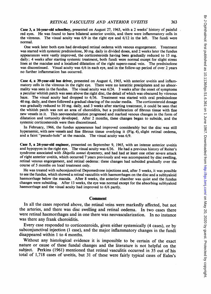
FIG. 4.-Case 4. Right eye, showing neovascu-larization, fibrous tissue overlying the optic disc,and retinal oedema.
retinal involvement, and also (in 1963) bilateral acute glaucoma, which was treated by bilateralScheie operations.When last seen on June 30, 1965, the visual acuity was 6/6 in the right eye, and 6/5 in the left.
The intra-ocular pressure was normal in each eye, inflammatory signs were absent, and the fundiwere within normal limits.Case 2, a 29-year-old housewife, presented on August 17, 1963, with bilateral anterior uveitis.Both fundi showed swollen optic discs and retinal oedema with "star" formations at the maculae.Visual acuity in the right eye was less than 6/60 and in the left 6/9. Treatment was started with oralprednisolone, 40 mg. daily in divided doses; 6 weeks later the fundi showed considerable improve-ment, the prednisolone having been reduced to 15 mg. daily. Systemic treatment was continuedfor a further 4 months, by which time the fundi had returned to normal except for slight stresslines at the maculae. The visual acuity had improved to 6/18 in the right eye, and 6/6 in the left.The patient has now been followed up for 2i years. Visual acuity has not improved further,
despite inactivity of the uveitis over this period.
362
on May 20, 2020 by guest. P
rotected by copyright.http://bjo.bm
j.com/
Br J O
phthalmol: first published as 10.1136/bjo.51.6.361 on 1 June 1967. D
ownloaded from
RETINAL VASCULITIS AND ANTERIOR UVEITIS
Case 3, a 16.year-old schoolboy, presented on August 27, 1963, with a 2 weeks' history of painfulred eyes. He was found to have bilateral anterior uveitis, and there were inflammatory cells inthe vitreous. The visual acuity was 6/9 in the right eye and 6/12 in the left. The fundi werenormal.One week later both eyes had developed retinal oedema with venous engorgement. Treatment
was started with systemic prednisolone, 30 mg. daily in divided doses, and 2 weeks later the fundusappearances were vastly improved, the corticosteroids having been gradually reduced to 15 mg.daily; 4 weeks after starting systemic treatment, both fundi were normal except for slight stresslines at the maculae and a localized dilatation of the right supero-nasal vein. The prednisolonewas discontinued. Visual acuity was 6/6 in each eye, and in the follow-up period of over 2 yearsno further inflammation has occurred.
Case 4, a 39-year-old bus driver, presented on August 6, 1965, with anterior uveitis and inflam-matory cells in the vitreous in the right eye. There were no keratitic precipitates and no abnor-mality was seen in the fundus. The visual acuity was 6/24. 3 weeks after the onset of symptomsa peculiar whitish patch was seen above the right disc, the detail of which was obscured by vitreoushaze. The visual acuity had dropped to 6/36. Treatment was started with oral prednisolone40mg. daily, and there followed a gradual clearing of the ocular media. The corticosteroid dosagewas gradually reduced to 10 mg. daily, and 3 weeks after starting treatment, it could be seen thatthe whitish patch was not an area of choroiditis, but a proliferation of fibrous tissue with finenew vessels in it. This neovascularization progressed and marked venous changes in the form ofdilatation and tortuosity developed. After 2 months, these changes began to subside, and thesystemic corticosteroids were then discontinued.
In February, 1966, the fundus appearance had improved considerably but the disc was stillhyperaemic, with new vessels and fine fibrous tissue overlying it (Fig. 4), slight retinal oedema,and a faint "pseudo-hole" at the macula. The visual acuity was 6/9.
Case 5, a 24-year-old engineer, presented on September 9, 1965, with an intense anterior uveitisand hypopyon in the right eye. The visual acuity was 6/36. He had a previous history of Reiter'ssyndrome associated with Shigella sonnei dysentery, and had had at least one other severe attackof right anterior uveitis, which occurred 7 years previously and was accompanied by disc swelling,retinal venous engorgement, and retinal oedema: these changes had subsided gradually over thecourse of 5 months on local treatment only.He was treated with subconjunctival Depomedrone injections and, after 5 weeks, it was possible
to see the fundus, which showed a retinal vasculitis with haemorrhages on the disc and a subhyaloidhaemorrhage below the macula. After 8 weeks, the anterior chamber was quiet and the funduschanges were subsiding. After 13 weeks, the eye was normal except for the absorbing subhyaloidhaemorrhage and the visual acuity had improved to 6/6 partly.
CommentIn all the cases reported above, the retinal veins were markedly affected, but not
the arteries, and there was disc swelling and retinal oedema. In two cases therewere retinal haemorrhages and in one there was neovascularization. In no instancewas there any frank choroiditis.Every case responded to corticosteroids, given either systemically (4 cases), or by
subconjunctival injection (1 case), and the major inflammatory changes in the fundidisappeared within 1 to 4 months.Without any histological evidence it is impossible to be certain of the exact
nature or cause of these fundal changes and the literature is not helpful on thesubject. Perkins (1961) mentioned that retinal vasculitis occurred in 35 out of histotal of 1,718 cases of uveitis, but 31 of these were fairly typical cases of Eales's
363
on May 20, 2020 by guest. P
rotected by copyright.http://bjo.bm
j.com/
Br J O
phthalmol: first published as 10.1136/bjo.51.6.361 on 1 June 1967. D
ownloaded from
J. NOLAN AND J. F. CULLEN
disease, and two others were examples of sickle-cell retinopathy. The remainingtwo cases are listed as "atypical vascular lesions", and these are the only ones inhis series which might be similar to those described above, but as no furtherdescriptive details are given, an exact comparison cannot be made.Woods (1961) stated that the ocular changes in Behcet's and the Vogt-Koyanagi-
Harada syndromes probably begin in the optic nerve, spreading to the retina andthence secondarily to the uveal tract. Early cases of Behcet's syndrome usuallyshow a narrowing of the retinal arterial bed and evidence of a periarteritis orendarteritis, sometimes with retinal haemorrhages. None of our five patients,however, had any other evidence of these syndromes.The explanation in our cases may be that vasodilator metabolites or toxins from
the anterior uveal inflammation diffuse across the vitreous body and affect the retinalvessels. The posterior pole shows the maximum changes because it is the mostvascular part of the retina (Warwick, 1963). Furthermore, the retinal oedemacaused by this inflammatory exudate would spread to involve the optic nerve head,thereby embarrassing the retinal venous return and causing the venous changeswhich been noted.Boyd (1961) pointed out that mild inflammation causes maximum dilatation of
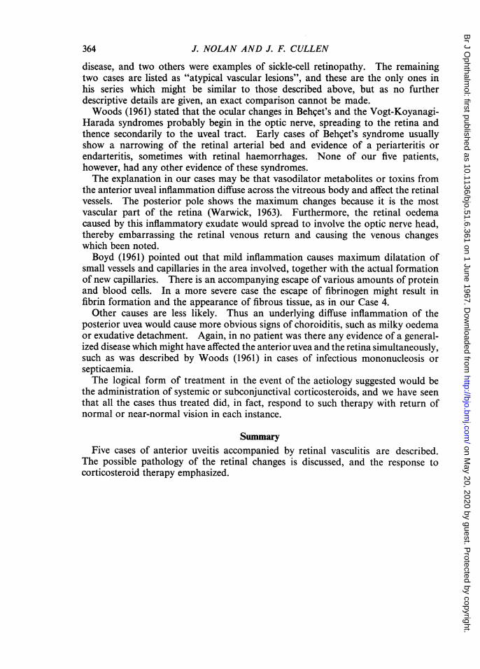
small vessels and capillaries in the area involved, together with the actual formationof new capillaries. There is an accompanying escape of various amounts of proteinand blood cells. In a more severe case the escape of fibrinogen might result infibrin formation and the appearance of fibrous tissue, as in our Case 4.
Other causes are less likely. Thus an underlying diffuse inflammation of theposterior uvea would cause more obvious signs of choroiditis, such as milky oedemaor exudative detachment. Again, in no patient was there any evidence of a general-ized disease which might have affected the anterior uvea and the retina simultaneously,such as was described by Woods (1961) in cases of infectious mononucleosis orsepticaemia.The logical form of treatment in the event of the aetiology suggested would be
the administration of systemic or subconjunctival corticosteroids, and we have seenthat all the cases thus treated did, in fact, respond to such therapy with return ofnormal or near-normal vision in each instance.
SummaryFive cases of anterior uveitis accompanied by retinal vasculitis are described.
The possible pathology of the retinal changes is discussed, and the response tocorticosteroid therapy emphasized.
364
on May 20, 2020 by guest. P
rotected by copyright.http://bjo.bm
j.com/
Br J O
phthalmol: first published as 10.1136/bjo.51.6.361 on 1 June 1967. D
ownloaded from