Embed Size (px)
Citation preview
2 0 2 0 V o l u m e 4 4https://doi.org/10.33321/cdi.2020.44.79
Australian Group on Antimicrobial Resistance (AGAR) Australian Gram-negative Sepsis Outcome Programme (GNSOP) Annual Report 2019Jan M Bell, Alicia Fajardo Lubian, Sally Partridge, Thomas Gottlieb, Jonathan Iredell, Denise A Daley, Geoffrey W Coombs
Communicable Diseases Intelligence ISSN: 2209-6051 Online
This journal is indexed by Index Medicus and Medline.
Creative Commons Licence - Attribution-NonCommercial-NoDerivatives CC BY-NC-ND
© 2020 Commonwealth of Australia as represented by the Department of Health
This publication is licensed under a Creative Commons Attribution- Non-Commercial NoDerivatives 4.0 International Licence from https://creativecommons.org/licenses/by-nc-nd/4.0/legalcode (Licence). You must read and understand the Licence before using any material from this publication.
Restrictions The Licence does not cover, and there is no permission given for, use of any of the following material found in this publication (if any):
• the Commonwealth Coat of Arms (by way of information, the terms under which the Coat of Arms may be used can be found at www.itsanhonour.gov.au);
• any logos (including the Department of Health’s logo) and trademarks;
• any photographs and images;
• any signatures; and
• any material belonging to third parties.
Disclaimer Opinions expressed in Communicable Diseases Intelligence are those of the authors and not necessarily those of the Australian Government Department of Health or the Communicable Diseases Network Australia. Data may be subject to revision.
Enquiries Enquiries regarding any other use of this publication should be addressed to the Communication Branch, Department of Health, GPO Box 9848, Canberra ACT 2601, or via e-mail to: [email protected]
Communicable Diseases Network Australia Communicable Diseases Intelligence contributes to the work of the Communicable Diseases Network Australia. http://www.health.gov.au/cdna
Communicable Diseases Intelligence (CDI) is a peer-reviewed scientific journal published by the Office of Health Protection, Department of Health. The journal aims to disseminate information on the epidemiology, surveillance, prevention and control of communicable diseases of relevance to Australia.
Editor Tanja Farmer
Deputy Editor Simon Petrie
Design and Production Kasra Yousefi
Editorial Advisory Board David Durrheim, Mark Ferson, John Kaldor, Martyn Kirk and Linda Selvey
Website http://www.health.gov.au/cdi
Contacts Communicable Diseases Intelligence is produced by: Health Protection Policy Branch Office of Health Protection Australian Government Department of Health GPO Box 9848, (MDP 6) CANBERRA ACT 2601
Email: [email protected]
Submit an Article You are invited to submit your next communicable disease related article to the Communicable Diseases Intelligence (CDI) for consideration. More information regarding CDI can be found at: http://health.gov.au/cdi.
Further enquiries should be directed to: [email protected].
1 of 10 health.gov.au/cdi Commun Dis Intell (2018) 2020;44 (https://doi.org/10.33321/cdi.2020.44.79) Epub 15/10/2020
Australian Group on Antimicrobial Resistance (AGAR) Australian Gram-negative Sepsis Outcome Programme (GNSOP) Annual Report 2019Jan M Bell, Alicia Fajardo Lubian, Sally Partridge, Thomas Gottlieb, Jonathan Iredell, Denise A Daley, Geoffrey W Coombs
Abstract
The Australian Group on Antimicrobial Resistance (AGAR) performs regular period-prevalence stud-ies to monitor changes in antimicrobial resistance in selected enteric gram-negative pathogens. The 2019 survey was the seventh year to focus on bloodstream infections, and included Enterobacterales, Pseudomonas aeruginosa and Acinetobacter species.
Eight thousand eight hundred and fifty-seven isolates, comprising Enterobacterales (7,983; 90.1%), P. aeruginosa (764; 8.6%) and Acinetobacter species (110; 1.2%), were tested using commercial auto-mated methods. The results were analysed using Clinical and Laboratory Standards Institute (CLSI) and European Committee on Antimicrobial Susceptibility Testing (EUCAST) breakpoints (January 2020). Of the key resistances, resistance to the third-generation cephalosporin ceftriaxone was found in 13.3%/13.3% (CLSI/EUCAST criteria) of Escherichia coli and 8.4%/8.4% of Klebsiella pneumo-niae. Resistance rates to ciprofloxacin were 16.0%/16.0% for E. coli, 10.2%/10.2% for K. pneumoniae complex, 5.9%/5.9% for Enterobacter cloacae complex, and 4.1%/9.3% for P. aeruginosa. Resistance rates to piperacillin-tazobactam were 3.2%/5.7%, 4.7%/8.5%, 14.8%/21.4%, and 6.9%/12.5% for the same four species/complex respectively. Twenty-nine isolates from 29 patients were shown to harbour a carbapenemase gene: 15 blaIMP-4, five blaOXA-181, four blaOXA-23 (one with blaOXA-58 also), three blaNDM-4/5, one blaGES-5, and one blaIMP-1.
Keywords: Australian Group on Antimicrobial Resistance (AGAR); antibiotic resistance; bacteraemia; gram-negative; Escherichia coli; Enterobacter; Klebsiella
Introduction
Emerging antimicrobial resistance (AMR) in common pathogenic members of the Enterobacterales is a world-wide phenomenon and presents therapeutic problems for practi-tioners, both in the community and in hospital practice. The Australian Group on Antimicrobial Resistance (AGAR) commenced surveillance of the key gram-negative pathogens, Escherichia coli and Klebsiella species, in 1992. Surveys have been conducted biennially until 2008 when
annual surveys commenced, alternating between community- and hospital-onset infections.i In 2004, another genus of gram-negative pathogens in which resistance can be of clinical impor-tance, Enterobacter species, was added. E. coli is the most common cause of community-onset urinary tract infection; Klebsiella species are less common but are known to harbour important resistances. Enterobacter species are less com-mon in the community, but of high impor-
i http://www.agargroup.org.au/agar-surveys.
Annual report
2 of 10 health.gov.au/cdiCommun Dis Intell (2018) 2020;44 (https://doi.org/10.33321/cdi.2020.44.79) Epub 15/10/2020
tance due to intrinsic resistance to first-line antimicrobials used in the community. Taken together, the three groups of species surveyed are considered to be valuable sentinels for multi-resistance and emerging resistance in enteric gram-negative bacilli. In 2013 AGAR com-menced the Enterobacteriaceae Sepsis Outcome Programme (EnSOP) which focused on the col-lection of resistance and some demographic data on all isolates prospectively from patients with bacteraemia. In 2015, Pseudomonas aeruginosa and Acinetobacter species were added, and the program has been referred to since that date as the Gram-negative Sepsis Outcome Program (GNSOP).
Resistances of particular interest include resistance to ß-lactams due to ß-lactamases, especially extended-spectrum ß-lactamases (ESBLs), which inactivate the third-generation cephalosporins that are normally considered reserve antimicrobials. Other resistances of interest are to agents important for treatment of these serious infections, such as gentamicin; and resistance to reserve agents such as ciprofloxa-cin, meropenem and colistin.
The objectives of the 2019 surveillance program were to:
• Monitor resistance in Enterobacterales, P. aeruginosa and Acinetobacter species isolated from blood cultures taken from patients presenting to the hospital or already in hospital;
• Examine the extent of co-resistance and mul-tidrug resistance in the major species;
• Detect emerging resistance to newer last-line agents such as carbapenems and colistin; and
• Examine the molecular basis of resistance to third-generation cephalosporins, quinolones and carbapenems.
Methods
Study design
From 1 January to 31 December 2019, 39 institu-tions across Australia collected either all or up to 200 isolates from different patient episodes of bacteraemia.
Species identification
Isolates were identified using the routine method for each institution: Vitek®, Phoenix™ automated microbiology systems, or where available matrix assisted laser desorption/ionisation – time of flight (MALDI-ToF) mass spectrometry.
Susceptibility testing
Testing was performed by two commercial semi-automated methods, Vitek 2 (BioMérieux, France) or Phoenix (Becton Dickinson, USA), which are calibrated to the ISO reference standard method of broth microdilution. Commercially available Vitek AST-N246, or Phoenix NMIC-404 and NMIC-422 cards were utilized by all participants throughout the survey period. The CLSI M100 and EUCAST v10.0 breakpoints from January 2020 have been employed in the analysis.1,2
Multidrug resistance
The definitions used by Magiorakos et al. were applied in this survey,3 where multidrug resist-ance was defined as resistance to one or more agent in three or more antimicrobial categories. For each species, antimicrobials were excluded from the count if they are affected by natural resistance mechanisms.
PCR screening and whole genome sequencing
E. coli, Klebsiella spp., Proteus spp. and Salmonella spp. with ceftazidime or ceftriaxone minimum inhibitory concentration (MIC) > 1 mg/L, or cefoxitin MIC > 8 mg/L; any other Enterobacterales with cefepime MIC > 1 mg/L;
3 of 10 health.gov.au/cdi Commun Dis Intell (2018) 2020;44 (https://doi.org/10.33321/cdi.2020.44.79) Epub 15/10/2020
Enterobacterales with ciprofloxacin MIC > 0.25 mg/L; Enterobacterales with meropenem MIC > 0.25 mg/L; P. aeruginosa or Acinetobacter spp. with meropenem MIC > 4 mg/L; all isolates with amikacin MIC > 32 mg/L; and all isolates with colistin MIC > 4 mg/L were referred to a cen-tral laboratory (Centre for Infectious Diseases and Microbiology, The Westmead Institute for Medical Research) and underwent polymerase chain reaction (PCR) to detect selected resist-ance genes (Centre for Infectious Diseases & Microbiology Laboratory Services, ICPMR, Westmead Hospital) and/or whole genome sequencing (WGS) (Antimicrobial Resistance Laboratory, Microbial Genomics Reference Laboratory, CIDMLS, ICPMR, Westmead Hospital).
All referred isolates, except P. aeruginosa, Acinetobacter spp., Salmonella spp., and Enterobacterales with meropenem MIC > 0.25 mg/L which underwent WGS, were screened using real-time multiplex PCR using published primers to detect ESBLs (blaSHV-ESBL with G→A substitution at position 700 and/or 703, blaCTX-M groups 1 and 9, blaVEB), plasmid-borne AmpC (blaCMY-2-like, blaDHA) and carbapenemase (blaIMP, blaNDM, blaVIM) genes.4
Assays for other ESBL targets (blaACT/MIR, blaKPC, blaOXA-48-like, blaGES, blaSME, blaSPM, blaAIM, blaGIM, blaSIM, blaOXA-23/24/58); aminoglycoside ribosomal methytransferases (armA, rmtA, rmtB, rmtC, rmtD, rmtE, rmtF, rmtG, rmtH); and mobile colistin resistance genes (mcr-1, mcr-2, mcr-3, mcr-4, mcr-5) were detected using in-house, NATA-accredited primers and probes in routine use by the Centre for Infectious Diseases & Microbiology Laboratory Services, ICPMR, at Westmead Hospital.
Genomic DNA for WGS was extracted using the DNeasy® Blood & Tissue Kit (Qiagen) according to the manufacturer’s instructions for gram-negative bacteria. WGS was performed by the Antimicrobial Resistance Laboratory, Microbial Genomics Reference Laboratory, CIDMLS, ICPMR, Westmead Hospital using the Illumina NextSeq 500 platform. Data were
analysed using a modification of the Nullarbor bioinformatic pipeline,5 incorporating searching contigs against the NCBI AMRFinder databaseii using ABRicate6 and AMRFinder7, followed by a custom AMR-specific pipeline which includes a read-based search using ARIBA8 against the CARD9 and NCBI databases.
Results
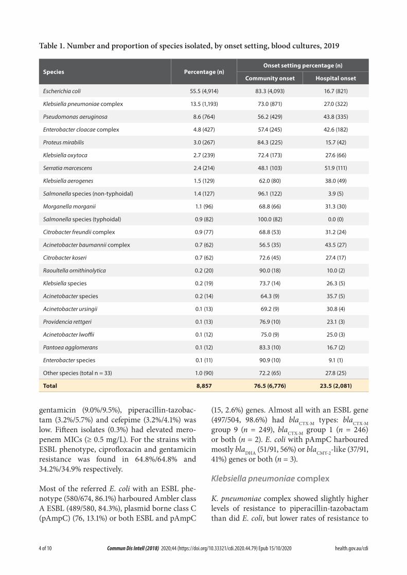
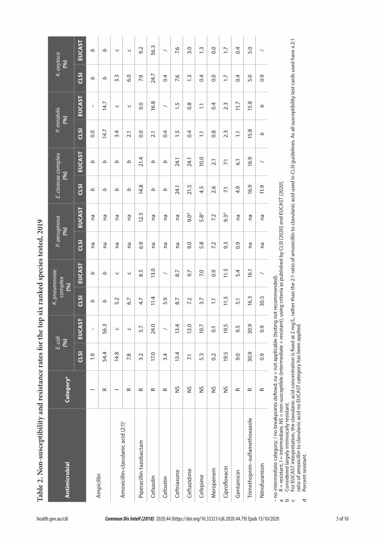
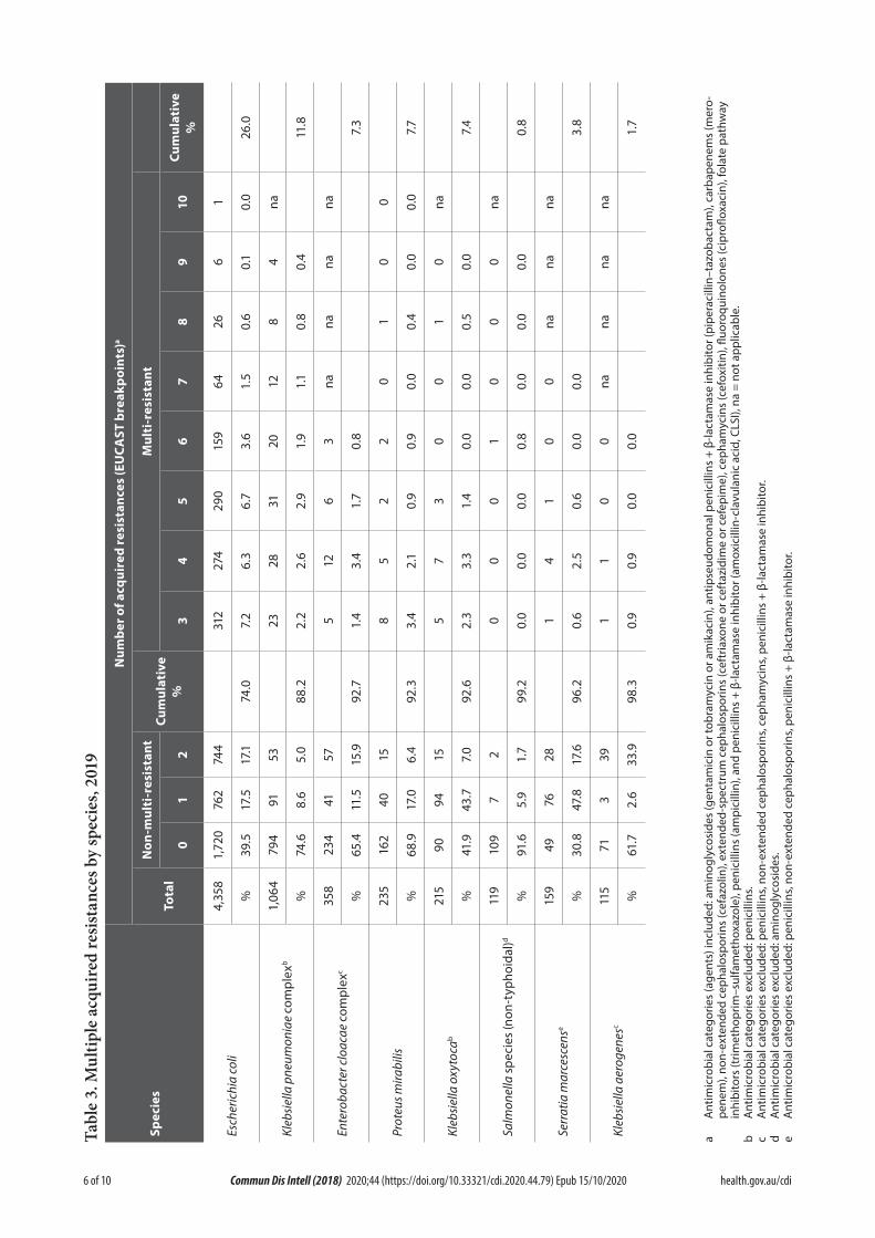
The species isolated, and the numbers of each by onset setting, are listed in Table 1. Enterobacterales accounted for 90.1%, followed by P. aeruginosa (8.6%) and Acinetobacter species (1.2%). Of the Enterobacterales, three genera—Escherichia (61.6%), Klebsiella (19.8%) and Enterobacter (5.5%)—contributed 86.9% of all isolates. Major resistances and non-suscepti-bilities for the top six ranked species are listed in Table 2. Non-susceptibility (which includes both intermediate and resistant isolates) has been included for some agents because these figures provide information about important emerging acquired resistances. Multiple acquired resist-ances by species are shown in Table 3. Multi-resistance was detected in 26.0% of E. coli iso-lates, 11.8% of K. pneumoniae complex, and 7.3% of E. cloacae complex. A more detailed break-down of resistances and non-susceptibilities by state and territory is provided in the online AGAR report.
Escherichia coli
Moderately high levels of resistance to ampicil-lin (and therefore amoxicillin) were maintained (54.4%/56.3%, CLSI/EUCAST criteria), with lower rates for amoxicillin-clavulanic acid (14.8%/– intermediate, 7.8%/– resistant). Non-susceptibility to third generation cephalospor-ins was maintained at similar levels to the 2018 survey (ceftriaxone 13.4%/13.4%, ceftazidime 7.1%/13.0%). Moderate levels of resistance were detected to cefazolin (17.0%/24.0%) and tri-methoprim–sulfamethoxazole (30.9%/30.9%). Ciprofloxacin non-susceptibility was found in 19.5%/19.5% of E. coli isolates. Resistance to
ii https://www.ncbi.nlm.nih.gov/bioproject/PRJNA313047.
4 of 10 health.gov.au/cdiCommun Dis Intell (2018) 2020;44 (https://doi.org/10.33321/cdi.2020.44.79) Epub 15/10/2020
Table 1. Number and proportion of species isolated, by onset setting, blood cultures, 2019
Species Percentage (n)Onset setting percentage (n)
Community onset Hospital onset
Escherichia coli 55.5 (4,914) 83.3 (4,093) 16.7 (821)
Klebsiella pneumoniae complex 13.5 (1,193) 73.0 (871) 27.0 (322)
Pseudomonas aeruginosa 8.6 (764) 56.2 (429) 43.8 (335)
Enterobacter cloacae complex 4.8 (427) 57.4 (245) 42.6 (182)
Proteus mirabilis 3.0 (267) 84.3 (225) 15.7 (42)
Klebsiella oxytoca 2.7 (239) 72.4 (173) 27.6 (66)
Serratia marcescens 2.4 (214) 48.1 (103) 51.9 (111)
Klebsiella aerogenes 1.5 (129) 62.0 (80) 38.0 (49)
Salmonella species (non-typhoidal) 1.4 (127) 96.1 (122) 3.9 (5)
Morganella morganii 1.1 (96) 68.8 (66) 31.3 (30)
Salmonella species (typhoidal) 0.9 (82) 100.0 (82) 0.0 (0)
Citrobacter freundii complex 0.9 (77) 68.8 (53) 31.2 (24)
Acinetobacter baumannii complex 0.7 (62) 56.5 (35) 43.5 (27)
Citrobacter koseri 0.7 (62) 72.6 (45) 27.4 (17)
Raoultella ornithinolytica 0.2 (20) 90.0 (18) 10.0 (2)
Klebsiella species 0.2 (19) 73.7 (14) 26.3 (5)
Acinetobacter species 0.2 (14) 64.3 (9) 35.7 (5)
Acinetobacter ursingii 0.1 (13) 69.2 (9) 30.8 (4)
Providencia rettgeri 0.1 (13) 76.9 (10) 23.1 (3)
Acinetobacter lwoffii 0.1 (12) 75.0 (9) 25.0 (3)
Pantoea agglomerans 0.1 (12) 83.3 (10) 16.7 (2)
Enterobacter species 0.1 (11) 90.9 (10) 9.1 (1)
Other species (total n = 33) 1.0 (90) 72.2 (65) 27.8 (25)
Total 8,857 76.5 (6,776) 23.5 (2,081)
gentamicin (9.0%/9.5%), piperacillin-tazobac-tam (3.2%/5.7%) and cefepime (3.2%/4.1%) was low. Fifteen isolates (0.3%) had elevated mero-penem MICs (≥ 0.5 mg/L). For the strains with ESBL phenotype, ciprofloxacin and gentamicin resistance was found in 64.8%/64.8% and 34.2%/34.9% respectively.
Most of the referred E. coli with an ESBL phe-notype (580/674, 86.1%) harboured Ambler class A ESBL (489/580, 84.3%), plasmid borne class C (pAmpC) (76, 13.1%) or both ESBL and pAmpC
(15, 2.6%) genes. Almost all with an ESBL gene (497/504, 98.6%) had blaCTX-M types: blaCTX-M group 9 (n = 249), blaCTX-M group 1 (n = 246) or both (n = 2). E. coli with pAmpC harboured mostly blaDHA (51/91, 56%) or blaCMY-2-like (37/91, 41%) genes or both (n = 3).
Klebsiella pneumoniae complex
K. pneumoniae complex showed slightly higher levels of resistance to piperacillin-tazobactam than did E. coli, but lower rates of resistance to
5 of 10 health.gov.au/cdi Commun Dis Intell (2018) 2020;44 (https://doi.org/10.33321/cdi.2020.44.79) Epub 15/10/2020
Tabl
e 2.
Non
-sus
cept
ibili
ty a
nd re
sist
ance
rate
s for
the
top
six
rank
ed sp
ecie
s tes
ted,
201
9
Ant
imic
robi
alCa
tego
rya
E. co
li (%
)
K. p
neum
onia
e co
mpl
ex
(%)
P. a
erug
inos
a (%
)E.
clo
acae
com
plex
(%
)P.
mira
bilis
(%
)K.
oxy
toca
(%
)
CLSI
EUCA
STCL
SIEU
CAST
CLSI
EUCA
STCL
SIEU
CAST
CLSI
EUCA
STCL
SIEU
CAST
Am
pici
llin
I1.
9–
bb
nana
bb
0.0
–b
b
R54
.456
.3b
bna
nab
b14
.714
.7b
b
Am
oxic
illin
-cla
vula
nic
acid
(2:1)
cI
14.8
c5.
2c
nana
bb
3.4
c3.
3c
R7.
8c
6.7
cna
nab
b2.
1c
6.0
c
Pipe
raci
llin-
tazo
bact
amR
3.2
5.7
4.7
8.5
6.9
12.5
14.8
21.4
0.0
0.0
7.9
9.2
Cefa
zolin
R17
.024
.011
.413
.0na
nab
b2.
116
.824
.756
.3
Cefo
xitin
R3.
4/
5.9
/na
nab
b0.
4/
0.4
/
Ceft
riaxo
neN
S13
.413
.48.
78.
7na
na24
.124
.11.
51.
57.
67.
6
Ceft
azid
ime
NS
7.113
.07.
29.
79.
09.
0d21
.524
.10.
40.
81.
33.
0
Cefe
pim
eN
S5.
310
.73.
77.
05.
85.
8d4.
510
.01.
11.
10.
41.
3
Mer
open
emN
S0.
20.
11.
10.
97.
27.
22.
62.
10.
80.
40.
00.
0
Cipr
oflox
acin
NS
19.5
19.5
11.5
11.5
9.3
9.3d
7.17.1
2.3
2.3
1.7
1.7
Gen
tam
icin
R9.
09.
55.
15.
40.
9na
4.9
6.1
1.1
11.7
0.4
0.4
Trim
etho
prim
–sul
fam
etho
xazo
leR
30.9
30.9
16.3
16.1
nana
16.9
16.9
15.8
15.8
5.0
5.0
Nitr
ofur
anto
inR
0.9
0.9
30.5
/na
na11
.9/
bb
0.9
/
– no
inte
rmed
iate
cat
egor
y; /
no b
reak
poin
ts d
efine
d; n
a =
not a
pplic
able
(tes
ting
not r
ecom
men
ded)
.a
R =
resi
stan
t, I =
inte
rmed
iate
, NS
= no
n-su
scep
tible
(int
erm
edia
te +
resi
stan
t), u
sing
crit
eria
as
publ
ishe
d by
CLS
I [20
20] a
nd E
UCA
ST [2
020]
.b
Cons
ider
ed la
rgel
y in
trin
sica
lly re
sist
ant.
c Fo
r EU
CAST
inte
rpre
tatio
n, th
e cl
avul
anic
aci
d co
ncen
trat
ion
is fi
xed
at 2
mg/
L, ra
ther
than
the
2:1
ratio
of a
mox
icill
in to
cla
vula
nic
acid
use
d in
CLS
I gui
delin
es. A
s al
l sus
cept
ibili
ty te
st c
ards
use
d ha
ve a
2:1
ra
tio o
f am
oxic
illin
to c
lavu
lani
c ac
id n
o EU
CAST
cat
egor
y ha
s be
en a
pplie
d.d
Perc
ent r
esis
tant
.
6 of 10 health.gov.au/cdiCommun Dis Intell (2018) 2020;44 (https://doi.org/10.33321/cdi.2020.44.79) Epub 15/10/2020
Tabl
e 3.
Mul
tiple
acq
uire
d re
sist
ance
s by
spec
ies,
2019
Spec
ies
Num
ber o
f acq
uire
d re
sist
ance
s (E
UCA
ST b
reak
poin
ts)a
Tota
l
Non
-mul
ti-r
esis
tant
Cum
ulat
ive
%
Mul
ti-r
esis
tant
01
23
45
67
89
10Cu
mul
ativ
e %
Esch
eric
hia
coli
4,35
81,
720
762
744
312
274
290
159
6426
61
%39
.517
.517
.174
.07.
26.
36.
73.
61.
50.
60.
10.
026
.0
Kleb
siella
pne
umon
iae
com
plex
b1,
064
794
9153
2328
3120
128
4na
%74
.68.
65.
088
.22.
22.
62.
91.
91.
10.
80.
411
.8
Ente
roba
cter
clo
acae
com
plex
c35
823
441
575
126
3na
nana
na
%65
.411
.515
.992
.71.
43.
41.
70.
87.
3
Prot
eus m
irabi
lis23
516
240
158
52
20
10
0
%68
.917
.06.
492
.33.
42.
10.
90.
90.
00.
40.
00.
07.
7
Kleb
siella
oxy
toca
b21
590
9415
57
30
01
0na
%41
.943
.77.
092
.62.
33.
31.
40.
00.
00.
50.
07.
4
Salm
onel
la s
peci
es (n
on-t
ypho
idal
)d11
910
97
20
00
10
00
na
%91
.65.
91.
799
.20.
00.
00.
00.
80.
00.
00.
00.
8
Serr
atia
mar
cesc
ense
159
4976
281
41
00
nana
na
%30
.847
.817
.696
.20.
62.
50.
60.
00.
03.
8
Kleb
siella
aer
ogen
esc
115
713
391
10
0na
nana
na
%61
.72.
633
.998
.30.
90.
90.
00.
01.
7
a A
ntim
icro
bial
cat
egor
ies
(age
nts)
incl
uded
: am
inog
lyco
side
s (g
enta
mic
in o
r tob
ram
ycin
or a
mik
acin
), an
tipse
udom
onal
pen
icill
ins
+ β-
lact
amas
e in
hibi
tor (
pipe
raci
llin–
tazo
bact
am),
carb
apen
ems
(mer
o-pe
nem
), no
n-ex
tend
ed c
epha
losp
orin
s (c
efaz
olin
), ex
tend
ed-s
pect
rum
cep
halo
spor
ins
(cef
tria
xone
or c
efta
zidi
me
or c
efep
ime)
, cep
ham
ycin
s (c
efox
itin)
, fluo
roqu
inol
ones
(cip
roflo
xaci
n), f
olat
e pa
thw
ay
inhi
bito
rs (t
rimet
hopr
im–s
ulfa
met
hoxa
zole
), pe
nici
llins
(am
pici
llin)
, and
pen
icill
ins
+ β-
lact
amas
e in
hibi
tor (
amox
icill
in-c
lavu
lani
c ac
id, C
LSI),
na
= no
t app
licab
le.
b A
ntim
icro
bial
cat
egor
ies
excl
uded
: pen
icill
ins.
c A
ntim
icro
bial
cat
egor
ies
excl
uded
: pen
icill
ins,
non-
exte
nded
cep
halo
spor
ins,
ceph
amyc
ins,
peni
cilli
ns +
β-la
ctam
ase
inhi
bito
r.d
Ant
imic
robi
al c
ateg
orie
s ex
clud
ed: a
min
ogly
cosi
des.
e A
ntim
icro
bial
cat
egor
ies
excl
uded
: pen
icill
ins,
non-
exte
nded
cep
halo
spor
ins,
peni
cilli
ns +
β-la
ctam
ase
inhi
bito
r.
7 of 10 health.gov.au/cdi Commun Dis Intell (2018) 2020;44 (https://doi.org/10.33321/cdi.2020.44.79) Epub 15/10/2020
amoxicillin-clavulanic acid, cefazolin, ceftriax-one, ciprofloxacin, gentamicin, and trimetho-prim-sulfamethoxazole. Twenty-one K. pneu-moniae complex isolates (1.8%) had elevated meropenem MICs (see below). A substantial majority of the referred K. pneumoniae complex with an ESBL phenotype (92/118; 78.0%) har-boured ESBL (76; 82.6%), pAmpC (15; 16.3%) or both ESBL and pAmpC (1; 1.1%) genes. The vast majority with an ESBL gene (72/77; 93.5%) had blaCTX-M types, mostly blaCTX-M group 1 (62/72; 86.1%). A substantial majority of the K. pneu-moniae complex with pAmpC harboured blaDHA (13/16; 81%).
Enterobacter cloacae complex
Acquired resistance was common among E. cloa-cae complex isolates, to piperacillin-tazobactam (14.8%/21.4%) ceftriaxone (23.4%/23.4%), ceftazidime (21.3%/21.5%) and trimethoprim–sulfamethoxazole (16.9%/16.9%). Cefepime, ciprofloxacin and gentamicin resistance remain at less than 10%. Seventeen (4.0%) E. cloacae complex isolates had elevated meropenem MICs.
Carbapenemases
Overall, 29 isolates (29 patients) in fourteen institutions from four states/territories were found to harbour a carbapenemase gene. blaIMP-4 was detected in 15 isolates: K. pneumoniae (five), E. cloacae (five), E. hormaechei (three), one K. variicola, and one E. coli. blaOXA-181 was detected in five K. pneumoniae. blaNDM-4 was detected in two K. pneumoniae and blaNDM-5 in one E. coli. blaOXA-23 was detected in three A. baumannii, one of which also harboured blaOXA-58, and one Proteus mirabilis. Among Pseudomonas aeruginosa, one blaGES-5 and one blaIMP-1 were detected. Just over one quarter of the carbapenemase-producing organisms were from one institution.
Discussion
AGAR has been tracking resistance in sentinel enteric gram-negative bacteria since 1992. From 2008, surveillance was segregated into hospital-
versus community-onset infections. The last year of hospital-onset only surveillance was 2011.10 In 2013, the first survey of antimicrobial resistance among Enterobacterales isolates from bacteraemic patients throughout Australia was conducted using an approach similar to that conducted by the European EARS-Net program. 2019 was the seventh survey of antimicrobial resistance among Enterobacterales, and the fifth for P. aeruginosa and Acinetobacter spp. from bacteraemic patients through Australia.
Relative to 2018, the percentage resistance in E. coli declined for almost two-thirds (7/11; 60%) of the antimicrobial agents tested, and for K. pneumoniae complex by half (5/10). AGAR data show a longitudinal trend of increasing E. coli resistance to key anti-gram-negative antimicrobial agents, such as ceftriaxone and ciprofloxacin. The steady rise in resistance to fluoroquinolones is more striking in hospital-onset bacteraemia, with a change from 13.7% to 21.3% between 2013 and 2019.
Carbapenem resistance attributable to acquired carbapenemase genes is still uncommon in patients with bacteraemia in Australia, although five different types (IMP, NDM, OXA-48-like, OXA-23, and GES-5) were detected in isolates from fourteen of the participating institu-tions. Compared with many other countries in our region, resistance rates in Australian gram-negative bacteria are still relatively low,11 but similar to those observed in 2018 in many Northern European countries.12 Resistance to third generation cephalosporins in E. coli from bacteraemic patients in Australia is similar to the European Union and European Economic Area average.12
One quarter of E. coli and 12% of K. pneumoniae complex were multi-resistant. This is likely to drive more broad-spectrum antibiotic use and increase the resistance selection pressure for important reserve classes, especially the carbap-enems.
8 of 10 health.gov.au/cdiCommun Dis Intell (2018) 2020;44 (https://doi.org/10.33321/cdi.2020.44.79) Epub 15/10/2020
Acknowledgments
This study was funded by a grant from the Australian Commission on Safety and Quality in Health care.
AGAR gratefully acknowledges the Centre for Infectious Diseases and Microbiology, The Westmead Institute for Medical Research; the Centre for Infectious Diseases and Microbiology Laboratory Services, ICPMR, Westmead Hospital [Justin Ellem and Mitchell Brown] for PCR screening of selected resistance genes; and the Antimicrobial Resistance Laboratory, Microbial Genomics Reference Laboratory, CIDMLS, ICPMR, Westmead Hospital [Jenny Drapper and Andrew Ginn] for performing whole genome sequencing.
Members of AGAR in 2019 were:
Australian Capital Territory
Peter Collignon and Susan Bradbury, Canberra Hospital
New South Wales
Thomas Gottlieb and Steven Siarakis, Concord Hospital
Rodney Givney and Kimberly Ross, John Hunter Hospital
Michael Maley and Helen Ziochos. Liverpool Hospital
James Branley and Linda Douglass, Nepean Hospital
Angela Wong, Royal North Shore Hospital
Sebastiaan van Hal and Alicia Beukers, Royal Prince Alfred Hospital
Jock Harkness and David Lorenz, St Vincent’s Hospital Sydney
Monica Lahra and Peter Huntington, Sydney Children’s Hospital
Jon Iredell and Andrew Ginn, Westmead Hospital
Peter Newton and Melissa Hoddle, Wollongong Hospital
Northern Territory
James McLeod, Alice Springs Hospital
Rob Baird and Jann Hennessy, Royal Darwin Hospital
Queensland
Enzo Binotto and Bronwyn Thomsett, Pathology Queensland Cairns Base Hospital
Graeme Nimmo and Narelle George, Pathology Queensland Central Laboratory, Royal Brisbane and Women’s Hospital
Clare Nourse Pathology Queensland Children’s Hospital
Petra Derrington and Cheryl Curtis, Pathology Queensland Gold Coast University Hospital
Robert Horvath and Laura Martin, Pathology Queensland Prince Charles Hospital
Naomi Runnegar and Joel Douglas, Pathology Queensland Princess Alexandra Hospital
Jennifer Robson and Georgia Peachey, Sullivan Nicolaides Pathology
South Australia
Kelly Papanaoum and Xiao Ming Chen, SA Pathology, Flinders Medical Centre
Morgyn Warner and Kija Smith, SA Pathology, Royal Adelaide Hospital and Women’s and Children’s Hospital
9 of 10 health.gov.au/cdi Commun Dis Intell (2018) 2020;44 (https://doi.org/10.33321/cdi.2020.44.79) Epub 15/10/2020
Tasmania
Pankaja Kalukottege and Kathy Wilcox, Launceston General Hospital
Louise Cooley and David Jones, Royal Hobart Hospital
Victoria
Denis Spelman and Chris Lee, Alfred Hospital
Marcel Leroi and Elizabeth Grabsch, Austin Health
Tony Korman, Despina Kotsanas and Kathryn Cisera, Monash Health, Dandenong Hospital, Monash Medical Centre, Monash Children’s Hospital
Andrew Daley and Gena Gonis, Royal Women’s and Children’s Hospital
Mary Jo Waters and Lisa Brenton, St Vincent’s Hospital
Western Australia
Shalinie Perera and Ian Meyer, Western Diagnostic Pathology, Joondalup Hospital
David McGechie and Denise Daley, PathWest Laboratory Medicine WA, Fiona Stanley Hospital
Chris Blyth, PathWest Laboratory Medicine WA, Perth Children’s Hospital
Ronan Murray and Jacinta Bowman, PathWest Laboratory Medicine WA, Sir Charles Gairdner Hospital
Michael Leung, PathWest Laboratory Medicine WA, Northwest WA
Owen Robinson and Geoffrey Coombs, PathWest Laboratory Medicine WA, Royal Perth Hospital
Sudha Pottumarthy-Boddu and Fay Kappler, Australian Clinical Laboratories, St John of God Hospital Murdoch
Author details
Ms Jan M Bell1
Dr Alicia Fajardo Lubian2,3
A/Prof Sally Partridge2,3,4
A/Prof Thomas Gottlieb3,5
Prof Jonathan Iredell2,3,4
Ms Denise A Daley6
Prof Geoffrey W Coombs7,8
1. University of Adelaide, Adelaide, South Aus-tralia, Australia
2. Westmead Institute for Medical Research, Westmead, New South Wales, Australia
3. The University of Sydney, New South Wales, Australia
4. Westmead Hospital, Westmead, New South Wales, Australia
5. Department of Microbiology and Infectious Diseases, Concord Hospital, Concord, New South Wales, Australia
6. Australian Group on Antimicrobial Resist-ance, Fiona Stanley Hospital, Murdoch, Western Australia, Australia
7. Antimicrobial Resistance and Infectious Diseases (AMRID) Research Laboratory, Murdoch University, Murdoch, Western Aus-tralia, Australia
8. Department of Microbiology, PathWest Labo-ratory Medicine-WA, Fiona Stanley Hospital, Murdoch, Western Australia, Australia

10 of 10 health.gov.au/cdiCommun Dis Intell (2018) 2020;44 (https://doi.org/10.33321/cdi.2020.44.79) Epub 15/10/2020
Corresponding Author
A/Prof Thomas Gottlieb
Telephone: (02) 9767 7533
Email: [email protected]
References
1. Clinical and Laboratory Standards Institute (CLSI). Performance Standards for Antimi-crobial Susceptibility Testing. 30th ed. CLSI supplement M100. Clinical and Laboratory Standards Institute, Wayne, Pennsylvania, USA; 2020.
2. European Committee on Antimicrobial Sus-ceptibility Testing (EUCAST). Clinical break-points – breakpoints and guidance. Version 10.0, 25 January 2020. Available at: http://www.eucast.org/clinical_breakpoints/.
3. Magiorakos AP, Srinivasan A, Carey RB, Car-meli Y, Falagas ME, Giske CG et al. Multid-rug-resistant, extensively drug-resistant and pandrug-resistant bacteria: an international expert proposal for interim standard defini-tions for acquired resistance. Clin Microbiol Infect. 2012;18(3):268–81.
4. Ellem J, Partridge SR, Iredell JR. Efficient direct extended-spectrum β-lactamase detec-tion by multiplex real-time PCR: accurate assignment of phenotype by use of a lim-ited set of genetic markers. J Clin Microbiol. 2011;49(8):3074–7.
5. Seemann T, Goncalves da Silva A, Bulach DM, Schultz MB, Kwong JC, Howden BP. Nullarbor. San Francisco; Github. Available from: https://github.com/tseemann/nullar-bor.
6. Seemann T. Abricate. San Francisco; Github. Available from: https://github.com/tsee-mann/abricate.
7. National Center for Biotechnology Informa-tion (NCBI). AMRFinderPlus. [Website.] Bethesda; United States National Library of Medicine, NCBI: 2020. Available from: https://ncbi.nlm.nih.gov/pathogens/antimi-crobial-resistance/AMRFinder/.
8. Hunt M, Mather AE, Sánchez-Busó L, Page AJ, Parkhill J, Keane JA et al. ARIBA: rapid antimicrobial resistance genotyping directly from sequencing reads. Microb Genom. 2017;3(10):e000131.
9. Alcock BP, Raphenya AR, Lau TTY, Tsang KK, Bouchard M, Edalatmand A et al. CARD 2020: antibiotic resistome surveil-lance with the comprehensive antibi-otic resistance database. Nucleic Acids Res. 2020;48(D1):D517–25.
10. Turnidge J, Gottlieb T, Mitchell D, Pearson J, Bell J, for the Australian Group for Anti-microbial Resistance. Gram-negative Survey 2011 Antimicrobial Susceptibility Report. Adelaide; 2011. Available from: http://www.agargroup.org/files/AGAR20GNB0820Re-port20FINAL.pdf.
11. Sheng WH, Badal RE, Hsueh PR, SMART Program. Distribution of extended-spectrum β-lactamases, AmpC β-lactamases, and carbapenemases among Enterobacteriaceae isolates causing intra-abdominal infections in the Asia-Pacific region: results of the study for Monitoring Antimicrobial Resistance Trends (SMART). Antimicrob Agents Chem-other. 2013;57(7):2981–8.
12. European Centre for Disease Prevention and Control (ECDC). Surveillance of antimicrobi-al resistance in Europe 2018. Solna: ECDC; 18 Nov 2019. Available from: https://www.ecdc.europa.eu/en/publications-data/surveillance-antimicrobial-resistance-europe-2018.