Embed Size (px)
Citation preview
Supplement to Paleopathology Newsletter
PALEOPATHOLOGY ASSOCIATION
SCIENTIFIC PROGRAM THIRTY-FIRST ANNUAL MEETING
(North America)
13 and 14 April, 2004
TAMPA, FLORIDA

PALEOPATHOLOGY ASSOCIATION 31ST ANNUAL NORTH AMERICA MEETING
APRIL 13TH AND 14TH 2004 TAMPA, FLORIDA
SCIENTIFIC PROGRAM TUESDAY APRIL 13TH
Morning Session (9am to 12 noon) Workshop 1 Porotic hyperostosis: what it is and what it isn’t (D. Ortner and B.
Ragsdale); from 9am to 12 noon Workshop 2 Bring your own slides (Brenda Baker and Elizabeth Miller); from
9am to 10.30am
Afternoon Session I (1:30pm to 3:20pm) – Chair: Dr. Christopher Knüsel 1.30pm Announcements and session opening 1.40pm J. L. Barta – TB or not TB: ancient DNA and the analysis of
paleopathology 2.00pm N. Bazarsad – Traumatic damage in ancient west Mongolian nomads 2.20pm P. Bennike and M. Lefort – Neither leprosy nor ergotism but…? 2.40pm H.R. Buckley and N.G.Tayles - Quantifying patterns of treponemal
disease in skeletal samples: A multi-level exploration of lesion, bone portion and individual patterns of disease expression
3.00pm I. Hershkovitz, V. Eshed, and A.Gopher - Health in the Levant at the advent of agriculture
3.20pm Break For Refreshments Afternoon Session II (3:40pm to 4:40pm) – Chair: Dr. Megan Brickley 3.40pm T. Jakob – Out of joint – prevalence of degenerative joint disease in early
Medieval populations from Britain and Germany 4.00pm C. Knüsel and A. Boylston - ‘Little Leaguer’s Elbow’ in the Medieval
period? 4.20pm N. Lynnerup and T. Gilbert – The 1711 plaque epidemic in Copenhagen:
epidemiology and DNA 4.40pm S. Mays - Hallux valgus in Mediaeval England 5.00-6.00pm Paleopathology Association Business Meeting 6.00-7.30pm Cocktails (Cash Bar) WEDNESDAY APRIL 14TH
Morning Session (9.10am to 11.40am) – Chair: Dr. Simon Mays 9:10am Announcements
2 31st Annual Meeting (North America), 2004
9.20am M. Schultz and T.H. Schmidt-Schultz - The great kurgan from Bajkara, Kazakhstan, and its necropolis: results of the paleopathological investigation
9.40am R.K. Wentz – Analysis of skeletal fractures and evidence of care in a 7,000-year-old population from Florida’s Archaic
10.00am Break For Refreshments 10.20am- Poster session (authors to be present with their posters during this time for Noon questions) Afternoon Session (1.50pm to 5.30pm) SPECIAL SESSION ON SOUTH AMERICAN PALEOPATHOLOGY: Current Research on Mummified and Skeletal remains (Organized by John Verano) This Symposium is Dedicated to the Memory of Eve Cockburn 1.50pm Announcements 2.00pm Introduction to the session (John Verano) 2.10pm C. Ceruti – Paleopathology of the Inca frozen mummies from Mount
Llullaillaco 2.30pm G. A. Cock and E. Goycochea - The Inca Cemetery at Puruchuco-
Huaquerones, Rimac Valley, Perú 2.50pm G. Conlogue, A.J. Bravo, S. Guillen and A. Nelson - Phantom Findings: A
Comparison between Autopsy and Radiographic Data 3.10pm H. D. Klaus, J. Centurión and M. Curo – Sacrifice victims in the
Lambayeque Valley, Peru: skeletal trauma and paleopathology in a biocultural context
3.30pm A. Lessa and S. Mendonça de Souza – Comparing violent trauma in different Atacama sites
3.50pm Break For Refreshments 4.10pm M.S. Murphy – Coca leaf chewing at Purichuco-Huaquerones, Peru 4.30pm J. M. Toyne – A fisherman’s signature? An observation of activity
marker. Patterns of a pre-Columbian coastal sample from Punta Lobos, Peru *
4.50pm J.W. Verano and H. Walde - A Mass Human Sacrifice at Punta Lobos, Huarmey River Valley, Northern Coastal Peru
5.10pm J. S. Williams - Investigating the relationship between diet and disease for a partially mummified infant from central Peru
5.30pm Closing remarks and Announcement of 2004 Cockburn Student Award winner
31st Annual Meeting (North America), 2004 3
ABSTRACTS SECTION 1: WORKSHOPS DIFFERENTIAL DIAGNOSIS IN SKELETAL DISEASE (XVI): "POROTIC HYPEROSTOSIS”: WHAT IT IS AND WHAT IT ISN'T Organized by Donald J. Ortner and Bruce D. Ragsdale Bone responds to a variety of diseases in a number of ways including forming new bone that is porous. There is no specific relationship between porous hypertrophic bone and a specific disease. Thus the common link that is made between porotic hyperostosis and anemia is only probable if there is also evidence of an increased demand for hematopoietic marrow (marrow hyperplasia). However, the type of porous hypertrophic bone, its location within the skeleton and its association with other skeletal abnormalities do provide additional criteria that can, in some cases, lead to a probable specific diagnosis. The workshop provided the participants with a variety of porous and porous hypertrophic bone cases that illustrate various manifestations of these abnormalities. Bruce Ragsdale summarized productive and lytic mechanisms that modify bone architecture in the face of disease and what soft tissue should be anticipated to have been in holes and on modified surfaces. Don Ortner reviewed each skeletal pathology case and highlighted what differences occur and what the significance of the differences is relative to diagnosis. BRING YOUR OWN SLIDES Organized by Brenda J. Baker and Elizabeth A. Miller Modeled after the workshop of the same name at the American Academy of Forensic Sciences meetings, this new PPA Workshop provided an opportunity for students and regular members to present slides of problematical cases and solicits diagnostic comments from the workshop participants. The workshop was 90 minutes long, allowing participants to attend the second half of the Ortner/Ragsdale workshop. SECTION 2: CONTRIBUTED PAPERS TB OR NOT TB – ANCIENT DNA AND THE ANALYSIS OF PALEOPATHOLOGY. Jodi L. Barta Recent advances in biotechnology have allowed the application of DNA extraction and amplification techniques to the study of pathological lesions in ancient skeletal remains. These techniques make it possible to identify the causative agent of macroscopically identified skeletal lesions. While these techniques are very useful to confirm the presence of pathogen DNA in a specimen or to assist in differential diagnoses, the results do not always make the picture clearer. Various scenarios can occur during ancient DNA (aDNA) analyses that target M. tuberculosis complex, which highlight the not so direct correlation between skeletal lesions and the presence of tuberculosis in an individual or
4 31st Annual Meeting (North America), 2004
population. This paper presents several cases from various contexts whose aDNA analyses provide unexpected results. TRAUMATIC DAMAGE IN ANCIENT WEST MONGOLIAN NOMADS. Naran Bazarsad This study presents a survey of human remains from the Bronze to Early Iron Age transition, a period termed “Scif” by Russian and Mongolian archaeologists. The burials were excavated in 1972-1974 at the Chandman Site cemetery in Western Mongolia, at Uvs aimag near Ulaangom city. Examples of traumatic injuries caused by sharp weapons were abundant, and numerous healed cases suggest that the Chandman culture successfully practiced surgical treatment of postcranial injuries. NEITHER LEPROSY NOR ERGOTISM BUT…? Pia Bennike and Michele Lefort A medieval skeleton with an intact skull from Aebelholt monastery, Denmark, plays an important role in the history of palaeopathology in Denmark. Møller-Christensen was particularly interested in this skeleton, because he thought that the pathological changes were due to leprosy. However, he found that the literature on leprous bone changes was very sparse, so he decided to excavate skeletons from a leprosy cemetery himself. His subsequent extensive skeletal studies enabled him to conclude that the skeleton from Aebelholt was definitely not affected by leprosy. He concluded that the most probable diagnosis for the disease was ergotism. During a recent re-evaluation of the pathological changes on this skeleton, ergotism could not be confirmed as the diagnosis, partly because the distal phalanges of the hands were normal. Several differential diagnoses are discussed, among them tuberculosis and rheumatoid arthritis, as suggested by Møller-Christensen. We are not convinced that either one of these diagnoses is correct, and suggest treponematosis, sarcoidosis and small-pox as more probable candidates. Our study revealed that the documentation of bone changes due to ergotism, smallpox and sarcoidosis is not abundant, and we hope to inspire future systematic studies. QUANTIFYING PATTERNS OF TREPONEMAL DISEASE IN SKELETAL SAMPLES: A MULTI-LEVEL EXPLORATION OF LESION, BONE PORTION AND INDIVIDUAL PATTERNS OF DISEASE EXPRESSION. Hallie R. Buckley and Nancy G. Tayles Recently Buckley and Tayles (2003) identified a high prevalence of treponemal disease, thought to be yaws, in a prehistoric skeletal sample from the Pacific Islands. The diagnosis of yaws was based on the type of bony lesions observed and the pattern of skeletal involvement of primarily osteoblastic lesions in the appendicular bones and cranial bones of adults. Three groups with lesions were differentiated as ‘probables’, ‘possibles’ and ‘non-specifics’, based on the type of lesion and skeletal pattern of involvement. This paper explores whether it is possible to identify a quantifiable pattern
31st Annual Meeting (North America), 2004 5
of lesions within skeletal elements of the ‘probables’ from this sample. If there is a quantifiable pattern of involvement in the ‘probables’, the question is raised whether it is possible to identify a similar pattern in individuals diagnosed as ‘possibles’, even in the absence of gummatous lesions. In order to address this, the diaphysis of each limb bone was divided into three units of observation: proximal, diaphyseal, and distal portions. The type of lesion, osteoblastic or osteoclastic, and lesion status, active or remodeled, were recorded. The severity of lesions, as represented by the amount of osteoblastic bone formation, was also recorded. By dividing each bone into portions and recording a number of characteristics of each lesion it may be possible to track the status and severity of infection through skeletal elements of each individual. Reference Buckley HR, Tayles NG. 2003. Skeletal pathology in a prehistoric Pacific Island sample: Issues in lesion recording, quantifying and interpretation. American Journal of Physical Anthropology 122 (4): 303-324. HEALTH IN THE LEVANT AT THE ADVENT OF AGRICULTURE. Israel Hershkovitz, Vered Eshed, and Avi Gopher The transition from foraging and hunting to farming was a rapid event in human evolution that forever changed the face of humanity (culturally and biologically). The 'point of no return' was crossed in the Levant ca. 10 millennia ago and the former lifeway, previously practiced for thousands of years, was abandoned. The Natufians (10, 500-8,300 BC) planted the seeds of change, followed by the Pre-Pottery Neolithic (ca. 8,300-ca. 5,500 BC) people who established the first farming communities. The present study evaluates the impact of this socio-economic transition on the health of these populations, through examination of 282 Neolithic skeletons and 231 Natufian skeletons. The major findings are as follows: infectious diseases were three times more common in the Neolithic group; non-inflammatory diseases (e.g. anemia) were more common in the Natufian; traumatic lesions to the skull were more common in the Natufian, while trauma to the post-cranial bones were more common in the Neolithic; the rate of arthritic lesions was similar in both populations; the rate of caries was similar in both populations; periodontal disease was more common in the Natufian; and ante-mortem incisor loss was more common in the Natufian but molar loss was more common in the Neolithic. OUT OF JOINT – PREVALENCE OF DEGENERATIVE JOINT DISEASE IN EARLY MEDIEVAL POPULATIONS FROM BRITAIN AND GERMANY. Tina Jakob Degenerative joint disease (DJD), or osteoarthritis (OA), is frequently observed in skeletal human populations, regardless of time period or geographic location. OA has been valued in the interpretation of activities and behaviour as “the most useful of all diseases for reconstructing the life style of early populations” (Wells 1982:152). However, the aetiology of OA is multifactorial and factors such as age, sex and traumatic injuries must be considered. This study compared the prevalence of degenerative joint
6 31st Annual Meeting (North America), 2004
disease in early medieval populations from Britain (433 individuals) and Germany (495 individuals). Differential preservation reduced the number of skeletons in each subsample. Subsamples were defined by sex and age categories according to methods outlined by Buikstra and Ubelaker (1994), and osteoarthritis was diagnosed using criteria described by Rogers and Waldron (1995). In both countries, osteoarthritis affected a high number of individuals and, as expected, prevalence increased with advancing age. Almost equal frequencies of extra-spinal degenerative joint disease were found, but Germans showed a statistically significant higher prevalence of spinal OA. Interpretations consider the likely influence of secondary osteoarthritis caused by joint dislocations and bone fractures, and possible differences in agricultural activities may have put higher stresses on the apophyseal joints of German spines. Pictorial and textual evidence is employed to strengthen this argument. References Buikstra JE, Ubelaker DH (editors). 1994. Standards forData Collection from Human Skeletal Remains. Fayetteville: Arkansas Archeological Survey Research Series No. 44. Rogers J, Waldron T. 1995. A Field Guide to Joint Disease in Archaeology. Chichester: Wiley. Wells, C. 1982. The human burials. In Cirencester Excavations II: Romano-British cemeteries at Cirencester (McWhirr A, Viner L, Wells C, editors) Cirencester: Cirencester Excavation Committee, pp. 135-201. ‘LITTLE LEAGUER’S ELBOW’ IN THE MEDIEVAL PERIOD? Christopher J. Knüsel and Anthea Boylston ‘Little Leaguer’s Elbow’, a partial or complete avulsion fracture of the medial epicondylar epiphysis of the humerus, occurs in recent times in youths engaged in strenuous throwing and arm-wrestling. This injury appears to be due to a valgus force produced by the repeated, powerful contraction of the common flexors (which have their origin on the medial epicondyle) with the arm abducted and forearm and wrist flexed. A healed injury, identified as an atrophic medial epicondyle, was identified in the hypertrophied left humerus of an individual excavated from a mass grave associated with the Battle of Towton (A.D. 1461) (Knüsel 2000). This individual also demonstrates localized hypertrophy of the distal left humerus, a feature seen in modern individuals who engage in strenuous activity involving unilateral limb use. Subsequently, additional lesions have been identified in four other Medieval populations. The presence of these injuries provides potential evidence for strenuous physical exertion in some individuals, whose burial positions allow insight into their social status. This contribution considers the differential diagnosis of such lesions and the potential etiologies to explain their occurrence in the Late Medieval period.
Reference Knüsel, C.J. 2000. Activity-related changes in casualties from the medieval battle of Towton, A.D. 1461. In Fiorato V, Boylston A, Knüsel CJ (editors), Blood Red Roses: the
31st Annual Meeting (North America), 2004 7
Archaeology of a Mass Grave from Towton, A.D 1461. Oxford: Oxbow Books, pp. 103-118. THE 1711 PLAGUE EPIDEMIC IN COPENHAGEN: EPIDEMIOLOGY AND DNA. Niels Lynnerup and Thomas Gilbert The last plague epidemic in Denmark was in AD 1711. The disease seemingly first arrived in Elsinore, north of Copenhagen, and reached Copenhagen inside a few weeks. The plague raged for four months, and then subsided. The city of Copenhagen had c. 60,000 inhabitants at that time, and the recorded plague deaths in Copenhagen number around 22,500 people. Hospitals (or rather houses for the sick) and burial plots were requisitioned by the municipal authorities, mainly in the immediate surrounding areas of Copenhagen. One such place, Vodroff Estate, a farm with a manor house, was used for this purpose during the epidemic. In 1991, part of the Vodroff plague cemetery was excavated archaeologically, and 57 exhumed skeletons were analyzed at our Laboratory. The Copenhagen plague of epidemic is one of the most fully recorded and documented plague epidemics. There are several contemporary accounts, as well as reasonably precise mortality statistics, and medical observations in terms of symptoms and morbidity. Alongside the existence of preserved skeletons from a documented plague cemetery, this makes the 1711 Copenhagen plague very suitable for paleopathological and paleoepidemiological investigations. This presentation focuses on the documentary evidence (mortality and morbidity statistics), the demographical profile of the skeletal material, and the search for plague bacteria aDNA analyses, which was unsuccessful at identification of Yersinia pestis as the etiological agent (Gilbert et. al., in press). Reference Gilbert TP, Cuccui J, White W, Lynnerup N, Titball RW, Cooper A, Prentice MB. 2004. Absence of Yersinia pestis-specific DNA in human teeth from five European excavations of putative plague victims. Microbiology 150: 341-354. HALLUX VALGUS IN MEDIAEVAL ENGLAND. Simon Mays
Hallux valgus is the abnormal lateral deviation of the great toe. The principal causative factor is biomechanical, specifically the habitual wearing of boots or shoes which constrict the toes. In this study, descriptions of the anatomical changes of hallux valgus from published cadaveric and clinical studies were used to generate criteria for identifying the condition in ancient skeletal remains. The value of systematic scoring of the hallux valgus in palaeopathology is illustrated using two British skeletal series, one dating from the earlier and one from the later Mediaeval period. Hallux valgus was restricted to the later Mediaeval burials. This appears consistent with archaeological and historical evidence for a rise in popularity during the late Mediaeval period (at least among the richer social classes), of narrow, pointed shoes which constricted the toes.
8 31st Annual Meeting (North America), 2004
THE GREAT KURGAN FROM BAJKARA, KAZAKHSTAN, AND ITS NECROPOLIS: RESULTS OF THE PALEOPATHOLOGICAL INVESTIGATION. Michael Schultz and Tyede H. Schmidt-Schultz Five burials excavated at the cemetery of the great kurgan from Bajkara, Kazakhstan, were examined by macroscopic, radiological, endoscopic, scanning-electron microscopic, light microscopic and biochemical techniques (extracellular bone matrix proteins). The burials date from the Bronze Age (Andronovo culture) and the Iron Age (Scythian and Sarmatian cultures). Individual age at death was determined using macroscopic and histomorphometric parameters. There is evidence of physical strain expressed in the micromorphology of the compact bone substance of long bones (e.g., inactivity atrophy). Evidence of deficiency diseases (e.g., scurvy) and unspecific infectious diseases (e.g., maxillary and frontal sinusitis and inflammatory processes of the deep venae of the lower extremities) as well as poor hygiene of the teeth and oral cavity are present. One case dating from the Iron Age shows evidence of a chronic inflammation of the lower limb bones suggestive of treponemal disease (polster, grenzstreifen, and sinuous lacunae).
ANALYSIS OF SKELETAL FRACTURES AND EVIDENCE OF CARE IN A 7,000-YEAR-OLD POPULATION FROM FLORIDA’S ARCHAIC. Rachel K. Wentz The skeletal remains from Windover (8BR246) provide a rare glimpse into the lifeways of the people of Florida’s Archaic period. Dated to over 7,000 BP, the exceptional preservation and broad population profile allow a detailed analysis of some of the earliest remains from North America. Comparisons to other population fracture frequencies provide insights into population differences with respect to biological stress. This research documents the presence, location, and frequency of skeletal fractures, which provide a mechanism for examining the lifeway of people living in eastern central Florida in the early mid-Holocene. This research reveals a population lacking evidence of frequent conflict and exhibiting low fracture frequencies among adults and sub-adults. The high frequency of well aligned, well healed fractures indicate the people of Windover had some knowledge of treatment of injuries and provided care and attention to those sustaining skeletal fractures, perhaps indicating one of the earliest examples of consistent treatment of fractures within the archaeological record.
SECTION 3: CONTRIBUTED POSTERS CRANIAL SURGICAL INTERVENTION IN ARCHAIC (7TH CENTURY B.C/B.C.E.) ABDERA. GREECE. Anagnostis Agelerakis A surgical operation for a cranial injury in the right occipito-parietal region (lambdoid suture) was masterfully executed during the Greek Archaic Period (7th c. BC) on an adult female individual. She was a member of a group of colonists from Klazomenai--one of the twelve cities of the Ionian Amphictyonic League in Greek Asia Minor and the Eastern
31st Annual Meeting (North America), 2004 9
Aegean Archipelago – who founded the city of Abdera in Aegean Thrace. Heretodus described native Thracian polemic activities against the Greek colonists (Agelarakis 2001). The wound, probably caused by a sling shot projectile, produced a compressed cranial fracture with radiating fissure(s) in the endocranial table, endangering the dura mater and necessitating surgical intervention. The resulting 19.91 x 9.19mm opening in the bone was made by scraping and not by trepanation, for the removal of bone splinters and possibly of the lodged object, as well as for the subsequent therapy of the wound. The female individual survived long after the surgical intervention, as indicated by well healed manifestations of an osteoblastic reparative process. Of great importance is the nexus between this Ionian cranial surgical intervention in the 7th c. BC, and the late 5th to early 4th c. BC Ionian procedural recommendations of the Hippocratic writings in the treatise “Surgery: On Cephalic Wounds”. The treatise contains recommendations for a protocol for examining the patient and differential diagnoses for more than 5 described types of cephalic wounds and fractures that consider the ‘signature’ wounds of a variety of weapon types, the trajectories of these weapons or projectiles, and the circumstances of impact. The selection of a conservative or pro-active method for surgical intervention is discussed, according to the diagnostic assessment, and recommendations are made for either the use of trepanation (with advice on the kind and size of trepanning bits and the mode and tempo of trepanation) or scraping, depending the nature of the wound and its cranial location. The application of pharmaceutical and curative treatments (Agelarakis, ongoing ethnographic study and ms. in preparation, Osawa et al. 1992) and prognostic potentials on the outcome of the treatment (depending on a variety of parameters including wound severity, application of prompt and appropriate treatment, complications resulting from malpractice of both surgical and post-operative procedures, and seasonality) are discussed. References Agelarakis, A. 2001. On the Clazomenean quest in Thrace during the 7th and 6th c. BC, as revealed through anthropological archaeology. Eulimene 2: 161-186. Agelarakis, A. Broth consumption of Hordeum vulgare seeds for treating “gastric pains and ulcers” in Greek folk medicine. (Ongoing ethnographic study). Agelarakis, A. On the antiseptic and antifungal abilities of the Hippocratic application of barley-vinegar plaster in cephalic wounds. Manuscript in preparation. Herodotus, Historia, Book 1: 128. Hippocrates, Surgery: On Cephalic Wounds, Vol. III-IV. Osawa T, Katsuzaki H, Hagiwara Y, Hagiwara H, Shibamoto T. 1992. A novel antioxidant isolated from young green barley leaves. Journal of Agricultural and Food Chemistry 40 (7): 1135-1138
BRUCELLOSIS IN ANCIENT NUBIA: IDENTIFICATION, DIFFERENTIAL DIAGNOSIS AND IMPLICATIONS FOR MORBIDITY. Mary M. Aubin* Brucellosis today causes considerable morbidity and economic distress worldwide (WHO 1997). However, there has been a minimal amount of paleopathological work on
10 31st Annual Meeting (North America), 2004
brucellosis (e.g., Ortner 2003) and only one published study of the disease in an ancient population (Capasso 1999). This lack of attention results partially from the paucity of established diagnostic criteria for lesion recognition in the anthropological literature, compared with better known diseases such as tuberculosis, with which it is sometimes confused. This study presents skeletal manifestations of brucellosis in an ancient Nubian skeletal sample from Semna South, Sudan, housed at Arizona State University. Differential diagnosis of these lesions, as opposed to those caused by other infectious or degenerative conditions, is aided by the use of clinical computed tomography (CT) (Rajapakse et al. 1987) and magnetic resonance imaging (MRI) (Özaksoy 2001) data; however, early disease manifestations are still problematic. Brucellosis lesions are documented in Meroitic (350 B.C. - A.D. 350, n = 374), Ballana (A.D. 350 - 550, n = 37) and Christian (A.D. 550 - 1200, n = 12) period adult Nubians. Analysis of the Semna South skeletal materials, in accordance with clinically defined criteria (Özaksoy 2001; Rajapakse et al. 1987), yields a total brucellosis frequency of 2.36% from vertebral lesions characteristic of long-term infections. All but one of those identified were from the Meroitic sub-sample; all but one were male, and the majority were at least 30 years old at time of death. Individuals with lesions that may represent early brucellosis infection total 12.65% of the sample. However, it is likely that a proportion of these potential “early” brucellosis lesions are actually early vertebral osteoarthritis, a degenerative condition found in more than 50% of these Nubians. Macroscopic and epidemiological examination show few similarities between brucellar and tuberculous spinal lesions and prove that the two diseases can be distinguished in archaeological material. Finally, all identified brucellosis infections appeared long-standing and may have caused considerable periodic debility in those affected. References Capasso L. 1999. Brucellosis at Herculaneum (79 AD). International Journal of Osteoarchaeology 9: 277-288. Ortner DJ. 2003. Identification of Pathological Conditions in Human Skeletal Remains. San Diego: Academic Press. Özaksoy D, Yücesoy K, Yücesoy M, Kovanlikaya Ï, Yüce A, Naderi S. 2001. Brucellar spondylitis: MRI findings. European Spine Journal 10: 529-533. Rajapakse CNA, Karim Al-Aska A, Al-Orainey I, Halim K, Arabi K. 1987. Spinal brucellosis. British Journal of Rheumatology 26: 28-31. WHO. 1997. Fact Sheet N173: Brucellosis. http://www.who.int/inf-fs/en/fact173.html: World Health Organization. BRUCELLOSIS AT ABYDOS, EGYPT. Brenda J. Baker The distribution of brucellosis and its impact on Old World populations is poorly understood. Brucellosis has rarely been documented in archaeological remains despite its common skeletal manifestations in clinical studies. The presence of principal reservoirs, including domestic sheep, goats, and cattle, suggest that brucellosis should be frequent in ancient Egyptian skeletal remains. Sacroiliac lesions in two disarticulated skeletal series
31st Annual Meeting (North America), 2004 11
dating to the Middle Kingdom and later are the only evidence for brucellosis advanced to date (Hodgkins 2003).
More secure identification of brucellosis in ancient Egypt is provided by a well-preserved burial excavated in late 2002 from the Abydos North Cemetery. The third and fourth lumbar vertebrae of this middle adult female present the pathognomonic Pedro-i-Pons sign, characterized by osteolysis of the anterosuperior margin of the vertebral body accompanied by reactive bone formation on the anterior aspect (Capasso 1999). Additional pathology indicative of infectious disease includes severe bilateral mastoiditis, lytic lesions on the proximal humeri, periosteal reactions on both fibulae, and nodular apposition on the visceral surfaces of ribs. While this case constitutes the first diagnostic evidence of brucellosis at Abydos, further examination of the skeletal series is required to estimate disease prevalence. The recent focus on diagnostic criteria and prevalence of brucellosis in well-preserved skeletal series from Africa, the Near East, and the Mediterranean (e.g., Aubin 2004; Capasso 1999; Fan et al. 2003) contributes to evaluation of its impact on ancient populations in these regions. References Aubin, MM. 2004. Brucellosis in ancient Nubia: Morbidity in biocultural perspective through time at Semna South, Sudan. Abstract of poster accepted for presentation at the 73rd Annual Meeting of the American Association of Physical Anthropologists, Tampa. Capasso L. 1999. Brucellosis at Herculaneum (79 A.D.). International Journal of Osteoarchaeology 9:277-288. Fan J, Ortner DJ, Frohlich B, Martin D. 2003. Brucellosis in antiquity: An analysis of possible cases of brucellar sacroiliitis. American Journal of Physical Anthropology Suppl. 36:94. Hodgkins JM. 2003. The tale that tail bones tell about the antiquity of the human disease brucellosis. American Journal of Physical Anthropology Suppl. 36:115. DIFFERENTIAL DIAGNOSIS OF MANDIBULAR TUMORS: TWO CASES FROM THE MAYA CITY OF TIKAL, GUATEMALA. Eric J. Bartelink and Lori E. Wright Tumors, or neoplasms, are abnormal masses of tissue that exceed normal development and persist following the stimulus that triggered their growth. Diagnosis of prehistoric tumors is limited by methods for distinguishing tumors from non-neoplastic bony growths. In this poster, we describe mandibular tumors in two adult Maya skeletons from the site of Tikal, Guatemala, excavated from domestic structures by the University of Pennsylvania’s Tikal Project and date to the Late Classic Period (A.D. 550-850). We observed each tumor macroscopically and by standard radiography. The tumor in burial PTP-017 is a large, radiodense, irregular mass centered in the medullary cavity of the anterior mandibular body and immediately inferior to the premolar alveoli. This expansion of the antero-buccal surface led to the anterior displacement of the right P3. The tumor in burial PTP-026A is a small, well-demarcated, cortical projection on the buccal aspect of the right mandibular body. This lesion extends from the alveolar border at the level of M1, and shows no evidence of dental involvement. The differential
12 31st Annual Meeting (North America), 2004
diagnosis in burial PNT-017 is consistent with a fibro-osseous or possibly odontogenic origin. The location and size of this lesion and evidence of tooth displacement suggests that the individual would have had some difficulty with mastication. The differential diagnosis in Burial 26A suggests a likely diagnosis of haematoma, exostosis, or osteoma based on morphological and radiographic features of the lesion. During life, this individual may have suffered from localized pain and swelling. IRON DEFICIENCY ANEMIAS IN EARLY MONGOLIAN NOMADS. Naran Bazarsad This paper describes pathological lesions attributed to iron deficiency anemia in human skeletal remains from archaeological sites representing the Chandman (Early Bronze and Iron Age, 7th century BC) and Hunuu (BC 209 - AD 93) nomadic peoples in Western and Central Mongolia. The various pathological changes provide useful information about nomadic living conditions in this time period and may be indicators of general health. The collections are curated in the Department of Anthropology, Institute of Archaeology of the Mongolian Academy of Sciences. Both sexes and all age categories are represented. Cribra orbitalia was observed in the samples from both historical periods at relatively high frequencies. This is suggestive of iron deficiency anemia during childhood, particularly affecting females. TENSION PNEUMOTHORAX IN A BLOCK HEAD. Ronald Beckett, Gerald Conlogue, George A. Stanley, Philip Holman and Larry Engel An extensive imaging examination, including endoscopy, conventional radiography, and computed tomography, was carried out on an individual known as the ‘carnival mummy’ now at the ‘Ripley’s Believe It or Not’ Museum in New Orleans, Louisiana. As with most sideshow mummies, an unsubstantiated history was associated with the mummified remains. Legend has it that the body was that of a carnival worker who performed the ‘block head act’ (hammering a nail up his nose) whose final wish was to be mummified. Conventional radiography documented a nail that extended into the nasal pharynx. Lateral Polaroid images of the thorax suggested a fractured sternum or ribs; however, due to the superimposition of the thoracic structures, a definitive description of the trauma was not possible. Computed tomographic images clearly demonstrated not only the relative position of the nail but also the extent of the trauma to the chest. Because of the mummy’s fine state of preservation, the position of the soft tissue structures within the thorax suggested the individual died of a tension pneumothorax. TREPONEMATOSIS IN CREMATED HUMAN REMAINS FROM PUEBLO GRANDE, ARIZONA. Frances M. Black and Brenda J. Baker It is well known that a form of treponemal infection affected many populations throughout the pre-Columbian New World, and several cases have been identified in the
31st Annual Meeting (North America), 2004 13
American Southwest (Baker and Armelagos 1988; Stodder in press). A cremated individual from the Classic period occupation at the Hohokam site of Pueblo Grande (A.D. 1150-1400) in Phoenix, Arizona, demonstrates lesions diagnostic of treponematosis according to Hackett’s (1976) criteria. This individual is one of 334 cremations recovered and analyzed during the 1997-2002 excavations conducted by Soil Systems, Inc. (SSI), at Pueblo Grande, and is the first of over 1800 individuals from all SSI excavations at the site to demonstrate treponemal lesions. The individual is a probable male, 35 to 45 years old. Due to extreme fragmentation, no complete bones are present. Pathognomonic lesions of the caries sicca sequence are found on several frontal and parietal fragments, consisting of confluent clustered pits, circumvallate cavitation, and radial scars. Postcranial lesions include superficial cavitation on an incomplete clavicle and extensive remodeling on several long bone fragments, although it is not possible to determine whether classic saber-shin tibiae are present. Diagnostic lesions on the cranium and the clavicle strongly support the diagnosis of treponematosis. This case is significant not only because it is the first from Pueblo Grande, but also because it is the only reported North American case identified in a cremation.
References Baker, B, Armelagos, GJ. 1988. The origin and antiquity of syphilis. Current Anthropology 29 (5):703-737. Hackett, CJ. 1976. Diagnostic criteria of syphilis, yaws, and treponarid (Treponematoses) and of some other diseases in dry bones. Berlin: Springer-Verlag. Stodder, ALW. (in press) Treponemal disease in the prehistoric Southwest. In Powell ML, Cook DC (editors): The Myth of Syphilis: The Natural History of Treponematosis in North America. Gainesville: University Press of Florida. A PRELIMINARY RADIOGRAPHIC SURVEY OF TEN MUMMIES IN MUSEO EL CARMEN IN MEXICO CITY, MEXICO. Gerald Conlogue, Josefina Mansilla Lory, Ronald Beckett, Ián Santiago Leboreiro Reyna and Amy Bucher A radiographic study was carried out on nine of the twelve fully clothed mummies on display at Museo El Carmen in San Angel, a suburb of Mexico City, Mexico. The project was made possible by collaboration between the National Institute of Anthropology and History (INAH) and Engel Brothers Media Inc. of New York and was included as an episode of the ‘Mummy Road Show’ for the National Geographic channel. Since only one day was granted to carry out the study, it was determined that a limited radiographic examination of as many mummies as possible could provide preliminary data that could be used to plan subsequent, more in-depth investigations. The time constraint established the operational parameters: minimal manipulation of the mummified remains limited the number of radiographs per mummy (lateral maxilla-mandible, thorax, and lumbar spine and an antero-posterior (AP) projection of the pelvis); and use of Polaroid photographic film that could be processed in 60 seconds on site. The radiographic findings and the value of preliminary surveys demonstrated dental pathology, including caries, evidence of abscess formation, and extensive bone resorption in all of the mummies. Degenerative changes were noted on the lateral spine radiographs of five individuals. A differential
14 31st Annual Meeting (North America), 2004
diagnosis considering ankylosing spondylitis versus diffuse idiopathic skeletal hyperostosis (DISH) is discussed for two of the mummies found to have degenerative changes. With minimal manipulation of the remains, a total of 48 images were obtained in less than eight hours. The project clearly demonstrates the potential of mobile radiography with Polaroid film as a preliminary screening mechanism. PERIMORTEM TRAUMA IN NORTHWEST COAST PREHISTORY: EVIDENCE FOR THE ANTIQUITY OF ENDEMIC WARFARE AND SLAVERY. A. Joanne Curtin Ethnohistoric accounts suggest that intertribal raiding for the purpose of capturing slaves and obtaining trophy heads or scalps for status displays and other rituals was endemic on the Northwest Coast at the time of European contact in the late 18th century. Whether these behaviors were part of the traditional social pattern, or a relatively recent reaction to the social and economic disruption resulting from European contact has been a matter for debate. Skeletal evidence of trauma may shed some light on this question. The crania of three young adult females, recovered from the Tsawwassen site in British Columbia, display perimortem cut marks on the periphery of their vaults suggestive of scalping. The archaeological context of the skeletons, in conjunction with certain distinctive osteological and dental characteristics, suggests that these were not members of the local group, but captured slaves. If this interpretation is correct, the ethnographic pattern of slave raiding and scalping may date back at least two thousand years in this region. A POSSIBLE RARE CASE OF ALBRIGHT HEREDITARY OSTEODYSTROPHY FROM THE ANASAZI SITE OF KINTIEL, ARIZONA. J. Christopher Dudar and Donald J. Ortner
Skeletal remains belonging to a young adult Native American female curated at the National Museum of Natural History, Smithsonian Institution (catalogue number 350241), exhibit characteristics of Albright Hereditary Osteodystrophy syndrome (AHO, a.k.a. pseudohypoparathyroidism and pseudopseudohypoparathyroidism). AHO is a rare genetic disorder resulting from various inactivating mutations in the GNAS1 gene that causes functional resistance of targeted tissues to parathyroid hormone, and usually mild to moderate mental retardation in over 70% of cases (Farfel & Friedman 1986). It is also characterized by variable somatic defects that may include: short stature, obesity, round face, subcutaneous ossifications, exostoses, calvarial thickening, spinal stenosis, bowing deformities, but especially brachydactyly (Germain-Lee et al. 2002; Resnick & Niwayama 2002; Wilson & Tremath 1994). This paper presents the various osseous defects found on 350241, as well as a comparison to the only other putative example of AHO in the paleopathological literature, a middle adult male skeleton from medieval Chichester, England (Ward 1996). Differential diagnoses are discussed, as well as future research involving ancient DNA amplification towards possible molecular confirmation of the syndrome.
31st Annual Meeting (North America), 2004 15
References Farfel Z, Friedman E. 1986. Mental deficiency in Pseudo-hypoparathyroidism Type I is associated with Ns-protein deficiency. Annals of Internal Medicine 105:197-199. Germain-Lee EL, Ding C-L, Deng A, Crane JL, Saji M, Ringel MD, Levine MA. 2002. Paternal imprinting of G-alpha-s in the human thyroid as the basis of TSH resistance in pseudohypoparathyroidism type 1a. Biochemical and Biophysical Research Communications 296: 67-72. Resnick D, Niwayama G. 2002. Diagnosis of Bone and Joint Disorders. 4th Edition Philadelphia: W.B. Saunders Company. Ward S. 1996. A Consideration of Mental Retardation in Pre-modern England: Historical and Anthropological Perspectives with a Case Study. Masters dissertation, Department of Archaeological Sciences, University of Bradford. Wilson LC, Tremath RC. 1994. Syndrome of the month: Albright’s Hereditary Osteodystrophy. Journal of Medical Genetics 31:779-784.
INDICATORS OF OCCUPATIONAL STRESS IN AN HISTORIC OHIO CEMETERY SAMPLE. Heather J.H. Edgar and Loren R. Lease A portion of a historic cemetery in downtown Columbus, Ohio was excavated in the spring of 2001. The cemetery was in use from 1813 to 1874. The partial remains of 38 individuals were disinterred. Complete, standard skeletal analyses were performed. While the fair to poor preservation of the skeletons prevents many conclusions about the sample as a whole, some patterns were observable in the better-preserved remains. Four relatively complete individuals exhibit bony changes associated with heavy, repetitive manual labor having been performed throughout the individuals’ lifetime. The most exaggerated development of the attachment sites is concentrated in shoulders, hands, and knees. Attachment sites for deltoid, trapezoid, and pectoralis major muscles are especially pronounced. One of the four skeletons has retained os acromiale, and two have degenerative changes in the shoulder joint. One of the four individuals was eight to ten years old; these remains indicate muscle development well beyond that usually seen in juveniles. These skeletal attributes are consistent with behaviors associated with constructing, lifting, and rowing, activities which would have been common among people living in the growing city of Columbus, Ohio during the mid nineteenth century.
ENTHESOPATHY FORMATION IN THE SERONGATIVE SPONDYLOARTHROPATHIES: DIFFERENTIAL DIAGNOSIS OF ACTIVITY-RELATED MUSCULO-SKELETAL MARKERS. Charlotte Henderson* Enthesopathies are bone spurs at attachment sites of tendons or ligaments to bones. Recent interest in these features in physical anthropology has focused on their use as indicators of physical stress. However, they do commonly occur in a number of other disorders and may be the result of increasing age or a natural predisposition to form bone. This study focuses on their occurrence in a collection of diseases called seronegative spondyloarthropathies, which include ankylosing spondylitis and reactive arthritis. There
16 31st Annual Meeting (North America), 2004
are a number of competing theories concerning the cause of these diseases, but genetic predisposition and bacterial triggers seem to be important factors. The aim is to present a review of clinical literature focusing on the possibility that the first manifestation of these diseases is enthesopathy formation. According to this theory, first proposed by McGonagle et al. (1998), attachment sites become inflamed and release pro-inflammatory cytokines and growth factors into neighbouring synovial tissues. This triggers synovial joint inflammation, one of the characteristic features of seronegative spondyloarthropathies. Enthesopathies are probably not formed subsequent to all inflammatory incidents at an attachment site. However, the fact that physical stress is not the sole cause of enthesopathy formation is an important point. This indicates that tendon and ligament attachment sites are affected by many factors, making this finding of significance to all those who study enthesopathies as indicators of physical stress. References McGonagle D, Gibbon W, Emery P. 1998. Classification of inflammatory arthritis by enthesitis. Lancet 352 (3): 1137-1140
TRICHINELLOSIS IN A GREENLAND CHILD MUMMY. Niels Lynnerup, Sten Holger Hansen, Christian Kapel, and Susanne Schmidt The remains of six mummified children and one adult female were found in a cave near Nuuk, the present day capital of Greenland. The mummies are dated to the 16-17th century. We took muscle tissue biopsies from preserved limb muscles and from the abdomen (psoas). In subsequent microscopical analyses of the tissue samples, foreign bodies closely resembling the nematode parasite Trichinella was found in one of the child mummies. Trichinellosis is a well known parasitic infestation among arctic peoples. It is mainly due to the ingestion of uncooked meat, especially of polar bear, polar fox and walrus. The parasite is adapted to arctic environments and is freeze-tolerant. The mummy in question is a 12-18 months old child. Since the average parasitic life cycle from ingestion until migration into muscle tissues takes one month, this means that the child must have ingested uncooked meat at the age 11-17 months, indicating that weaning had started at this age. The poster illustrates the child mummy, microscopy slides indicative of parasitic infestation, and trichinellosis epidemiology and pathology, as well as the possible implications for weaning pattern discussions in native, arctic populations.
A CASE OF EXTERNAL EAR CANAL DISEASE IN A 19TH CENTURY WOMAN. Simon Mays
The skeleton of an elderly female shows erosion of both external auditory meati. It is suggested that the changes are due to external ear canal cholesteatomas - accumulations of dead epithelial cells in the external auditory meati. To date, palaeo-otology has concentrated on diseases of the middle ear; there is almost nothing on disease originating in the external ear in earlier populations, and I have been unable to locate any other cases of external canal cholesteatoma. The causes and significance of the condition are
31st Annual Meeting (North America), 2004 17
discussed, as is its differential diagnosis from other diseases which may cause erosion of the bony external ear canal. HEALTH AND DISEASE IN BRITAIN PREHISTORY TO THE PRESENT: TEMPORAL TRENDS. Charlotte A. Roberts and Margaret Cox
This poster summarizes Roberts and Cox (2003) which aimed to consider the health of the British through a long period of time using a biocultural perspective. The project considered data on health from c.35, 000 skeletons from archaeological sites in Britain dating from the Late Upper Palaeolithic (10,500BC-8000BC) to the post-Medieval period (c.1550-1850). While periods of time before the Roman period have not produced great numbers of skeletal remains, from the Roman period onwards (3rd-4th century AD) data from large sample populations were available. The overall general picture is one of declining health through time. Dental, infectious, metabolic and joint disease, along with trauma, congenital and neoplastic problems all increase through time. Increases in population density, poverty, access to sugar, local and general environmental ‘pollution’, and changes in occupation likely contributed to this pattern. Today we see more diseases of old age, a decline in infant mortality, better medical care, an increase in the consumption of fats and sugar (and less exercise) leading to heart disease, and drug, alcohol and tobacco abuse increasing. While it is impossible to comment on some of these aspects of people’s lifestyle in the past, it appears clear that as society has got more complex the British population have had more opportunity to become less healthy. Reference Roberts, CA, Cox M. 2003. Health and disease in Britain. From prehistory to the present day. Stroud, Gloucestershire: Sutton Publishing. THE PROFILE OF POROTIC HYPEROSTOSIS IN PREHISTORIC SOCIETIES IN GREECE: A RE-EVALUATION OF DATA AND A SHIFT TO A NEW PARADIGM. Eleni Stravopodi Paleopathological research in Greece has long focused on cause-effect associations between disease and socio-cultural mechanisms, with anthropological research being dependent on archaeology. Within this framework, porotic hyperostosis was identified by many scholars as a genetic or iron-deficiency anaemia, and its epidemic occurrence was considered to be connected with the "agricultural revolution" in Neolithic Greece. In the last few decades, however, the intensification of research on newly discovered human skeletal collections from prehistoric sites has questioned this finding (Stuart-Macadam 1992), and the need to revisit concepts and methodologies became inevitable (Buikstra and Ubelaker 1994). The present project aims to investigate the identity, biogeography and possible etiology of porotic hyperostosis, as seen in prehistoric skeletons in Greece. The working hypothesis is that this condition, of multi-factorial etiology, is not necessarily associated with Neolithic "agricultural" societies, but it rather suggests an idiopathic nature, of unpredictable frequency distribution pattern, in different regions in
18 31st Annual Meeting (North America), 2004
prehistory. Consequently, samples from skeletons from various geomorphological and archaeological settings in Greece were studied as micro-regional units. A new methodological approach, based upon investigation of the histological features of porotic hyperostosis, is attempted. As a more reliable method of diagnosis (Schultz 2001, Stout 1979), it aims to establish differential diagnoses, build up a histological data bank of pathological tissues from Greek prehistoric collections, and introduce a new analytical technique for paleopathological research. The preliminary results from gross morphological observation indicate that there is no significant relationship between an increased prevalence of porotic hyperostosis and "agricultural" Neolithic Greece. The same discriminating factors, environmental and/or biocultural, operate sometimes to predispose to disease in time and space. Histological analysis has begun to answer specific questions in Greek history, such as the importance of porotic hyperostosis at the time of the transition to agriculture (Schultz 2003). References Buikstra J, Ubelaker DH (editors). 1994. Standards for Data Collection from Human Skeletal Remains. Fayetteville: Arkansas Archaeological Survey Research Series 44. Schultz M. 2001. Paleohistopathology of bone: a new approach to the study of ancient diseases. Yearbook of Physical Anthropology, 44:106-148 Schultz M. 2003. Workshop of Paleopathology Association: "Differential Diagnosis of bone in ancient diseases at the microstructural level'. 210/7-2/8. Germany, University of Göttingen. Stout S. 1979. Use of histology in ancient bone research. Yearbook of Physical Anthropology 22:228-249. Stuart-Macadam P. 1992. Porotic hyperostosis: A new perspective. American Journal of Physical Anthropology 87:39-47.
EVIDENCE OF STARVATION ON THE SKELETAL REMAINS OF A MAYA INFANT RECOVERED IN RURAL GUATEMALA. Jason Wiersema, Mario Vasquez, Luis Rios and Lori E. Wright Evidence of the deliberate starvation of a large number of children in a Maya village in rural Guatemala in 1982 led to the excavation of 11 subadult skeletons in the summer of 2003 by the Oficina de Derechos Humanos del Arzobizpado de Guatemala (ODHAG). A number of the remains exhibited a variety of pathological lesions associated with malnourishment. The skeleton of one extremely malnourished 4-month-old infant is discussed here in detail. The nearly complete skeleton displays evidence of a constellation of pathological conditions suggestive of deficient nourishment. Porous lesions, as well as healed and unhealed fractures, were pervasive throughout the skeleton. Evidence of deficiencies in vitamin D and C intake, as well as active porosity associated with iron-deficiency anemia, was noted in abundance. This poster illustrates each of the affected elements, with comparisons to an unaffected individual of the same age from the same population, and presents differential diagnoses that support the notion that this child endured a prolonged exposure to dramatically insufficient nourishment. Additional
31st Annual Meeting (North America), 2004 19
evidence from the other skeletons recovered from the same cemetery is also discussed in support of this hypothesis. Over and above its value in a potential criminal investigation, this skeleton represents an unusually well preserved example of the skeletal manifestations of starvation that may be of considerable comparative value to paleopathologists.
SECTION 4: SPECIAL SESSION - SOUTH AMERICAN PALEOPATHOLOGY: CURRENT RESEARCH ON MUMMIFIED AND SKELETAL REMAINS Organizer: John W. Verano
This Symposium is Dedicated to the Memory of Eve Cockburn PALEOPATHOLOGY OF THE INCA FROZEN MUMMIES FROM MOUNT LLULLAILLACO. Constanza Ceruti
The frozen bodies of a young woman and two infants were found at an altitude of 6,715 m. during archaeological excavations conducted by Johan Reinhard and the author on the summit of Volcano Llullaillaco in the Andes of Northwestern Argentina. The mummies were buried together with more than one hundred sumptuary objects that included textiles, gold and silver statues, pottery and feathered headdresses. The individuals had been sacrificed approximately five hundred years ago during the Inca Empire. The summit of Llullaillaco is considered to be the highest archaeological site in the world. This paper provides an overview of the paleopathology of these frozen bodies, which are among the best preserved mummies known. Interdisciplinary studies on the Llullaillaco mummies have been coordinated by the author at the Institute of High Mountain Research at the Catholic University of Salta, and conducted by scholars from the Department of Bioanthropology of the IHMR. Radiological studies by Carlos Previgliano included conventional radiographs and CT scans which provided information about cranial deformation, bone mineralisation, condition and pathology of the internal organs and probable causes of death. Dental studies by Facundo Arias and Josefina Gonzalez Diez comprised metric analysis, diagnosis of dental health, and estimations of the ages of the three Llullaillaco individuals at the time of death. Ancient DNA and hair analyses are also being performed in cooperation with academic institutions in the U.S. and Europe. Cranial trauma was ruled out as the cause of death in the three mummies of Llullaillaco. Intentional cranial deformation was present in the two younger individuals, as well as evidence of excessive dental abrasion but no cavities. An abnormal condition observable in the CT scans of the lungs of the young woman was diagnosed as a probable constrictive bronchiolitis (cf. Previgliano et al. 2003). For comparative purposes, reference will be made to mummified remains of other Inca sacrificial victims recovered from mounts Chañi, Aconcagua, El Toro, Chuscha and Quehuar in the Andes of Argentina. The extraordinary preservation of the Inca frozen bodies from Mount
20 31st Annual Meeting (North America), 2004
Llullaillaco provide new details about Inca mountaintop sacrifices, including information on the age and sex profile of the selected victims, inferences about their health, and glimpses of their social and ethnic origin. References Previgliano C, Ceruti C, Reinhard J, Arias FA, Diez J.G. 2003. Radiologic evaluation of the Llullaillaco mummies. American Journal of Radiology 181:1473-1479. THE INCA CEMETERY AT PURUCHUCO-HUAQUERONES, RIMAC VALLEY, PERÚ. Guillermo A. Cock and Elena Goycochea The Rímac Valley, the location of the capitol of Peru, is one of the lesser understood regions in Peru in archaeological terms. Although as in the case of most Peruvian coastal valleys, excellent archaeological preservation was a characteristic of the Rímac Valley, disorderly urban growth, city expansion, and the lack of enforcement of the cultural protection laws have produced the disappearance of practically all of the pre-Hispanic cemeteries in the valley. The Puruchuco-Huaquerones cemetery was occupied by squatters in October, 1989. Ten years later, 1240 families were living on top of it when we began an archaeological evaluation in order to establish if there were any burials left after a decade of human occupation. To our surprise, during the evaluation and the subsequent archaeological salvage excavations carried out between 1999 and 2001, we were able to recover, from streets, avenues, parks, and the school playground, a total of 1286 recognizable human burials, and an estimate of between 2200 and 2400 individuals. The Puruchuco-Huaquerones cemetery became not only the single-period largest cemetery ever excavated in Peru, but because it contained individuals from all ages and from all ranks in society, it is today one of the best available samples of pre-Hispanic Andean populations to study. Current work by a team of both physical anthropologists and dentists is revealing new information about the health, diet, population relationships/affinity, growth and development, and mortuary treatment of the subjects of the Inca empire from the central coast of Peru. PHANTOM FINDINGS: A COMPARISON BETWEEN AUTOPSY AND RADIOGRAPHIC DATA. Gerald Conlogue, Anthony J. Bravo, Sonia Guillén and Andrew Nelson Beginning in May 1997, mummy bundles that were partially or totally disturbed by looters were recovered from the Laguna de los Condores in the northern Andes of Peru. In the spring of 1998, a field radiographic facility was established in a remote village located near the recovery site. The initial radiographic survey, which spanned several years, included antero-posterior (AP) and lateral x-rays of the skull, thorax and abdominal-pelvic regions. Due to poor positioning of the bundles prior to radiography, superimposition of structures, and/or inadequate radiographic exposure settings, preliminary radiographic findings were reported for only 188 of the bundles. In July 2003, one of the bundles was selected for repeat radiographic examination and autopsy.
31st Annual Meeting (North America), 2004 21
The initial radiographic impressions were compared to the autopsy findings and to radiographs that were obtained immediately prior to, during, and following the dissection. Results suggest that great care needs to be taken in the interpretation of radiographs of archaeological material, particularly when the subjects are not in standard radiographic positions. SACRIFICE VICTIMS IN THE LAMBAYEQUE VALLEY, PERU: SKELETAL TRAUMA AND PALEOPATHOLOGY IN A BIOCULTURAL CONTEXT. Hagen D. Klaus, Jorge Centurión and Manuel Curo Human sacrifice has long been a topic of interest in the ancient Andes, especially with recent discoveries of sacrifice victims in northern Peru. In this paper, we seek to understand the cultural and ritual significance of the most recently documented sample of Andean sacrifice victims, using paleopathological data. Dating to the Middle Sicán culture (AD 900-1100, north coast of Peru), thirty-two skeletons of juveniles and young adult males were recovered in late 2002 from a small mountaintop temple in the Lambayeque valley. Perimortem cut mark distribution shows seven or more permutations of ritual killing: combinations of slitting the throat, incisions into the anterior wall of the chest, and semi-decapitation. The minimal presence of infectious lesions indicates the victims lived healthy lives, yet nearly all displayed evidence of developmental stress. Minor to moderate arthropathies in the robust young adults underscore physically active lifestyles. Frequencies of dental caries serve as a significant biological correlate indicating the probable ethnic identity of the victims. In sum, this paper aims to empirically contribute to a holistic understanding of human sacrifice in the late pre-Hispanic era, which has been largely overlooked. Our findings underscore a contextual and biocultural approach to interpret paleopathological data to promote understandings of behavior and ritual from bioarchaeological phenomena. This research was supported by Nextel Peru and The Ohio State University. COMPARING VIOLENT TRAUMA IN DIFFERENT ATACAMA SITES. A. Lessa and Sheila Mendonça de Souza Comparative analysis of violent trauma in skulls or complete skeletons from Coyo Oriente, Coyo 3, Quitor 6, Solcor 3 and Catarpe in San Pedro de Atacama, Chile, is the subject of this presentation. More than 600 individuals from different samples belonging to the Pre-Tiwanaku, Tiwanaku and Post-Tiwanaku periods are compared to test hypotheses about lifeway change and its impact on violent trauma prevalence in that region. Exploratory analysis and segregation of samples offer interesting additional results to the usual aggregation of results for each period. In different cemeteries, quantitative and qualitative analysis of trauma enable us to explore the complexity of the cultural system expressed by violence. Violence against women is a common observation in different periods and sites. Violence against young men (47%) and their premature death characterizes the Solcor 3 Tiwanaku period, being almost absent in the previous period (5.8%). Ritual violence, represented by 10% of men with broken noses, contrasts
22 31st Annual Meeting (North America), 2004
with about 2% of other skull fractures in the Coyo Oriente. Between 10% and 20% of the fractures in the chest suggests that violence could be underestimated, especially for women. Based on the paleoepidemiological data, the biocultural significance is discussed in each case.
BIOCULTURAL MODEL TO EXPLAIN HEALTH AND DISEASE IN FURNA DO ESTRAGO BRAZILIAN SAVANA PEOPLE. (poster) Sheila Mendonça de Souza and Claudia Rodrigues-Carvalho
Eighty-four burials with partial or complete human skeletons, most of them well preserved, representing a natural population, were excavated in a cemetery at Furna do Estrago rock shelter, in Pernambuco State, Brazil. Funerary goods provide insight into the cultural context, dated between 1860 +-50BP(Beta145954) and 1610 +-70BP(Beta 145955). Genetic data (epigenetic traits and aDNA) help characterize the group and identify biological similarities between individuals. Human coprolites provide evidence of parasitic infestation distinct to that of non-human coprolites in the same stratigraphic levels. The most common pathologies observed were healed fractures: 44% of the adults had between 1 and 5 fractured vertebrae. Arthritic changes were intense and of early onset, especially in the vertebral column and lower limbs. Developmental anomalies were found in 33% to 55% of the individuals. Osteomyelitis occured in only one individual, although periostitis was observed in 44% of the adults. Slight porotic hyperostosis, as well as cribra orbitalia, affected 16% of the skulls. Intensive tooth abrasion, pulp exposure and dental decay were present from infancy onwards. A dry environment, dispersed settlement pattern, hard, fibrous non-domesticated savanna diet and an elevated risk of accidents are some of the cultural and environmental characteristics that may be associated with the prehistoric context and paleopathological findings in Furna do Estrago. An exploratory paleoepidemiological analysis suggests changes during the period the prehistoric group used the site as a burial place. Biocultural models aid in the interpretation of the paleopathological data.
COCA LEAF CHEWING AT PURUCHUCO-HUAQUERONES, PERU. Melissa S. Murphy Research in biological anthropology, paleopathology, archaeology, and ethnohistory has indicated that coca leaf chewing has a long tradition in the Andean region of South America. Recently published studies have identified possible indicators of coca leaf chewing. The strongest indicator was the presence of large wide cervical-root caries on the buccal surface of lower molar teeth accompanied by root exposure (Indriati 1998, Indriati and Buikstra 2001). Secondary indicators included cervical-root caries on maxillary molars, cervical-root caries on the buccal surface of premolars accompanying the antemortem loss of adjacent molars, antemortem loss of molars, and the presence of molar roots only (Indriati 1998; Indriati and Buikstra 2001). As part of a larger study of health and disease at the Inca cemetery of Puruchuco-Huaquerones, a sample of 128 adults and late adolescents were systematically examined for evidence of coca leaf chewing. The following variables were scored: tooth presence, root exposure,
31st Annual Meeting (North America), 2004 23
antemortem tooth loss, postmortem tooth loss, stage of dental wear, caries location by tooth surface, the presence and location of abscesses, and degree of calculus formation. The dental evidence from this study indicates that less than 20% of this sample evinces indicators of coca leaf chewing. The frequency of coca leaf chewing at Puruchuco-Huaquerones is lower than in other published studies from the Andean region. Future comparative studies are needed to investigate the frequency of the coca leaf chewing from additional archaeological samples of human remains from the central coast of Peru. References Indriati E. 1998. A Dental Anthropological Approach to Coca-Leaf Chewing in the Andes. Ph.D., University of Chicago, Chicago. Indriati E, Buikstra J. 2001. Coca chewing in prehistoric coastal Peru: dental evidence. American Journal of Physical Anthropology 114: 242-257. TUMOR, MYOSITIS, OR CYST: DIFFERENTIAL DIAGNOSIS OF A TIBIAL LESION FROM PREHISTORIC PERU. (poster) Sara S. Phillips and John W. Verano This poster presents the differential diagnosis of an unusual tibial lesion found on an adult male from the archaeological site of Punta Lobos in the Huarmey river valley of northern coastal Peru. The lesion is located on the exterior antero-lateral portion of the proximal diaphysis. There is an elliptical area of cortical erosion associated with a buttress of thin, shell-like bone, which forms a half dome antero-medially from the surface of the tibia. Several possible etiologies including bone tumors, myositis ossificans traumatica, simple bone cysts, and aneurysmal bone cysts are discussed. The lesion is diagnosed as a subperiosteal bone cyst based on its location, the presence of cortical erosion, and the dome of shell-like bone.
PARASITISM IN PRE-COLUMBIAN AMERICA: THE FINDING OF ECHINOSTOMA SP. IN COPROLITES FROM A BRAZILIAN MUMMY. (poster) Luciana Sianto, M. Chame, S. Chaves, Sheila Mendonça de Souza, Karl Reinhard, Marcelo L. C. Gonçalves, A. Fernandes, A. Prous, Luiz Fernando Rocha Ferreira da Silva, and Adauto Jose G. de Araújo
We report the finding of Echinostoma sp. in coprolites from a naturally mummified male body found in Lapa do Boquete, Minas Gerais State, Southwestern Brazil, dated between 600 to 1200 years before present. The coprolites were rehydrated by immersion in a 0.5% aqueous solution of trisodium phosphate for 72 hours according to Callen & Cameron’s technique (1960). The technique proposed by Lutz (1919) for parasitological diagnosis was used to examine the rehydrated material. Drops of each sediment were used for microscopic examination. The samples were examined at magnification of up to X 400. Eggs of Echinostoma sp. and of ancylostomids were found. Although human ancylostomids have already been found in the New World in pre-Columbian times (Goncalves et al. 2003), this is the first time Echinostoma sp eggs have been found in coprolites. There is no record of Echinostoma sp. human infection in Brazil. Humans are
24 31st Annual Meeting (North America), 2004
usually infected by eating raw mussels or snails. According the egg’s size, the species involved could be E. ilocanum, although this species is only found in humans in Asia. False parasitism, or pseudoparasitism, should also be considered. False parasitism occurs whenever eggs from a non-human parasite are passed out without infecting the host. Such eggs could have been introduced in human digestive tract by consumption of some definitive parasite host. References Callen EO, Cameron TWM. 1960. A prehistoric diet revealed in coprolites. New Scientist 8: 35-40. Goncalves MLC, Araujo A, Ferreira LF. 2003. Human intestinal parasites in the past: new findings and a review. Memoirs Inst Oswaldo Cruz 98 (suppl 1): 103-118. Lutz A. 1919. O Schistosomum mansoni e a schistosomatose segundo observacoes feitas no Brasil. Memoirs Inst Oswaldo Cruz 11: 121-150. A FISHERMAN’S SIGNATURE? AN OBSERVATION OF ACTIVITY MARKER PATTERNS OF A PRE-COLUMBIAN COASTAL SAMPLE FROM PUNTA LOBOS, PERU. J. Marla Toyne* (Winner, Cockburn Student Award, 2004) The archaeological interpretation of the identity of the individuals buried on the Peruvian coast at Punta Lobos has leaned towards describing them as fishermen. This hypothesis is based on the close proximity of the bodies to the Pacific Ocean, the proposed ritual significance of the body orientation towards the water, and possible association of a small cache of artifacts including fishing nets and shells. Osteological analysis confirmed that where identifiable all individuals were males. More significantly, skeletal evidence of muscle markings and spinal pathology demonstrated a link to those patterns observed in other populations who also relied on marine resources, thus lending support to the marine occupation hypothesis.
A MASS HUMAN SACRIFICE AT PUNTA LOBOS, HUARMEY RIVER VALLEY, NORTHERN COASTAL PERU. John W. Verano and Hector Walde In 1997, an archaeological survey in the Huarmey River Valley encountered a mass deposit of sacrificial victims at the coastal promontory of Punta Lobos. Excavations during early 1998 eventually produced the partially mummified remains of over 200 individuals, one of the largest sacrificial sites ever found in Peru. The hands and feet of the victims were bound with cord and cloth, and cloth blindfolds were found still in place on many of the bodies. Analysis of textiles found with the bodies suggests a Late Intermediate Period date (c. AD 1000-1470) for the deposit, subsequently confirmed by radiocarbon dates. In this paper we provide an overview of the sample, focusing on the demographic characteristics of the victims and evidence of perimortem trauma and other skeletal pathology.
31st Annual Meeting (North America), 2004 25
INVESTIGATING THE RELATIONSHIP BETWEEN DIET AND DISEASE FOR A PARTIALLY MUMMIFIED INFANT FROM CENTRAL PERU. (podium) Jocelyn S. Williams
Puruchuco-Huaquerones is a Late Horizon cemetery in central Peru, outside Lima in the Rímac Valley. Excavations from 1999-2001 have recovered 1286 burials representing 2200-2400 individuals. As many as 40-60% of these individuals are subadults. This paper presents a case study of an infant younger than two years of age: 1 year ± 6 months based on diaphyseal bone length and 18 months ± 6 months based on dental eruption. Overall, the preservation is excellent with the retention of hair and skin. A suite of pathological lesions characterize this skeleton including: stage three and four active porotic hyperostosis and cribra orbitalia, coalescing porosity on the vertebrae, proximal rib ends, pubis, ilium and proximal femur, and flaring of the distal tibiae and femora. The dental enamel is a brown-orange color, possibly indicating erythroblastosis fetalis or neonatal hepatitis. Lastly, the hair is red which could be indicative of kwashiorkor but may also be due to diagenesis. A variety of diagnoses are explored including: scurvy, anemia, kwashiorkor, and tuberculosis. As part of a larger dissertation project investigating lifetime diet, the hair, skin and bone of this infant have been analyzed isotopically. The isotopic data from the hair and skin provide information about diet in the weeks prior to death, whereas the isotopic data from bone represents the average diet during fetal development and infancy. These measures permit the investigation of the role, if any, of diet in the disease experience of this infant.
* = entered in the Cockburn Student Award competition List of Authors: Anagnostis Agelarakis, Dept of Anthropology, Adelphi University, Garden City NY
11530-4299, USA; [email protected] Adauto Jose G. de Araujo, Escola Nacional de Saúde Publica, Fundação Oswaldo Cruz,
Rua Leopoldo Bulhoes 1480, 21041-210 Rio de Janeiro RJ, BRASIL; [email protected]
Mary McClain Aubin, 5343 E. Taylor St., #301, Phoenix, AZ 85008, USA; [email protected]
Brenda J. Baker, Dept of Anthropology, Arizona State University, Box 872402, Tempe AZ 85287-2402, USA; [email protected]
Jodi Lynn Barta, 341 Fiddler’s Green Rd., Ancaster, ON L9G 1W9, CANADA; [email protected]
Eric Bartelink, Dept of Anthropology, 234 Anthropology Building, 43552 Texas A&M University, College Station TX 77843-4352, USA; [email protected]
Naran Bazarsad, Institute of Archaeology, Department of Anthropology, Mongolian Academy of Sciences, Ulaanbaatar-51, MONGOLIA; [email protected] G. Beckett, Bioanthropology Research Institute, Quinnipiac University, Mount
Carmel Ave., Hamden CT 06518, USA; [email protected]
26 31st Annual Meeting (North America), 2004
Pia Bennike, Laboratory of Biological Anthropology, University of Copenhagen, Blegdamsvej 3, DK-2200 Copenhagen N DENMARK; [email protected]
Frances Black, 1653 N. Circle Dr., Tempe AZ 85281, USA; [email protected] Anthea Boylston, Brandie Close, West Chevin Road , Otley, LS21 3HA, UNITED
KINGDOM; [email protected] Anthony J. Bravo, 160 Red Oak Rd., Fairfield CT 06430, USA; [email protected] Amy Bucher, Engel Brothers Media, 535 Eighth Ave., 19th Floor, New York NY 10018,
USA Hallie Buckley, Dept of Anatomy and Structural Biology, University of Otago, PO Box
913, Dunedin NEW ZEALAND; [email protected] Jorge Centurion, Museo Nacional Arqueológico Bruüning de Lambayeque, PERU Constanza Ceruti, Institute of High Mountain Research, Universidad Católica de Salta, 25
de Mayo 391-1o “A”, 4400 Salta, ARGENTINA; [email protected] M. Chame, Escola Nacional de Saúde Publica, Fundação Oswaldo Cruz, Rua Leopoldo
Bulhoes 1480, 21041-210 Rio de Janeiro RJ, BRASIL S. Chaves, Escola Nacional de Saúde Publica, Fundação Oswaldo Cruz, Rua Leopoldo
Bulhoes 1480, 21041-210 Rio de Janeiro RJ, BRASIL Guillermo Cock, Avenida Monte de los Olivos 778, Surco, Lima 33, PERU;
[email protected] Gerald Conlogue, Respiratory Care Program, Quinnipiac University, 275 Mount Carmel
Ave., Hamden CT 06518, USA; [email protected] Margaret Cox, School of Forensic and Bioarchaeological Sciences, Bournemouth
University, Fern Barrow, Poole, Dorset BH12 5 BB, UNITED KINGDOM; [email protected]
A. Joanne Curtin, Dept of Sociology & Anthropology, University of West Florida,11000 University Parkway, Pensacola FL 32514-5751, USA; [email protected]
Manuel Curo, Museo Nacional Arqueológico Bruüning de Lambayeque, PERU J. Christopher Dudar, Smithsonian Institution, NMNH MRC 138, Repatriation Osteology
Laboratory, Washington DC 20560, USA; [email protected] Heather J. H. Edgar, Laboratory of Human Osteology, Maxwell Museum of
Anthropology/MSC01 1050, University of New Mexico, Albuquerque NM 87131-0001 USA; [email protected]
Larry Engel, Engel Brothers Media, 535 Eighth Ave, 19th Floor, New York NY 10018, USA
Vered Eshed, Dep. of Anatomy and Anthropology, Sackler Faculty of Medicine, Tel Aviv University, Tel Aviv ISRAEL 69978
Luiz Fernando Rocha Ferreira da Silva, Escola Nacional de Saúde Publica, Fundação Oswaldo Cruz, Rua Leopoldo Bulhoes 1480, 21041-210 Rio de Janeiro RJ, BRASIL
A. Fernándes, Escola Nacional de Saúde Publica, Fundação Oswaldo Cruz, Rua Leopoldo Bulhoes 1480, 21041-210 Rio de Janeiro RJ, BRASIL
Thomas P. Gilbert, Ecology and Evolutionary Biology, University of Arizona, 1041 E Lowell St., Tucson AZ 85721, USA; [email protected]
Marcelo L. C. Gonçalves, Escola Nacional de Saúde Publica, Fundação Oswaldo Cruz, Rua Leopoldo Bulhoes 1480, 21041-210 Rio de Janeiro RJ, BRASIL
Avi Gopher, Institute of Archaeology, Tel Aviv University, Tel Aviv ISRAEL 69978 Elena Goycochea, ConsultPatCu, PERU
31st Annual Meeting (North America), 2004 27
Sonia Guillén, Ave. Arnaldo Marquez 2014, Jesus Maria, Lima 11, PERU; [email protected]
Sten Holger Hansen, Institute of Forensic Medicine, University of Copenhagen , DK-1200 Copenhagen K, DENMARK
Charlotte Henderson, 12 Park House Rd., Merryoaks, DH1 3QF Durham UNITED KINGDOM; [email protected]
Israel Hershkovitz, Dept of Anatomy and Anthropology, Sackler Faculty of Medicine Tel Aviv University, Tel Aviv ISRAEL 69978; [email protected]
Philip Holman, University Diagnostic Institute - Winter Park, Winter Park FL, USA Tina Jacob, Dept. of Archaeology, University of Dirham, South Road, Dirham DH1 3LE,
UNITED KINGDOM; [email protected] Christian Kapel, Royal Veterinary School, Copenhagen, DENMARK Hagen D. Klaus, Dept. of Anthropology, 244 Lord Hall, 124 W. 17th Ave., Ohio State
University, Columbus OH 43210, USA; [email protected] Christopher J. Knüsel, Biological Sciences Research Laboratory, Dept of Archaeological
Sciences, University of Bradford, Bradford West Yorks BD7 1DP, UNITED KINGDOM; [email protected]
Loren R. Lease, 3027 Sunset Dr., Columbus OH 43202, USA; [email protected] Michele Lefort, Laboratory of Biological Anthropology, University of Copenhagen,
Blegdamsvej 3, DK-2200 Copenhagen N, DENMARK Andrea Lessa, Escola Nacional de Saúde Publica, Fundação Oswaldo Cruz, Rua
Leopoldo Bulhoes 1480, 21041-210 Rio de Janeiro RJ, BRASIL Josefina Mansilla Lory, Instituto Nacional de Antropología e Historia, Reforma y Gandhi
s/n, Polanco, México D.F., MEXICO Niels Lynnerup, Laboratory of Biological Anthropology, The Panum Institute, Blegdamsvej 3, DK-2200 Copenhagen N DENMARK; [email protected] Simon Mays, Ancient Monuments Laboratory, English Heritage, Fort Cumberland
Fort Cumberland RD, Eastney, Portsmouth PO4 9LD UNITED KINGDOM; [email protected]
Sheila Maria Mendonça de Souza, Escola Nacional de Saúde Publica, Fundação Oswaldo Cruz, Rua Leopoldo Bulhoes 1480, 21041-210 Rio de Janeiro RJ, BRASIL; [email protected]
Elizabeth Miller, Dept. of Anthropology, California State University – Los Angeles, 5151 State University Dr., Los Angeles CA 90032, USA; [email protected]
Melissa Murphy, 1409 18th St., Apt A, Sacramento CA 95814, USA; [email protected]
Andrew Nelson, Dept of Anthropology, University of Western Notario, London, ON, N6A5C2, CANADA; [email protected]
Donald J. Ortner, Dept of Anthropology, NMNH 345, MRC 112, Smithsonian Institution, Washington DC 20560-0112, USA; [email protected] 1975
Sara S. Phillips, Dept of Anthropology, Tulane University, 1021 Audubon St., New Orleáns LA 70118, USA; [email protected]
A. Prous, Escola Nacional de Saúde Publica, Fundação Oswaldo Cruz, Rua Leopoldo Bulhoes 1480, 21041-210 Rio de Janeiro RJ, BRASIL
Bruce Ragsdale, 1555 Bridle Ridge Trail, San Luis Obispo CA 93405, USA
28 31st Annual Meeting (North America), 2004
Karl J. Reinhard, School of Natural Resource Sciences, 126 BH, University of Nebraska – Lincoln, Lincoln NE 68588-0340, USA; [email protected]
Ilán Santiago Leboreiro Reyna, Instituto Nacional de Antropología e Historia, Reforma y Gandhi s/n, Polanco, México D.F., MEXICO
Luis Rios, Dept of Anthropology, 234 Anthropology Building, 43552 Texas A&M University, College Station TX 77843-4352, USA
Charlotte Ann Roberts, Dept of Archaeology, University of Durham Science Site, South Road, Durham DH1 3LE, UNITED KINGDOM; [email protected]
Claudia Rodrigues-Carvalho, Nacional Museum, Federal University of Rio de Janeiro, Rio de Janeiro RJ, BRASIL
Michael Schultz, Zentrum Anatomie, University of Göttingen, Kreuzbergring 36, D-37075 Göttingen GERMANY; [email protected]
S. Schmidt, Laboratory of Biological Anthropology, Institute of Forensic Medicine, Frederiksholms kanal, 4 University of Copenhagen, DK-1200 Copenhagen K, DENMARK
Tyede Schmidt-Schultz, Zentrum Anatomie, University of Göttingen, Kreuzbergring 36, D-37075 Göttingen GERMANY; [email protected]
Luciana Sianto, Departamento de Endemias Samuel Pessoa, Escola Nacional de Saúde Publica, Fundação Oswaldo Cruz, Rua Leopoldo Bulhoes 1480, 21041-210 Rio de Janeiro RJ, BRASIL; [email protected]
George A. Stanley, University Diagnostic Institute - Winter Park, Winter Park FL, USA Elena Stravopodi, Ephory of Paleoanthropology-Speleology, Ministry of Culture, 34B
Ardittou Street, Athens 11636 GREECE; [email protected] Nancy Tayles, Dept of Anatomy & Structural Biology, University of Otago Medical
School, P.O. Box 913, Dunedin NEW ZEALAND; [email protected]
J. Marla Toyne, 6030 Prytania St, New Orleans LA 70118, USA; [email protected] Mario Vasquez, Dept of Anthropology, 234 Anthropology Building, 43552 Texas A&M
University, College Station TX 77843-4352, USA John W. Verano, Dept of Anthropology, Tulane University, New Orleans LA 70118-
5698, USA; [email protected] Hector Walde, Proyecto Arqueológico Punta Lobos, Lima, PERU; [email protected] Rachel Wentz, 402 Wilson Ave, Apt B, Tallahassee FL 32303-6255, USA;
[email protected] Jocelyn S. Williams, Dept of Archaeology, University of Calgary, 2500 University Dr.
NW, T2N 1N4 Calgary AB, CANADA; [email protected] Jason Wiersema, Dept of Anthropology, 234 Anthropology Building, 43552 Texas A&M
University, College Station TX 77843-4352, USA; [email protected] Lori E Wright, Dept of Anthropology, 234 Anthropology Building, 43552 Texas A&M
University, College Station TX 77843-4352, USA; [email protected]
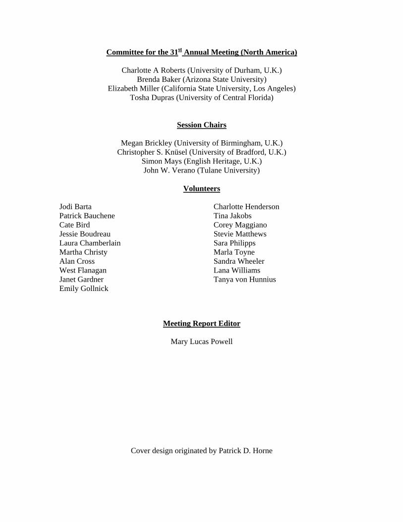
Committee for the 31st Annual Meeting (North America)
Charlotte A Roberts (University of Durham, U.K.) Brenda Baker (Arizona State University)
Elizabeth Miller (California State University, Los Angeles) Tosha Dupras (University of Central Florida)
Session Chairs
Megan Brickley (University of Birmingham, U.K.) Christopher S. Knüsel (University of Bradford, U.K.)
Simon Mays (English Heritage, U.K.) John W. Verano (Tulane University)
Volunteers
Jodi Barta Patrick Bauchene Cate Bird Jessie Boudreau Laura Chamberlain Martha Christy Alan Cross West Flanagan Janet Gardner Emily Gollnick
Charlotte Henderson Tina Jakobs Corey Maggiano Stevie Matthews Sara Philipps Marla Toyne Sandra Wheeler Lana Williams Tanya von Hunnius
Meeting Report Editor
Mary Lucas Powell
Cover design originated by Patrick D. Horne

![paleopathology soft tissues what mummies can the histological processing of mummified tissues provided by Sir Armand Ruffer and Sandison [8, 9]. were paramount for the study of soft](https://img.pdfslide.us/doc/110x75/60628c923141ee2359120123/paleopathology-soft-tissues-what-mummies-can-the-histological-processing-of-mummified.jpg)