Embed Size (px)
Citation preview
Volume 6: 1-8
September 2020 Vol. 6:1-8 Undergraduate Research Article https://jemi.microbiology.ubc.ca/ 1
Commercial Stevia does not inhibit growth or biofilm formation of Escherichia coli strain MG1655 Thomas Soroski, Boyan K. Tsankov, Al R. Hossain, Derek Lee Department of Microbiology and Immunology, University of British Columbia, Vancouver, British Columbia, Canada SUMMARY Stevia is a readily available, non-caloric sugar alternative with demonstrated non-carcinogenic and non-genotoxic activity. Stevia is gaining worldwide popularity as a non-nutritive sweetener, with the global stevia market growing about 8% yearly since 2016. However, recent findings suggest that some non-stevia sweeteners may cause adverse health consequences by altering the gut microbiome’s composition and diversity, raising concerns about stevia. Growth and adhesion are fundamental processes in the microbiome, and previous observations suggest that stevia may impact these processes. Therefore, we investigated the effects of commercial stevia on growth, adhesion, and biofilm formation of Escherichia coli MG1655. E. coli growth and biofilm formation was not significantly inhibited when treated with 1%, 10% or 20% (v/v) stevia (p > 0.05), compared to the untreated control. Attachment was not inhibited in any growth condition. These findings suggest that low to medium concentrations of stevia (≤20% v/v) do not affect E. coli surface-based biofilm formation, or planktonic growth. INTRODUCTION
tevia is used to describe a group of active compounds (steviol glycosides) extracted from Stevia rebaudiana, a shrub native to South America, and is produced by a number
of companies including Cargill Inc., PureCircle, and Ingredion Inc. (1). It is used worldwide as a non-nutritive sweetener (NNS) in a variety of food products. Stevia is mainly used as a plant-derived, relatively safe sweetener that has been demonstrated to be non-carcinogenic and non-genotoxic at recommended intake levels (2). However, recent evidence suggests that NNSs (in particular stevia, sucralose, and saccharin) can affect the gut microbiome (3, 4). Some investigations demonstrate that stevia, in particular, can alter the microbiome by selectively inhibiting the growth of some commensal populations and reducing microbiome diversity in mice (5, 6), and humans (7). NNS-mediated microbiome alterations observed in other, non-stevia sweeteners have been shown to have serious health implications including glucose intolerance (8) (a condition with an estimated 70% lifetime conversion rate to diabetes mellitus in adult humans (9)), obesity (10), and mouse immunity (showing increased cytokine production and lymphocyte numbers in the murine gut (11)). While these effects have not been documented as an effect of stevia, it has also not been studied as extensively as other NNSs. Increasing usage of stevia as an NNS thus raises interest in the possible effects of stevia on commensal gut bacteria.
A number of commensal characteristics can have implications on host health. While the majority of commensal studies to date have focused on commensal diversity and composition (12), there is evidence that other commensal characteristics can have host health implications as well, including gut immune dysfunction and regulation of homeostasis (12, 13). For
S
Published Online: September 2020
Citation: Thomas Soroski, Boyan K. Tsankov, Al R. Hossain, Derek Lee. 2020. Commercial Stevia does not inhibit growth or biofilm formation of Escherichia coli strain MG1655. UJEMI+ 5:1-8
Editor: Daniela Morales and Julia Huggins, University of British Columbia
Copyright: © 2020 Undergraduate Journal of Experimental Microbiology and Immunology.
All Rights Reserved.
Address correspondence to: https://jemi.microbiology.ubc.ca/
TheUndergraduateJournalofExperimentalMicrobiology&Immunology(+PeerReviewed)
UJEMI+ Soroski et al.
September 2020 Volume 6: 1-8 Undergraduate Research Article https://jemi.microbiology.ubc.ca/ 2
example, low growth and absolute count of commensals have been associated with Crohn’s disease (13). Commensal interactions (i.e. biofilm formation and intestinal quorum sensing) are associated with microbiome recovery and homeostasis following antibiotic treatment (14). Therefore, this study investigated whether off-the-shelf commercial stevia has any effect on Escherichia coli MG1655 (E. coli) growth and biofilm formation, to guide future research into possible effects of stevia on the microbiome. METHODS AND MATERIALS
Strains and media. E. coli K-12 MG1655 (CGSC#:6300) was purchased from the Yale Coli Genetic Stock Centre. NOW® BetterStevia® Organic Liquid (60mL) was used in all subsequent bacterial assays. E. coli was first streaked on Luria Bertani (LB) agar (1% peptone, 0.5% yeast extract, 1% NaCl, 2% agar). Isolated colonies were then cultured in LB broth (1% peptone, 0.5% yeast extract, 1% NaCl) overnight. Turbid cultures were then standardized to an OD600 of 0.05 in liquid M9 medium (10% M9 salts, 0.2% 1M MgSO4, 0.01% 1M CaCl2, 0.2% D-Glucose) supplemented with either 0%, 1%, 10%, 20% or 40% (v/v) NOW® BetterStevia® Organic Liquid (stevia) and grown overnight for subsequent assays. M9 minimal medium was used in this study as it provides the nutrient-deprived conditions necessary for biofilm formation, while still allowing for bacterial growth (15). Growth curves. Three independent overnight cultures of E. coli in LB-broth were diluted to an OD600 of 0.05 with M9 and subsequently supplemented with either 0%, 1%, 10%, 20%, or 40% (v/v) stevia (n=3 biological replicates). 200µL of each culture was plated in duplicate in a flat-bottom 96-well plate and subsequently incubated at 37°C for 16 hours in an automatic BioTek Epoch 2 microplate reader. Growth curves were generated by measuring OD600 every 10 minutes. Biofilm assay. Three independent overnight cultures of E. coli grown in LB-broth were diluted to an OD600 of 0.05 with M9 and subsequently supplemented with either 0%, 1%, 10%, 20%, or 40% (v/v) stevia. 100µL of the diluted cultures were plated in a flat-bottom 96-well plate and grown for 48hrs at 37°C without shaking (n=3 biological replicates). Media was aspirated after 24hrs of incubation and 100µL of fresh M9, supplemented with either 0%, 1%, 10%, 20%, or 40% (v/v) stevia, was added in order to prevent well desiccation and replenish nutrients available for growth and biofilm formation. After 48hrs of incubation, the supernatant was discarded, and the extracellular biofilm matrix was stained with 125µL of 0.1% crystal violet (Fisher Scientific catalog number: C581-25) for 15 minutes at room temperature. Excess crystal violet was washed away with dH2O, and biofilms were solubilized in 125µL of 30% acetic acid in water. The solubilized biofilm and crystal violet stain were transferred to a new 96-well flat bottom plate, and an OD550 reading was taken with a spectrophotometer to quantify biofilm formation. Biofilm attachment assay. E. coli cells were grown in a 96-well flat-bottom plate in M9 supplemented with either 0%, 1%, 10%, 20%, or 40% (v/v) stevia for 48hrs as described previously in our biofilm assay using 3 biological replicates (n=3). The OD600 of the wells was subsequently taken as previously described, and the supernatants containing planktonic bacteria were then discarded. The remaining adherent biofilm-forming bacteria were resuspended in 200µL of M9 media supplemented with the appropriate concentration of stevia. Subsequent OD600 of the resuspended wells was measured. To approximate the proportion of well-adherent cells in the biofilm assay, the ratio of resuspended cell absorbance to whole-well absorbance was calculated. Statistical Analysis. Student’s t-tests were performed in order to analyze experimental data. The results are expressed as mean ± standard deviation of at least three biological replicates, as indicated by n. A P value <0.05 was considered as statistically significant.
UJEMI+ Soroski et al.
September 2020 Volume 6: 1-8 Undergraduate Research Article https://jemi.microbiology.ubc.ca/ 3
RESULTS
Stevia concentration does not inhibit planktonic growth of E. coli. Conflicting results on the effect of stevia derivatives on the growth of E. coli exist, ranging from bacteriostatic (6) to bactericidal (16). In a 1997 paper from Tomita et al., a colony forming assay demonstrated that fermented stevia extract concentrations of 40% (v/v) reduced the colony formation of eight different pathogenic E. coli strains, implying that stevia extract reduces E. coli viability in a dose-dependent manner (16). In a 2018 paper by Wang et al., the growth of E. coli HB101 incubated with Rebaudioside A (Reb A, one of several key components in stevia extract) was assessed (6). It was found that 2.5% (w/v) Reb A reduced the number of E. coli HB101 CFUs by 83% but did not significantly impact E. coli K-12 CFUs. As these studies investigated different E. coli strains, in addition to testing different compounds, a general effect on the effects of stevia on E. coli growth or viability cannot be established. We adapted these experiments to determine the growth effects of commercial stevia on E. coli.
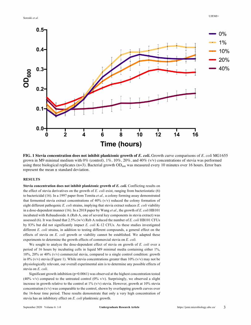
We sought to analyze the dose-dependent effect of stevia on growth of E. coli over a period of 16 hours by incubating cells in liquid M9 minimal media containing either 1%, 10%, 20% or 40% (v/v) commercial stevia, compared to a single control condition: growth in 0% (v/v) stevia (Figure 1). While stevia concentrations greater than 10% (v/v) may not be physiologically relevant, our overall experimental aim is to determine any possible effects of stevia on E. coli.
Significant growth inhibition (p=0.0061) was observed at the highest concentration tested (40% v/v) compared to the untreated control (0% v/v). Surprisingly, we observed a slight increase in growth relative to the control at 1% (v/v) stevia. However, growth at 10% stevia concentration (v/v) was comparable to the control, shown by overlapping growth curves over the 16-hour time period. These results demonstrate that only a very high concentration of stevia has an inhibitory effect on E. coli planktonic growth.
FIG. 1 Stevia concentration does not inhibit planktonic growth of E. coli. Growth curve comparisons of E. coli MG1655 grown in M9 minimal medium with 0% (control), 1%, 10%, 20%, and 40% (v/v) concentrations of stevia was performed using three biological replicates (n=3). Bacterial growth OD600 was measured every 10 minutes over 16 hours. Error bars represent the mean ± standard deviation.
UJEMI+ Soroski et al.
September 2020 Volume 6: 1-8 Undergraduate Research Article https://jemi.microbiology.ubc.ca/ 4
Biofilm formation is not impacted by the presence of stevia. To investigate stevia effects in a community setting, we examined the effect of stevia treatment on E. coli biofilm formation. Biofilm formation is closely related to growth, as biofilm maturation is effectively three-dimensional growth, shaped by community interactions and quorum sensing (17), key processes in the host microbiome (18). A 2017 study by Abdul Razak et al. showed that 10% stevia treatment reduced the solid-surface adherence and biofilm forming ability of Streptococcus spp. (19). The importance of microbial adherence, biofilm formation, and a lack of evidence into stevia-mediated biofilm effects led us to investigate E. coli biofilm formation (18). To this end, we investigated biofilm formation of E. coli incubated in differing concentrations of commercial stevia. To validate growth effects of previous studies (16) we chose to incubate E. coli in 0%, 1%, 10%, 20%, and 40% (v/v) concentrations of commercial stevia extract.
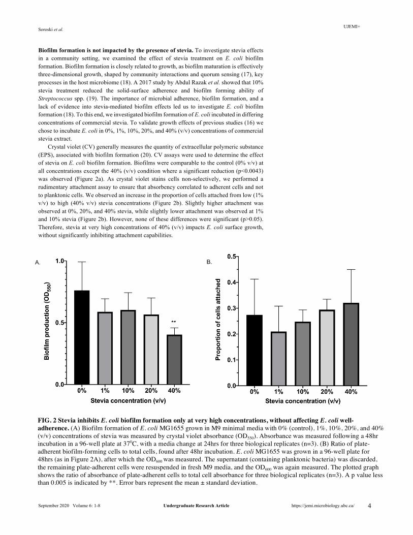
Crystal violet (CV) generally measures the quantity of extracellular polymeric substance (EPS), associated with biofilm formation (20). CV assays were used to determine the effect of stevia on E. coli biofilm formation. Biofilms were comparable to the control (0% v/v) at all concentrations except the 40% (v/v) condition where a significant reduction (p<0.0043) was observed (Figure 2a). As crystal violet stains cells non-selectively, we performed a rudimentary attachment assay to ensure that absorbency correlated to adherent cells and not to planktonic cells. We observed an increase in the proportion of cells attached from low (1% v/v) to high (40% v/v) stevia concentrations (Figure 2b). Slightly higher attachment was observed at 0%, 20%, and 40% stevia, while slightly lower attachment was observed at 1% and 10% stevia (Figure 2b). However, none of these differences were significant (p>0.05). Therefore, stevia at very high concentrations of 40% (v/v) impacts E. coli surface growth, without significantly inhibiting attachment capabilities.
FIG. 2 Stevia inhibits E. coli biofilm formation only at very high concentrations, without affecting E. coli well-adherence. (A) Biofilm formation of E. coli MG1655 grown in M9 minimal media with 0% (control), 1%, 10%, 20%, and 40% (v/v) concentrations of stevia was measured by crystal violet absorbance (OD550). Absorbance was measured following a 48hr incubation in a 96-well plate at 370C, with a media change at 24hrs for three biological replicates (n=3). (B) Ratio of plate-adherent biofilm-forming cells to total cells, found after 48hr incubation. E. coli MG1655 was grown in a 96-well plate for 48hrs (as in Figure 2A), after which the OD600 was measured. The supernatant (containing planktonic bacteria) was discarded, the remaining plate-adherent cells were resuspended in fresh M9 media, and the OD600 was again measured. The plotted graph shows the ratio of absorbance of plate-adherent cells to total cell absorbance for three biological replicates (n=3). A p value less than 0.005 is indicated by **. Error bars represent the mean ± standard deviation.
UJEMI+ Soroski et al.
September 2020 Volume 6: 1-8 Undergraduate Research Article https://jemi.microbiology.ubc.ca/ 5
DISCUSSION
Previous studies have shown that NNSs can affect the gut microbiome, altering the microbial physiology in a manner that can have implications on host health (8, 10, 11). Yet, the effect of stevia has not been studied extensively in the context of microbial communities, or in the context of microbial model organisms (i.e. E. coli). In this study, we aimed to understand whether stevia has any effect on E. coli growth and biofilm formation. To validate prior growth findings, we used similar stevia concentrations as in previous studies (16).
Growth curves revealed growth similar to the no-stevia control at all concentrations, except the 40% stevia condition. It is possible that the increased growth of E. coli in 1% (v/v) stevia was due to cells simultaneously metabolizing both glucose and stevia, or cells metabolizing stevia following glucose depletion, causing the observed increase in growth (7). While the ability of E. coli to metabolize stevia has not been researched, a previous study has shown that human fecal microflora are able to completely metabolize key stevia molecules (stevioside, and rebaudioside A) in vitro (7), suggesting that this increased growth could be attributed, in part, to E. coli metabolizing stevia. However, optical density is not sufficient to evaluate bactericidal or bacteriostatic effects of compounds. A growth curve should be accompanied by live/dead staining, measuring metabolically active cells, or plating on rich media to determine whether the lack of growth is due to dead cells or dormant cells.
Stevia-mediated increased growth was likely offset by the presence of a toxic compound in the media, inhibiting growth in a dose-dependent manner at concentrations above 1%. NOW® BetterStevia® contains 11% organic cane alcohol (equivalent to 11% ethanol) as a solvent (22). Previous studies have shown that concentrations of ethanol as low as 4% (v/v) can reduce the survival of E. coli by half (23). Mechanisms that mediate this effect include disruptions in cell division (24), protein and fatty acid composition differentiations (25), and variations in membrane composition that impact associated processes such as nutrient uptake (26). At our treatment conditions, stevia concentrations of 1%, 10%, 20% and 40% (v/v) correspond to a final ethanol concentration of 0.1%, 1.1%, 2.2%, and 4.4%, respectively. This study aimed to test off-the-shelf commercial stevia, often prepared with an alcoholic solvent. Future studies should aim to conduct a pure alcohol or cane alcohol vehicle control to determine possible stevia-only growth effects. The reduced growth of E. coli at high concentrations of stevia suggests an inhibitory effect on planktonic growth, although whether these effects are bacteriostatic or bactericidal are yet to be resolved.
We next sought to evaluate the effects of stevia on biofilm formation, a key process of gut microbiota physiology that mediates cellular responses including modulation of the immune response in the intestinal mucosa (27). Studies have shown that disruption of gut biofilms can result in the development of inflammatory diseases such as inflammatory bowel disease, attributed to the release of planktonic bacteria with biofilm-related phenotypes (28). Biofilm production was inferred to be significantly inhibited at only 40% (v/v) stevia, with minor non-significant reductions observed at other concentrations, measured with a CV assay. In general, CV measures EPS production without distinguishing between live or dead bacteria, thus limiting definitive interpretation of biofilm effects. Nonetheless, multiple biological replicates yielded similar results, reinforcing our initial finding that biofilm production is only inhibited at 40% (v/v) stevia treatment. Inhibited biofilm formation may be attributed to 40% (v/v) stevia inhibiting growth from the aforementioned toxicity or metabolic changes in response to stevia, reducing the total amount of viable cells capable of producing EPS.
Additionally, the air-liquid interface is a substantial component of the biofilm in addition to solid-liquid interfaces (29). The CV assay is inherently limited as the washing step and media change step could inadvertently disrupt the air-liquid or solid-liquid biofilm. To address the proportion of adherent biofilm cells remaining, a rudimentary plate-attachment assay was performed by calculating the absorbance ratio of plate-adherent cells (following the removal of supernatant with planktonic cells) to total cells in the well (adhered and planktonic), following a 48-hour incubation. The trend observed in our attachment assay did not correlate with the decrease in biofilm formation seen at 40% (v/v) stevia treatment. Instead, we observed an inverse relationship between CV staining and the ratio of adhered cells. Across bacterial populations, the EPS composition can differ significantly with multiple polysaccharides and is widely complex containing a variety of proteins, glycoproteins, and
UJEMI+ Soroski et al.
September 2020 Volume 6: 1-8 Undergraduate Research Article https://jemi.microbiology.ubc.ca/ 6
glycolipids (30). Thus, as CV staining targets negatively charged molecules found within the EPS, compositional differences result in a lack of sensitivity and specificity of the dye when performing biofilm quantification via EPS production (20). In addition, the presence of ethanol from the stevia product potentially caused additional solubilization of the biofilm in conjunction with the 30% acetic acid, possibly resulting in further discrepancy between the observed inverse relationship between CV staining and ratio of adhered cells (31). Previous studies have also shown that EPS production in E. coli is not required as a prerequisite for successful surface attachment but rather induced post-attachment, as opposed to bacterial species such as S. epidermidis (32). In this context, the initial attachment phase of biofilms may be captured without detectable amounts of EPS production, and thus potentially contributing to the observed discrepancy. It is possible that increasing stevia concentration promotes greater attachment to the surface (although data is insignificant), but also inhibits biofilm maturation, resulting in an overall decrease in EPS production. In total, these factors may serve as an explanation for the observed discrepancy between increasing attachment yet lower biofilm formation. Therefore, stevia treatments lower than 40% (v/v) may cause subtler effects that are unable to be captured due to the limitations of the CV assay, particularly at 20% (v/v) stevia with known inhibitory growth effects as shown in our earlier findings. Conclusions Based on our findings, we conclude that commercial stevia does not inhibit the growth or biofilm formation of E. coli. Altogether, only the 40% (v/v) stevia condition, which is unlikely to be physiologically relevant, had a negative effect on the growth and biofilm formation of E. coli. Future Directions A major limitation in this study is the lack of vehicle controls. While this study aimed to investigate the effect of commercially available stevia, liquid preparations of stevia are made with an alcohol solvent. Repeating these investigations using powdered stevia, or by using liquid stevia compared to an alcohol-only control would generate clearer conclusions about the effect of stevia itself.
It has been previously noted that different brands of stevia extract vary in their composition, with some brands containing over 99% Reb A, and others containing less than 20% Reb A (33). For the purposes of this study, only NOW® BetterStevia® organic liquid sweetener was used. Future studies could investigate other brands of stevia, or the effects of a single stevia molecule, similar to the work of Nettleton et al. investigating the effect of only Reb A on the mouse microbiome (5).
The CV assay is inherently limited as it does not distinguish between live or dead cells, and relies upon the non-specific uptake of the dye into the extracellular matrix (31). This makes repeated CV assays variable and poorly reproducible due to variations in cell viability and extracellular matrix composition (31). There are also variables that can introduce batch variability (including incubation time and temperature), further clouding the results (31). Future studies should validate CV biofilm assays by measuring cell viability (e.g. CFU), or by using a separate biofilm assay to corroborate results (e.g. Tetrazolium Salt Assay, Total organic carbon assay (31).
This investigation aimed to shed light on possible mechanisms by which stevia could dysregulate the gut microbiome, using a model organism. Future studies should investigate the effect of stevia on other human microbiome species (e.g. Enterobacter spp., Lactobacillus spp.), or on microbial co-cultures (similar to the co-culture work investigating sucralose, a different NNS, by Corder et al. (4). The European Food Safety Authority (EFSA) has recommended a daily intake of 388 milligrams of stevia equivalents per kilogram of body weight per day, based on long-term (2 year) rat carcinogenicity studies (34). It has been shown that human digestive enzymes are not able to degrade stevioside into steviol (21), suggesting that the microbiome would be exposed to the majority of host-consumed stevia. However, it is unlikely that the gut microbiome would be exposed to stevia levels as high as the ones used in this investigation (i.e. 10%, 20%, 40%), especially in hosts following EFSA intake recommendations. Future studies should investigate the effects at stevia concentrations that model in vivo conditions, to better understand stevia-mediated microbial interactions that would happen in a typical stevia consuming host. Studies should also aim to confirm the observed pro-growth effect of 1% (v/v) stevia exposure found in the present study.
UJEMI+ Soroski et al.
September 2020 Volume 6: 1-8 Undergraduate Research Article https://jemi.microbiology.ubc.ca/ 7
ACKNOWLEDGEMENTS
Funding and institutional support for the research was provided by the University of British Columbia, department of Microbiology and Immunology. The project was supported intellectually by Dr. Evelyn Sun and Dr. David Oliver. Special thanks to Craig Kornak, Reynold Farrera Calderon, and to our colleagues in the MICB447 lab. We would also like to thank two anonymous reviewers for constructive feedback on this manuscript.
REFERENCES
1. Global Stevia Market. Mordor Intelligence. 2. Ferrazzano GF, Cantile T, Alcidi B, Coda M, Ingenito A, Zarrelli A, Di Fabio G, Pollio A.
2015. Is Stevia rebaudiana Bertoni a Non Cariogenic Sweetener? A Review. Molecules 21:E38. 3. Ruiz-Ojeda FJ, Plaza-Díaz J, Sáez-Lara MJ, Gil A. 2019. Effects of Sweeteners on the Gut
Microbiota: A Review of Experimental Studies and Clinical Trials. Adv Nutr 10:S31–S48. 4. Corder B, Knobbe A. 2018. The effects of the artificial sweetener sucralose on the gut bacteria
Escherichia coli and Enterobacter aerogenes. JEMI 4:1–9. 5. Nettleton JE, Klancic T, Schick A, Choo AC, Shearer J, Borgland SL, Chleilat F, Mayengbam
S, Reimer RA. 2019. Low-Dose Stevia (Rebaudioside A) Consumption Perturbs Gut Microbiota and the Mesolimbic Dopamine Reward System. Nutrients 11.
6. Wang Q-P, Browman D, Herzog H, Neely GG. 2018. Non-nutritive sweeteners possess a bacteriostatic effect and alter gut microbiota in mice. PLoS One 13:e0199080.
7. Gardana C, Simonetti P, Canzi E, Zanchi R, Pietta P. 2003. Metabolism of stevioside and rebaudioside A from Stevia rebaudiana extracts by human microflora. J Agric Food Chem 51:6618–6622.
8. Suez J, Korem T, Zeevi D, Zilberman-Schapira G, Thaiss CA, Maza O, Israeli D, Zmora N, Gilad S, Weinberger A, Kuperman Y, Harmelin A, Kolodkin-Gal I, Shapiro H, Halpern Z, Segal E, Elinav E. 2014. Artificial sweeteners induce glucose intolerance by altering the gut microbiota. Nature 514:181–186.
9. Nathan DM, Davidson MB, DeFronzo RA, Heine RJ, Henry RR, Pratley R, Zinman B, American Diabetes Association. 2007. Impaired fasting glucose and impaired glucose tolerance: implications for care. Diabetes Care 30:753–759.
10. Farup PG, Lydersen S, Valeur J. 2019. Are Nonnutritive Sweeteners Obesogenic? Associations between Diet, Faecal Microbiota, and Short-Chain Fatty Acids in Morbidly Obese Subjects. J Obes 2019:4608315.
11. Martínez-Carrillo BE, Rosales-Gómez CA, Ramírez-Durán N, Reséndiz-Albor AA, Escoto-Herrera JA, Mondragón-Velásquez T, Valdés-Ramos R, Castillo-Cardiel A. 2019. Effect of Chronic Consumption of Sweeteners on Microbiota and Immunity in the Small Intestine of Young Mice. Int J Food Sci 2019:9619020.
12. Cani PD. 2018. Human gut microbiome: hopes, threats and promises. Gut 67:1716–1725. 13. Vandeputte D, Kathagen G, D’hoe K, Vieira-Silva S, Valles-Colomer M, Sabino J, Wang J,
Tito RY, De Commer L, Darzi Y, Vermeire S, Falony G, Raes J. 2017. Quantitative microbiome profiling links gut community variation to microbial load. Nature 551:507–511.
14. Bivar Xavier K. 2018. Bacterial interspecies quorum sensing in the mammalian gut microbiota. C R Biol 341:297–299.
15. Wood TK. 2009. Insights on Escherichia coli biofilm formation and inhibition from whole-transcriptome profiling. Environ Microbiol 11:1–15.
16. Tomita T, Sato N, Arai T, Shiraishi H, Sato M, Takeuchi M, Kamio Y. 1997. Bactericidal activity of a fermented hot-water extract from Stevia rebaudiana Bertoni towards enterohemorrhagic Escherichia coli O157:H7 and other food-borne pathogenic bacteria. Microbiol Immunol 41:1005–1009.
17. Beloin C, Roux A, Ghigo JM. 2008. Escherichia coli biofilms. Curr Top Microbiol Immunol 322:249–289.
18. Tytgat HLP, Nobrega FL, van der Oost J, de Vos WM. 2019. Bowel Biofilms: Tipping Points between a Healthy and Compromised Gut? Trends Microbiol 27:17–25.
19. Abdul Razak F, Baharuddin BA, Akbar EFM, Norizan AH, Ibrahim NF, Musa MY. 2017. Alternative sweeteners influence the biomass of oral biofilm. Arch Oral Biol 80:180–184.
20. Magana M, Sereti C, Ioannidis A, Mitchell CA, Ball AR, Magiorkinis E, Chatzipanagiotou S, Hamblin MR, Hadjifrangiskou M, Tegos GP. 2018. Options and Limitations in Clinical Investigation of Bacterial Biofilms. Clin Microbiol Rev 31.
21. Koyama E, Sakai N, Ohori Y, Kitazawa K, Izawa O, Kakegawa K, Fujino A, Ui M. 2003. Absorption and metabolism of glycosidic sweeteners of stevia mixture and their aglycone, steviol, in rats and humans. Food Chem Toxicol 41:875–883.
22. 2016. BetterStevia® Liquid, Organic. NOW Foods. 23. Ingram LO, Vreeland NS, Eaton LC. 1980. Alcohol tolerance in Escherichia coli. Pharmacol
Biochem Behav 13 Suppl 1:191–195.
UJEMI+ Soroski et al.
September 2020 Volume 6: 1-8 Undergraduate Research Article https://jemi.microbiology.ubc.ca/ 8
24. Fried VA, Novick A. 1973. Organic solvents as probes for the structure and function of the bacterial membrane: effects of ethanol on the wild type and an ethanol-resistant mutant of Escherichia coli K-12. J Bacteriol 114:239–248.
25. Chiou RY-Y, Phillips RD, Zhao P, Doyle MP, Beuchat LR. 2004. Ethanol-mediated variations in cellular fatty acid composition and protein profiles of two genotypically different strains of Escherichia coli O157:H7. Appl Environ Microbiol 70:2204–2210.
26. Bowles LK, Ellefson WL. 1985. Effects of butanol on Clostridium acetobutylicum. Appl Environ Microbiol 50:1165–1170.
27. Ellermann M, Sartor RB. 2018. Intestinal bacterial biofilms modulate mucosal immune responses. J Immunol Sci 2:13–18.
28. Buret AG, Motta J-P, Allain T, Ferraz J, Wallace JL. 2019. Pathobiont release from dysbiotic gut microbiota biofilms in intestinal inflammatory diseases: a role for iron? J Biomed Sci 26:1.
29. Sinibaldi G, Iebba V, Chinappi M. 2018. Swimming and rafting of E.coli microcolonies at air-liquid interfaces. Microbiologyopen 7.
30. Flemming H-C, Neu TR, Wozniak DJ. 2007. The EPS matrix: the “house of biofilm cells.” J Bacteriol 189:7945–7947.
31. Wilson C, Lukowicz R, Merchant S, Valquier-Flynn H, Caballero J, Sandoval J, Okuom M, Huber C, Brooks TD, Wilson E, Clement B, Wentworth CD, Holmes AE. 2017. Quantitative and Qualitative Assessment Methods for Biofilm Growth: A Mini-review. Res Rev J Eng Technol 6.
32. Danese PN, Pratt LA, Kolter R. 2000. Exopolysaccharide production is required for development of Escherichia coli K-12 biofilm architecture. J Bacteriol 182:3593–3596.
33. Formigoni M, Milani P, Zorzenon M, Da Silva Avincola A, Dacome A, Pilau EJ, Costa S. 2019. Analysis of Commercial Stevia Extracts Composition by HPLC and UHPLC-MS-MS-QTOF. 1 75:355–360.
34. F. Aguilar, U.R. Charrondiere, B. Dusemund, P. Galtier, J. Gilbert, D.M. Gott, S. Grilli, R. Gürtler, J. König, C. Lambré, J- C. Larsen, J- C. Leblanc, A. Mortensen, D. Parent- Massin, I. Pratt, I.M.C.M. Rietjens, I. Stankovic, P. Tobback, T. Verguieva, R.A. Woutersen. 2010. Scientific Opinion on the safety of steviol glycosides for the proposed uses as a food additive. EFSA Journal.


![CULTIVATION AND USES OF STEVIA (Stevia rebaudiana Bertoni ... · Stevia [Stevia rebaudiana Bertoni; Family Asteraceae] is a natural sweetener plant that is grown commercially in many](https://img.pdfslide.us/doc/110x75/5e72492d6311fa6493415583/cultivation-and-uses-of-stevia-stevia-rebaudiana-bertoni-stevia-stevia-rebaudiana.jpg)