Embed Size (px)
Citation preview
RESEARCH ARTICLE
Combined Inhibition of the Renin-Angiotensin
System and Neprilysin Positively Influences
Complex Mitochondrial Adaptations in
Progressive Experimental Heart Failure
Laura Grois1☯, Julian Hupf1☯, Jorg Reinders2, Josef Schroder3, Alexander Dietl1, Peter
M. Schmid1, Carsten Jungbauer1, Markus Resch1, Lars S. Maier1, Andreas Luchner4,
Christoph Birner1*
1 Department of Internal Medicine II, University Hospital Regensburg, Regensburg, Germany, 2 Institute of
Functional Genomics, University Regensburg, Regensburg, Germany, 3 Electron Microscopy Core Facility,
Institute for Pathology, University Hospital Regensburg, Regensburg, Germany, 4 Department of Internal
Medicine I, Clinic St. Marien, Amberg, Germany
☯ These authors contributed equally to this work.
Abstract
Background
Inhibitors of the renin angiotensin system and neprilysin (RAS-/NEP-inhibitors) proved to be
extraordinarily beneficial in systolic heart failure. Furthermore, compelling evidence exists
that impaired mitochondrial pathways are causatively involved in progressive left ventricular
(LV) dysfunction. Consequently, we aimed to assess whether RAS-/NEP-inhibition can
attenuate mitochondrial adaptations in experimental heart failure (HF).
Methods and Results
By progressive right ventricular pacing, distinct HF stages were induced in 15 rabbits, and 6
animals served as controls (CTRL). Six animals with manifest HF (CHF) were treated with
the RAS-/NEP-inhibitor omapatrilat. Echocardiographic studies and invasive blood pressure
measurements were undertaken during HF progression. Mitochondria were isolated from
LV tissue, respectively, and further worked up for proteomic analysis using the SWATH tech-
nique. Enzymatic activities of citrate synthase and the electron transfer chain (ETC) com-
plexes I, II, and IV were assessed. Ultrastructural analyses were performed by transmission
electron microscopy. During progression to overt HF, intricate expression changes were
mainly detected for proteins belonging to the tricarboxylic acid cycle, glucose and fat metabo-
lism, and the ETC complexes, even though ETC complex I, II, or IV enzymatic activities were
not significantly influenced. Treatment with a RAS-/NEP-inhibitor then reversed some mal-
adaptive metabolic adaptations, positively influenced the decline of citrate synthase activity,
and altered the composition of each respiratory chain complex, even though this was again
not accompanied by altered ETC complex enzymatic activities. Finally, ultrastructural
PLOS ONE | DOI:10.1371/journal.pone.0169743 January 11, 2017 1 / 21
a1111111111
a1111111111
a1111111111
a1111111111
a1111111111
OPENACCESS
Citation: Grois L, Hupf J, Reinders J, Schroder J,
Dietl A, Schmid PM, et al. (2017) Combined
Inhibition of the Renin-Angiotensin System and
Neprilysin Positively Influences Complex
Mitochondrial Adaptations in Progressive
Experimental Heart Failure. PLoS ONE 12(1):
e0169743. doi:10.1371/journal.pone.0169743
Editor: Edward J. Lesnefsky, Virginia
Commonwealth University, UNITED STATES
Received: April 13, 2016
Accepted: December 21, 2016
Published: January 11, 2017
Copyright: © 2017 Grois et al. This is an open
access article distributed under the terms of the
Creative Commons Attribution License, which
permits unrestricted use, distribution, and
reproduction in any medium, provided the original
author and source are credited.
Data Availability Statement: All relevant data are
within the paper.
Funding: This work was supported by an
institutional research grant (ReForM-A) of the
University Hospital Regensburg.
Competing Interests: The authors have declared
that no competing interests exist.
evidence pointed to a reduction of autophagolytic and degenerative processes with omapatri-
lat-treatment.
Conclusions
This study describes complex adaptations of the mitochondrial proteome in experimental
tachycardia-induced heart failure and shows that a combined RAS-/NEP-inhibition can ben-
eficially influence mitochondrial key pathways.
Introduction
Systolic heart failure is characterized by a detrimental activation of the sympathetic nervous
system (SNS) and the renin-angiotensin system (RAS) [1–3], whose pharmacologic blockade
has proven to be prognostically beneficial, respectively [4–7]. Nevertheless, facing a five-year
survival rate of about 50% prognosis remains very poor [8] thereby indicating that the thera-
peutic potential has by far not been realized yet.
Having said this, increasing evidence points to a new pathophysiologic paradigm, where
the true driving force for progressive left ventricular dysfunction is now seen in a deleterious
imbalance between maladaptive (i.e., SNS and RAS) and protective (mainly the natriuretic
peptide system, NPS) mechanisms [9], which means, that beneficial effects were to expect not
only from inhibiting the former, but also from augmenting the later ones. Consequently, a new
pharmacologic class has been developed which inhibits both the angiotensin converting enzyme
and the natriuretic peptides degrading enzyme neprilysin [10]. The leading substance of this
“vasopeptidase inhibitors” (VPIs) named class, omapatrilat, was thoroughly evaluated [11–13],
but failed to be launched due to its rare, but relevant side effects (mainly angioedema). Subse-
quently, a neprilysin inhibitor was combined with an angiotensin-receptor blocker instead of
an ACE-inhibitor, thereby introducing the class of ARNIs (angiotensin receptor neprilysin-
inhibitors). Its leading substance, LCZ696, has recently shown beneficial effects with better tol-
erance and convincingly confirmed the new pathophysiological concept behind this combined
RAS-/NEP-inhibition [14]. By further evaluating this principle, our group was able to demon-
strate a positive impact of omapatrilat on structural cardiac remodeling and neurohumoral acti-
vation [15], which both could provide a pathophysiologic fundament for the beneficial clinical
effects.
Besides this new paradigm of neurohumoral imbalance, a rapidly growing body of evidence
points to a central role of mitochondrial impairment in progressive heart failure resulting in
detrimental energetic deprivation and deleterious oxidative stress [16]. This was also con-
firmed by our work group when evaluating proteomic alterations in left ventricles [17] and
atria [18]. But despite recognition of its importance, mitochondrial adaptations remain never-
theless insufficiently characterized during progression to overt heart failure and therefore
deserve further evaluation to potentially identify new therapeutic targets. Furthermore, it is
unknown which impact a combined RAS-/NEP-inhibition has on energetically relevant path-
ways and whether these two mechanisms are interlinked to result in beneficial clinical effects.
We therefore evaluated in our well established model of progressive, pacing-induced heart
failure in rabbits [17,19,20,15,21,22], which structural, functional and proteomic alterations car-
diac mitochondria undergo in different stages of heart failure, and whether these adaptations
are influenced by combined RAS-/NEP-inhibition. Facing the evident importance of both neu-
rohumoral and energetic mechanisms, we hypothesized that mitochondrial adaptations which
Mitochondrial Effects of a Combined RAS-/NEP-Inhibition in Experimental Heart Failure
PLOS ONE | DOI:10.1371/journal.pone.0169743 January 11, 2017 2 / 21
develop in progressive heart failure should be reversed or at least mitigated by RAS-/NEP-
inhibition.
Methods
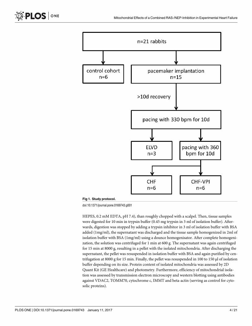
Model of progressive pacing-induced heart failure
All experiments were approved by the institutional and governmental animal care committees,
respectively. A total of 21 male rabbits (chinchilla bastard; Charles River Laboratories Interna-
tional, Inc.) was used for this study (see Fig 1). The animals were exposed to a 12:12 h light:
dark rhythm and received standard chow and water ad libitum. 15 animals underwent implan-
tation of a cardiac pacemaker (Medtronic Minix 8340, Minneapolis, MN or Vitatron Model
810, Dieren, NL) and a transvenous right ventricular lead with 3 and 12 of them being paced
until early left ventricular dysfunction (ELVD) and congestive heart failure (CHF) was gener-
ated, respectively. Surgical procedures were conducted under general anesthesia (Ketamine 60
mg/kg and Xylazine 5 mg/kg i.m.), and a standardized pharmacological protocol was applied in
the early post-surgery period (4 mg/kg BW Rimadyl s.c. and 5 mg/kg BW Baytril s.c. for 3 days,
respectively). Six untreated animals served as controls (CTRL). ELVD and CHF was induced by
a standardized protocol of progressive rapid right ventricular pacing as described previously
[17,19,20,15,18]. In brief, animals of the ELVD group were paced with 330 beats per minute
(bpm) for 10 days, and animals of the CHF group underwent an additional pacing period at
360 bpm for further 10 days. Drinking water of CHF animals was either substituted with Oma-
patrilat to reach a daily dose of 50 mg/kg BW (CHF-VPI group), or remained untreated (CHF
group). Pharmacological intervention was started after initiation of cardiac pacing and was sus-
tained during the whole pacing period. At the end of the experiments, rabbits were euthanized
by i.v. pentobarbital injection and tissue was rapidly harvested and deep-frozen. During the
experiments, rabbits were monitored at least daily, which also included an evaluation regarding
prespecified early endpoints (e.g., reduced food or liquid intake, dyspnea, impaired mobility,
untreatable surgical complications such as severe infections). No early endpoint was met during
the experiments and no animals died prior to the experimental endpoint. All experiments were
approved by the institutional and governmental animal care committees, respectively (Univer-
sity Hospital Regensburg and Regierung der Oberpfalz).
Echocardiography and hemodynamic evaluation
Measurements were done as described previously [15]: Under light sedation (5 mg midazolam
i.m.) a long and short-axis echocardiogram (using HP Sonos 5500 with a 12 MHz probe) was
performed in a supine position from the left parasternal window. LV enddiastolic (LVEDd)
and LV endsystolic (LVESd) diameters, diastolic and systolic thickness of interventricular sep-
tum (IVSd, IVSs) and posterior wall (LVPWd, LVPWs) as well as left atrial diameters (LAd)
were determined from three repeated 2D-guided M-mode tracings using the ASE conventions.
Fractional shortening (FS) was calculated as: FS = (LVEDd-LVESd)/LVEDd.
Conscious arterial blood pressure was measured invasively via the medial ear artery under
light sedation and after pausing the pacemaker stimulus.
Echocardiographic and hemodynamic evaluations were done at baseline, after each pacing
period and at the end of the experiments.
Isolation of cardiac mitochondria
A modified protocol by Schaeffer et al. [23] was used for mitochondrial isolation. Left ventric-
ular tissue samples were first washed in 3 mL of isolation buffer (300 mM succrose, 10 mM
Mitochondrial Effects of a Combined RAS-/NEP-Inhibition in Experimental Heart Failure
PLOS ONE | DOI:10.1371/journal.pone.0169743 January 11, 2017 3 / 21
HEPES, 0.2 mM EDTA, pH 7.4), than roughly chopped with a scalpel. Then, tissue samples
were digested for 10 min in trypsin buffer (0.45 mg trypsin in 3 ml of isolation buffer). After-
wards, digestion was stopped by adding a trypsin inhibitor in 3 ml of isolation buffer with BSA
added (1mg/ml), the supernatant was discharged and the tissue sample homogenized in 2ml of
isolation buffer with BSA (1mg/ml) using a dounce homogenisator. After complete homogeni-
zation, the solution was centrifuged for 1 min at 600 g. The supernatant was again centrifuged
for 15 min at 8000 g, resulting in a pellet with the isolated mitochondria. After discharging the
supernatant, the pellet was resuspended in isolation buffer with BSA and again purified by cen-
trifugation at 8000 g for 15 min. Finally, the pellet was resuspended in 100 to 150 μl of isolation
buffer depending on its size. Protein content of isolated mitochondria was assessed by 2D
Quant Kit (GE Healthcare) and photometry. Furthermore, efficiency of mitochondrial isola-
tion was assessed by transmission electron microscopy and western blotting using antibodies
against VDAC2, TOMM70, cytochrome c, IMMT and beta-actin (serving as control for cyto-
solic proteins).
Fig 1. Study protocol.
doi:10.1371/journal.pone.0169743.g001
Mitochondrial Effects of a Combined RAS-/NEP-Inhibition in Experimental Heart Failure
PLOS ONE | DOI:10.1371/journal.pone.0169743 January 11, 2017 4 / 21
Protein expression of CPT1A (carnitine palmitoyltransferase 1 A)
The expression level of CPT1A, which is an essential enzyme for the beta oxidation of long
chain fatty acids by mediating their transport from the cytosol into the mitochondrial inter-
membrane space, was determined by western blot analysis. For this purpose, anti-CPT1A
(PA5-29995, Thermo Scientific, Waltham, USA) was used as primary antibody, and ab97085
Dnk pAb to rabbit-HRP (Abcam, Cambridge, UK) as secondary antibody.
Enzymatic activites of citrate synthase, NADH dehydrogenase (complex
I), succinate dehydrogenase (complex II), and cytochrome c oxidase
(complex IV)
Activity of citrate synthase was assessed following the protocol of Srere et al. [24] In brief,
mitochondrial membranes were destroyed by Triton X100, and acetyl-CoA, oxalacetate and
DTNB were added. After reaction of oxalacetate with acetyl-CoA, which is mediated by citrate
synthase (at 30˚C), free CoA can convert DTNB into TNB. The amount of TNB can then be
assessed by photometry at 412 nm over 200 s.
Activity of the ETC complex I-enzyme NADH dehydrogenase was determined as previ-
ously described [25]. In brief, this assay photometrically measures oxidation of NADH at a
wavelength of 340 nm and additionally determines rotenone-insensitive complex I activity for
control purposes. To assess enzymatic activity of the ETC complex II-enzyme succinate dehy-
drogenase, 20 μl of a mitochondrial isolate were mixed with warmed reaction medium con-
taining 500mM phosphate buffer (pH 7.5), 50 mg/ml BSA, 200mM succinate, 5mM DCPIP,
10mM KCN, 10mM ATP, and H2O. Photometric measurements were then performed using a
wavelength of 600nm. Finally, enzymatic activity of the ETC complex IV-enzyme cytochrome
c oxidase was determined by adding a mitochondrial isolate to a solution containing DTT-
reduced cytochrome c and assessing the absorbance at a wavelength of 550nm.
Protein identification by mass spectrometry using the SWATH technique
For mass spectrometry proteins of mitochondrial isolates have been precipitated by ethanol,
than homogenized in ammonium bicarbonate with a FastPrep-24 device (MP Biomedicals).
Protein solution was diluted to match a protein concentration of 2μg/μl. Protein samples were
reduced, carbamidomethylated and digested according to the RapidACN protocol [26]. 1 μg of
the resulting peptide mixtures were subjected to SWATH-MS measurements as published pre-
viously [27]. The SWATH-library was build using the NCBInr database and the Protein Pilot
4.5 software (Sciex GmbH, Darmstadt, Germany) employing a 1% false discovery rate. Func-
tional data for protein matches was attained using the UniProt DB.
Transmission electron microscopy
Frozen myocardial samples were thawed and fixed in Karnovsky-fixative, then dehydrated
in graded ethanols, and afterwards embedded in epoxy resin (EmBed812). 80 nm sections
were double contrasted with uranyl and lead salts, and examined using the EFTEM LEO912
AB (Zeiss/Oberkochen) electron microscope operating at 100 kV acceleration in zero loss
mode. Images were acquired with a 1kx1k pixel side-entry mounted CCD camera controlled
with the iTEM software (OSIS/Muenster), and both qualitatively and semi-quantitatively
analyzed.
Mitochondrial Effects of a Combined RAS-/NEP-Inhibition in Experimental Heart Failure
PLOS ONE | DOI:10.1371/journal.pone.0169743 January 11, 2017 5 / 21
Statistical analysis
Data are expressed as mean±S.E.M. or mean±SD. Differences between two analyzed groups
were assessed by the Student‘s t-test or ANOVA, when appropriate. Statistical significance was
defined as P<0.05. SPSS Statistics (version 22, IBM, Armonk, USA) was used for data analysis.
Results
Hemodynamic and structural changes
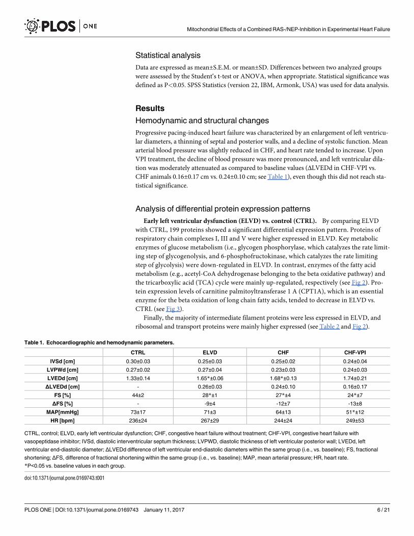
Progressive pacing-induced heart failure was characterized by an enlargement of left ventricu-
lar diameters, a thinning of septal and posterior walls, and a decline of systolic function. Mean
arterial blood pressure was slightly reduced in CHF, and heart rate tended to increase. Upon
VPI treatment, the decline of blood pressure was more pronounced, and left ventricular dila-
tion was moderately attenuated as compared to baseline values (ΔLVEDd in CHF-VPI vs.
CHF animals 0.16±0.17 cm vs. 0.24±0.10 cm; see Table 1), even though this did not reach sta-
tistical significance.
Analysis of differential protein expression patterns
Early left ventricular dysfunction (ELVD) vs. control (CTRL). By comparing ELVD
with CTRL, 199 proteins showed a significant differential expression pattern. Proteins of
respiratory chain complexes I, III and V were higher expressed in ELVD. Key metabolic
enzymes of glucose metabolism (i.e., glycogen phosphorylase, which catalyzes the rate limit-
ing step of glycogenolysis, and 6-phosphofructokinase, which catalyzes the rate limiting
step of glycolysis) were down-regulated in ELVD. In contrast, enzymes of the fatty acid
metabolism (e.g., acetyl-CoA dehydrogenase belonging to the beta oxidative pathway) and
the tricarboxylic acid (TCA) cycle were mainly up-regulated, respectively (see Fig 2). Pro-
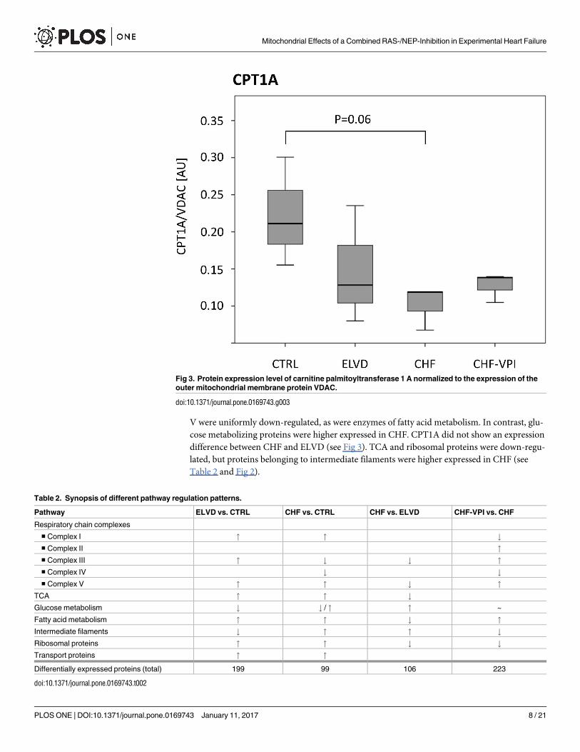
tein expression levels of carnitine palmitoyltransferase 1 A (CPT1A), which is an essential
enzyme for the beta oxidation of long chain fatty acids, tended to decrease in ELVD vs.
CTRL (see Fig 3).
Finally, the majority of intermediate filament proteins were less expressed in ELVD, and
ribosomal and transport proteins were mainly higher expressed (see Table 2 and Fig 2).
Table 1. Echocardiographic and hemodynamic parameters.
CTRL ELVD CHF CHF-VPI
IVSd [cm] 0.30±0.03 0.25±0.03 0.25±0.02 0.24±0.04
LVPWd [cm] 0.27±0.02 0.27±0.04 0.23±0.03 0.24±0.03
LVEDd [cm] 1.33±0.14 1.65*±0.06 1.68*±0.13 1.74±0.21
ΔLVEDd [cm] - 0.26±0.03 0.24±0.10 0.16±0.17
FS [%] 44±2 28*±1 27*±4 24*±7
ΔFS [%] - -9±4 -12±7 -13±8
MAP[mmHg] 73±17 71±3 64±13 51*±12
HR [bpm] 236±24 267±29 244±24 249±53
CTRL, control; ELVD, early left ventricular dysfunction; CHF, congestive heart failure without treatment; CHF-VPI, congestive heart failure with
vasopeptidase inhibitor; IVSd, diastolic interventricular septum thickness; LVPWD, diastolic thickness of left ventricular posterior wall; LVEDd, left
ventricular end-diastolic diameter; ΔLVEDd difference of left ventricular end-diastolic diameters within the same group (i.e., vs. baseline); FS, fractional
shortening; ΔFS, difference of fractional shortening within the same group (i.e., vs. baseline); MAP, mean arterial pressure; HR, heart rate.
*P<0.05 vs. baseline values in each group.
doi:10.1371/journal.pone.0169743.t001
Mitochondrial Effects of a Combined RAS-/NEP-Inhibition in Experimental Heart Failure
PLOS ONE | DOI:10.1371/journal.pone.0169743 January 11, 2017 6 / 21
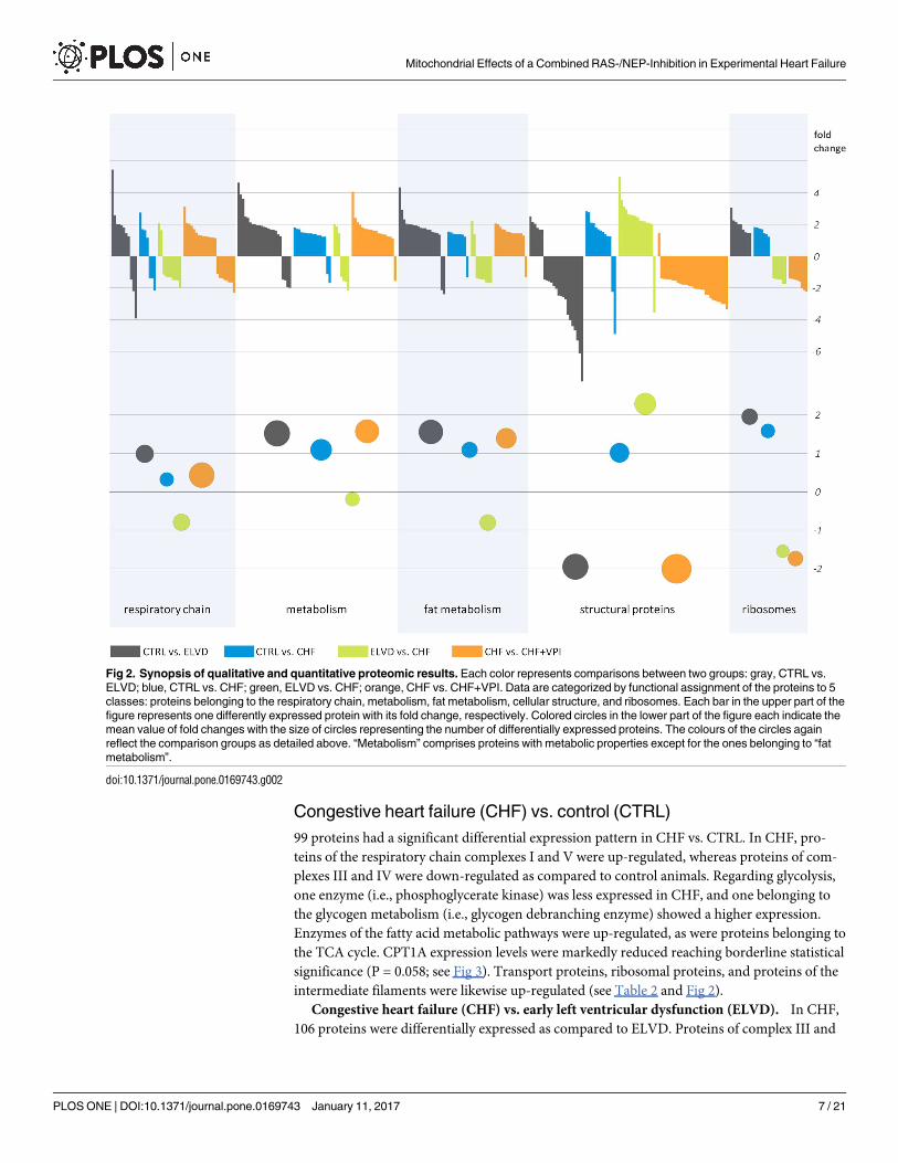
Congestive heart failure (CHF) vs. control (CTRL)
99 proteins had a significant differential expression pattern in CHF vs. CTRL. In CHF, pro-
teins of the respiratory chain complexes I and V were up-regulated, whereas proteins of com-
plexes III and IV were down-regulated as compared to control animals. Regarding glycolysis,
one enzyme (i.e., phosphoglycerate kinase) was less expressed in CHF, and one belonging to
the glycogen metabolism (i.e., glycogen debranching enzyme) showed a higher expression.
Enzymes of the fatty acid metabolic pathways were up-regulated, as were proteins belonging to
the TCA cycle. CPT1A expression levels were markedly reduced reaching borderline statistical
significance (P = 0.058; see Fig 3). Transport proteins, ribosomal proteins, and proteins of the
intermediate filaments were likewise up-regulated (see Table 2 and Fig 2).
Congestive heart failure (CHF) vs. early left ventricular dysfunction (ELVD). In CHF,
106 proteins were differentially expressed as compared to ELVD. Proteins of complex III and
Fig 2. Synopsis of qualitative and quantitative proteomic results. Each color represents comparisons between two groups: gray, CTRL vs.
ELVD; blue, CTRL vs. CHF; green, ELVD vs. CHF; orange, CHF vs. CHF+VPI. Data are categorized by functional assignment of the proteins to 5
classes: proteins belonging to the respiratory chain, metabolism, fat metabolism, cellular structure, and ribosomes. Each bar in the upper part of the
figure represents one differently expressed protein with its fold change, respectively. Colored circles in the lower part of the figure each indicate the
mean value of fold changes with the size of circles representing the number of differentially expressed proteins. The colours of the circles again
reflect the comparison groups as detailed above. “Metabolism” comprises proteins with metabolic properties except for the ones belonging to “fat
metabolism”.
doi:10.1371/journal.pone.0169743.g002
Mitochondrial Effects of a Combined RAS-/NEP-Inhibition in Experimental Heart Failure
PLOS ONE | DOI:10.1371/journal.pone.0169743 January 11, 2017 7 / 21
V were uniformly down-regulated, as were enzymes of fatty acid metabolism. In contrast, glu-
cose metabolizing proteins were higher expressed in CHF. CPT1A did not show an expression
difference between CHF and ELVD (see Fig 3). TCA and ribosomal proteins were down-regu-
lated, but proteins belonging to intermediate filaments were higher expressed in CHF (see
Table 2 and Fig 2).
Fig 3. Protein expression level of carnitine palmitoyltransferase 1 A normalized to the expression of the
outer mitochondrial membrane protein VDAC.
doi:10.1371/journal.pone.0169743.g003
Table 2. Synopsis of different pathway regulation patterns.
Pathway ELVD vs. CTRL CHF vs. CTRL CHF vs. ELVD CHF-VPI vs. CHF
Respiratory chain complexes
■ Complex I " " #
■ Complex II "
■ Complex III " # # "
■ Complex IV # #
■ Complex V " " # "
TCA " " #
Glucose metabolism # # / " " ~
Fatty acid metabolism " " # "
Intermediate filaments # " " #
Ribosomal proteins " " # #
Transport proteins " "
Differentially expressed proteins (total) 199 99 106 223
doi:10.1371/journal.pone.0169743.t002
Mitochondrial Effects of a Combined RAS-/NEP-Inhibition in Experimental Heart Failure
PLOS ONE | DOI:10.1371/journal.pone.0169743 January 11, 2017 8 / 21
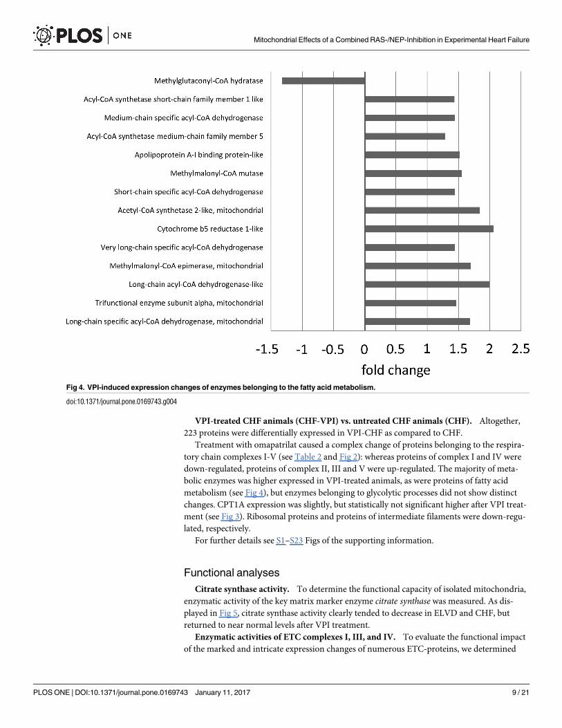
VPI-treated CHF animals (CHF-VPI) vs. untreated CHF animals (CHF). Altogether,
223 proteins were differentially expressed in VPI-CHF as compared to CHF.
Treatment with omapatrilat caused a complex change of proteins belonging to the respira-
tory chain complexes I-V (see Table 2 and Fig 2): whereas proteins of complex I and IV were
down-regulated, proteins of complex II, III and V were up-regulated. The majority of meta-
bolic enzymes was higher expressed in VPI-treated animals, as were proteins of fatty acid
metabolism (see Fig 4), but enzymes belonging to glycolytic processes did not show distinct
changes. CPT1A expression was slightly, but statistically not significant higher after VPI treat-
ment (see Fig 3). Ribosomal proteins and proteins of intermediate filaments were down-regu-
lated, respectively.
For further details see S1–S23 Figs of the supporting information.
Functional analyses
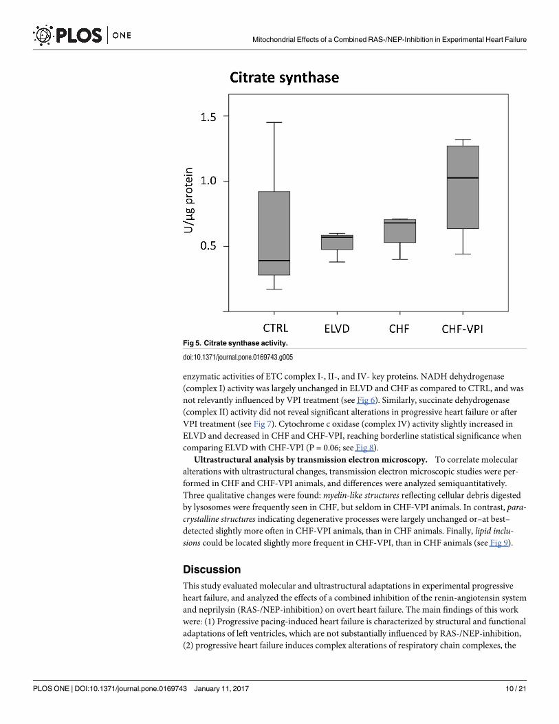
Citrate synthase activity. To determine the functional capacity of isolated mitochondria,
enzymatic activity of the key matrix marker enzyme citrate synthase was measured. As dis-
played in Fig 5, citrate synthase activity clearly tended to decrease in ELVD and CHF, but
returned to near normal levels after VPI treatment.
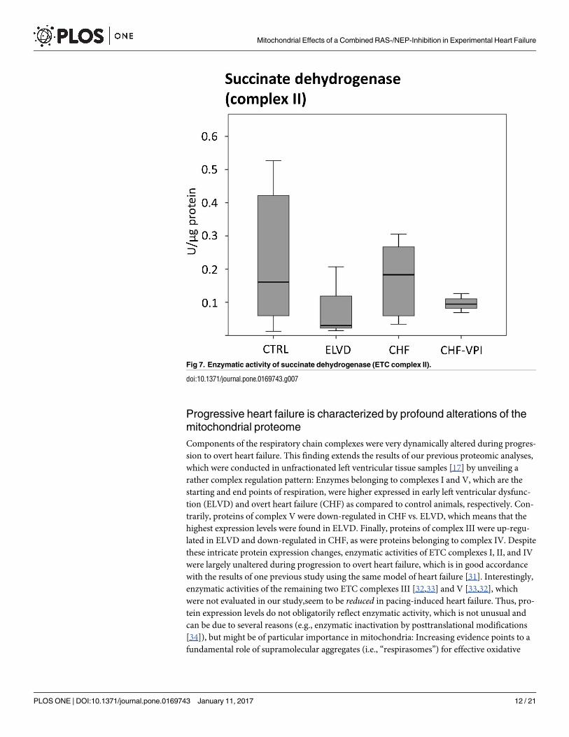
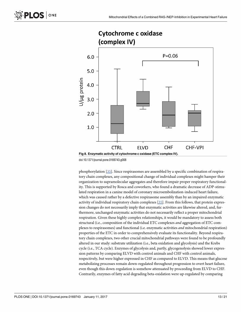
Enzymatic activities of ETC complexes I, III, and IV. To evaluate the functional impact
of the marked and intricate expression changes of numerous ETC-proteins, we determined
Fig 4. VPI-induced expression changes of enzymes belonging to the fatty acid metabolism.
doi:10.1371/journal.pone.0169743.g004
Mitochondrial Effects of a Combined RAS-/NEP-Inhibition in Experimental Heart Failure
PLOS ONE | DOI:10.1371/journal.pone.0169743 January 11, 2017 9 / 21
enzymatic activities of ETC complex I-, II-, and IV- key proteins. NADH dehydrogenase
(complex I) activity was largely unchanged in ELVD and CHF as compared to CTRL, and was
not relevantly influenced by VPI treatment (see Fig 6). Similarly, succinate dehydrogenase
(complex II) activity did not reveal significant alterations in progressive heart failure or after
VPI treatment (see Fig 7). Cytochrome c oxidase (complex IV) activity slightly increased in
ELVD and decreased in CHF and CHF-VPI, reaching borderline statistical significance when
comparing ELVD with CHF-VPI (P = 0.06; see Fig 8).
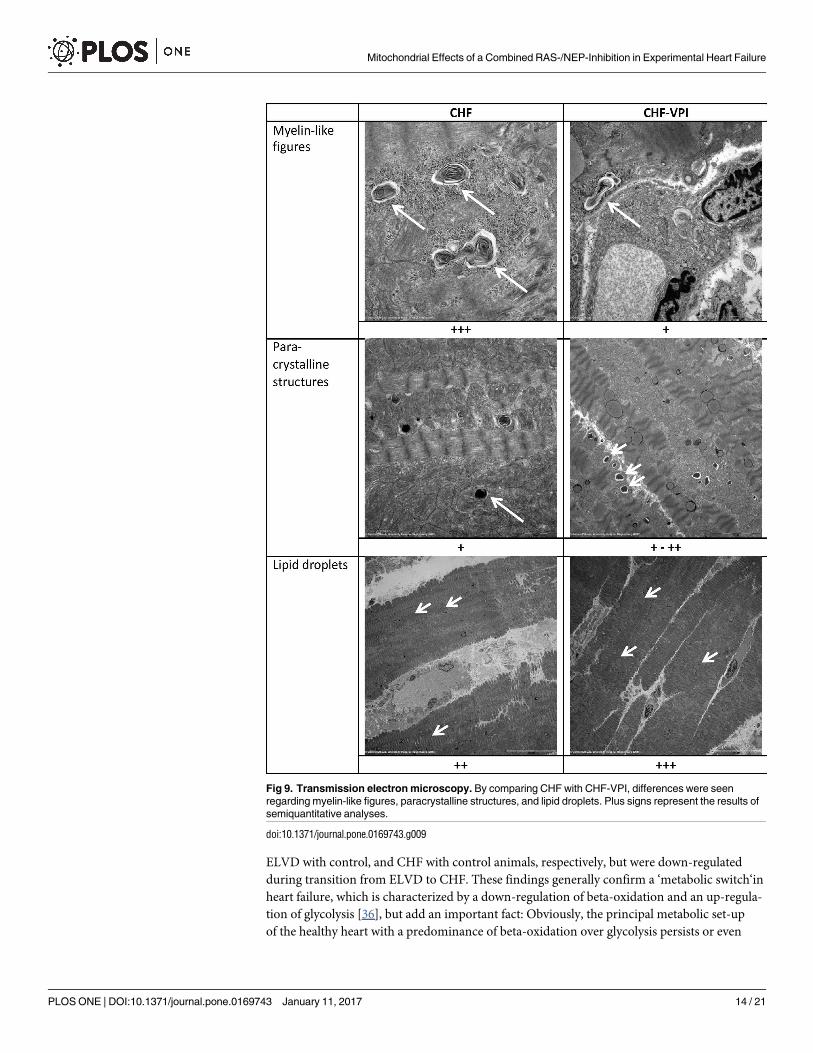
Ultrastructural analysis by transmission electron microscopy. To correlate molecular
alterations with ultrastructural changes, transmission electron microscopic studies were per-
formed in CHF and CHF-VPI animals, and differences were analyzed semiquantitatively.
Three qualitative changes were found: myelin-like structures reflecting cellular debris digested
by lysosomes were frequently seen in CHF, but seldom in CHF-VPI animals. In contrast, para-crystalline structures indicating degenerative processes were largely unchanged or–at best–
detected slightly more often in CHF-VPI animals, than in CHF animals. Finally, lipid inclu-sions could be located slightly more frequent in CHF-VPI, than in CHF animals (see Fig 9).
Discussion
This study evaluated molecular and ultrastructural adaptations in experimental progressive
heart failure, and analyzed the effects of a combined inhibition of the renin-angiotensin system
and neprilysin (RAS-/NEP-inhibition) on overt heart failure. The main findings of this work
were: (1) Progressive pacing-induced heart failure is characterized by structural and functional
adaptations of left ventricles, which are not substantially influenced by RAS-/NEP-inhibition,
(2) progressive heart failure induces complex alterations of respiratory chain complexes, the
Fig 5. Citrate synthase activity.
doi:10.1371/journal.pone.0169743.g005
Mitochondrial Effects of a Combined RAS-/NEP-Inhibition in Experimental Heart Failure
PLOS ONE | DOI:10.1371/journal.pone.0169743 January 11, 2017 10 / 21
TCA cycle and key metabolic processes, and (3) a combined inhibition of the renin-angioten-
sin system and neprilysin intricately influences the composition of all respiratory chain com-
plexes, enhances the normal substrate utilizing pathway, and preserves the otherwise reduced
citrate synthase activity. Furthermore, autophagolytic processes seem to be markedly reduced
upon RAS-/NEP-inhibition.
RAS-/NEP-inhibition does not substantially influence left ventricular
structural remodeling
Progressive tachycardia-induced heart failure was characterized by structural and functional
adaptations, which are also hallmarks of the human disease [28,29]. Interestingly, left ventricu-lar remodeling was not substantially influenced by combined RAS-/NEP-inhibition in this
study, whereas left atrial remodeling was markedly attenuated in our previous work investigat-
ing omapatrilat in the same animal model [15]. It is noteworthy that very similar findings were
recently reported for the RAS-/NEP-inhibitor LCZ696 in the PARAMOUNT trial [30], which
compared LCZ696 to valsartan in patients with HFPEF (i.e., heart failure with preserved ejec-
tion fraction): in this study parameters of left atrial remodeling were significantly improved
after 36 weeks, whereas indicators for left ventricular remodeling remained unchanged. From
this follows that a combined RAS-/NEP-inhibition seems to influence left atrial rather than left
ventricular structural remodeling, whereas profound molecular alterations are also evident in
left ventricles.
Fig 6. Enzymatic activity of NADH dehydrogenase (ETC complex I).
doi:10.1371/journal.pone.0169743.g006
Mitochondrial Effects of a Combined RAS-/NEP-Inhibition in Experimental Heart Failure
PLOS ONE | DOI:10.1371/journal.pone.0169743 January 11, 2017 11 / 21
Progressive heart failure is characterized by profound alterations of the
mitochondrial proteome
Components of the respiratory chain complexes were very dynamically altered during progres-
sion to overt heart failure. This finding extends the results of our previous proteomic analyses,
which were conducted in unfractionated left ventricular tissue samples [17] by unveiling a
rather complex regulation pattern: Enzymes belonging to complexes I and V, which are the
starting and end points of respiration, were higher expressed in early left ventricular dysfunc-
tion (ELVD) and overt heart failure (CHF) as compared to control animals, respectively. Con-
trarily, proteins of complex V were down-regulated in CHF vs. ELVD, which means that the
highest expression levels were found in ELVD. Finally, proteins of complex III were up-regu-
lated in ELVD and down-regulated in CHF, as were proteins belonging to complex IV. Despite
these intricate protein expression changes, enzymatic activities of ETC complexes I, II, and IV
were largely unaltered during progression to overt heart failure, which is in good accordance
with the results of one previous study using the same model of heart failure [31]. Interestingly,
enzymatic activities of the remaining two ETC complexes III [32,33] and V [33,32], which
were not evaluated in our study,seem to be reduced in pacing-induced heart failure. Thus, pro-
tein expression levels do not obligatorily reflect enzymatic activity, which is not unusual and
can be due to several reasons (e.g., enzymatic inactivation by posttranslational modifications
[34]), but might be of particular importance in mitochondria: Increasing evidence points to a
fundamental role of supramolecular aggregates (i.e., “respirasomes”) for effective oxidative
Fig 7. Enzymatic activity of succinate dehydrogenase (ETC complex II).
doi:10.1371/journal.pone.0169743.g007
Mitochondrial Effects of a Combined RAS-/NEP-Inhibition in Experimental Heart Failure
PLOS ONE | DOI:10.1371/journal.pone.0169743 January 11, 2017 12 / 21
phosphorylation [35]. Since respirasomes are assembled by a specific combination of respira-
tory chain complexes, any compositional change of individual complexes might hamper their
organization to supramolecular aggregates and therefore impair proper respiratory functional-
ity. This is supported by Rosca and coworkers, who found a dramatic decrease of ADP-stimu-
lated respiration in a canine model of coronary microembolization-induced heart failure,
which was caused rather by a defective respirasome assembly than by an impaired enzymatic
activity of individual respiratory chain complexes [35]. From this follows, that protein expres-
sion changes do not necessarily imply that enzymatic activities are likewise altered, and, fur-
thermore, unchanged enzymatic activities do not necessarily reflect a proper mitochondrial
respiration. Given these highly complex relationships, it would be mandatory to assess both
structural (i.e., composition of the individual ETC complexes and aggregation of ETC com-
plexes to respirasomes) and functional (i.e. enzymatic activities and mitochondrial respiration)
properties of the ETC in order to comprehensively evaluate its functionality. Beyond respira-
tory chain complexes, two other crucial mitochondrial pathways were found to be profoundly
altered in our study: substrate utilization (i.e., beta oxidation and glycolysis) and the Krebs
cycle (i.e., TCA cycle). Enzymes of glycolysis and, partly, glycogenolysis showed lower expres-
sion patterns by comparing ELVD with control animals and CHF with control animals,
respectively, but were higher expressed in CHF as compared to ELVD. This means that glucose
metabolizing processes remain down-regulated throughout progression to overt heart failure,
even though this down-regulation is somehow attenuated by proceeding from ELVD to CHF.
Contrarily, enzymes of fatty acid degrading beta-oxidation were up-regulated by comparing
Fig 8. Enzymatic activity of cytochrome c oxidase (ETC complex IV).
doi:10.1371/journal.pone.0169743.g008
Mitochondrial Effects of a Combined RAS-/NEP-Inhibition in Experimental Heart Failure
PLOS ONE | DOI:10.1371/journal.pone.0169743 January 11, 2017 13 / 21
ELVD with control, and CHF with control animals, respectively, but were down-regulated
during transition from ELVD to CHF. These findings generally confirm a ‘metabolic switch‘in
heart failure, which is characterized by a down-regulation of beta-oxidation and an up-regula-
tion of glycolysis [36], but add an important fact: Obviously, the principal metabolic set-up
of the healthy heart with a predominance of beta-oxidation over glycolysis persists or even
Fig 9. Transmission electron microscopy. By comparing CHF with CHF-VPI, differences were seen
regarding myelin-like figures, paracrystalline structures, and lipid droplets. Plus signs represent the results of
semiquantitative analyses.
doi:10.1371/journal.pone.0169743.g009
Mitochondrial Effects of a Combined RAS-/NEP-Inhibition in Experimental Heart Failure
PLOS ONE | DOI:10.1371/journal.pone.0169743 January 11, 2017 14 / 21
increases throughout progression to heart failure, and is just contrarily modulated as soon
as LV dysfunction reaches its terminal stage. Similarly, components of the TCA cycle were
up-regulated in ELVD and CHF as compared to control animals, but were down-regulated in
CHF vs. ELVD indicating a likewise attenuation of this persistently up-regulated pathway in
later stages of the disease. Interestingly, enzymatic activity of citrate synthase, which catalyzes
the first step of the Krebs cycle in a pace-making fashion, was reduced in both ELVD and
CHF. Against this background it would be tempting to speculate that the initial up-regulation
of TCA cycle enzymes would have to compensate for the reduced substrate flow through the
enzymatic starting point of this pathway representing a mechanism which is exhausted with
further progression to overt heart failure. This could be supported by Dodd and coworkers,
who investigated Krebs cycle activity in a model of myocardial-infarction induced heart failure
by demonstrating a functional TCA cycle impairment, which began to correlate with the
degree of cardiac dysfunction not until 6 weeks after induction of myocardial infarction [37].
Thus, in the early stages of heart failure compensatory mechanisms could probably counterbal-
ance the reduced citrate synthase activity, which was also seen in the study of Dodd et al. [37],
but this could be exhausted in later stages.
Combined RAS-/NEP-inhibition has profound impact on mitochondrial
key pathways
It is noteworthy that the highest number of differentially expressed proteins was found by
comparing the CHF-VPI with the CHF group, which underlines the far-reaching impact of
RAS-/NEP-inhibition on the mitochondrial molecular setup. In detail, as compared to CHF
animals, RAS-/NEP-inhibition did affect three central mitochondrial pathways (i.e., electron
transfer chain (ETC), substrate utilization, and TCA cycle), and severely influenced autopha-
golytic processes. Regarding ETC, treatment with omapatrilat reduced the expression of pro-
teins belonging to complexes I and IV, and increased components of complexes II, III, and V.
This partly reversed the adaptations of complexes I and III (which are main sources of reactive
oxygen species in mitochondria [38]) in progressive heart failure and aggravated those of com-
plexes IV and V. Interestingly, a deficiency of complex II and III has been linked to human
dilated cardiomyopathy [39] and ischemic cardiomyopathy [40], respectively, so a restoration
of complex II and III content might probably counteract LV dysfunction. Besides RAS-/NEP-
inhibition, also TNF-alpha blockade by etanercept [32], cardiac resynchronization by biventri-
cular pacing [41], treatment with resveratrol in doxorubicin-induced heart failure [42], or
application of trimetazidine [43] were likewise followed by ETC adaptations, thereby indicat-
ing a therapeutic involvement of the ATP-generating apparatus in these instances. But given
the intricate processes around oxidative phosphorylation, which include the complex func-
tionality of supramolecular aggregates (as mentioned above), the true functional relevance of
these findings (e.g., reduction of oxidative stress or augmented efficiency of ATP generation)
is difficult to delineate and has clearly to be evaluated in focused future experiments.
Beyond ETC adaptations, RAS-/NEP-inhibition increased the expression of proteins
involved in the metabolism of free fatty acids (FFA), but did not relevant influence glycolytic
pathways in our study. This might counteract the relative down-regulation of beta-oxidation
in progressive heart failure and suggests some restoration of the normal substrate utilization
processes. Interestingly, a similar effect has been shown for failing human hearts which have
been unloaded by left ventricular assist devices [44] or for recovering canine hearts after halt-
ing the tachypacing stimulus [45], so this metabolic change might indicate some kind of car-
diac regeneration by RAS-/NEP-inhibition. Similarly, activity of the key TCA enzyme citrate
synthase, which was reduced in both ELVD and CHF, clearly tended to normal levels in
Mitochondrial Effects of a Combined RAS-/NEP-Inhibition in Experimental Heart Failure
PLOS ONE | DOI:10.1371/journal.pone.0169743 January 11, 2017 15 / 21
omapatrilat-treated animals, even though protein expression of TCA components per se was
not altered. A likewise reconstitution of enzymatic activity was again seen in unloaded human
[44] and recovering canine hearts [45], so this omapatrilat-induced change might in turn mir-
ror some extent of molecular reverse remodeling.
Finally, we could demonstrate a remarkable reduction of myelin-like figures along with an
increase of paracrystalline structures in cardiomyocytes of omapatrilat-treated animals,
whereas lipid droplets, which were already highly expressed in CHF animals, did show only
marginal changes. Myelin-like figures, which were also found in cardiomyopathic hamsters in
addition to signs of autophagic cell death [46] or in cyclophosphamide-damaged cardiomyo-
cytes [47], are supposed to be rather late products of autophagic lysosomal digestion [48]. In
contrast, lipid droplets seem to rapidly evolve in damaged skeletal muscle cells [48] and are
indicative for a disturbance of lipid metabolism leading to lipid accumulation in the heart [49].
Lastly, paracrystalline structures have been linked to mitochondrial diseases [50] and could be
a sign of degeneration, though their significance might not easily be interpreted in our study.
Taken together, these omapatrilat-induced ultrastructural adaptations most probably point to
a beneficial decrease of otherwise detrimental autophagic processes, which usually accompany
progressive left ventricular dysfunction.
Limitations
Our work might have some limitations: Firstly, even though we conducted our experiments by
using a cutting edge proteomic approach with some additional functional tests, this was never-
theless a classical proteomic study, so we cannot clearly specify the functional relevance of our
findings. Otherwise, this was actually beyond the scope of our work, which was rather con-
ceived to depict a global view of molecular alterations in properly isolated cardiac mitochon-
dria thereby providing a starting point for subsequent functional analyses. Secondly, the
combined RAS-/NEP-inhibition by omapatrilat significantly lowered blood pressure, which is
a well known effect of this compound. Consequently, one might ascribe the beneficial action
of omapatrilat to this hemodynamic property rather than to an intrinsic modulatory effect on
neurohumoral activation. That being said very recent evidence with the closely related sub-
stance LCZ696, which blocks neprilysin and the angiotensin receptor instead of the angioten-
sin converting enzyme, rather argue for an intrinsic neurohumoral modulatory action as a
cause of the beneficial drug effects [51]. Facing these hemodynamic side effects of a combined
RAS-/NEP-inhibition, but also considering the current standard of care (which is represented
by an ACE inhibitor much more than by a placebo preparation), an active comparator such as
enalapril could be advantageous. Thirdly, we used the vasopeptidase inhibitor (VPI) omapatri-
lat instead of the newer angiotensin receptor neprilysin inhibitor (ARNI) LCZ696 to execute a
combined RAS-/NEP-inhibition, since the latter one was not available at the beginning of our
experiments. Given the closely related modes of action, one could argue for a class effect,
which means that our results should be transferable. But this has clearly to be confirmed as
soon as LCZ696 becomes available for research purposes.
Conclusion
Progressive pacing-induced left ventricular dysfunction is characterized by profound constitu-
tional alterations of the electron transfer chain, the TCA cycle, and substrate utilization pro-
cesses. These adaptations are at least partly reversed by RAS-/NEP-inhibition, which also
seems to reduce detrimental subcellular degeneration in cardiomyocytes. Thus, RAS-/NEP-
inhibition unfolds beneficial effects on energetically relevant pathways in a model of progres-
sive pacing-induced heart failure.
Mitochondrial Effects of a Combined RAS-/NEP-Inhibition in Experimental Heart Failure
PLOS ONE | DOI:10.1371/journal.pone.0169743 January 11, 2017 16 / 21
Supporting Information
S1 Fig. Mitochondrial ETC proteins in CTRL vs. ELVD.
(TIF)
S2 Fig. Metabolic enzymes in CTRL vs. ELVD.
(TIF)
S3 Fig. Intermediate filaments in CTRL vs. ELVD.
(TIF)
S4 Fig. Fat metabolism in CTRL vs. ELVD.
(TIF)
S5 Fig. Ribosomal proteins in CTRL vs. ELVD.
(TIF)
S6 Fig. Transport proteins in CTRL vs. ELVD.
(TIF)
S7 Fig. Mitochondrial ETC proteins in CTRL vs.CHF.
(TIF)
S8 Fig. Metabolic enzymes in CTRL vs. CHF.
(TIF)
S9 Fig. Intermediate filaments in CTRL vs. CHF.
(TIF)
S10 Fig. Fat metabolism in CTRL vs. CHF.
(TIF)
S11 Fig. Ribosomal proteins in CTRL vs. CHF.
(TIF)
S12 Fig. Transport proteins in CTRL vs. CHF.
(TIF)
S13 Fig. Mitochondrial ETC proteins in CHF vs. CHF+VPI.
(TIF)
S14 Fig. Metabolic enzymes in CHF vs. CHF+VPI.
(TIF)
S15 Fig. Intermediate filaments in CHF vs. CHF+VPI.
(TIF)
S16 Fig. Ribosomal proteins in CHF vs. CHF+VPI.
(TIF)
S17 Fig. Transport proteins in CHF vs. CHF+VPI.
(TIF)
S18 Fig. Mitochondrial ETC proteins in ELVD vs. CHF.
(TIF)
S19 Fig. Metabolic enzymes in ELVD vs. CHF.
(TIF)
Mitochondrial Effects of a Combined RAS-/NEP-Inhibition in Experimental Heart Failure
PLOS ONE | DOI:10.1371/journal.pone.0169743 January 11, 2017 17 / 21
S20 Fig. Intermediate filaments in ELVD vs. CHF.
(TIF)
S21 Fig. Fat metabolism in ELVD vs. CHF.
(TIF)
S22 Fig. Ribosomal proteins in ELVD vs. CHF.
(TIF)
S23 Fig. Transport proteins in ELVD vs. CHF.
(TIF)
Acknowledgments
This work was supported by an institutional research grant (ReForM-A) of the University
Hospital Regensburg. We greatly appreciate the outstanding technical assistance of Ms. Ingrid
Winkel. This paper contains parts of the MD thesis of Ms. Laura Grois.
Author Contributions
Conceptualization: LG LM AL CB.
Formal analysis: LG JH CB.
Funding acquisition: CB.
Investigation: LG JH JR JS PS AD.
Methodology: LG AL CB.
Project administration: CB.
Supervision: LM AL CB.
Validation: JH CB.
Visualization: LG JH CB.
Writing – original draft: LG JH CJ MR CB.
Writing – review & editing: LG JH MR AL CB.
References1. Zucker IH, Xiao L, Haack KKV (2014) The central renin-angiotensin system and sympathetic nerve
activity in chronic heart failure. Clinical science (London, England: 1979) 126 (10): 695–706.
2. Zhang DY, Anderson AS (2014) The sympathetic nervous system and heart failure. Cardiology clinics
32 (1): 33–45, vii. doi: 10.1016/j.ccl.2013.09.010 PMID: 24286577
3. Lymperopoulos A, Rengo G, Koch WJ (2013) Adrenergic nervous system in heart failure: pathophysiol-
ogy and therapy. Circulation research 113 (6): 739–753. doi: 10.1161/CIRCRESAHA.113.300308
PMID: 23989716
4. (1999) The Cardiac Insufficiency Bisoprolol Study II (CIBIS-II): a randomised trial. Lancet (London,
England) 353 (9146): 9–13.
5. Packer M, Coats AJ, Fowler MB, Katus HA, Krum H, Mohacsi P et al. (2001) Effect of carvedilol on sur-
vival in severe chronic heart failure. The New England journal of medicine 344 (22): 1651–1658. doi:
10.1056/NEJM200105313442201 PMID: 11386263
6. (1999) Effect of metoprolol CR/XL in chronic heart failure: Metoprolol CR/XL Randomised Intervention
Trial in Congestive Heart Failure (MERIT-HF). Lancet (London, England) 353 (9169): 2001–2007.
Mitochondrial Effects of a Combined RAS-/NEP-Inhibition in Experimental Heart Failure
PLOS ONE | DOI:10.1371/journal.pone.0169743 January 11, 2017 18 / 21
7. (1987) Effects of enalapril on mortality in severe congestive heart failure. Results of the Cooperative
North Scandinavian Enalapril Survival Study (CONSENSUS). The CONSENSUS Trial Study Group.
The New England journal of medicine 316 (23): 1429–1435. doi: 10.1056/NEJM198706043162301
PMID: 2883575
8. Go AS, Mozaffarian D, Roger VL, Benjamin EJ, Berry JD, Blaha MJ et al. (2014) Heart disease and
stroke statistics—2014 update: a report from the American Heart Association. Circulation 129 (3): e28–
e292. doi: 10.1161/01.cir.0000441139.02102.80 PMID: 24352519
9. McMurray JJV (2015) Neprilysin inhibition to treat heart failure: a tale of science, serendipity, and sec-
ond chances. European journal of heart failure 17 (3): 242–247. doi: 10.1002/ejhf.250 PMID: 25756942
10. Trindade PT RJL (2001) Vasopeptidase inhibitors: potential role in the treatment of heart failure. Heart
Fail Monit. 2 (1): 2–7. PMID: 12634892
11. Packer M, Califf RM, Konstam MA, Krum H, McMurray JJ, Rouleau JL et al. (2002) Comparison of oma-
patrilat and enalapril in patients with chronic heart failure: the Omapatrilat Versus Enalapril Randomized
Trial of Utility in Reducing Events (OVERTURE). Circulation 106 (8): 920–926. PMID: 12186794
12. McClean DR, Ikram H, Garlick AH, Richards AM, Nicholls MG, Crozier IG (2000) The clinical, cardiac,
renal, arterial and neurohormonal effects of omapatrilat, a vasopeptidase inhibitor, in patients with
chronic heart failure. Journal of the American College of Cardiology 36 (2): 479–486. PMID: 10933361
13. Trippodo NC, Fox M, Monticello TM, Panchal BC, Asaad MM (1999) Vasopeptidase inhibition with oma-
patrilat improves cardiac geometry and survival in cardiomyopathic hamsters more than does ACE inhi-
bition with captopril. Journal of cardiovascular pharmacology 34 (6): 782–790. PMID: 10598120
14. McMurray JJV, Packer M, Desai AS, Gong J, Lefkowitz MP, Rizkala AR et al. (2014) Angiotensin-nepri-
lysin inhibition versus enalapril in heart failure. The New England journal of medicine 371 (11): 993–
1004. doi: 10.1056/NEJMoa1409077 PMID: 25176015
15. Birner C, Ulucan C, Bratfisch M, Gotz T, Dietl A, Schweda F et al. (2012) Antihypertrophic effects of
combined inhibition of the renin-angiotensin system (RAS) and neutral endopeptidase (NEP) in pro-
gressive, tachycardia-induced experimental heart failure. Naunyn-Schmiedeberg’s archives of pharma-
cology 385 (11): 1117–1125. doi: 10.1007/s00210-012-0791-6 PMID: 22895639
16. Marın-Garcıa J, Goldenthal MJ (2008) Mitochondrial centrality in heart failure. Heart failure reviews 13
(2): 137–150. doi: 10.1007/s10741-007-9079-1 PMID: 18185992
17. Birner C, Dietl A, Deutzmann R, Schroder J, Schmid P, Jungbauer C et al. (2012) Proteomic profiling
implies mitochondrial dysfunction in tachycardia-induced heart failure. Journal of cardiac failure 18 (8):
660–673. doi: 10.1016/j.cardfail.2012.06.418 PMID: 22858083
18. Dietl A, Winkel I, Deutzmann R, Schroder J, Hupf J, Riegger G et al. (2014) Interatrial differences of
basal molecular set-up and changes in tachycardia-induced heart failure-a proteomic profiling study.
European journal of heart failure 16 (8): 835–845. doi: 10.1002/ejhf.122 PMID: 25045083
19. Birner C, Hierl S, Dietl A, Hupf J, Jungbauer C, Schmid PM et al. (2014) Experimental heart failure
induces alterations of the lung proteome—insight into molecular mechanisms. Cellular physiology and
biochemistry: international journal of experimental cellular physiology, biochemistry, and pharmacology
33 (3): 692–704.
20. Birner C, Husser O, Jeron A, Rihm M, Fredersdorf S, Resch M et al. (2012) Differential expression of
potassium channels and abnormal conduction in experimental tachycardia-induced heart failure. Nau-
nyn-Schmiedeberg’s archives of pharmacology 385 (5): 473–480. doi: 10.1007/s00210-011-0723-x
PMID: 22311348
21. Birner CM, Ulucan C, Fredersdorf S, Rihm M, Lowel H, Stritzke J et al. (2007) Head-to-head compari-
son of BNP and IL-6 as markers of clinical and experimental heart failure: Superiority of BNP. Cytokine
40 (2): 89–97. doi: 10.1016/j.cyto.2007.08.009 PMID: 17920926
22. Luchner A, Muders F, Dietl O, Friedrich E, Blumberg F, Protter AA et al. (2001) Differential expression
of cardiac ANP and BNP in a rabbit model of progressive left ventricular dysfunction. Cardiovascular
research 51 (3): 601–607. PMID: 11476751
23. Boehm EA, Jones BE, Radda GK, Veech RL, Clarke K (2001) Increased uncoupling proteins and
decreased efficiency in palmitate-perfused hyperthyroid rat heart. American journal of physiology. Heart
and circulatory physiology 280 (3): H977–83. PMID: 11179038
24. Srere PA (1973) [1] Citrate synthase. In: Lowenstein JM, editor. Citric acid cycle. New York: Academic
Press. pp. 3–11.
25. Kramer KA, Oglesbee D, Hartman SJ, Huey J, Anderson B, Magera MJ et al. (2005) Automated spec-
trophotometric analysis of mitochondrial respiratory chain complex enzyme activities in cultured skin
fibroblasts. Clinical chemistry 51 (11): 2110–2116. doi: 10.1373/clinchem.2005.050146 PMID:
16141288
Mitochondrial Effects of a Combined RAS-/NEP-Inhibition in Experimental Heart Failure
PLOS ONE | DOI:10.1371/journal.pone.0169743 January 11, 2017 19 / 21
26. Bluemlein K, Ralser M (2011) Monitoring protein expression in whole-cell extracts by targeted label-
and standard-free LC-MS/MS. Nature protocols 6 (6): 859–869. doi: 10.1038/nprot.2011.333 PMID:
21637204
27. Reinders Y, Voller D, Bosserhoff A, Oefner PJ, Reinders J (2016) Testing Suitability of Cell Cultures for
SILAC-Experiments Using SWATH-Mass Spectrometry. Methods in molecular biology (Clifton, N.J.)
1394: 101–108.
28. Cioffi G, Tarantini L, Feo S de, Pulignano G, Del Sindaco D, Stefenelli C et al. (2004) Dilated versus
nondilated cardiomyopathy in the elderly population treated with guideline-based medical therapy for
systolic chronic heart failure. Journal of cardiac failure 10 (6): 481–489. PMID: 15599838
29. Andersson B, Caidahl K, Waagstein F (1995) An echocardiographic evaluation of patients with idio-
pathic heart failure. Chest 107 (3): 680–689. PMID: 7874937
30. Solomon SD, Zile M, Pieske B, Voors A, Shah A, Kraigher-Krainer E et al. (2012) The angiotensin
receptor neprilysin inhibitor LCZ696 in heart failure with preserved ejection fraction: a phase 2 double-
blind randomised controlled trial. Lancet (London, England) 380 (9851): 1387–1395.
31. Marın-Garcıa J, Goldenthal MJ, Moe GW (2001) Abnormal cardiac and skeletal muscle mitochondrial
function in pacing-induced cardiac failure. Cardiovascular research 52 (1): 103–110. PMID: 11557238
32. Moe GW, Marin-Garcia J, Konig A, Goldenthal M, Lu X, Feng Q (2004) In vivo TNF-alpha inhibition
ameliorates cardiac mitochondrial dysfunction, oxidative stress, and apoptosis in experimental heart
failure. Am J Physiol Heart Circ Physiol. 287 (4): H1813–20. doi: 10.1152/ajpheart.00036.2004 PMID:
15205165
33. Marın-Garcıa J, Goldenthal MJ, Damle S, Pi Y, Moe GW (2009) Regional distribution of mitochondrial
dysfunction and apoptotic remodeling in pacing-induced heart failure. Journal of cardiac failure 15 (8):
700–708. doi: 10.1016/j.cardfail.2009.04.010 PMID: 19786259
34. Wende AR (2015) Post-translational modifications of the cardiac proteome in diabetes and heart failure.
Proteomics. Clinical applications.
35. Rosca MG, Vazquez EJ, Kerner J, Parland W, Chandler MP, Stanley W et al. (2008) Cardiac mitochon-
dria in heart failure: decrease in respirasomes and oxidative phosphorylation. Cardiovascular research
80 (1): 30–39. doi: 10.1093/cvr/cvn184 PMID: 18710878
36. Doenst T, Nguyen TD, Abel ED (2013) Cardiac metabolism in heart failure: implications beyond ATP
production. Circulation research 113 (6): 709–724. doi: 10.1161/CIRCRESAHA.113.300376 PMID:
23989714
37. Dodd MS, Atherton HJ, Carr CA, Stuckey DJ, West JA, Griffin JL et al. (2014) Impaired in vivo mitochon-
drial Krebs cycle activity after myocardial infarction assessed using hyperpolarized magnetic resonance
spectroscopy. Circulation. Cardiovascular imaging 7 (6): 895–904. doi: 10.1161/CIRCIMAGING.114.
001857 PMID: 25201905
38. Tsutsui H, Kinugawa S, Matsushima S (2011) Oxidative stress and heart failure. American journal of
physiology. Heart and circulatory physiology 301 (6): H2181–90. doi: 10.1152/ajpheart.00554.2011
PMID: 21949114
39. Jain-Ghai S, Cameron JM, Al Maawali A, Blaser S, MacKay N, Robinson B et al. (2013) Complex II defi-
ciency—a case report and review of the literature. American journal of medical genetics. Part A 161A
(2): 285–294. doi: 10.1002/ajmg.a.35714 PMID: 23322652
40. Marin-Garcia J, Hu Y, Ananthakrishnan R, Pierpont ME, Pierpont GL, Goldenthal MJ (1996) A point
mutation in the cytb gene of cardiac mtDNA associated with complex III deficiency in ischemic cardio-
myopathy. Biochemistry and molecular biology international 40 (3): 487–495. PMID: 8908357
41. Agnetti G, Kaludercic N, Kane LA, Elliott ST, Guo Y, Chakir K et al. (2010) Modulation of mitochondrial
proteome and improved mitochondrial function by biventricular pacing of dyssynchronous failing hearts.
Circulation. Cardiovascular genetics 3 (1): 78–87. doi: 10.1161/CIRCGENETICS.109.871236 PMID:
20160199
42. Dolinsky VW, Rogan KJ, Sung MM, Zordoky BN, Haykowsky MJ, Young ME et al. (2013) Both aerobic
exercise and resveratrol supplementation attenuate doxorubicin-induced cardiac injury in mice. Ameri-
can journal of physiology. Endocrinology and metabolism 305 (2): E243–53. doi: 10.1152/ajpendo.
00044.2013 PMID: 23695218
43. Dedkova EN, Seidlmayer LK, Blatter LA (2013) Mitochondria-mediated cardioprotection by trimetazi-
dine in rabbit heart failure. Journal of molecular and cellular cardiology 59: 41–54. doi: 10.1016/j.yjmcc.
2013.01.016 PMID: 23388837
44. Gupte AA, Hamilton DJ, Cordero-Reyes AM, Youker KA, Yin Z, Estep JD et al. (2014) Mechanical
unloading promotes myocardial energy recovery in human heart failure. Circulation. Cardiovascular
genetics 7 (3): 266–276. doi: 10.1161/CIRCGENETICS.113.000404 PMID: 24825877
Mitochondrial Effects of a Combined RAS-/NEP-Inhibition in Experimental Heart Failure
PLOS ONE | DOI:10.1371/journal.pone.0169743 January 11, 2017 20 / 21
45. Qanud K, Mamdani M, Pepe M, Khairallah RJ, Gravel J, Lei B et al. (2008) Reverse changes in cardiac
substrate oxidation in dogs recovering from heart failure. American journal of physiology. Heart and cir-
culatory physiology 295 (5): H2098–105. doi: 10.1152/ajpheart.00471.2008 PMID: 18820029
46. Miyata S, Takemura G, Kawase Y, Li Y, Okada H, Maruyama R et al. (2006) Autophagic cardiomyocyte
death in cardiomyopathic hamsters and its prevention by granulocyte colony-stimulating factor. The
American journal of pathology 168 (2): 386–397. doi: 10.2353/ajpath.2006.050137 PMID: 16436654
47. Lushnikova EL, Nepomnyashchikh LM, Sviridov EA, Klinnikova MG (2008) Ultrastructural signs of
cyclophosphamide-induced damage to cardiomyocytes. Bulletin of experimental biology and medicine
146 (3): 366–371. PMID: 19240862
48. Duncan CJ (1991) Calcium, oxygen radicals, and cellular damage. Cambridge [England], New York:
Cambridge University Press. 1 online resource (x, 224 p.
49. Goldberg IJ, Trent CM, Schulze PC (2012) Lipid metabolism and toxicity in the heart. Cell metabolism
15 (6): 805–812. doi: 10.1016/j.cmet.2012.04.006 PMID: 22682221
50. Meyers DE, Basha HI, Koenig MK (2013) Mitochondrial cardiomyopathy: pathophysiology, diagnosis,
and management. Texas Heart Institute journal / from the Texas Heart Institute of St. Luke’s Episcopal
Hospital, Texas Children’s Hospital 40 (4): 385–394.
51. Jhund PS, Claggett B, Packer M, Zile MR, Voors AA, Pieske B et al. (2014) Independence of the blood
pressure lowering effect and efficacy of the angiotensin receptor neprilysin inhibitor, LCZ696, in patients
with heart failure with preserved ejection fraction: an analysis of the PARAMOUNT trial. European jour-
nal of heart failure 16 (6): 671–677. doi: 10.1002/ejhf.76 PMID: 24692284
Mitochondrial Effects of a Combined RAS-/NEP-Inhibition in Experimental Heart Failure
PLOS ONE | DOI:10.1371/journal.pone.0169743 January 11, 2017 21 / 21