Embed Size (px)
Citation preview
Colonic polypsColonic polyps
By assistant lecturer: By assistant lecturer: Waleed Waleed FouadFouad
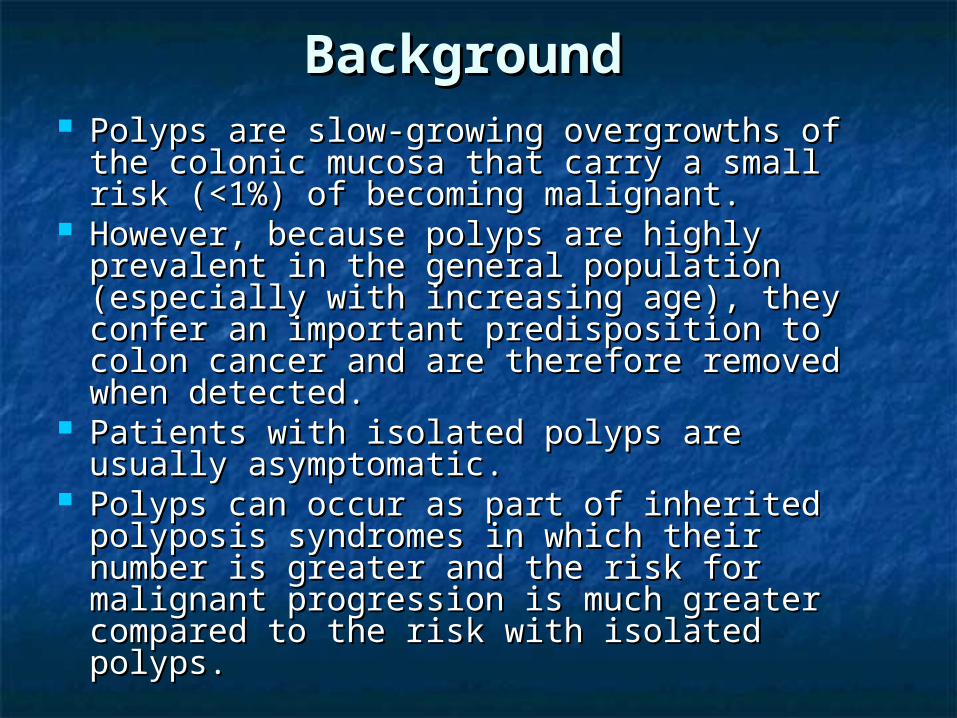
BackgroundBackground Polyps are slow-growing overgrowths of the Polyps are slow-growing overgrowths of the
colonic mucosa that carry a small risk colonic mucosa that carry a small risk (<1%) of becoming malignant. (<1%) of becoming malignant.
However, because polyps are highly However, because polyps are highly prevalent in the general population prevalent in the general population (especially with increasing age), they (especially with increasing age), they confer an important predisposition to colon confer an important predisposition to colon cancer and are therefore removed when cancer and are therefore removed when detected.detected.
Patients with isolated polyps are usually Patients with isolated polyps are usually asymptomatic.asymptomatic.
Polyps can occur as part of inherited Polyps can occur as part of inherited polyposis syndromes in which their number polyposis syndromes in which their number is greater and the risk for malignant is greater and the risk for malignant progression is much greater compared to progression is much greater compared to the risk with isolated polyps. the risk with isolated polyps.
PathophysiologyPathophysiology Colonic polyps, or adenomas, are Colonic polyps, or adenomas, are
benign epithelial neoplasms that arise benign epithelial neoplasms that arise from the epithelial cells lining the colon. from the epithelial cells lining the colon.
Polyps are traditionally divided into 4 Polyps are traditionally divided into 4 groups.groups. Hyperplastic polyps.Hyperplastic polyps. Adenomas.Adenomas. Polyposis syndromes.Polyposis syndromes. Miscellaneous.Miscellaneous.
Hyperplastic polypsHyperplastic polyps Hyperplastic polyps comprise about Hyperplastic polyps comprise about
90% of all polyps and are totally 90% of all polyps and are totally benign protrusions. benign protrusions.
They are usually less than 0.5 cm in They are usually less than 0.5 cm in diameter. diameter.
They most commonly occur in the They most commonly occur in the rectosigmoid region during adulthood.rectosigmoid region during adulthood.
The lesions tend to be smaller than The lesions tend to be smaller than adenomas and do not appear to be adenomas and do not appear to be related to the adenoma-carcinoma related to the adenoma-carcinoma sequence. sequence.
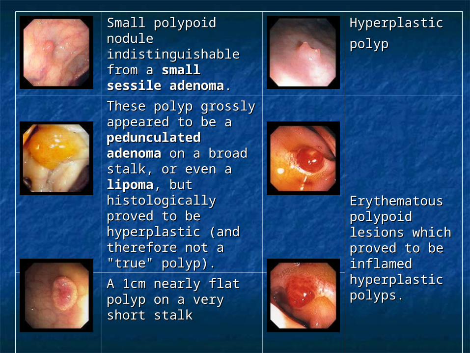
Small polypoid nodule Small polypoid nodule indistinguishable from indistinguishable from a a small sessile small sessile adenomaadenoma. .
Hyperplastic Hyperplastic
polyppolyp
These polyp grossly These polyp grossly appeared to be a appeared to be a pedunculated pedunculated adenomaadenoma on a broad on a broad stalk, or even a stalk, or even a lipomalipoma, but , but histologically proved histologically proved to be hyperplastic to be hyperplastic (and therefore not a (and therefore not a "true" polyp). "true" polyp).
Erythematous Erythematous polypoid lesions polypoid lesions which proved to which proved to be inflamed be inflamed hyperplastic hyperplastic polyps. polyps.
A 1cm nearly flat A 1cm nearly flat polyp on a very short polyp on a very short stalk stalk
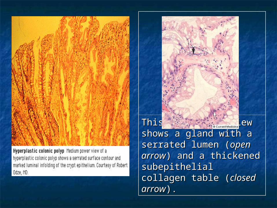
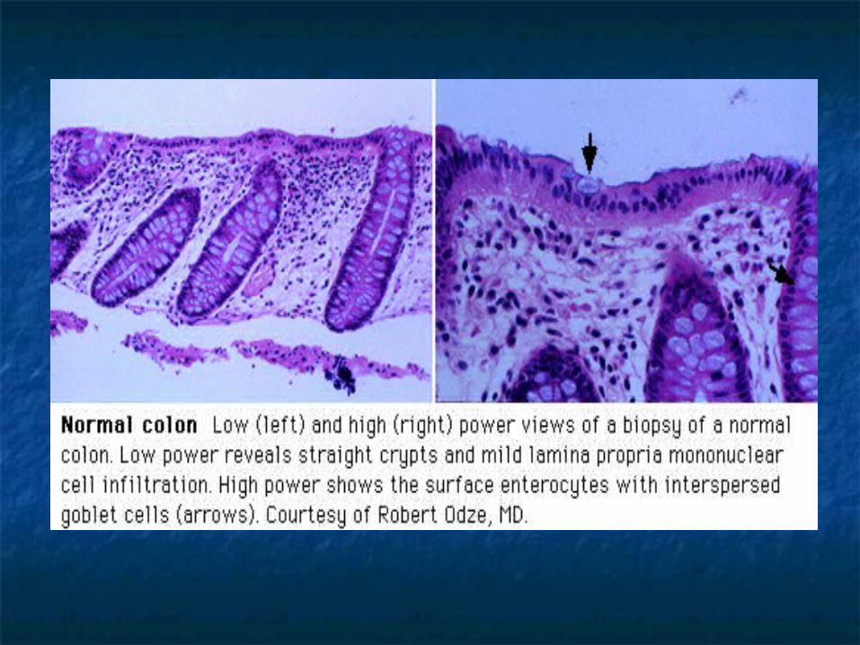
This high-power view This high-power view shows a gland with a shows a gland with a serrated lumen (serrated lumen (open open arrowarrow) and a thickened ) and a thickened subepithelial collagen subepithelial collagen table (table (closed arrowclosed arrow). ).

AdenomasAdenomas
Adenomas comprise approximately 10% of Adenomas comprise approximately 10% of polyps. polyps.
Most (~90%) are small, usually less than Most (~90%) are small, usually less than 1.5 cm in diameter, and have a very small 1.5 cm in diameter, and have a very small potential for malignancy. potential for malignancy.
The remaining 10% of adenomas are The remaining 10% of adenomas are larger than 1.5 cm and have about a 10% larger than 1.5 cm and have about a 10% chance of containing invasive cancer. chance of containing invasive cancer.
Adenomas are traditionally divided into 3 Adenomas are traditionally divided into 3 types: tubular, tubulovillous, and villous. types: tubular, tubulovillous, and villous.
Tubular adenomas are the most common Tubular adenomas are the most common of the 3 types and can be found anywhere of the 3 types and can be found anywhere in the colon.in the colon.
Those with a distinct stalk are Those with a distinct stalk are termed pedunculated; those without termed pedunculated; those without a stalk are termed sessile. a stalk are termed sessile.
The risk of progression to carcinoma The risk of progression to carcinoma is related to the size of the adenoma.is related to the size of the adenoma.
Tubulovillous adenomas are most Tubulovillous adenomas are most commonly found in the rectal area.commonly found in the rectal area.
The degree of villous component of The degree of villous component of these adenomas is correlated with these adenomas is correlated with the risk of progression to carcinoma.the risk of progression to carcinoma.
Villous adenomas most commonly occur Villous adenomas most commonly occur in the rectal area.in the rectal area.
They tend to be larger than the other two They tend to be larger than the other two types; and tend to be nonpedunculated, types; and tend to be nonpedunculated, velvety, or cauliflower-like in appearance. velvety, or cauliflower-like in appearance.
Villous adenomas are associated with the Villous adenomas are associated with the highest morbidity and mortality rates of highest morbidity and mortality rates of all polyps. all polyps.
They can cause hypersecretory They can cause hypersecretory syndromes characterized by hypokalemia syndromes characterized by hypokalemia and profuse mucous discharge and can and profuse mucous discharge and can harbor carcinoma in situ or invasive harbor carcinoma in situ or invasive carcinoma more frequently than other carcinoma more frequently than other adenomas. adenomas.

Sessile Tubular Adenoma Sessile Tubular Adenoma
A small, sessile, A small, sessile, multilobulated lesion multilobulated lesion which proved to be a which proved to be a benign tubular benign tubular adenoma. adenoma.
A smooth sessile A smooth sessile polyp on a broad polyp on a broad base. base.
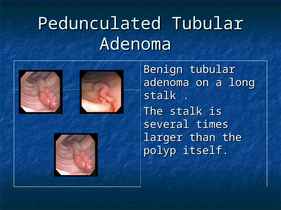
Pedunculated Tubular Pedunculated Tubular Adenoma Adenoma
Benign tubular Benign tubular adenoma on a long adenoma on a long stalk .stalk .
The stalk is several The stalk is several times larger than the times larger than the polyp itself.polyp itself.
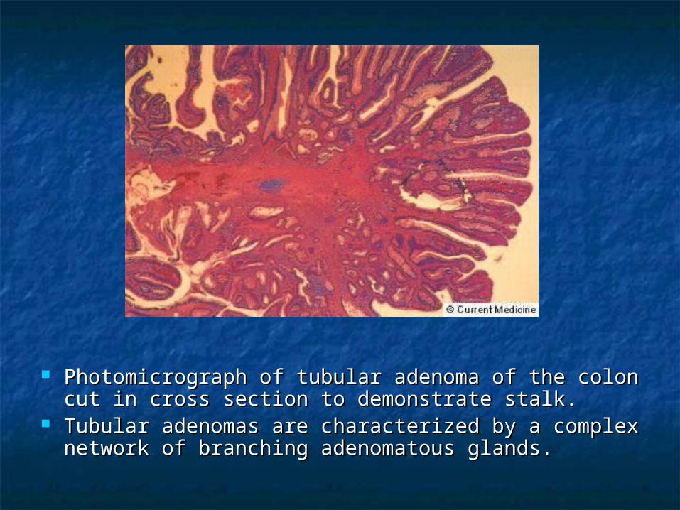
Photomicrograph of tubular adenoma of the colon Photomicrograph of tubular adenoma of the colon cut in cross section to demonstrate stalk. cut in cross section to demonstrate stalk.
Tubular adenomas are characterized by a complex Tubular adenomas are characterized by a complex network of branching adenomatous glands. network of branching adenomatous glands.
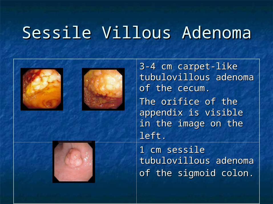
Sessile Villous AdenomaSessile Villous Adenoma
3-4 cm carpet-like 3-4 cm carpet-like tubulovillous adenoma of tubulovillous adenoma of the cecum.the cecum.
The orifice of the The orifice of the appendix is visible in the appendix is visible in the image on the left.image on the left. 1 cm sessile tubulovillous 1 cm sessile tubulovillous adenoma of the sigmoid adenoma of the sigmoid colon.colon.
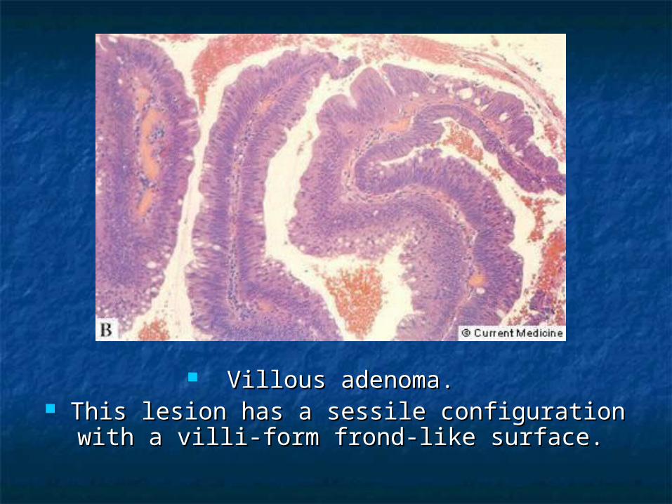
Villous adenoma. Villous adenoma. This lesion has a sessile configuration with This lesion has a sessile configuration with
a villi-form frond-like surface. a villi-form frond-like surface.
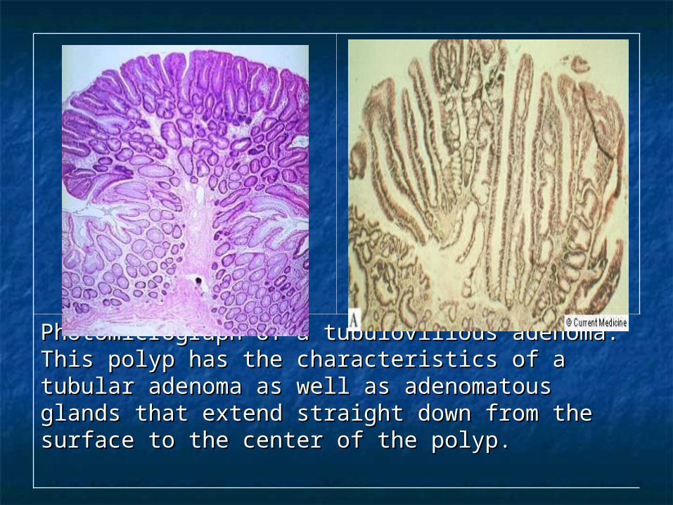
Photomicrograph of a tubulovillous adenoma. This Photomicrograph of a tubulovillous adenoma. This polyp has the characteristics of a tubular adenoma polyp has the characteristics of a tubular adenoma as well as adenomatous glands that extend straight as well as adenomatous glands that extend straight down from the surface to the center of the polyp. down from the surface to the center of the polyp.
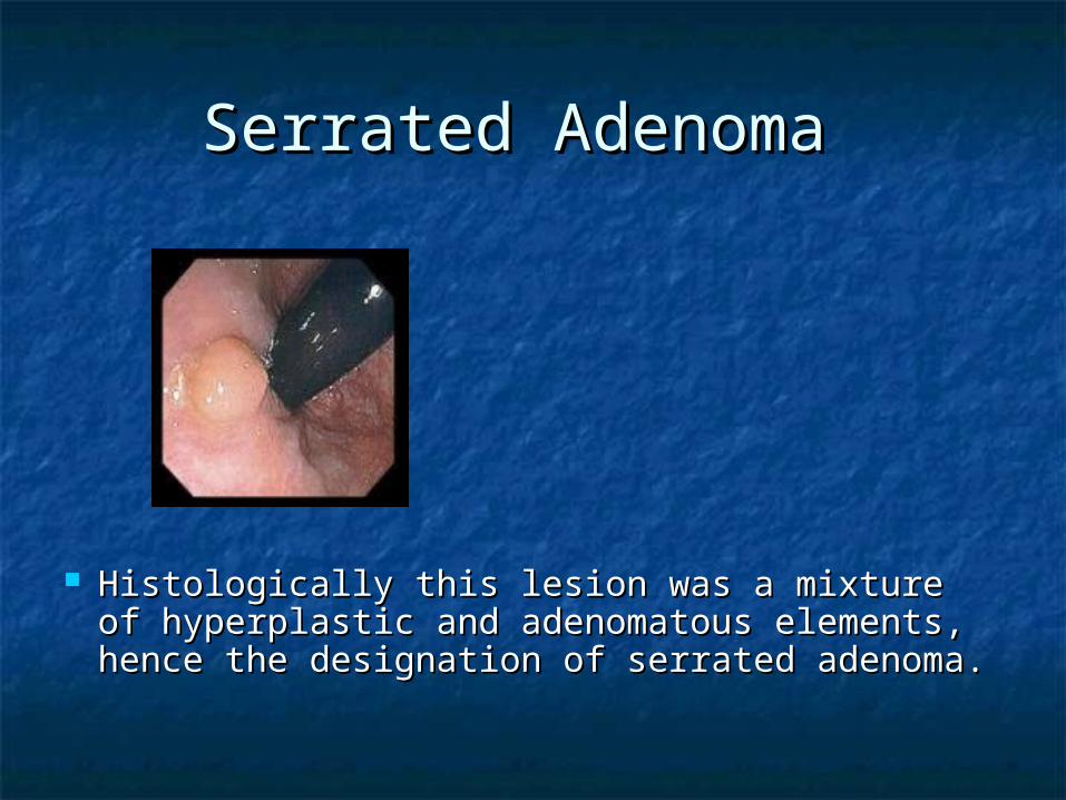
Serrated Adenoma Serrated Adenoma
Histologically this lesion was a mixture of Histologically this lesion was a mixture of hyperplastic and adenomatous elements, hyperplastic and adenomatous elements, hence the designation of serrated adenoma. hence the designation of serrated adenoma.
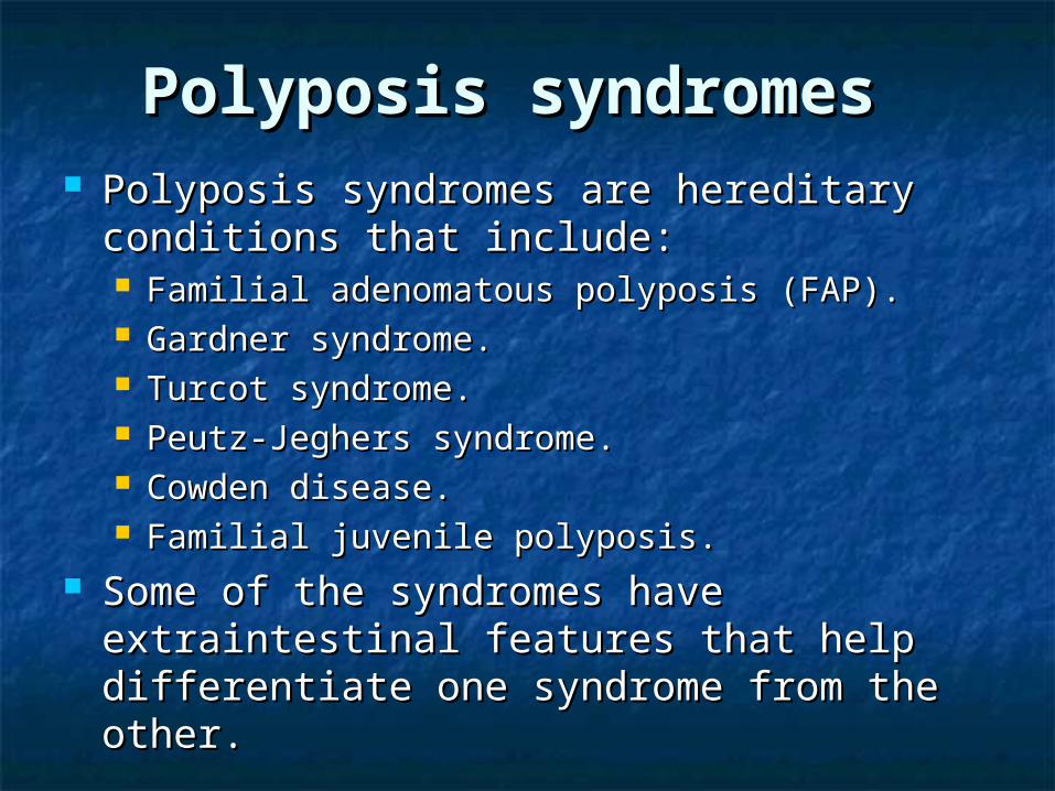
Polyposis syndromesPolyposis syndromes Polyposis syndromes are hereditary Polyposis syndromes are hereditary
conditions that include: conditions that include: Familial adenomatous polyposis (FAP).Familial adenomatous polyposis (FAP). Gardner syndrome.Gardner syndrome. Turcot syndrome.Turcot syndrome. Peutz-Jeghers syndrome.Peutz-Jeghers syndrome. Cowden disease.Cowden disease. Familial juvenile polyposis. Familial juvenile polyposis.
Some of the syndromes have Some of the syndromes have extraintestinal features that help extraintestinal features that help differentiate one syndrome from the other.differentiate one syndrome from the other.
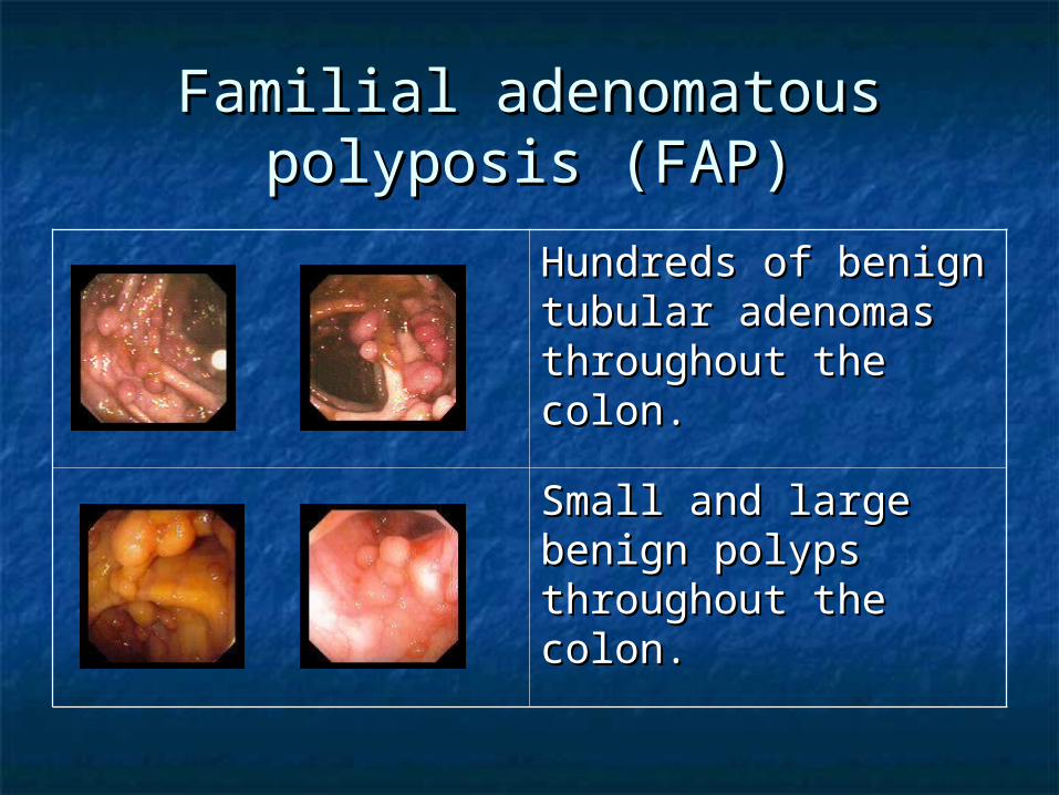
Familial adenomatous Familial adenomatous polyposis (FAP)polyposis (FAP)
Hundreds of benign Hundreds of benign tubular adenomas tubular adenomas throughout the colon.throughout the colon.
Small and large Small and large benign polyps benign polyps throughout the colon. throughout the colon.
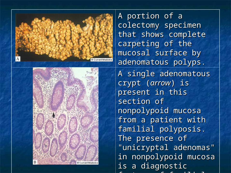
A portion of a colectomy A portion of a colectomy specimen that shows specimen that shows complete carpeting of complete carpeting of the mucosal surface by the mucosal surface by adenomatous polyps. adenomatous polyps.
A single adenomatous A single adenomatous crypt (crypt (arrowarrow) is present ) is present in this section of in this section of nonpolypoid mucosa nonpolypoid mucosa from a patient with from a patient with familial polyposis. The familial polyposis. The presence of "unicryptal presence of "unicryptal adenomas" in adenomas" in nonpolypoid mucosa is a nonpolypoid mucosa is a diagnostic feature of diagnostic feature of familial adenomatous familial adenomatous polyposis. polyposis.
Gardner syndrome (GS) Gardner syndrome (GS) Is the association of colonic adenomatous Is the association of colonic adenomatous
polyposis, osteomas, and soft tissue tumors polyposis, osteomas, and soft tissue tumors (epidermoid cysts, fibromas, desmoid tumors). (epidermoid cysts, fibromas, desmoid tumors).
Turcot syndrome Turcot syndrome Includes polyps, medulloblastoma, congenital Includes polyps, medulloblastoma, congenital
hypertrophy of the retinal pigmented hypertrophy of the retinal pigmented epithelium [CHRPE], and glioblastoma epithelium [CHRPE], and glioblastoma multiforme).multiforme).
Cowden syndrome Cowden syndrome Includes polyps, fibrocystic disease, breast Includes polyps, fibrocystic disease, breast
cancer, and thyroid cancer).cancer, and thyroid cancer).
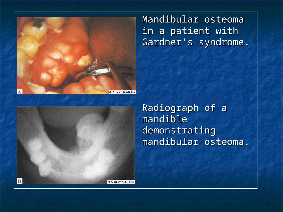
Mandibular osteoma in Mandibular osteoma in a patient with a patient with Gardner's syndrome.Gardner's syndrome.
Radiograph of a Radiograph of a mandible mandible demonstrating demonstrating mandibular osteoma. mandibular osteoma.
Peutz-Jeghers syndrome.Peutz-Jeghers syndrome. Inherited in an Inherited in an autosomal dominantautosomal dominant
manner, PJS is characterized by the manner, PJS is characterized by the association of gastrointestinal polyposis and association of gastrointestinal polyposis and mucocutaneous pigmentation. mucocutaneous pigmentation.
Peutz-Jeghers type hamartomatous polyps Peutz-Jeghers type hamartomatous polyps are most prevalent in the small intestine are most prevalent in the small intestine (jejenum, ileum, and duodenum, (jejenum, ileum, and duodenum, respectively), but can occur elsewhere in the respectively), but can occur elsewhere in the GI tract.GI tract.
Mucocutaneous hyperpigmentation presents Mucocutaneous hyperpigmentation presents in children under the age of five years as in children under the age of five years as dark blue to dark brown mucocutaneous dark blue to dark brown mucocutaneous macules around the mouth, eyes, and macules around the mouth, eyes, and nostrils, in the perianal area, on the buccal nostrils, in the perianal area, on the buccal mucosa, and on the fingers. mucosa, and on the fingers.

Females are at risk for sex cord Females are at risk for sex cord tumors with annular tubules (SCTAT), tumors with annular tubules (SCTAT), a benign neoplasm of the ovaries. a benign neoplasm of the ovaries.
Males occasionally develop calcifying Males occasionally develop calcifying Sertoli cell tumors of the testes, Sertoli cell tumors of the testes, which secrete estrogen and can lead which secrete estrogen and can lead to gynecomastia. to gynecomastia.
Individuals with Peutz-Jeghers Individuals with Peutz-Jeghers syndrome are at increased risk for syndrome are at increased risk for intestinal and extraintestinal intestinal and extraintestinal malignancies, including colorectal, malignancies, including colorectal, esophageal, gastric, breast, ovarian, esophageal, gastric, breast, ovarian, and pancreatic cancers. and pancreatic cancers.
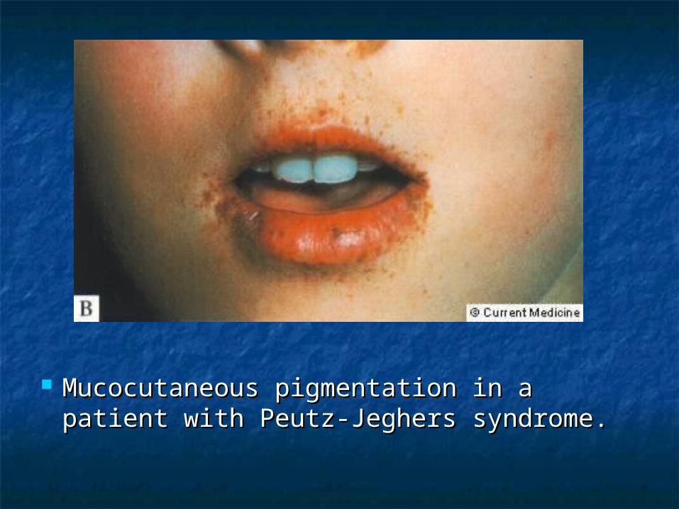
Mucocutaneous pigmentation in a Mucocutaneous pigmentation in a patient with Peutz-Jeghers syndrome. patient with Peutz-Jeghers syndrome.
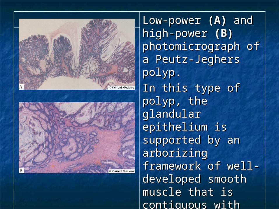
Low-power Low-power (A)(A) and and high-power high-power (B)(B) photomicrograph of a photomicrograph of a Peutz-Jeghers polyp. Peutz-Jeghers polyp.
In this type of polyp, In this type of polyp, the glandular the glandular epithelium is epithelium is supported by an supported by an arborizing framework arborizing framework of well-developed of well-developed smooth muscle that smooth muscle that is contiguous with the is contiguous with the muscularis mucosae. muscularis mucosae.
Juvenile polyposis Juvenile polyposis syndrome (JPS)syndrome (JPS)
Juvenile polyposis syndrome (JPS) is Juvenile polyposis syndrome (JPS) is characterized by predisposition for characterized by predisposition for hamartomatous polyps in the hamartomatous polyps in the gastrointestinal (GI) tract, specifically in the gastrointestinal (GI) tract, specifically in the stomach, small intestine, colon, and rectum. stomach, small intestine, colon, and rectum.
JPS is diagnosed if any one of the following JPS is diagnosed if any one of the following is present: is present: More than five juvenile polyps of the colorectum. More than five juvenile polyps of the colorectum.
OR OR Multiple juvenile polyps throughout the GI tract. Multiple juvenile polyps throughout the GI tract.
OROR Any number of juvenile polyps. Any number of juvenile polyps.
and aand a Family history of juvenile polyps. Family history of juvenile polyps.
The term "juvenile" refers to the type of The term "juvenile" refers to the type of polyp, not the age of onset of polyps. polyp, not the age of onset of polyps.
Most individuals with JPS have some Most individuals with JPS have some polyps by 20 years of age. polyps by 20 years of age.
Some individuals may only have four or Some individuals may only have four or five polyps over their lifetimes, whereas five polyps over their lifetimes, whereas others in the same family may have others in the same family may have over a hundred. over a hundred.
Most juvenile polyps are benign; Most juvenile polyps are benign; however, malignant transformation can however, malignant transformation can occur. occur.
Estimates of developing GI cancers in Estimates of developing GI cancers in families with JPS range from 9-50%. families with JPS range from 9-50%.
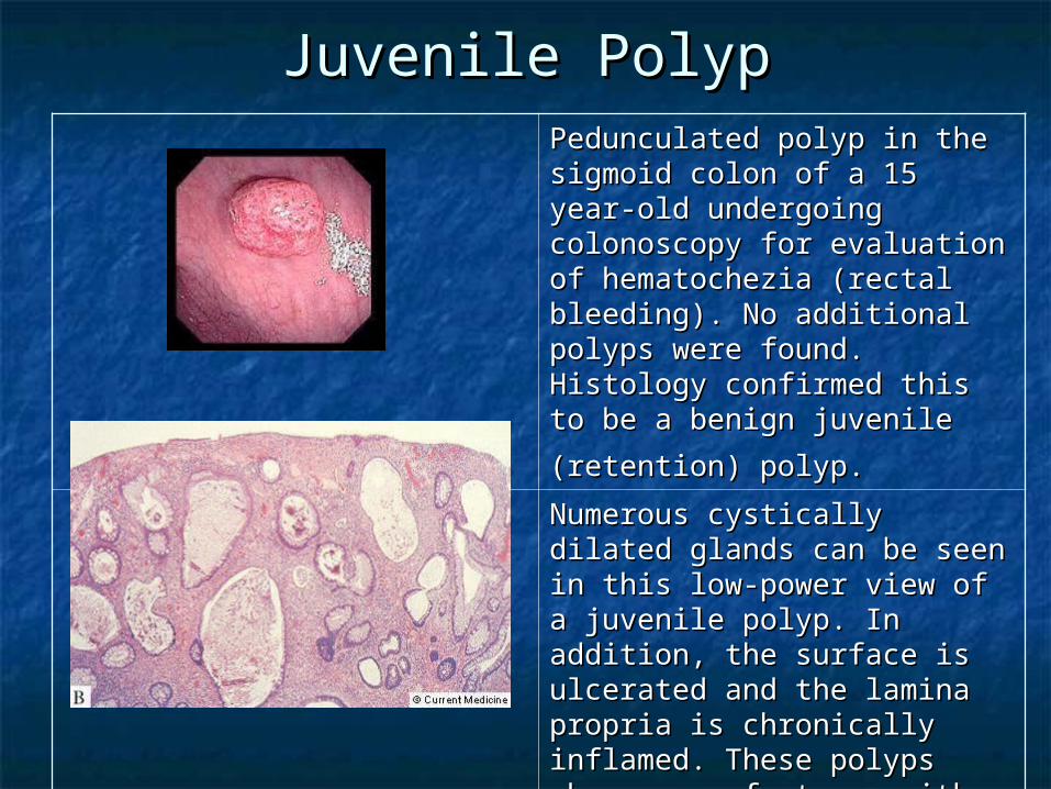
Juvenile PolypJuvenile PolypPedunculated polyp in the Pedunculated polyp in the sigmoid colon of a 15 year-old sigmoid colon of a 15 year-old undergoing colonoscopy for undergoing colonoscopy for evaluation of hematochezia evaluation of hematochezia (rectal bleeding). No additional (rectal bleeding). No additional polyps were found. Histology polyps were found. Histology confirmed this to be a benign confirmed this to be a benign
juvenile (retention) polyp.juvenile (retention) polyp. Numerous cystically dilated Numerous cystically dilated glands can be seen in this low-glands can be seen in this low-power view of a juvenile polyp. power view of a juvenile polyp. In addition, the surface is In addition, the surface is ulcerated and the lamina ulcerated and the lamina propria is chronically inflamed. propria is chronically inflamed. These polyps share many These polyps share many features with inflammatory features with inflammatory polyps, however, the degree of polyps, however, the degree of cystic dilatation in juvenile cystic dilatation in juvenile polyps is usually not seen in polyps is usually not seen in inflammatory polyps. inflammatory polyps.
Other hereditary syndromesOther hereditary syndromes Hereditary mixed polyposis Hereditary mixed polyposis
syndromesyndrome Mode of inheritanceMode of inheritance is unknown. is unknown. The syndrome is characterized by atypical The syndrome is characterized by atypical
juvenile polyps, polyps containing mixed juvenile polyps, polyps containing mixed histology, or multiple polyps of more than histology, or multiple polyps of more than one histologic type in an individual. one histologic type in an individual.
Neurofibromatosis type 1 (NF1)Neurofibromatosis type 1 (NF1) Individuals with NF1 may exhibit multiple Individuals with NF1 may exhibit multiple
intestinal polypoid neurofibromas or intestinal polypoid neurofibromas or ganglioneuromas in the small bowel, ganglioneuromas in the small bowel, stomach, and colon. stomach, and colon.
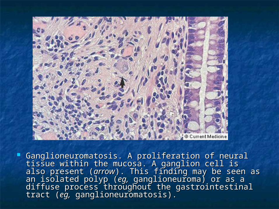
Ganglioneuromatosis. A proliferation of neural tissue Ganglioneuromatosis. A proliferation of neural tissue within the mucosa. A ganglion cell is also present within the mucosa. A ganglion cell is also present ((arrowarrow). This finding may be seen as an isolated polyp ). This finding may be seen as an isolated polyp ((eg,eg, ganglioneuroma) or as a diffuse process ganglioneuroma) or as a diffuse process throughout the gastrointestinal tract (throughout the gastrointestinal tract (eg,eg, ganglioneuromatosis). ganglioneuromatosis).
Hereditary non-polyposis Hereditary non-polyposis colon cancer (HNPCC)colon cancer (HNPCC)
It is an It is an autosomal dominantautosomal dominant colon cancer colon cancer syndrome with proximal colonic syndrome with proximal colonic predominance. predominance.
Few colonic adenomas are present. Few colonic adenomas are present. Other malignancies include cancer of the Other malignancies include cancer of the
endometrium, ovary, stomach, small intestine, endometrium, ovary, stomach, small intestine, and urinary tract. and urinary tract.
It may be difficult to distinguish between It may be difficult to distinguish between HNPCC and attenuated FAP in individuals and HNPCC and attenuated FAP in individuals and families who have few adenomatous colonic families who have few adenomatous colonic polyps. polyps.
Acquired syndromeAcquired syndrome
Cronkite-Canada syndromeCronkite-Canada syndrome Generalized gastrointestinal Generalized gastrointestinal
hamartomatous polyposis, cutaneous hamartomatous polyposis, cutaneous hyperpigmentation, hair loss, and nail hyperpigmentation, hair loss, and nail atrophy. atrophy.
MiscellaneousMiscellaneous
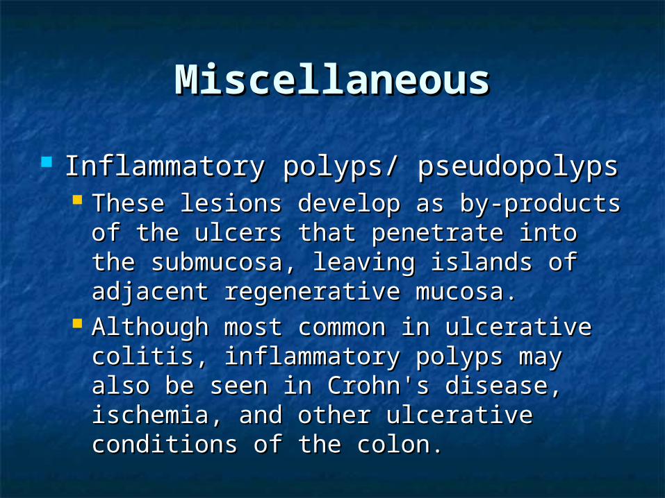
Inflammatory polyps/ pseudopolypsInflammatory polyps/ pseudopolyps These lesions develop as by-products of These lesions develop as by-products of
the ulcers that penetrate into the the ulcers that penetrate into the submucosa, leaving islands of adjacent submucosa, leaving islands of adjacent regenerative mucosa. regenerative mucosa.
Although most common in ulcerative Although most common in ulcerative colitis, inflammatory polyps may also be colitis, inflammatory polyps may also be seen in Crohn's disease, ischemia, and seen in Crohn's disease, ischemia, and other ulcerative conditions of the colon. other ulcerative conditions of the colon.
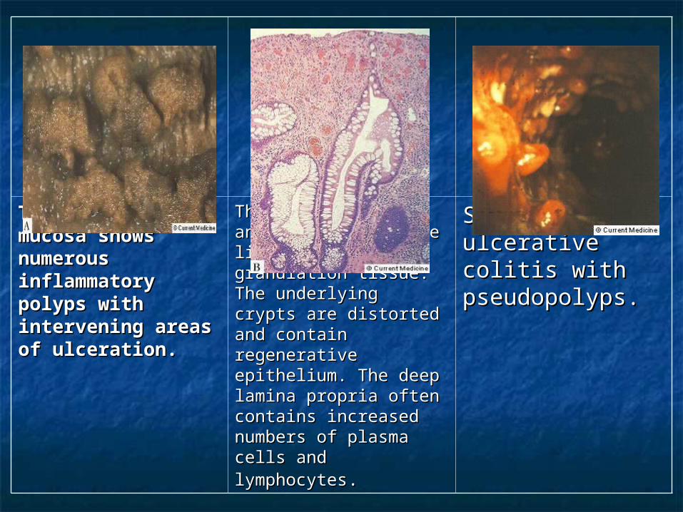
The colonic The colonic mucosa shows mucosa shows numerous numerous inflammatory inflammatory polyps with polyps with intervening areas intervening areas of ulceration. of ulceration.
The pseudopolyp has an The pseudopolyp has an ulcerated surface lined ulcerated surface lined with granulation tissue. with granulation tissue. The underlying crypts The underlying crypts are distorted and are distorted and contain regenerative contain regenerative epithelium. The deep epithelium. The deep lamina propria often lamina propria often contains increased contains increased numbers of plasma cells numbers of plasma cells and lymphocytesand lymphocytes..
Severe ulcerative Severe ulcerative colitis with colitis with pseudopolyps. pseudopolyps.
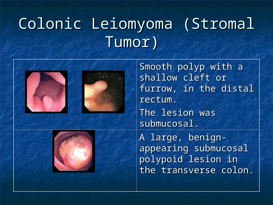
Colonic Leiomyoma (Stromal Colonic Leiomyoma (Stromal Tumor) Tumor)
Smooth polyp with a Smooth polyp with a shallow cleft or furrow, in shallow cleft or furrow, in the distal rectum.the distal rectum.
The lesion was The lesion was submucosal.submucosal.
A large, benign-A large, benign-appearing submucosal appearing submucosal polypoid lesion in the polypoid lesion in the transverse colon. transverse colon.
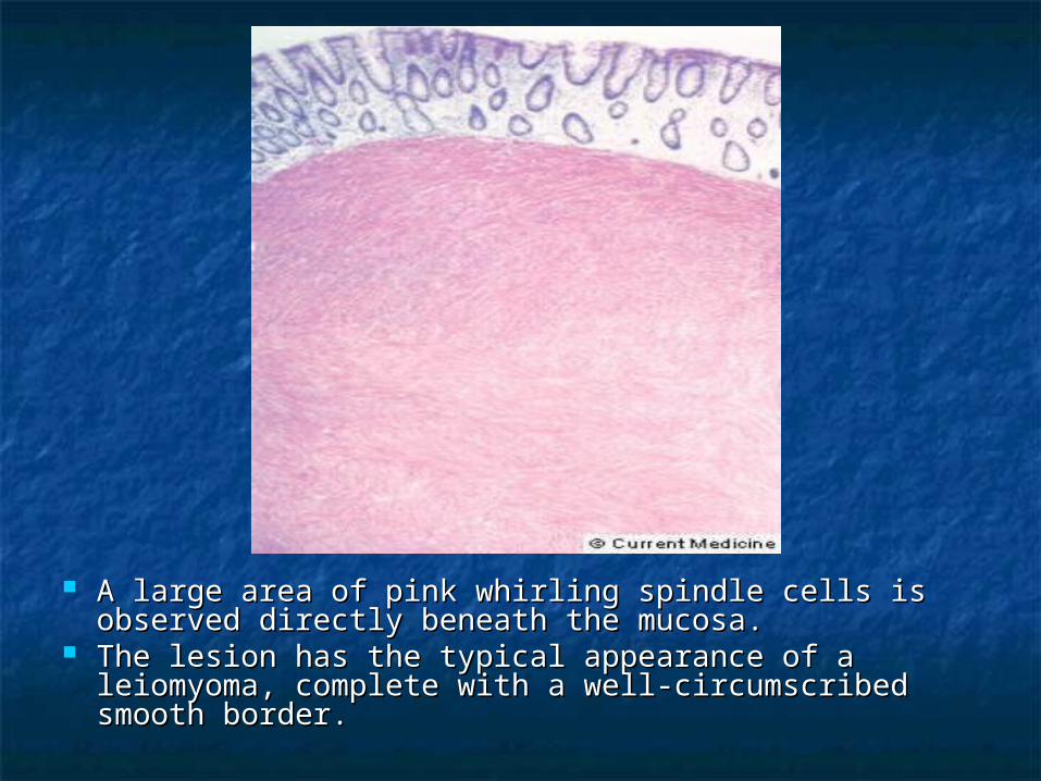
A large area of pink whirling spindle cells is observed A large area of pink whirling spindle cells is observed directly beneath the mucosa. directly beneath the mucosa.
The lesion has the typical appearance of a leiomyoma, The lesion has the typical appearance of a leiomyoma, complete with a well-circumscribed smooth border. complete with a well-circumscribed smooth border.
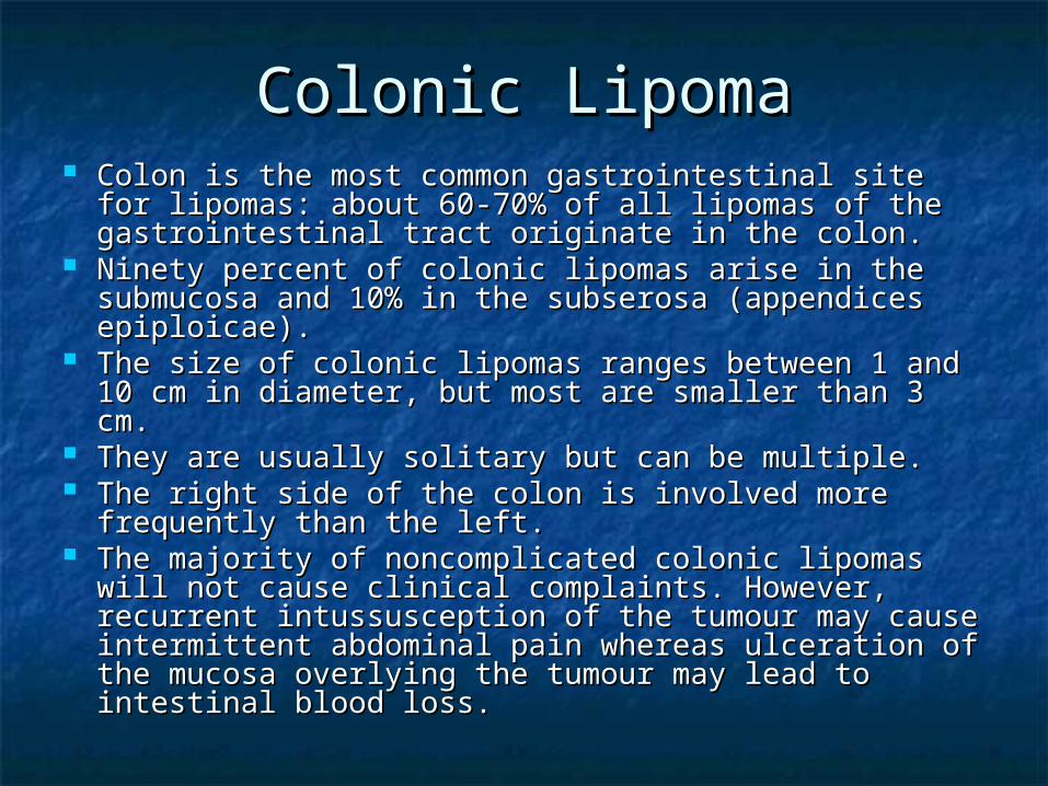
Colonic LipomaColonic Lipoma Colon is the most common gastrointestinal site for Colon is the most common gastrointestinal site for
lipomas: about 60-70% of all lipomas of the lipomas: about 60-70% of all lipomas of the gastrointestinal tract originate in the colon. gastrointestinal tract originate in the colon.
Ninety percent of colonic lipomas arise in the Ninety percent of colonic lipomas arise in the submucosa and 10% in the subserosa (appendices submucosa and 10% in the subserosa (appendices epiploicae). epiploicae).
The size of colonic lipomas ranges between 1 and 10 The size of colonic lipomas ranges between 1 and 10 cm in diameter, but most are smaller than 3 cm. cm in diameter, but most are smaller than 3 cm.
They are usually solitary but can be multiple. They are usually solitary but can be multiple. The right side of the colon is involved more The right side of the colon is involved more
frequently than the left. frequently than the left. The majority of noncomplicated colonic lipomas will The majority of noncomplicated colonic lipomas will
not cause clinical complaints. However, recurrent not cause clinical complaints. However, recurrent intussusception of the tumour may cause intussusception of the tumour may cause intermittent abdominal pain whereas ulceration of intermittent abdominal pain whereas ulceration of the mucosa overlying the tumour may lead to the mucosa overlying the tumour may lead to intestinal blood loss.intestinal blood loss.
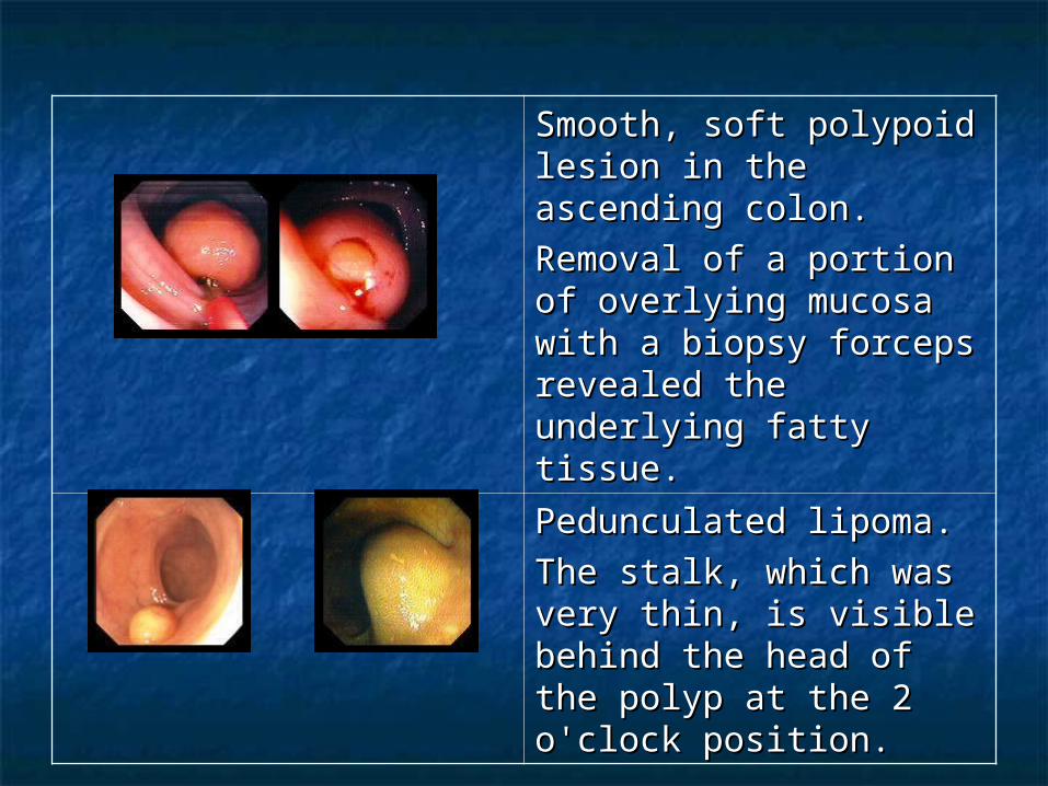
Smooth, soft polypoid Smooth, soft polypoid lesion in the ascending lesion in the ascending colon.colon.
Removal of a portion of Removal of a portion of overlying mucosa with a overlying mucosa with a biopsy forceps revealed biopsy forceps revealed the underlying fatty the underlying fatty tissue. tissue.
Pedunculated lipoma.Pedunculated lipoma.
The stalk, which was The stalk, which was very thin, is visible very thin, is visible behind the head of the behind the head of the polyp at the 2 o'clock polyp at the 2 o'clock position. position.
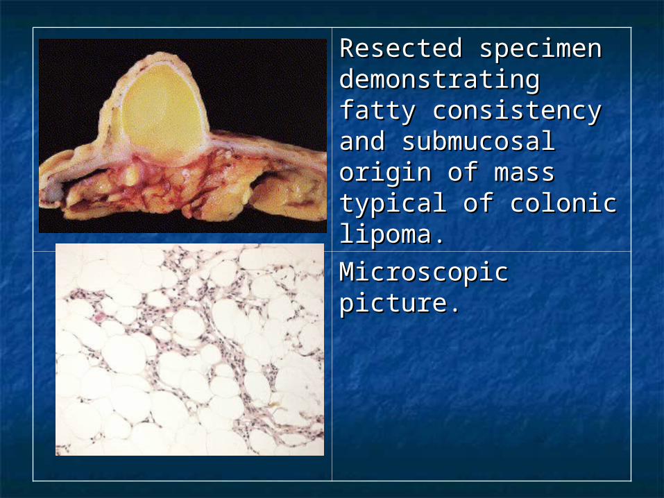
Resected specimen Resected specimen demonstrating fatty demonstrating fatty consistency and consistency and submucosal origin of submucosal origin of mass typical of mass typical of colonic lipoma.colonic lipoma.
Microscopic picture.Microscopic picture.
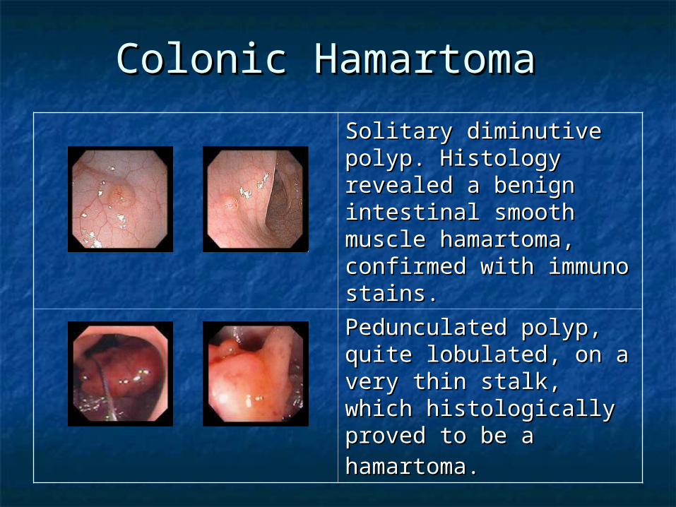
Colonic Hamartoma Colonic Hamartoma
Solitary diminutive polyp. Solitary diminutive polyp. Histology revealed a Histology revealed a benign intestinal smooth benign intestinal smooth muscle hamartoma, muscle hamartoma, confirmed with immuno confirmed with immuno stains. stains.
Pedunculated polyp, Pedunculated polyp, quite lobulated, on a quite lobulated, on a very thin stalk, which very thin stalk, which histologically proved to histologically proved to be a hamartoma.be a hamartoma.
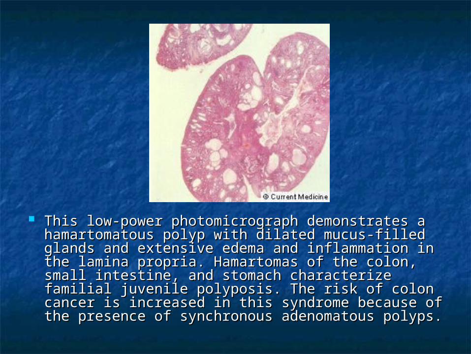
This low-power photomicrograph demonstrates a This low-power photomicrograph demonstrates a hamartomatous polyp with dilated mucus-filled hamartomatous polyp with dilated mucus-filled glands and extensive edema and inflammation in glands and extensive edema and inflammation in the lamina propria. Hamartomas of the colon, small the lamina propria. Hamartomas of the colon, small intestine, and stomach characterize familial intestine, and stomach characterize familial juvenile polyposis. The risk of colon cancer is juvenile polyposis. The risk of colon cancer is increased in this syndrome because of the increased in this syndrome because of the presence of synchronous adenomatous polyps. presence of synchronous adenomatous polyps.
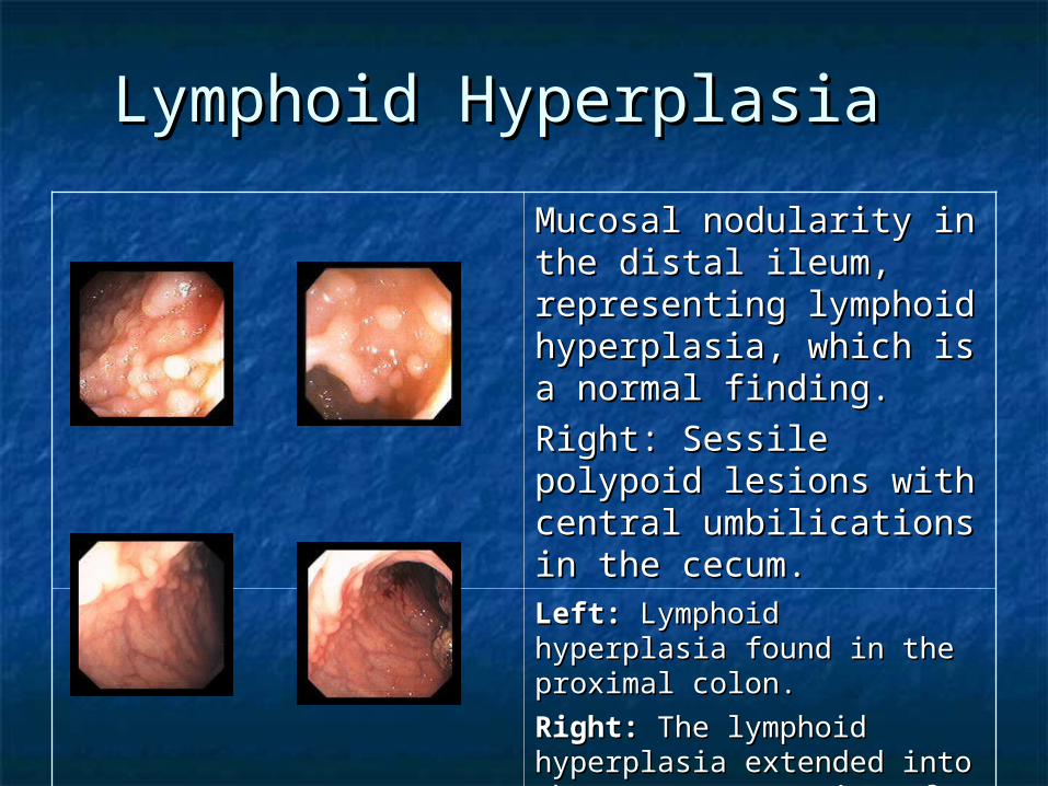
Lymphoid Hyperplasia Lymphoid Hyperplasia
Mucosal nodularity in the Mucosal nodularity in the distal ileum, representing distal ileum, representing lymphoid hyperplasia, lymphoid hyperplasia, which is a normal finding. which is a normal finding.
Right: Sessile polypoid Right: Sessile polypoid lesions with central lesions with central umbilications in the umbilications in the cecum. cecum. Left: Left: Lymphoid hyperplasia Lymphoid hyperplasia found in the proximal colon.found in the proximal colon.
Right: Right: The lymphoid The lymphoid hyperplasia extended into the hyperplasia extended into the cecum; a portion of the cecal cecum; a portion of the cecal mass can be seen at the right.mass can be seen at the right.
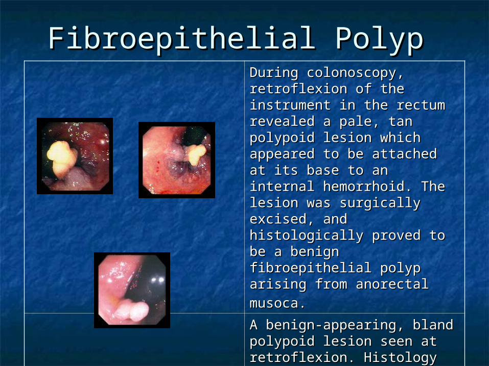
Fibroepithelial Polyp Fibroepithelial Polyp During colonoscopy, During colonoscopy, retroflexion of the instrument retroflexion of the instrument in the rectum revealed a pale, in the rectum revealed a pale, tan polypoid lesion which tan polypoid lesion which appeared to be attached at its appeared to be attached at its base to an internal base to an internal hemorrhoid. The lesion was hemorrhoid. The lesion was surgically excised, and surgically excised, and histologically proved to be a histologically proved to be a benign fibroepithelial polyp benign fibroepithelial polyp arising from anorectal arising from anorectal musoca.musoca. A benign-appearing, bland A benign-appearing, bland polypoid lesion seen at polypoid lesion seen at retroflexion. Histology of the retroflexion. Histology of the surgically-excised specimen surgically-excised specimen revealed fibroepithelial revealed fibroepithelial
papilloma.papilloma.
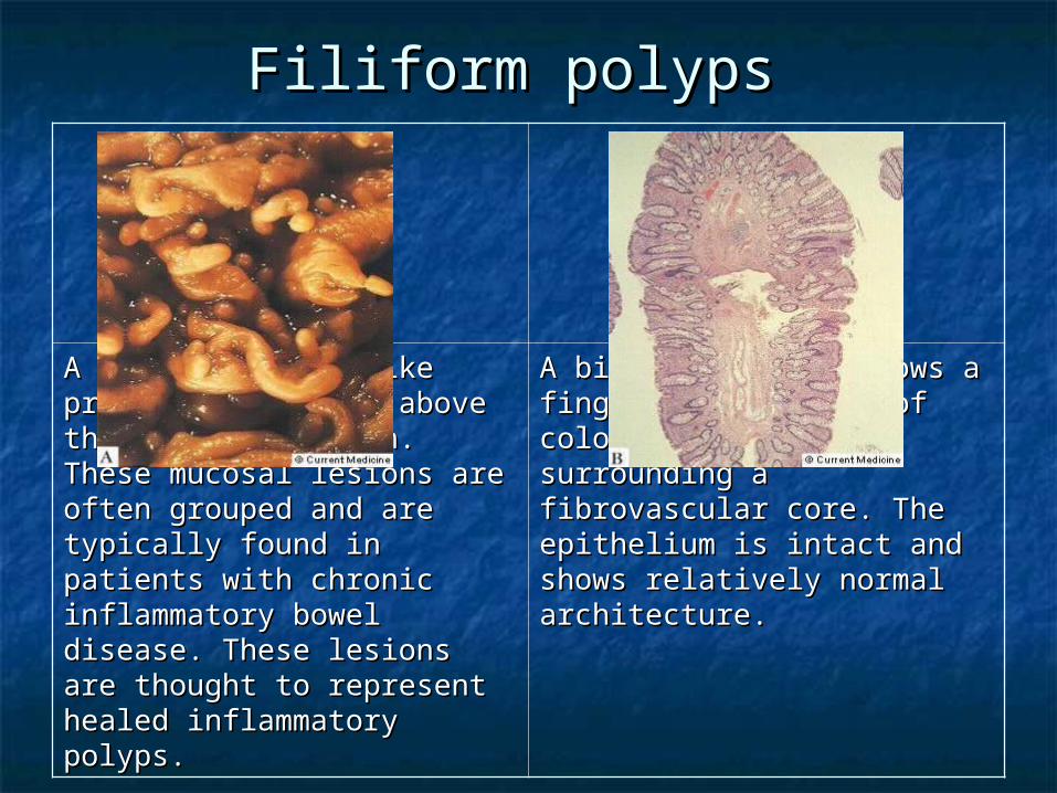
Filiform polyps Filiform polyps
A numerous fingerlike A numerous fingerlike projections rising above the projections rising above the mucosa is shown. These mucosa is shown. These mucosal lesions are often mucosal lesions are often grouped and are typically grouped and are typically found in patients with chronic found in patients with chronic inflammatory bowel disease. inflammatory bowel disease. These lesions are thought to These lesions are thought to represent healed represent healed inflammatory polyps. inflammatory polyps.
A biopsy specimen shows a A biopsy specimen shows a finger like portion of colonic finger like portion of colonic mucosa surrounding a mucosa surrounding a fibrovascular core. The fibrovascular core. The epithelium is intact and shows epithelium is intact and shows relatively normal architecture.relatively normal architecture.
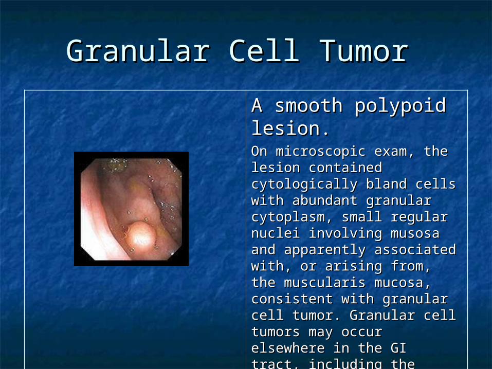
Granular Cell Tumor Granular Cell Tumor
A smooth polypoid A smooth polypoid lesion.lesion.On microscopic exam, the On microscopic exam, the lesion contained cytologically lesion contained cytologically bland cells with abundant bland cells with abundant granular cytoplasm, small granular cytoplasm, small regular nuclei involving regular nuclei involving musosa and apparently musosa and apparently associated with, or arising associated with, or arising from, the muscularis mucosa, from, the muscularis mucosa, consistent with granular cell consistent with granular cell tumor. Granular cell tumors tumor. Granular cell tumors may occur elsewhere in the GI may occur elsewhere in the GI tract, including the tract, including the esophagusesophagus. .
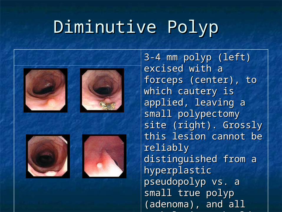
Diminutive Polyp Diminutive Polyp
3-4 mm polyp (left) 3-4 mm polyp (left) excised with a forceps excised with a forceps (center), to which (center), to which cautery is applied, cautery is applied, leaving a small leaving a small polypectomy site (right). polypectomy site (right). Grossly this lesion cannot Grossly this lesion cannot be reliably distinguished be reliably distinguished from a hyperplastic from a hyperplastic pseudopolyp vs. a small pseudopolyp vs. a small true polyp (adenoma), true polyp (adenoma), and all such lesions and all such lesions should be removed for should be removed for microscopic analysis. microscopic analysis.

Rectal CondylomaRectal CondylomaTwo small, hypopigmented Two small, hypopigmented polypoid lesions seen at polypoid lesions seen at retroflexion in the rectum.retroflexion in the rectum.
Histologically the lesions were Histologically the lesions were squamous papillomas, with squamous papillomas, with mild to moderate dysplasia, mild to moderate dysplasia, and features consistent with and features consistent with Human Papilloma Virus (HPV) Human Papilloma Virus (HPV)
infection.infection. Sessile bland polypoid Sessile bland polypoid lesion seen at lesion seen at retroflexion in the retroflexion in the rectum.rectum.
Lymphomatous polyposisLymphomatous polyposis
Occurrence of primary extranodal Occurrence of primary extranodal lymphomas in the gastrointestinal lymphomas in the gastrointestinal tract. tract.
Two types include: Two types include: Multiple lymphomatous polyposis. Multiple lymphomatous polyposis. Mediterranean-type lymphoma. Mediterranean-type lymphoma.
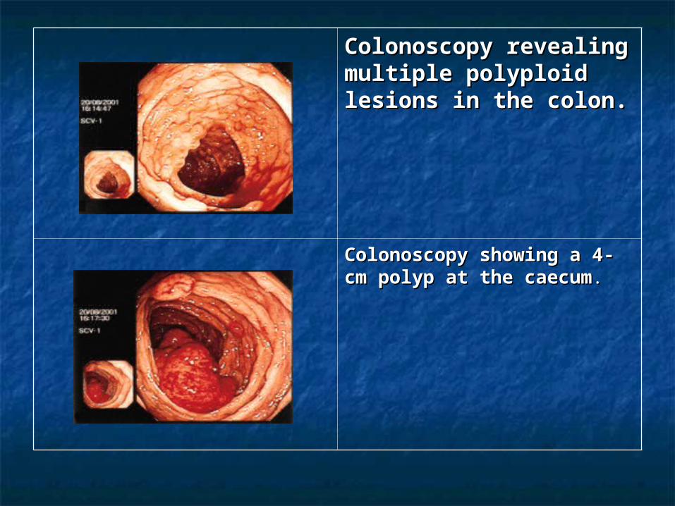
Colonoscopy revealing Colonoscopy revealing multiple polyploid multiple polyploid lesions in the colon.lesions in the colon.
Colonoscopy showing a 4-Colonoscopy showing a 4-cm polyp at the caecumcm polyp at the caecum..
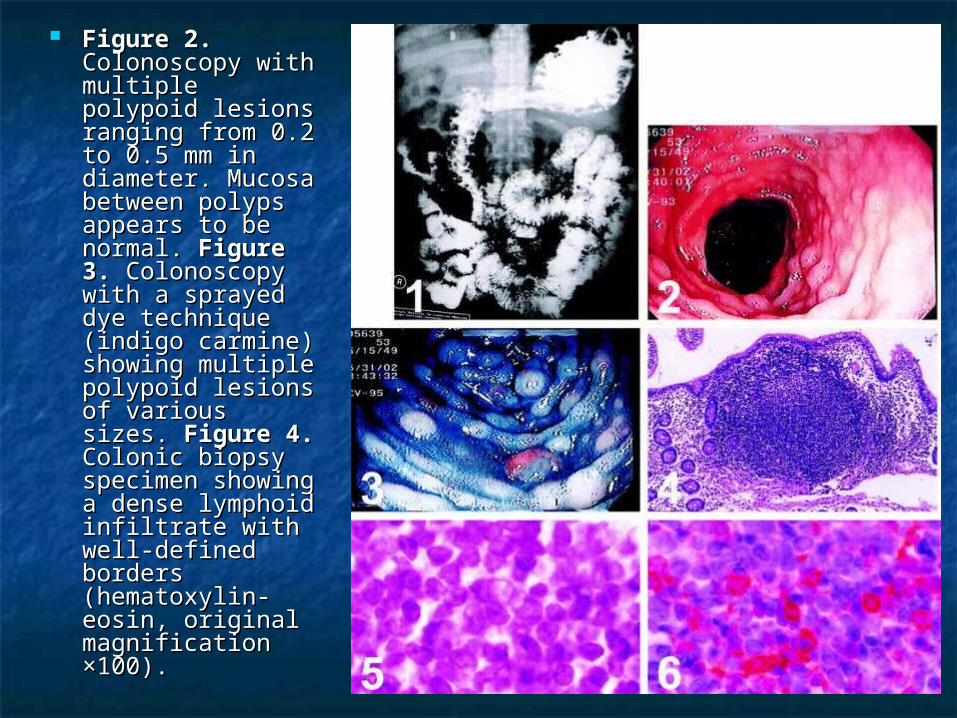
Figure 2.Figure 2. Colonoscopy with Colonoscopy with multiple polypoid multiple polypoid lesions ranging lesions ranging from 0.2 to 0.5 from 0.2 to 0.5 mm in diameter. mm in diameter. Mucosa between Mucosa between polyps appears to polyps appears to be normal. be normal. Figure 3.Figure 3. Colonoscopy with Colonoscopy with a sprayed dye a sprayed dye technique (indigo technique (indigo carmine) showing carmine) showing multiple polypoid multiple polypoid lesions of various lesions of various sizes. sizes. Figure 4.Figure 4. Colonic biopsy Colonic biopsy specimen specimen showing a dense showing a dense lymphoid lymphoid infiltrate with infiltrate with well-defined well-defined borders borders (hematoxylin-(hematoxylin-eosin, original eosin, original magnification magnification ×100).×100).
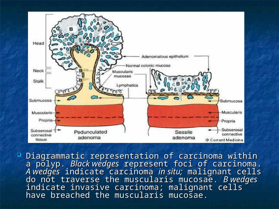
Diagrammatic representation of carcinoma within a Diagrammatic representation of carcinoma within a polyp. polyp. Black wedgesBlack wedges represent foci of carcinoma. represent foci of carcinoma. A A wedgeswedges indicate carcinoma indicate carcinoma in situ;in situ; malignant cells do malignant cells do not traverse the muscularis mucosae. not traverse the muscularis mucosae. B wedgesB wedges indicate invasive carcinoma; malignant cells have indicate invasive carcinoma; malignant cells have breached the muscularis mucosae. breached the muscularis mucosae.
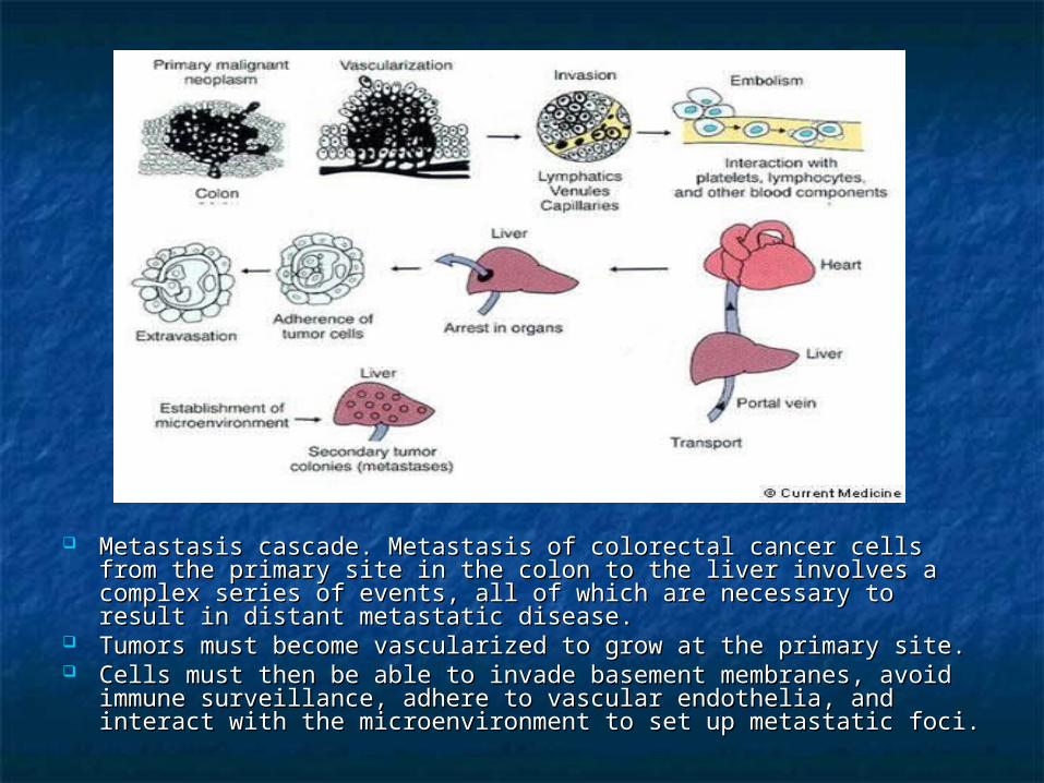
Metastasis cascade. Metastasis of colorectal cancer cells from the Metastasis cascade. Metastasis of colorectal cancer cells from the primary site in the colon to the liver involves a complex series of primary site in the colon to the liver involves a complex series of events, all of which are necessary to result in distant metastatic events, all of which are necessary to result in distant metastatic disease. disease.
Tumors must become vascularized to grow at the primary site. Tumors must become vascularized to grow at the primary site. Cells must then be able to invade basement membranes, avoid Cells must then be able to invade basement membranes, avoid
immune surveillance, adhere to vascular endothelia, and interact immune surveillance, adhere to vascular endothelia, and interact with the microenvironment to set up metastatic foci. with the microenvironment to set up metastatic foci.

Thank youThank you