Embed Size (px)
Citation preview

College of medicine
Department of pathology
3rd year

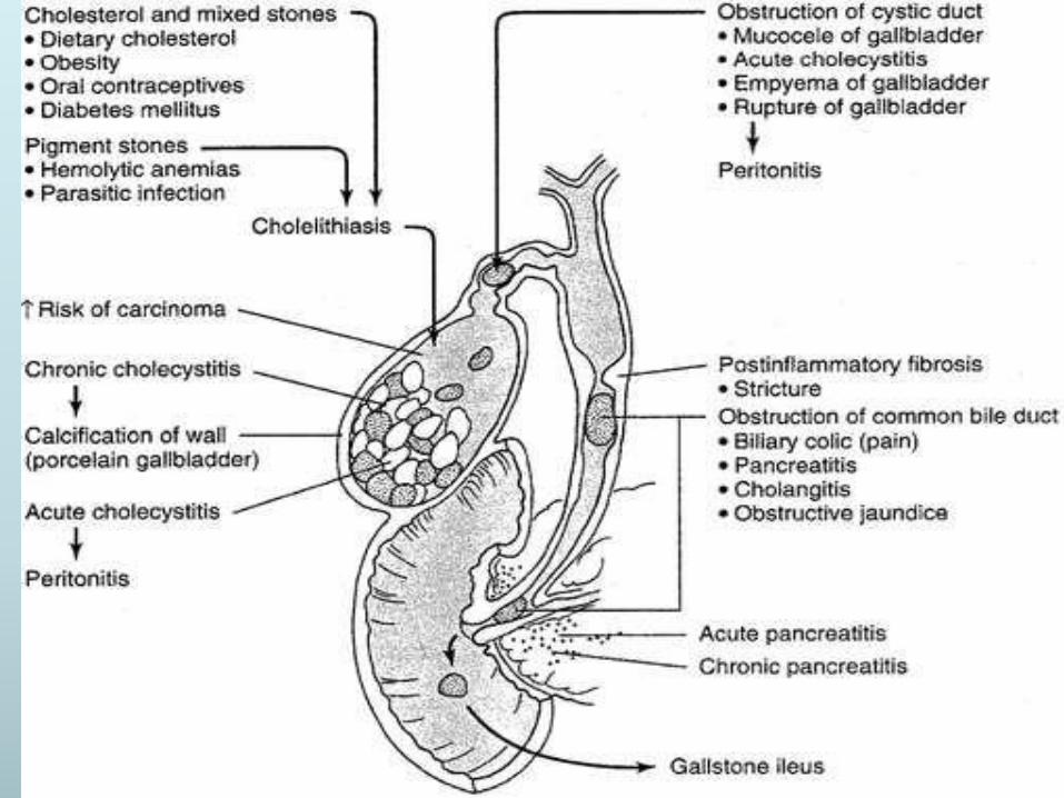
Symptoms of gallstones.
70% to 80% 0f patients with gallstones remain
asymptomatic throughout life,
The remainder becoming symptomatic at the rate of
1% to 3% per year.
The symptoms are biliary colic at right
hypochondrium, vomiting.
Biliary pain of constant or colicky, spasmodic in nature due
to obstruction.

Complications of gallstones
.
1. 1-2% have acute or chronic cholecystitis
2. Choledocholithiasis (stones in the common bile
duct).
3.Cholangitis (inflammation of biliary tree).
4.Empyema (impaction of stone at the neck of
gallbladder).
5.Gallstone ileus ( a large stone may erode directly
into the adjacent loop of small bowel, generating intestinal
obstruction).
6.Acute pancreatitis.
7.Biliary fistulae.
Summary:

Summary

Impacted stone at ampulla within marked dilated common
bile duct (CBD).
duodenum
CBD

Papillary adenocarcinoma of the gallbladder in a patient with
gallstones.

Cholecystitis• Def: Inflammation of the gallbladder
• Can be dividedinto
– Acutecholecystitis
– Chronic cholecystitis
– Acute superimposed on chronic
Acute: fever, leukocytosis, RUQ pain
Chronic: Subclinical or pain
Ultrasound can detect stones well
Go hand in hand with stones in gallbladder or
ducts
Cholecystitis predisposes to cholelithiasis, and VICE VERSA!
If surgery is required, most is laparoscopic

Cholecystitis: Acute, Chronic, & acute superimposed on Chronic.
Acute cholecystitis:
1-Acute calculous cholecystitis:
90% of cases
caused by gallstone obstruction of the neck or the cystic duct
2- Acute acalculus cholecystitis Which occurs in the absence of
gallstones
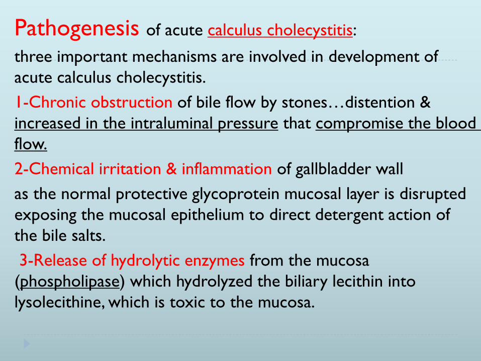
Pathogenesis of acute calculus cholecystitis:
three important mechanisms are involved in development of
acute calculus cholecystitis.
1-Chronic obstruction of bile flow by stones…distention &
increased in the intraluminal pressure that compromise the blood
flow.
2-Chemical irritation & inflammation of gallbladder wall
as the normal protective glycoprotein mucosal layer is disrupted
exposing the mucosal epithelium to direct detergent action of
the bile salts.
3-Release of hydrolytic enzymes from the mucosa
(phospholipase) which hydrolyzed the biliary lecithin into
lysolecithine, which is toxic to the mucosa.

Acute cholecystitis:

Gross: enlarged, distended gallbladder; congested vessels .
Serosal and mucosal exudates,
thickened wall with edema and hemorrhage; ulcers with
blood clot, pus and bile.
In 90% of cases, stones are present; obstruct the neck of
gallbladder or the cystic duct.
When the lumen of gallbladder is filled with frank pus. This
condition is called empyema of gallbladder.
In more severe cases the gallbladder is transformed into a
green- black necrotic organ, termed gangeranous
cholecyetitis.

Microscopically:
Initially :edema,
Vascular congestion, hemorrhage,
later mucosal and mural necrosis
with neutrophils; variable
reactive epithelial changes
resembling Dysplasia.
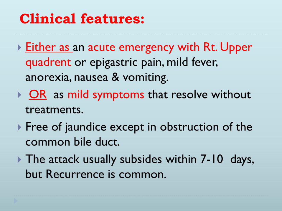
Clinical features:
Either as an acute emergency with Rt. Upper
quadrent or epigastric pain, mild fever,
anorexia, nausea & vomiting.
OR as mild symptoms that resolve without
treatments.
Free of jaundice except in obstruction of the
common bile duct.
The attack usually subsides within 7-10 days,
but Recurrence is common.

Complications of acute cholecystitis
1-mucocele of the gallbladder
2-Empyema: local abscess formation of the gallbladder.
3-Secondary bacterial infection of the biliary tree
(Ascending bacterial cholangitis).
4-Gallbladder perforation or ruptures: escape of the
contents into the peritoneal cavity leads to localized
or generalized peritonitis.
5-Biliary – enteric fistula. due to ulceration of the
gallbladder by a large cholesterol stone through the
duodenum or the colon.
6- Pancreatitis.

7- Obstructive choleycystitis, small gall stones more
dangerous because enter the cystic duct or common bile
duct lead to obstruction & secondary biliary cirrhosis.
8- increase the risk of carcinoma of the gallbladder.

Acute Acalculus cholecystitis. no
gallstones.
Represent 10% of cases.
Causes.
(1) The postoperative state after major nonbiliary surgery.
(2) Severe trauma (e.g. car accident……etc).
(3) Severe burns.
(4) Sepsis.
There are multiple events are thought to contribute to
acalculus cholecystitis. Like dehydration, gallbladder stasis,
shock & bacterial contamination.

Chronic cholecystitis:
Results from repeated attacks of acute cholecystitis,
usually insidious, accompanied by dyspeptic
symptoms or biliary colic.
Gallstones are almost always present.
95% of cases are associated with gallstones.
• Bacteria present in 11-30%, similar organisms as in
acute cholecystitis (Escherichia coli & enterococci).
• 75% of cases are in female within the fourth decade
of life

Morphology :
Gross:
Variable(may be normal size, contracted or enlarged )
thickening of gallbladder wall, variable adhesions.
Ulceration of mucosa is may be due to pressure by
stones.
Mic.
Mucosa shows variable degrees of mononuclear
inflammatory cells infiltration & fibrosis.
Surface epithelium may be relatively normal, atrophic, or
shows hyperplastic or metaplastic changes.
The gallbladder wall may show fibrosis, smooth muscle
hypertrophy.

Chronic cholecystitis:

Complications of chronic cholecystitis
:
1. Acute cholecystitis.
2. Choledocholithiasis.
3. Acute pancreatitis.
4. Gallstone ileus.
5. Biliary fistulas.
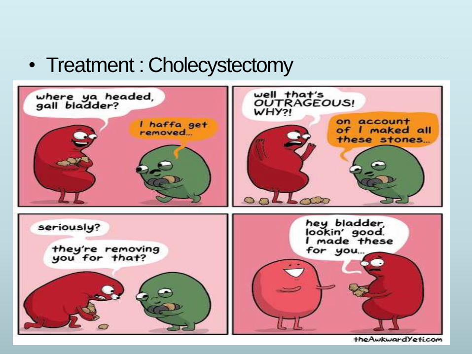
• Treatment : Cholecystectomy
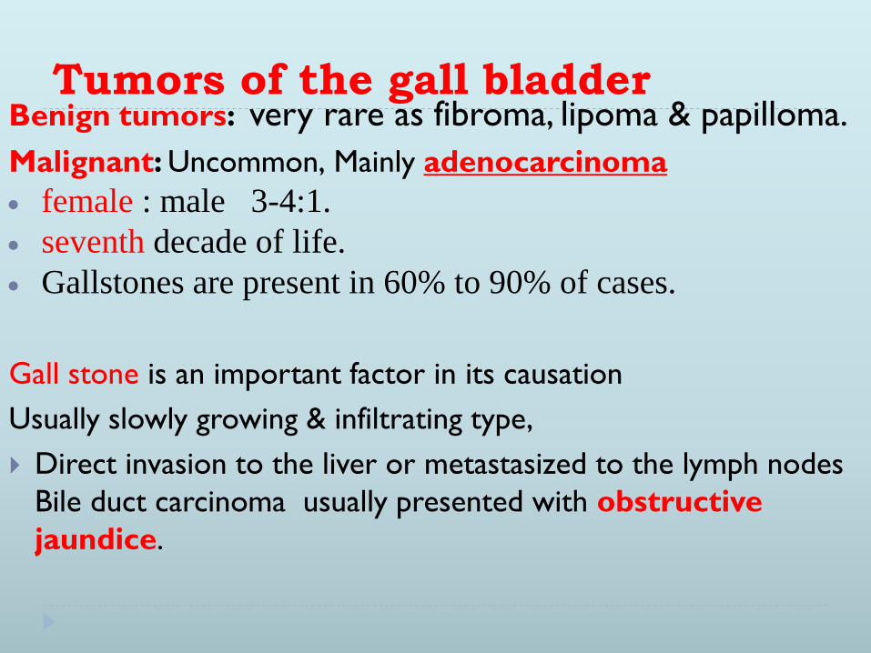
Tumors of the gall bladder Benign tumors: very rare as fibroma, lipoma & papilloma.
Malignant: Uncommon, Mainly adenocarcinoma
female : male 3-4:1.
seventh decade of life.
Gallstones are present in 60% to 90% of cases.
Gall stone is an important factor in its causation
Usually slowly growing & infiltrating type,
Direct invasion to the liver or metastasized to the lymph nodes
Bile duct carcinoma usually presented with obstructive
jaundice.

Morphology of gall bladder ca
Gross: either (1) Infiltrative (diffuse).
(2) Exophytic (irregular, cauliflower mass).
Sometimes contain gallstones
Micro: most cases are adencarcinoma. Some are
papillary & other are poorly differentiated carcinoma.

Adenocarcinoma of the
gallbladder

Biliary tree pathology

INTRAHEPATIC
BILE DUCTS
Choledochlithiasis:
It is means the presence of stones within biliary tree
(common bile duct).
Either primary stones or secondary stones
(commonest).
Primary stones are formed within common bile duct
(CBD). & while secondary stones are formed within
gallbladder & then enter the CBD.

Complications of
Choledocholithiasis:
1- 10% are asymptomatic.
2. Biliary obstruction.
3. Pancreatitis.
4. Cholangitis
5. Hepatic abscess.
6. Chronic liver diseases. …secondary biliary
cirrhosis.
CholangitisIt is referred to acute inflammation of the wall of bile ducts,
which always caused by bacterial infection of the normally
sterile lumen.
Causes:
1. Gallstones.
2. Complications of biliary surgery.
3. Tumors.
4.catherterization of biliary.
5. Acute pancreatitis
6. Benign strictures.
7. Parasitic infections.

Pathogenesis of cholangitis
: two mechanisms are involved.
A.
Obstruction of biliary tree. (stones, tumors, ….etc)
B.
Bacterial infection. (Most likely enter biliary tree
through the sphincter of oddi rather than hematogenous
route. The bacteria are usually G-ve aerobes such as E.coli,
Klebsiella, clostridium, bacteroides).
These two mechanisms are must be occur
together.
Symptoms of cholangitis: include fever, abdominal pain,
and jaundice

Carcinoma of biliary tree.(cholangiocarcinoma).
It is referred to carcinoma of intrahepatic &extrahepatic
ducts.
Those of intrahepatic bile ducts are closely resemble to
HCC.
While those of extrahepatics ducts are usually cause
painless, progressive deepening jaundice.
more in elderly male.
Risk factors of chlangiocarcinoma:
1-Primary sclerosing cholangitis.
2-Inflammatory bowel diseases.
3-Gallstones.
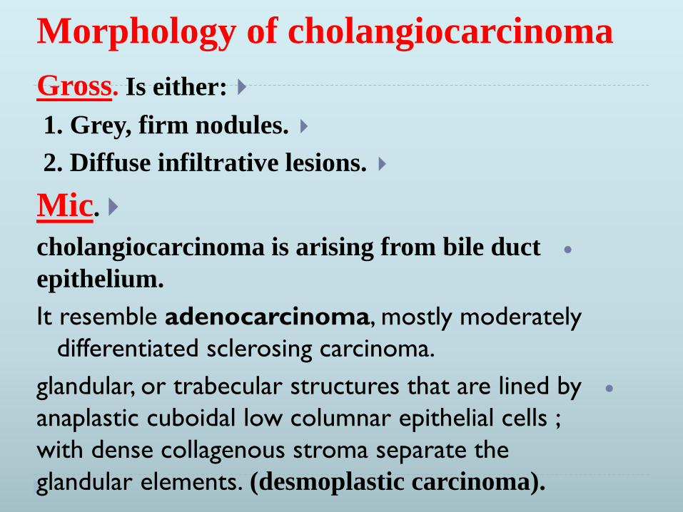
Morphology of cholangiocarcinoma
Gross. Is either:
1. Grey, firm nodules.
2. Diffuse infiltrative lesions.
Mic.
cholangiocarcinoma is arising from bile duct
epithelium.
It resemble adenocarcinoma, mostly moderately
differentiated sclerosing carcinoma.
glandular, or trabecular structures that are lined by
anaplastic cuboidal low columnar epithelial cells ;
with dense collagenous stroma separate the
glandular elements. (desmoplastic carcinoma).

Cholangiocarcinoma
Hematogenous metastasis to the lung, bones (mainly
vertebrae), adrenals & brain in 50% of the cases., but less
frequent with hepatocellular carcinoma.
Also 50% of cholangiocarcinoma spread by lymph node
metastasis, mainly peri-hilar, peri-pancreatic & Para-aortic
lymph nodes above & below the diaphragm, also less
frequently with hepatocellular carcinoma.

CHOLANGIOCARCINOMA

cholangiocarcinoma
19نيسان، 20

Clinical features:
Undefined upper abdominal pain, malaise, fatigue,
weight loss, abdominal fullness.
Sometimes hepatomegaly with irregularity or
nodularity or as abdominal mass.
High level of serum alpha-feto protein marker in
60 -75 % of hepatocellular carcinoma.
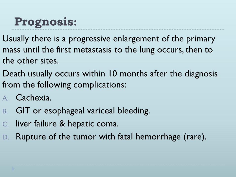
Prognosis:
Usually there is a progressive enlargement of the primary
mass until the first metastasis to the lung occurs, then to
the other sites.
Death usually occurs within 10 months after the diagnosis
from the following complications:
A. Cachexia.
B. GIT or esophageal variceal bleeding.
C. liver failure & hepatic coma.
D. Rupture of the tumor with fatal hemorrhage (rare).