Embed Size (px)
Citation preview

Sede Amministrativa: Università degli Studi di Padova
Dipartimento di Scienze Cardiologiche, Toraciche e Vascolari
CORSO DI DOTTORATO DI RICERCA IN: SCIENZE MEDICHE, CLINICHE E SPERIMENTALI
CURRICOLO: NEUROSCIENZE
CICLO 29°
COGNITIVE PROCESSING AND BRAIN
COMMUNICATION IN AMYOTROPHIC
LATERAL SCLEROSIS
Tesi redatta con il contributo finanziario della Fondazione CARIPARO
Coordinatore: Ch.mo Prof. Gaetano Thiene
Supervisore: Ch.ma Prof.ssa Elena Pegoraro
Co-Supervisore: Ch.mo Prof. Niels Birbaumer
Valutatori: Prof. Christoph Braun and Prof. Boris Kleber
Dottoranda : María Ángeles Prats Sedano

Acknowledgements
First of all, I would like to thank Prof. Birbaumer for giving me the opportunity to work
with him during my PhD project. He guided me during my PhD with his expertise and
patience, and above all, he always supported me, encouraged my research and allowed
me to grow as a scientist. I am deeply grateful for his motivation, sense of humor and
invaluable advices.
Inoltre vorrei ringraziare la mia relatrice la Prof. Pegoraro per la supervisione nella stesura
della tesi e per la sua toleranza. Il suo è un essempio di ciò che significa passione e
dedizione per la ricerca.
Un ringraziamento va a tutti i colleghi del dell’IRCCS Fondazione Ospedale San Camillo per
il loro prezioso contributo e sostegno. In particolare a i miei cari colleghi, a Stefano Silvoni
per l’aiuto nell’elaborazione dei dati neurofisiologici e il suo sostegno, Chiara Volpato per
avermi introdotto nel mondo della SLA e essere sempre disponibile a darmi una mano,
Marianna Cavinato per avermi insegnato tantisimo nel mondo della neurofisiologia e
essere sempre gentile e ottimista, e Andrea Paggiaro e Daniele de Massari per la sua
disponibilità e amicizia.
Inoltre vorrei ringraziare e miei amici italiani che mi hanno fatto sentire sempre a casa
(nonostante la lontananza): Marcello, Antonio, Raffaella, Sophie, Marco, Irene, Elisabetta,
Andrea Polli.
I also would like to thank the BCI team in Tübingen for giving me the opportunity to
participate in this amazing project. Thanks to Ujwal, Bin, Katharina and Azim. I want to
specially thank Aygul, who is doing an outstanding work and she has been an amazing
travel partner in our visit to the patients. Together we had made a great team.
I also want to thank all my friends and colleagues of Tübingen whom I have shared life
experiences through different periods of my PhD adventure: Aurelie, Carlos, Anna,

Andreita, Kolja, Florian, Inaki, Ainitze, Trevor, Sebastian, Jim, Laura, Max, Alessandra,
Ander, Farid, Andreas, Thiago, Beatrice, Giulia.
Gracias especiales al salad group de Tübingen por vuestra amistad. Sin vosotros esta
experiencia no hubiese sido la misma. A Edu, por su incondicional apoyo en el trabajo y
fuera de él, a Ainhoa por darle sal a mi vida y llenarla de risas, a Andrea por sus grandes
zascas y por ser la mejor companera de habitación que se podía tener, a Nerea por ser
una amiga leal y con la que puedo contar siempre, y a Boris por su disponibilidad y sus
sabios consejos.
Y sobretodo, gracias a Jordi, sin duda alguna el mejor regalo que me llevo de esta
aventura. Gracias por hacerme el camino más bonito con tu amor y apoyo incondicional.
No podía dejar sin dar las gracias a mi familia (mis padres, mi hermana, mi abuela, mi
madrina y mis primas) por estar siempre allí; sin vosotros nada de esto hubiese sido
posible. Dicen que la familia no se elige, pero yo he tenido la gran suerte de caer en la
mejor familia que nadie podría tener. També volia donar ses gràcies a sa meva altra
familia, es meus amics: Marga, Angela, Tanit, Francina, Marian i Berto. Gràcies per es
vostre suport durant aquests anys tot i sa distància que mos separa.
Finally, I would like to express my gratitude to the ALS patients and their relatives who
took part in the studies. Their enthusiasm, generosity and strength have touched my
heart and inspired me throughout this process and will never leave me. This thesis is
dedicated to them and their families.

Glossary
AAT Aphasia Aachener Test
ALS Amyotrophic Lateral Sclerosis
ALSFRS-R ALS functional rating scale-revised
BCI Brain Computer Interface
C9ORF72 Chromosome 9 open reading frame 72
CLIS Completely Locked-in State
EEG Electroencephalography
EOG Electrooculography
ERPs Event-related potentials
fALS Familial Amyotrophic Lateral Sclerosis
fNIRS Functional Near Infrared Spectroscopy
FTD Fronto-temporal Dementia
LIS Locked-in State
LMN Lower Motor Neuron
M-WCST Modified Wisconsin Card Sorting test
MCI Mild cognitive impairment
MND Motor Neuron Disease
PSD Power spectrum density
QoL Quality of life
RCPM Raven coloured progressive matrices
RON Re-orienting negativity
RVMD Rivermead memory test
SOD1 Superoxide dismutase 1
SSEPs Somatosensory evoked potentials
SVM Support Vector Machine
ToM Theory of Mind
UMN Upper Motor Neuron

ABSTRACT
Amyotrophic Lateral Sclerosis (ALS) is a fatal neurodegenerative disease characterized by
progressive paralysis of limbs and bulbar musculature. This severe physical impairment
makes cognitive evaluation a big challenge, thus there is a great need for an assessment
that does not require overt motor responses. Moreover, we need of augmentative
communication strategies because the disease generally leads to complete paralysis and,
therefore, patients are unable to communicate with the external world by any means. For
this purpose, Brain Computer Interfaces (BCIs) seem a promising approach to facilitate
communication with these patients.
The aim of this thesis is twofold. First, assessing cognitive processing in ALS by means of a
novel evaluation tool. Second, allowing brain communication in completely paralyzed ALS
patients who had lost their vision in order to eliminate the unbearable loss of
communication in paralysis (“unlocking the locked-in”).
The first study introduces a novel approach for assessing cognitive functions in ALS. This
approach uses neuropsychological tests that require minimal overt motor or verbal
responses; together with vibro-tactile P300s. Results indicate mild cognitive impairment
in oral language comprehension tasks and reduced vibro-tactile P300 amplitudes in
patients compared to healthy controls. Importantly, correlations between the vibro-
tactile P300 latency and psychometric test results suggest that the former measure could
serve as a neurophysiological marker of cognitive decline in ALS patients.

The second study introduces a distraction paradigm based in auditory event-related
potentials (ERPs) to evaluate the ability of change detection, focusing, and re-orientation
of attention in ALS. The results revealed a modification of the amplitude and the latency
of the N200, the P300 and the re-orienting negativity (RON) components. This could
suggest an alteration of the endogenous mechanism that controls the detection of
change, thus resulting in a reduction of the allocation and the re-orientation of
attentional resources.
The third study aimed at testing the feasibility of a Near Infrared Spectroscopy (NIRS) -
based BCI communication approach for patients in the Completely Locked-in Stage (CLIS)
due to ALS. For this purpose two CLIS patients were trained to control their cerebral-
cortex´s functional-activations in response to auditory processing of correct or incorrect
statements assessed with NIRS. The results of the study are very promising, showing that
both CLIS patients communicated with fronto-cortical oxygenation based BCI at an
average correct response rate of 70% over a period of several weeks. We conclude that
this novel approach of brain-communication is safe and, reliable, representing, so far, the
best communication possible for patients in completely locked-in state.
In conclusion we propose a) the novel combination of vibro-tactile or acoustic ERPs and
motor-independent neuropsychological tests as an alternative and easily implementable
way for assessing cognitive functions in ALS and b) we confirm the usefulness and
effectiveness of above mentioned electrophysiological approaches in the late stage of ALS
either to assess cognitive processing or to establish communication with a BCI system.

TABLE OF CONTENTS
1. AN INTRODUCTION TO AMYOTROPHIC LATERAL SCLEROSIS .............................................................. 1
1.1. TERMINOLOGY AND CLINICAL FEATURES ..................................................................................................... 1
1.2. EPIDEMIOLOGY ..................................................................................................................................... 2
1.3. PATHOLOGY AND PATHOGENESIS ............................................................................................................. 4
1.4. DIAGNOSIS .......................................................................................................................................... 6
1.5. QUALITY OF LIFE ................................................................................................................................... 8
1.6. LATE STAGE OF ALS AND END OF LIFE MANAGEMENT: ................................................................................ 12
2. COGNITIVE AND BEHAVIORAL IMPAIRMENT IN ALS ......................................................................... 14
2.1. SPECTRUM OF ALS AND DEMENTIA ........................................................................................................ 14
2.2. COGNITIVE DEFICITS IN ALS .................................................................................................................. 15
2.2.1. Executive dysfunctions .......................................................................................................... 15
2.2.2. Memory ................................................................................................................................. 17
2.2.3. Language ............................................................................................................................... 18
2.2.4. Visuo-perceptual functions .................................................................................................... 19
2.3. SOCIAL COGNITION AND EMOTIONAL PROCESSING ..................................................................................... 20
2.4. BEHAVIOUR ....................................................................................................................................... 21
3. EVENT-RELATED POTENTIALS AND BRAIN COMMUNICATION IN ALS ............................................... 23
3.1. EVENT-RELATED POTENTIALS IN ALS ....................................................................................................... 23
3.1.1. Event-related potentials ........................................................................................................ 23
3.1.2. Event-related potentials studies in ALS: ................................................................................ 29
3.2. BRAIN COMMUNICATION IN AMYOTROPHIC LATERAL SCLEROSIS ................................................................... 31
3.2.1. Introduction to Brain Computer Interfaces ........................................................................... 31
3.2.2. Brain computer interfaces for communication in the late stage of ALS ................................ 36
3.2.3. fNIRS-EEG based BCIs for communication in the late stage of ALS ....................................... 38
4. ASSESSING COGNITIVE FUNCTIONING IN AMYOTROPHIC LATERAL SCLEROSIS WITH A NOVEL
COMBINATION OF VIBRO-TACTILE P300 AND NEUROPSYCHOLOGICAL TESTING ...................................... 40
4.1. INTRODUCTION ................................................................................................................................... 40
4.2. MATERIALS AND METHODS ......................................................................................................................... 43
4.2.1. Participants .................................................................................................................................. 43
4.2.2. Neuropsychological Assessment .................................................................................................. 45
4.2.3. ERPs assessment and data acquisition and processing of the EEG .............................................. 48

4.2.4 Median-nerve somato-sensory evoked potentials (SSEPs) ............................................................ 50
4.2.5 Statistical analysis ......................................................................................................................... 50
4.3. RESULTS .................................................................................................................................................. 52
4.3.1. Demographic and clinical data ..................................................................................................... 52
4.3.2. Neuropsychological performance ................................................................................................ 52
4.3.3. P300 components ........................................................................................................................ 53
4.3.4. Median nerve somato-sensory evoked potentials data ................................................................ 55
4.3.5. Relationship between patient´s clinical characteristics and neuropsychological performance .... 55
4.3.6 Relationship between demographic/clinical data and P300 components ..................................... 56
4.3.7. Relationship between clinical data and median-nerve somatosensory evoked potentials ......... 56
4.3.8. Relationship between P300 and neuropsychological performance ............................................. 56
4.4. DISCUSSION ............................................................................................................................................. 57
4.4.1. Neuropsychological differences in ALS ......................................................................................... 57
4.4.2. Neurophysiological differences in ALS .......................................................................................... 58
4.4.3. Relationship between test outcome and clinical data ................................................................. 58
5. SELECTIVE ATTENTION IMPAIRMENT IN AMYOTROPHIC LATERAL SCLEROSIS ....................................... 61
5.1. INTRODUCTION ........................................................................................................................................ 61
5.2 METHODS ................................................................................................................................................. 62
5.2.1. Participants .................................................................................................................................. 62
5.2.2. Neuropsychological assessment ................................................................................................... 63
5.2.3. ERPs paradigm ............................................................................................................................. 63
5.2.4. Acquisition and analysis of the electroencephalogram (EEG) ..................................................... 64
5.3 RESULTS ................................................................................................................................................... 65
5.3.1. Neuropsychological test ................................................................................................................ 65
5.3.2 ERPs paradigm ............................................................................................................................... 65
5.3.3. N200.............................................................................................................................................. 66
5.3.5. RON ............................................................................................................................................... 67
5.4. DISCUSSION ............................................................................................................................................. 72
6. BRAIN COMPUTER INTERFACE FOR COMMUNICATION IN THE LATE STAGE OF AMYOTROPHIC LATERAL
SCLEROSIS ................................................................................................................................................. 77
6.1. INTRODUCTION ........................................................................................................................................ 77
6.2. MATERIALS AND METHODS ......................................................................................................................... 78
6.2.1. Patients ................................................................................................................................. 78
6.2.2. Instrumentation and data acquisition .................................................................................. 79

6.2.3. Experimental Procedures ....................................................................................................... 81
6.2.4 Data analysis ............................................................................................................................. 83
6.3. RESULTS ............................................................................................................................................ 85
6.4. DISCUSSION ............................................................................................................................................. 90
DISCUSSION AND CONCLUSIONS .............................................................................................................. 92
SUPPLEMENTARY MATERIAL .................................................................................................................... 97
REFERENCES ............................................................................................................................................ 100
Cognitive processing and Brain communication in ALS
1
1. AN INTRODUCTION TO AMYOTROPHIC LATERAL SCLEROSIS
1.1. Terminology and clinical features
Amyotrophic lateral sclerosis (ALS) is a fatal motor neurodegenerative disease (MND)
characterized by a progressive degeneration of motor neurons in the primary motor cortex,
brain stem and spinal cord, which results into total paralysis. The term ALS was first
described by Jean-Martin Charcot (a french neurologist) in the 19th century. In the United
States of America the disease is also known as “Lou Gehrig” because of a famous baseball
player who suffered from ALS.
ALS disease is the most common form of motor neuron disease (MND) accounting for
approximately 85% of cases. The disease is characterized by lower and upper neuron motor
dysfunction. Lower motor neuron dysfunction includes signs and symptoms as fatigue,
muscular weakness, cramps, muscle atrophy, fasciculations, hyporreflexia and hypotonia,
while upper motor neuron dysfunction includes: slowing of distal movements, stiffness,
spasticity, pathological hyperreflexia, tonic-flexor spams and pseudo-bulbar affect.
The most common region of symptom’s onset is in the limbs, where distal or proximal
weakness of the upper or lower limb muscles may appear. Patients might notice symptoms
as focal muscle wasting, slowness in the execution of fine motor skills, inco-ordination and
foot drop (1). Bulbar onset arises in approximately 25% of cases and manifests in the form of
dysarthria (motor speech disorder), dysphagia (swallowing difficulty), sialorrhea (excessive
drooling), laringospam and pseudo-bulbar palsy. ALS patients with bulbar involvement may
have prominent emotional liability showing uncontrollable crying or laughing. The bulbar
onset is associated to a worst disease prognosis. Approximately 5% of patients show

María Ángeles Prats Sedano
2
respiratory onset, displaying signs of diaphragmatic weakness in the absence of significant
bulbar or limb symptoms.
About 30 to 50% of the patients with ALS showed mild cognitive deficits or/and behavioural
disturbances, whom 10-15% meet criteria for frontotemporal degeneration.
During the course of ALS disease the anal sphincter muscle and the striated urinary are
relatively preserved until the late stage of the disease. Ocular muscles also seem to remain
intact until the last stage of the disease being one of the only channels for patients to
communicate with the external world.
1.2. Epidemiology
The worldwide incidence of ALS is approximately 1.2-1.8 per 100.000 (2) The worldwide
mortality rate of the disease is around 1.89 -1.91 per 100.00/year. The mean age of onset for
sporadic ALS is about 55-65 years. Only 5% of cases have an onset before the age of 30 years
(3). Overall, male prevalence is higher than women (M:F ratio 1.5:1). The survival mean is
around 36 months. ALS causes progressive paralysis and leads to death within 2–3 years for
bulbar onset cases and 3–5 years for limb onset ALS cases (4).
In the European population Logroscino et al. (5) found that the crude annual incidence rate
of ALS in the general European population was 2.16 per 100.000 person-years. Their study
was based on data from six prospective, population-based ALS registers from different
countries: Ireland, Scotland, England and Italy. A vast number of ALS patients were collected
in their study (n = 1,028) and allowed the epidemiology of ALS to be precisely quantified
(Table 1). The incidence was higher among men (3.0 per 100,000 person-years); than among
women (2.4 per 100,000 person-years).

Cognitive processing and Brain communication in ALS
3
Table 1. Incidence of ALS per age (>18) and gender, per 100,000 person-years, according to Logroscino et al. (3).
In Italy the ALS disease has an incidence of 2.96 per 100.000 and a prevalence of 7.89 per
100.000 (6). Chiò et al., (6) described the incidence of ALS in Piemonte and Valle d’Aosta,
Italy, in the 10-year period 1995 through 2004 and they reported no changes in the
incidence over the 10-year period of the study. Table 2 describes the demographics and
clinical features of ALS patients, in 1995–1999 and 2000–2004 cohorts, and of the prevalent
cohort.

María Ángeles Prats Sedano
4
Table 2. Demographics and clinical features of ALS patients, in 1995–1999 and 2000–2004 cohorts, and of the prevalent cohort. Image from (6).
1.3. Pathology and Pathogenesis
ALS is a heterogeneous disease with diverse genetic causes and complex pathology. The
most predominant pathological features of ALS are degeneration of the corticospinal tracts
and extensive loss of lower motor neurons (LMNs) or anterior horn cells (7), degeneration
and loss of Betz cells and other pyramidal cells in the primary motor cortex (8,9) and reactive
gliosis in the motor cortex and spinal cord (10,11).

Cognitive processing and Brain communication in ALS
5
In the last decade, huge progress was made in unravelling the aetiology of the disease, with
the identification of ALS-causing mutations in new genes, as well as significant molecular
causes involved in the origin or progression of ALS.
While the vast of ALS cases are classed as sporadic (around 90%), the 10% of cases are
familial (fALS); with a Mendelian pattern of inheritance. fALS is caused by mutations in
different genes that trigger pathogenesis. The most important genes and proteins that are
linked to ALS are: SOD1; TP-43, FUS and C9ORF72.
Superoxide dismutase 1 (SOD1) is the first ALS-linked gene that was identified (12).
Nowadays, more than 170 mutations in the SOD1 gene have been linked to the pathogenesis
of ALS. The role of these SOD1 mutations in fALS is clear; however the presence of misfolded
SOD1 in sporadic ALS remains controversial.
The discovery of ALS-linked mutations in the DNA/RNA-binding proteins as TDP-43 (13) and
FUS (14) contribute to the understanding of the ALS pathogenesis. In the case of TDP-43
more than 35 mutations in the gene which encodes TDP-43 are the causes of ALS; being the
responsible of around the 5% of fALS and less the 1% of sporadic ALS (sALS). More recently
mutations in FUS gene have been described as a cause of around 4% of fALS and rare sALS
(14,15).
In 2011, the most common genetic cause of ALS was identified to be a gene called C9ORF72
(16). C9ORF72 is the most prevalent genetic change identified in ALS patients. These
C9ORF72 expansions are specifically found in patients with FTD. Expansions are found in
around 39% of fALS cases and around the 7% of s ALS. However significant differences are
not found between world populations.

María Ángeles Prats Sedano
6
In sALS, there is no indication of genetic inheritance. The cause of most sALS is not well-
characterized, but recently studies have shown that fALS-linked mutations may also trigger
disease in sporadic cases. Certainly a small percentage of sALS patients revealed to carry de
novo mutations in known ALS-causing genes. C9ORF72 repeat expansions, which were also
found in a considerable part (~7%) of sALS patients, probably are not occurring de novo, but
rather represent cases with insufficient family history. Taken together, approximately
around the 10% of apparently sporadic ALS cases are caused by known genetic mutations,
while the aetiology of the rest ~90% sALS remains enigmatic (17). Figure 1 illustrates the
gene mutations in familial and sporadic ALS.
Figure 1. Gene mutations in familial and sporadic ALS. Image from (17)
1.4. Diagnosis
Nowadays the diagnostic of ALS is based on the presence of very characteristic clinical
features. A suggestion of a diagnose of ALS disease can be made after finding signs of
combined upper motor neuron and lower motor neuron involvement, that cannot be
explained by other disease process, together with progression of symptoms or signs within a
region or to other regions.

Cognitive processing and Brain communication in ALS
7
The only two available and validated diagnostic criteria are “El Escorial Diagnostic Criteria”,
which was defined in Spain in 1994 (18); and the revised Airlie House criteria (19).
According to the Revised El Escorial Research Diagnostic Criteria for ALS (18) the diagnosis of
ALS requires:
1) Evidence of LMN degeneration by clinical, electrophysiological or neuropathological
examination.
2) Evidence of UMN degeneration by clinical examination, and 3 Progressive spread of
symptoms or signs within a region or to other regions, as determined by history or
examination.
Together with the absence of:
1) Electrophysiological and pathological evidence of other disease that might explain
the signs of LMN and/or UMN degeneration.
2) Neuroimaging evidence of other disease processes that might explain the observed
clinical and electrophysiological signs.
El Escorial Research Diagnostic Criteria for ALS revised use of clinical, electrophysiological,
genetic and neuroimaging modalities to apply a level of certainty to the diagnosis of ALS. The
diagnostic categories of ALS include clinically definite, probable, possible and suspected and
are described in the Table 3.

María Ángeles Prats Sedano
8
Table 3. Diagnostic categories for ALS. Image from (20)
1.5. Quality of life
Quality of life (QoL) is an important issue for patients with ALS and their families. Although
early thoughts frequently are that quality of life in ALS will be poor, there are strong data to
support a moderately good quality of life despite the unavoidable deterioration of physical
function with disease progression (20–25).
Quality of life of ALS patients uses to be underestimated. Caretakers and relatives of severely
paralyzed patients usually tend to think that the emotional status of the patient is worse
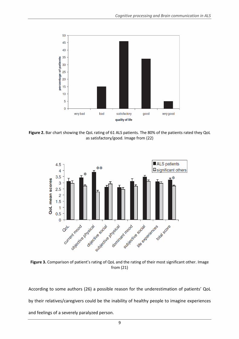
than it really is. Kübler et al.,(22) evaluated 76 ALS patients and assess the level of QoL.
According to the previous hypothesis, they found that the vast majority of the ALS sample
(80%) rated their QoL as satisfactory or good (see Figure 2). Contrarily, partners or
caregivers were also asked to rate patients’ quality of life and the outcome was significantly
lower (see Figure 3).
Cognitive processing and Brain communication in ALS
9
Figure 2. Bar chart showing the QoL rating of 61 ALS patients. The 80% of the patients rated they QoL as satisfactory/good. Image from (22)
Figure 3. Comparison of patient’s rating of QoL and the rating of their most significant other. Image from (21)
According to some authors (26) a possible reason for the underestimation of patients’ QoL
by their relatives/caregivers could be the inability of healthy people to imagine experiences
and feelings of a severely paralyzed person.

María Ángeles Prats Sedano
10
Moreover, depression symptoms have to be taken into account when studying QoL in
neurodegenerative diseases. Some studies have focused in investigating QoL and the
emotional status of ALS patients (21,27) . Despite what is commonly thought that physical
deterioration affect patient’s mood, the link between depression and physical impairment in
ALS is not clear. Some studies support a relationship between depressive mood and disease
stage (22,28,29) while others do not (21,30,31). Lulé et al., (21) carried out a longitudinal
study within the ALS patients and a comparative study between ALS group and healthy
participants. The results of these studies highlighted that physical disability do not correlate
with depression in ALS patients (see Figure 4). More importantly, they found that the
emotional status in ALS is similar to healthy controls. Those results provide evidence that
ALS patients can experience a pleasing QoL without depressive mood even if they are
severely physically impaired.
Figure 4. Scattered Plot of depressive symptoms and physical impairment (measured with the ALS Functional Rating Scale, ALSFRS) in ALS patients. Image from (21).
However, QoL of life in ALS remains a controversial issue. For instance, contrary to those
previous studies, Körner et al., (27) reported QoL was reduced in ALS specially based the
Cognitive processing and Brain communication in ALS
11
positive correlation found between the disease progression and physical impairment.
However, there was no significant correlation between depression and disease duration.
This finding is in line with a previous literature (22) and suggest that patients can develop
strategies to cope with the diagnosis and the physical impairment; therefore depressive
mood is less frequent or at least not progressive with longer disease duration despite
increasing disability.
In conclusion, even if depression occurs among ALS patients, it can be prevented. Patients
can maintain a good QoL and remain free of depression. Thus, factors that contribute to
patient’s QoL, depression, and approaches toward treatment options need to be regularly
examined throughout the course of the disease. Depression should be treated, and patients
have to be provided with unbiased information, including their medical and palliative care
options.
For this purpose, some recommendations can be taken into account to prevent depression
and increase QoL in ALS.
- Assistive devices to improve mobility and autonomy.
- Pharmacological treatment of depressive symptoms.
- Psychotherapeutic support for patients and their relatives.
- Use of augmentative and alternative communication strategies in the late stage of
the disease.
Regardless of depression and physical disability, other areas such as social functioning and
spirituality/religiousness need to be considered when assessing QoL.
María Ángeles Prats Sedano
12
1.6. Late stage of ALS and end of life management:
ALS has a fatal prognosis and death results usually from respiratory failure within 3 to 5
years of symptom onset. Other causes of death include cardiac arrest, coronary disease,
asphyxia and pulmonary embolism (32).
Different areas need to be considered in the end-of life management of ALS patients; these
areas include respiration, nutrition, therapy and exercise, pain, depression and suicide,
spirituality and religion, cognitive changes, the development of advance directives, and care
at the end of life (33). From the pharmacological point of view, the use of riluzole can
prolong survival only by about two to three months in ALS patients (34). Also non-
pharmacological treatments including ventilation and gastrostomy can improve the survival
and quality of life, respectively.
When a patient can no longer tolerate non-invasive ventilation or it stops being effective,
invasive mechanical ventilation with tracheostomy can be used (when available) or patients
can be managed with supportive symptomatic care together with palliative care facilities
(32). If patients accepted to be artificially mechanical ventilated and fed, their life
expectancy can be increased.
ALS disease may lead to severe or complete motor paralysis rendering communication
impossible. The late stage of ALS is divided in two phases: the state of severely paralyzed
patients with residual voluntary control of particular muscles (e.g. eye muscles, lips, fingers)
is known as locked-in state (LIS) (35,36). Patients with several remaining functional motor
channels (as those in LIS) can benefit from assistive communication that uses a range of
augmentative and alternative communication (AAC) strategies, such as eye trackers with
speech-generating devices. However, there are also patients who lose all motor control
Cognitive processing and Brain communication in ALS
13
resulting in the completely locked-in state (CLIS) (37). Thus, these patients have the greatest
need to reestablish communication and interaction with the social environment.
The total number of LIS/CLIS cases is not known for sure, because a high percentage of
patients do not receive assistance in hospitals or in specialized rehabilitations institutes and
instead are treated at home. The lack of prevalence of LIS/CLIS is also explained by the high
misdiagnosis among these patients as being in a vegetative state. The clinical population in
the late stage of the ALS has not been thoroughly investigated, so exhaustive investigations
of this clinical population are required.

María Ángeles Prats Sedano
14
2. COGNITIVE AND BEHAVIORAL IMPAIRMENT IN ALS
The impairment due to ALS is traditionally known to be physical only, but there is a growing
body of literature that suggests the presence of cognitive impairment in ALS patients.
Although the degeneration in ALS disease predominantly affects the motor system, cognitive
and behavioural deficits have been also reported indicating an extra-motor cerebral
involvement in motor neuron disease. The conception of ALS of a heterogeneous disease is
based on the phenotypic variability together with the presence or absence of cognitive
deficits, and the diverse ranges in survivorship (3).
2.1. Spectrum of ALS and dementia
The link between ALS and fronto-temporal dementia was postulated in 1932 (38) and since
then several studies have reported strong clinical, pathological, radiological and genetic
evidence of this association. In addition, several studies suggested that ALS disease and
frontotemporal dementia form a clinical spectrum based in neuropsychological and genetic
tests.
Recently, this continuum between FTD and motor neuron disease has gain support after the
discovery of the C9ORF72 mutation as one of the most frequent genetic linkage in familial
variants of FTD and ALS (39–42). Moreover, few clinical features of cognitive impairment in
ALS as irritability, personality changes, obsessions and deficits in tests that are related to the
executive system (verbal fluency, working memory, attention) are consistent with the
features of frontotemporal dementia.
The study performed by Lillo et al. (41), aimed at investigating the continuum between
bvFTD and ALS based on the performance on neuropsychological and behavioral tests. They
assessed a group of 20 ALS patients and 20 bvFTD patients and a similar cognitive profile was

Cognitive processing and Brain communication in ALS
15
found in both groups in tests related to behaviour, inhibitory control and working memory.
Thus, this result together with the Rasch analysis that was performed exposed an overlap
between bvFTD and ALS.
From a neuroanatomic point of view, neuroimaging investigations have showed frontal and
temporal lobe atrophy (anterior cingular gyrus and frontotemporal areas) in ALS patients
who present cognitive deficits. Those neuroimaging investigations found similar patterns of
atrophy including neuronal loss, superficial linear spongiosis and ubiquitinated taunegative
and synuclein-negative intraneuronal inclusions. These studies revealed an involvement of
some areas of the brain other than the motor cortex and provide some evidence of that
possible continuum between FTD and ALS.
However, this continuum nowadays still remains controversial and needs of a further
investigation.
2.2. Cognitive deficits in ALS
A total of 30 to 50% of ALS patients suffer from cognitive disorders, whom 10-15% meet
criteria for frontotemporal degeneration (43). The current section will provide an overview
of the literature concerning cognitive and behavioural changes in non–demented ALS
patients.
2.2.1. Executive dysfunctions
Executive functions are usually known as high mental processes that control cognitive
processes such as attention, working memory, abstraction, cognitive flexibility, problem
solving and multitasking.

María Ángeles Prats Sedano
16
Traditionally, executive dysfunctions have represented a prominent feature of ALS. ALS
patients have shown deficits related to different components of the executive system
such as verbal fluency, selective attention and mental flexibility (44–49).
Verbal fluency is one of the most salient impairment in the executive domain and have
been reported in the majority of the studies about cognitive impairment in ALS
(39,44,48–51). The most well know neuropsychological tests to assess verbal fluency are
Controlled Oral Word Association Test (COWA) (52) and the Thurstone’s Word Fluency
Test (TWF) (52). However, as those verbal tests require speech motor skills, the
frequency of impaired cognition might have been misrepresented. Thus, Abrahams
designed a modified version of the TWF to control the impact of verbal disabilities in the
outcome of the test. In this modified version the patient is asked to copy the words that
were previously generated. After that, a verbal fluency index was calculated by the
𝑉𝑒𝑟𝑏𝑎𝑙 𝐹𝑙𝑢𝑒𝑛𝑐𝑦 𝐼𝑛𝑑𝑒𝑥: (𝑡𝑖𝑚𝑒 𝑓𝑜𝑟 𝑔𝑒𝑛𝑒𝑟𝑎𝑡𝑖𝑜𝑛 𝑐𝑜𝑛𝑑𝑖𝑡𝑖𝑜𝑛)– (𝑡𝑖𝑚𝑒 𝑓𝑜𝑟 𝑐𝑜𝑝𝑦 𝑐𝑜𝑛𝑑𝑖𝑡𝑖𝑜𝑛)
𝑇𝑜𝑡𝑎𝑙 𝑛𝑢𝑚𝑏𝑒𝑟 𝑜𝑓 𝑤𝑜𝑟𝑑𝑠 𝑔𝑒𝑛𝑒𝑟𝑎𝑡𝑒𝑑
The robustness of this methodology to detect verbal impairments in ALS has been
proved in some studies (44,46,53,54) and the findings of verbal impairments remained
after the application of this procedure. Moreover, verbal fluency deficits seem to be
correlated with neural markers of frontal or striato-frontal lobe dysfunction, so it could
be a good marker to detect frontal impairments (53,55).
Deficits in cognitive flexibility have been reported in some ALS studies. One of the most
popular tests to assess cognitive flexibility is the Wisconsin Card Sorting Test (WCST).
Several studies have described that MND patients showed lower performance in the

Cognitive processing and Brain communication in ALS
17
WCST compared to age-matched healthy participants (50,54,56). However, those results
seem not to be consistent with the ones found in further investigations.
Attention deficits have been also found in different studies (57,58). The assessment of
those deficits seem to be extremely important as some disinhibited patients (a clinical
feature of executive dysfunction) might have normal results in usual neuropsychological
tests of executive system but dysfunctions in selective attention tests. Abnormal
dysfunctions in the attentional network in ALS have been also associated with lesions on
the frontal lobes.
2.2.2. Memory
There is some controversy regarding memory impairment in ALS. Previously, studies
revealing impairments in the memory domain in ALS were less prevalent in comparison
with those reporting executive dysfunctions. Memory impairment in ALS has been
previously ignored or considered as a failure in the memory encoding process specific of
the executive system. However, there are several studies describing memory deficits in
ALS, tested using picture recall, word list learning, pair association learning or story recall
(40,46,56,59–62). These studies have exposed that memory deficits are specially related
to short immediate recall and encoding processing (48,63). Deficits in delay recall are
highly variable and are considered as an abnormality in the encoding rather than in the
speed of forgetting (64). Recent studies in ALS have displayed the involvement of the
temporal lobe reporting significant volume reduction in some medial-temporal lobe
related areas as the basal ganglia (65) and hippocampal TPD-43 pathology (66). As
temporal lobe atrophy is a key feature of Alzheimer disease, Macths and colleagues (67)
aimed at investigating the relationship between memory dysfunctions in ALS and Mild
cognitive impairment (MCI) patients and they found a different pattern in temporal lobe

María Ángeles Prats Sedano
18
dysfunction in between both pathologies. Moreover, memory deficits beyond executive
dysfunctions were found in ALS.
The implications of memory impairment in ALS in clinical management (as medication or
treatment) have been little studied; urging for a great need of further evaluation of
amnesic deficits in ALS which are equally important as executive dysfunctions.
2.2.3. Language
There is some neuro-imaging (PET and MRI studies) evidence that language areas seem
to be damaged in ALS (55), which give support to the fact that ALS disease affects extra-
motor networks of the brain. Recently several studies have highlighted that language
deficits in ALS are more common than previously thought. The most described language
impairments in ALS are associated to word generation (measured by verbal fluency tasks)
(46,59), sentence comprehension and verb processing (59,68) .
The possibility that those language deficits are related to aphasia-like impairment or to
executive dysfunctions is not clear. Primarily language impairments were observed in
association with executive dysfunctions (48,69). However, recently some large
population-based studies showed some evidence that non-demented ALS patients
without executive dysfunctions can have language deficits. Taylor and colleagues (70)
performed a comprehensive language assessment and found mild language impairments
in about the 43% of a sample of 51 non-demented ALS patients. Moreover they found
that the performance in executive and language domain shared only 44% of variance,
and more than the 40% of the patients who had impaired language composite did not
have executive dysfunctions. Taken all the results together, they concluded that
language impairment in ALS could be more common than deficits in the executive system

Cognitive processing and Brain communication in ALS
19
and that those language dysfunctions could also occur in absence of executive deficits.
Abrahams in an editorial commentary (71) suggested that the area of language
impairment in ALS could have been neglected during the past years and propose that
further researches in this area should clarify the nature of language deficits in ALS and
specially determine whether those language deficits are seen as subclinical language
feature of FTD or a specific pattern of impairment in ALS disease.
Former cognitive studies assessing language deficits in ALS performed typical
neuropsychological test batteries that require of physical and verbal abilities, and did not
take in account that speech dysfunctions may interfere with the performance of the
psychometric tests and therefore could pose a problem of masking or over-emphasized
the results. Different authors started to take this problem into account and the tests they
used were designed to control for motor/speech speed and to accommodate for the
range of disabilities that are characteristic of ALS patients (60) or they used
neuropsychological tests that not require verbalizations (72,73). Recently, Abrahams and
colleagues (51) designed a questionnaire to assess cognitive functions in ALS called “the
Edinburgh Cognitive and Behavioural ALS Screen ECAS”, developed for ALS patients with
physical and speech disability. The ECAS was validated in different countries and it seems
to be a sensitive screening tool with high sensitivity and specificity ALS impairments.
Either in the UK and the German-Swing version of the ECAS language was one of the
most impaired domains.
2.2.4. Visuo-perceptual functions
Visuo-perceptual functions are a set of skills we use to gather visual information from the
environment and integrate them with our other senses. These visuo-perceptual
functions include visual discrimination, figure perception and attention.

María Ángeles Prats Sedano
20
Visuo-perceptual dysfunctions seem not to be very frequent in ALS. However, Strong
and colleagues (62) have reported unexpected difficulties in ALS patients on some non-
verbal tests including verbal-perceptual ability.
2.3. Social cognition and emotional processing
There is a growing body of literature suggesting deficits in social cognition and emotional
processing in ALS. Deficits in emotional perception have also been reported in other
neurological disorders such as Alzheimer (74–76), Parkinson (77–79) and mild cognitive
impairment.
Social cognition refers to cognitive processes that facilitate the encoding and decoding of
socially salient information, such as the emotions and intentions of others (80). Previous
studies of social cognition in ALS were basically focused on basic emotion recognition
and Theory of Mind (ToM). ToM is the capacity to attribute and infer to oneself and
other emotional and mental states (thoughts, feelings, desires, intentions) in order to
understand and predict their behaviour (81). Mostly all the studies regarding theory of
mind in ALS were focused in emotional perception. Some authors reported changes in
emotional perception (i.e emotional faces recognition) in both in ALS and FTD patients
(82–84). Papps et al., (85) revealed a deficit for emotional material in ALS, more
concretely, they found a selective failure to show the normative pattern of enhanced
recognition memory for emotional words in comparison to neutral words.
In other hand, the study of Lulé et al, (86) investigating the emotional response to visual
socio-emotional stimuli with different valence and arousal concluded that emotional
processing in ALS tends to be altered showing a more positive response to neural stimuli
and a neutralizing response to extreme stimuli. These results minimize the global impact

Cognitive processing and Brain communication in ALS
21
of the disease in the emotional processing in ALS and suggest a compensatory
mechanism leading to cognitive and/or changes in neuroplasticity of the brain.
In conclusion, the potential clinical implications of dysfunctions in emotional processing
in ALS patients might be a crucial issue because this impairment may aggravate mood
and behavioural disturbance in ALS, as well as increasing the caregiver’s burden.
Importantly, emotional dysfunctions have to be taken into account when planning the
patient’s care or experimental trials, specifically, focusing on the patient’s ability to
evaluate emotional consequences and implications of therapeutic approaches, for them
and their families.
2.4. Behaviour
Behavioural impairment is recognized as a feature of ALS, occurring in up to the 67% of
that clinical population. There is a close link between behavioural changes and FTD
reported by some numerous studies suggesting that ALS patients suffering from FTD
have a propensity to manifest behavioural abnormalities as executive dysfunctions and
disinhibition. Different questionnaires as the Neuropsychiatry inventory, Frontal
Behaviour Inventory and Frontal System Behaviour Scale (87,88) have been used to
assess behavioural changes in ALS, and have identified apathy as the most prominent
behavioral trait in these patients (41).
An important contribution to the field was done by Murphy et al. (89) that assessed a
sample of 274 ALS patients using behavioural validated interviews with accompanying
caregivers and they reported that 27% to 66% of the patients presented higher levels of
apathy, irritability, emotional indifference and poor frustration tolerance. Those
behavioural changes increased the emotional burden in their caregivers. Moreover a

María Ángeles Prats Sedano
22
relationship between cognitive impairment and emotional indifference, aphasia/apraxia
and logopenia was found, showing that ALS patients that present these behavioural
traits are more likely to show cognitive problems. So, the presence of behavioural traits
could alert the clinicians about the occurrence of cognitive decline and thus could signal
the need of an exhaustive neuropsychological assessment and treatment strategies.
Abrahams et al (51) performed a behavioral screening test battery and found behavioral
changes in 40% of a sample of 20 ALS patients. The most common behavioral change was
apathy, followed by loss of empathy and changes in eating behaviour. In contrast to
previous studies, they took into account ALS motor deficits and for this purpose they
used an assessment that was specifically design to this pathology. The result was in line
with the previous literature indicating that apathy is the most prevalent behavioural trait
in ALS (89–91) .
However, apathy could be confused and swap with depression, fatigue and respiratory
function so it is important to distinguish between them by using validated scales and an
exhaustive examination of the medical history of the patient. In addition to apathy,
changes as irritability and lack of inhibition are also found (62) and these behavioural
traits might not be related to cognitive impairment (measured by standardized
neuropsychological testing).

Cognitive processing and Brain communication in ALS
23
3. EVENT-RELATED POTENTIALS AND BRAIN COMMUNICATION IN ALS
3.1. Event-related potentials in ALS
3.1.1. Event-related potentials
Event-related potentials (ERPs) are electrophysiological responses recorded over the
scalp that are related to an internal cognitive event. ERPs can provide significant
information about how the human brain processes information and has been
demonstrated that abnormalities in this processing can give us some information about
neurological or psychiatric disorders.
It is commonly well-known that ERPs reflect synchronous changes of slow postsynaptic
potential occurring within a large number of similarly oriented cortical pyramidal
neurons in a certain area of the cortex. ERPs are short monophasic deflections that are
shown in the background EEG. These deflections are characterized by their polarity
(positive or negative), peak latency (relative to the onset of the event), peak amplitude
(relative to a baseline or peak-to-peak) and scalp distribution (92).
The main advantages of using ERPs to assess cognitive functions/dysfunctions in ALS are
its high temporal resolution and its applicability in cases of severely paralyzed patients
unable to performed neuropsychological tests (72,93,94).
The most studied ERPs are the following ones:
P100: It is a positive deflection in the EEG that it shows up around 50ms/100ms after the
stimulus onset. P100 is not usually easy to identified/visualized. P100 component is a
neurophysiological marker of attentional processing to the sensorial stimulus and it
provides information about the integrity of the sensorial channel used for the

María Ángeles Prats Sedano
24
stimulation. The auditory P100 usually appears before 100ms after the stimulus and it
has its biggest amplitude on the frontal/central areas of the cortex. Contrarily the visual
P100 usually has biggest amplitude in the occipital part of the brain and it varies
regarding the degree of attentional demand to the task.
N100: The N100 is an early ERP component whose characteristics are associated with
cognitive/endogenous processing of the stimuli. N1 is large negative deflection at central
sites around 100 ms after stimulus onset. The N1 is evoked by a relatively unexpected
change that violates a sensorial pattern (95). N100 classically appears together with the
P200 wave and it is usually called “N100-P200 complex”. The N100 in heathy adults,
peaks between 80 and 120 ms after the stimulus onset and it is predominantly
distributed over the fronto-central region of the scalp.
P200: P200 is a positive deflection in the EEG around 200ms after a target stimulus
appears. P200 could be obtained by auditory stimuli or visual stimuli. Auditory P200 is
usually merged with the N100 (N100-P200 complex) and its higher amplitudes are
usually found in the frontal regions of the cortex. Visual P200 is predominantly seen in
the frontal regions and its amplitudes vary when increasing the complexity of the visual
stimulus or of the cognitive task.
The P200 wave of the ERP is assumed to represent a later stage of stimulus processing
and is viewed as an index of both sensory processing and cognitive demands as the
stimulus classification process (96).
Mismatch negativity (MMN): The MMN is a negative component of the ERPs elicited by
any discriminable change in the auditory system, concretely, to a violation of pattern
regularity. The MMN is a negative deflection over the fronto-central and central scalp
electrodes in the difference wave obtained by subtracting the event related potential

Cognitive processing and Brain communication in ALS
25
(ERP) to frequent, ‘‘target’’, stimuli from that to deviant stimuli (97). The MMN
frequently appears around 150–250 ms after the frequent stimulus onset, with this peak
latency getting shorter with the increasing magnitude of stimulus change. MMN
represents a pre-perceptual electrophysiological measure of the accuracy of the central
sound representation in the human brain (98).
In many neurological disorders, MMN amplitude to sound distractor has been revealed
to be smaller or the peak-latency of the component delayed compared to controls.
Figure 5. Mismatch negativity (MMN) evoked during active detection of a deviant stimulus is elicited along with attention-related ERP components N200 and P300. Image extracted from
Nääatänen et at., 2014 (99).
N200: N200 is a negative wave peaking between 180 and 350 ms after stimulus onset in
and oddball task. It is the second negative peak in the averaged ERP waveform and it is
usually observed as a prominent frontocentral negative peak at around 100 ms in the
auditory modality or a prominent temporo-occipital negative peak at around 180 ms in
the visual modality (100). N200 typically appears together with the P300 wave and it is
often referred as the “N2-P3 complex”. N200 reflects cognitive mechanisms that are

María Ángeles Prats Sedano
26
related to the discrimination of the characteristics of the relevant stimulus of the oddball
task. The N200 displays the automatic detection of the novel stimuli in and oddball task
(101). The N200 is a neurophysiological marker of cognitive control and it is associated to
inhibition responses, conflict responses and error monitoring.
P300: One of the most studied ERP components is the P300 wave, a large, broad,
positive deflection of the EEG that typically appears about 250 to 700 ms after the
perception of a “meaningful” target stimulus in an odd-ball task (101,102) (See Figure 6).
Figure 6. Example of an oddball task: two different stimuli are presented in a random
sequence, with one occurring less frequently than the other does (target=T, standard=S). Image
from Polich 2007 (103).
After initial sensory processing, attention-driven comparison process evaluates the
representation of the previous event in working memory. In an oddball paradigm if there
is no identification of the stimulus as the novel one, the current mental schema of the
stimulus context is well-maintained, and only sensory evoked potentials are recorded
(N100, P200, N200). By contrast, if a new stimulus is detected, attentional processes lead
into a change of the stimulus representation that is associated with P300 (See Figure 7).

Cognitive processing and Brain communication in ALS
27
Figure 7. Schema of the context updating theory of P300. Image from Polich 2003 (78)
P300 scalp distribution is defined as the amplitude change over the midline electrodes
(Fz, Cz, Pz), which typically increases in magnitude from the frontal to parietal electrode
sites (104).
There have been discovered two different peaks that characterized the P300:
- P3a that is originated from stimulus driven frontal attention mechanisms during task
processing.
- P3b originates from temporal parietal activity associated with attention and appears
related to subsequent memory processing.
The P300 wave it have been demonstrated to be a sensitive neurophysiological
marker of cognitive processes including attentional demand and cognitive workload
(54,103,105).
N400: The N400 is a negative deflection in the EEG that occurs approximately 400 ms
after a meaningful stimulus onset. It has been linked to the semantic integration of a
given stimulus into a previous context (106).

María Ángeles Prats Sedano
28
This component was first discovered in 1980 by Kutas and Hillyard (106) as a response to
semantic anomalous sentence endings in linguistic paradigms (see figure 8). However,
similar effects were lately observed related to non-linguistic material involving
meaningful actions (107). N400 could be elicited by different kind of stimulus including
written, spoken, and signed (pseudo)words, drawings, photos, and videos of faces,
objects and actions, sounds, and mathematical symbols.
In the linguistic domain, the N400 is a robust neurophysiological marker of semantic
processing. Studies investigating the N400 have reported that its latency remains
remarkably constant. Contrarily its amplitude seems to be very variable and it is sensitive
not only to the degree of semantic incongruity per se but also to several other factors.
Thus, the N400 it is not just a marker of a violation of a pattern; it gives us information
about the processing of the meaning. There is some growing evidence that the
meaningful/non-meaningful dimension might be more important than the
linguistic/nonlinguistic dimension (108).
Figure 8. Image of the N400 effect. Image modified from Steinhauer 2014 (84)

Cognitive processing and Brain communication in ALS
29
3.1.2. Event-related potentials studies in ALS:
Event-related potentials (ERPs) have been used to assess cognitive and clinical status of ALS
patients whose cognitive functions cannot be easily expressed in their behavior (109).
Different ERP components have been studied in ALS disease using different paradigms (93).
Studies concerned with differences in ERPs between ALS patients and healthy controls have
been controversially discussed. Literature reporting abnormalities in the ERPs in ALS is
mixed: there are several studies reporting significantly longer latencies in the ERPs
components (110–112), smaller amplitudes (93,113), and both latencies and amplitudes
(58,61).
In contrast, other studies reported comparable ERPs amplitudes and latencies between ALS
patients and healthy participants (114).
Table 4 summarized the main studies regarding ERPs abnormalities in ALS.

María Ángeles Prats Sedano
30
Authors ALS Patients/Controls
ERPs paradigm used ERPs abnormalities in ALS patients
Gil et al., 1995 (110) 20/20 Auditory oddball paradigm. Delayed latencies in the N200 and P300.
Münte et al., 1998 (113) 8/8 Visual recognition memory test: immediate and delay recall.
Reduced amplitudes of the N400 and enhancement of a late positive component (LPC) for repeated items.
Vieregge et al., 1999 (115) 8/8 Auditory oddball. Processing negativity (PN) was recorded.
PN was smaller in ALS patients.
Hanagasi et al., 2002 (61) 20/13 P3b classical auditory oddball paradigm, P3a in a novelty paradigm, CNV and MMN.
Smaller P3a and P3b amplitudes. Delayed P3a latencies. Mean CNV were higher.
Paulus et al., 2002 (111) 16/30 Visual and auditory oddball. Delayed latencies in the P300 in both paradigms.
Kotchoubey et al., 2003 (116)
3 ALS patients in CLIS
Passive auditory oddball, emotional oddball, word-pairs, learning paradigm, movement intention.
Patient 1: P300 wave was observed in the auditory and word-pair tasks. Patient 2: N300 observed in the auditory and emotional oddball. Movement intention showed adequate preparation of the motor areas. Patient 3: ERPs responses were not found.
Raggi et al., 2008 (93) 10/10 Passive auditory three stimuli oddball.
Lower amplitudes in the N100, P3a and MMN.
Pinkhardt et al., 2008 (117)
20/20 Auditory oddball using four types of tones. N100, Nd, MMN and P300 were recorded.
Decrease of the fronto-precentral negative difference wave (Nd). Analysis of the P300 showed increased processing of non-relevant stimuli.
Ogawa et al., 2009 (112) 19/19 Active auditory oddball. Prolonged N1/N2/P3 GFP latencies.
Volpato et al., 2010 (54) 24/17 Active auditory oddball. Delayed N1/P2/N2 latencies.
Silvoni et al., 2015 (118) 14/10 Vibro-tactile oddball. Delayed N2
Volpato et al., 2016 (58) 15/15 Active auditory oddball with four types of tones. N200, P300 and re-orienting negativity (RON) components were analyzed.
Reduced amplitudes and delayed latencies of N200, P300 and RON:
Table 4. Main ERPs studies in non-demented ALS patients.

Cognitive processing and Brain communication in ALS
31
The results of the aforementioned studies demonstrate significant alterations in the
different stages of the information processing in ALS. Those results suggest that ERPs
evaluation could be of central importance to assess the clinical and cognitive profile of
ALS patients. Moreover, some studies (54,116) proposed that ERPs could serve as a
strategy for the evaluation of cognitive impairment in cases of severely paralyzed
patients (such as ALS patients) unable to perform cognitive test that requires overt
motor/verbal responses.
3.2. Brain communication in Amyotrophic Lateral Sclerosis
3.2.1. Introduction to Brain Computer Interfaces
Brain Computer Interfaces (BCI) or brain–machine interfaces (BMI) are systems that
decode brain activity and transform it into commands to control external devices. Some
of the applications of BCIs are to achieve direct brain communication in completely
paralyzed patients (such as those suffering from ALS) and restoration of movement in
paralyzed limbs (i.e in patients with severe stroke) through the transmission of brain
signals to the muscles or to external prosthetic devices (119).
From an historical perspective, Hans Berger, who discovered the EEG, was the first to talk
about the possibility of reading thoughts from the EEG signal by using refined
mathematical analyses. About 40 years ago, Jacques Vidal coined the term “Brain-
Computer Interface” when presented a system capable of decoding EEG signals and
transform it into computer control commands. He predicted in this visionary paper (120):
“As the reader undoubtedly realizes, direct brain-computer communication still lies
somewhat in the future. Even the relatively modest experimental program outlined in this
paper may take several years to reach maturity, at which time new directions probably

María Ángeles Prats Sedano
32
will have emerged. In summary, it can be said that the feasibility of the communication
concept rests on three basic assumptions. The first assumption is that mental decisions
and reactions can be probed, in a dimension that both transcends and complements overt
behavior, from the array of observable bioelectric signals and, in particular, from the
electroencephalographic potential fluctuations as measured on the human scalp. A
second assumption is that all meaningful EEG phenomena should be viewed as a complex
structure of elementary wavelets, similar in nature to components of evoked responses
that sequentially reflect individual cortical events and create a continuous flow of
neuroelectric messages. The third assumption is that operant conditioning procedures
can increase the reliability and stability of these time signatures and patterns. Admittedly
the validity and implications of these assumptions are far from universally accepted”.
Types of BCIs
Invasive BCIs: Invasive BCIs use activity recorded by brain implanted micro or
macro-electrodes. Different types of brain activity could be measured with
invasive BCIs: local field potentials (LFPs) (121,122), single-unit activity (SUA)
(123–125), multi-unit activity (MUA)(121), electrocorticographic activity (ECoG)
(126,127), and calcium channel permeability (128).
Non-invasive BCIs: Non-invasive BCIs use brain signals recorded using sensors
over the scalp. Different brain signals are used to control a BCI as: Slow cortical
potentials (129,130), sensory motor rhythms (SMR) and motor-related beta
rhythms (131), event-related potentials (ERPs) (i.e the well-known P300 (132)),
steady-state visual or auditory-evoked potential (133,134), blood oxygenation
level-dependent (BOLD) imaging using functional magnetic resonance imaging

Cognitive processing and Brain communication in ALS
33
(fMRI) (135,136), concentration changes of oxy/deoxy hemoglobin using near-
infrared spectroscopy (NIRS)) (137).
Figure 9. General framework of brain–computer interface (BCI) systems. Invasive BCI approaches (left) include the measurement of local field potentials (LFPs), single-unit activity (SUA), multi-
unit activity (MUA), and electrocorticography (ECoG). Noninvasive BCI approaches (right) include EEG, near-infrared spectroscopy (NIRS) and blood oxygenation level-dependent (BOLD)
functional MRI. Brain signals are processed to extract features relevant to the aim of the BCI (for example, communication) and then classified using a translational algorithm to construct a
control signal that drives the BCI. BCIs can be classified as assistive to help patients with communication or movement, or as rehabilitative to help recover neural function. Image and
legend from (138).
Additionally, a BCI system can be characterized according to the control task required to
the user, which is the mental task that the participant is asked to perform in order to
generate the brain signal used to drive the BCI. We can differentiate between two types
of brain signal to control a BCI:
Exogenous (evoked) brain signals to drive BCIs: consists of brain responses
generated in response to specific stimuli. For instance, external stimuli might

María Ángeles Prats Sedano
34
trigger evoked potentials (EP) in the EEG which can be identified by the BCI
system. For instance, in the P300 paradigm different external stimuli can be
employed both visual (114,132,139), auditory (140,141) or tactile (142). During
the BCI experiment the user is asked to pay attention to a particular stimulus.
Endogenous (self-generated) brain signals: consists of brain activation pattern
occurring during normal brain function and initiated by the participant. In this
case BCIs require of a training period in which the user has to learn how to
produce a certain brain oscillatory pattern that is associate with the control of
the system. In this type of BCI the feedback plays an essential role since it
produces a change in the brain signals required by the paradigm. On the other
hand, this type of BCI is more sensitive to physiological and psychological state
of the participant, i.e. motivation, fatigue, etc. The most employed endogenous
EEG signals for BCIs are characterized by slow cortical potentials (SCPs) (129)
and brain oscillations associated with sensory processing and motor behavior.
The parts of a BCI:
BCIs requires of an input (e.g. electrophysiological activity from the user), output (i.e. device
commands), components that decode inputs into outputs, and a procedure that determines
the onset, offset, and timing of operation (131). A functional BCI system is divided in the
following parts:
Signal acquisition: In the signal-acquisition part, the chosen input is acquired (by the
recording sensors), amplified, and digitized.
Signal processing and feature extraction: After digitalizing the signals and pre-
processing the data, feature extraction procedures are required (e.g spatial filtering,

Cognitive processing and Brain communication in ALS
35
voltage amplitude measurements, spectral analyses, or single-neuron separation).
This analysis extracts the signal features that encode the participants’ commands.
BCIs can use signal features that are in the time domain, in the frequency domain or
both. In general, the signal features used in present-day BCIs reflect distinguishable
brain events.
Pattern Classification: After extracting the signal features, a translation algorithm is
required to translate these signal features into device commands that carry out the
user's orders. This algorithm might use linear models (e.g. linear discriminant analysis
(LDA) or support vector machine (SVM)) or nonlinear methods (e.g. neural networks).
The output device: This output is the feedback that serves to the participant to learn
how to modulate his/her brain activity and to improve the accuracy in the
performance.
A schematic view of the BCIs parts and its functionality is shown in Figure 10.

María Ángeles Prats Sedano
36
Figure 10. Schematic diagram representing the information stream in a BCI, image from (143).
3.2.2. Brain computer interfaces for communication in the late stage of ALS
BCI-based communication involves generation of brain signals by the patient to drive
alphanumeric grids, binary cursers, and/or web browsing tools to formulate sentence or
express feelings or desires to the caregivers/family members (138).
In the past several BCI applications for communication (invasive and non-invasive) had
been used in severely paralyzed patients as those with ALS (144–148) or with severe
brain damage, such as stroke or spinal cord injury (139,149,150). Nowadays, the target
population to use a BCI for communication is represented mainly by patients in the late
stage of ALS who suffer the completely locked-in syndrome.
Since the first application of BCIs using non-invasive EEG-recordings in the LIS patients
published in “Nature” in 1999 by Prof. Birbaumer and co-workers (129), several groups
have shown that different types of non-invasive brain-computer interfaces allow
communication with locked-in patients. However, in a meta-analysis it have been shown

Cognitive processing and Brain communication in ALS
37
that while brain-computer interfaces allow communication with locked-in patients, all
attempts to communicate with a patient in a completely locked-in state (CLIS) failed
(37,119,147). Kübler and Birbaumer in 2008 (36) exposed that CLIS patients do not
achieve adequate control of their brain activity to enable communication using EEG
signals. Specific cognitive problems and abnormal neurophysiological signatures in the
EEG might be, at least in part, responsible for the failure in CLIS. In this meta-analysis of
all completely locked-in patients at that point in time, they hypothesized that complete
paralysis with no output-channel available leads to extinction of goal-directed thinking
and extinction of intentional communication and thought. If no attempt to express
desires and wishes is followed by the anticipated consequences, extinction of this
specific type of behavior and thinking should follow.
However, De Massari and co-workers in 2013 provide evidence that communication
could be achieved in the CLIS (151). In their study, a novel paradigm was introduced
using Pavlovian semantic conditioning for online classification of EEG signals to
discriminate between covert (cognitive) ‘yes’ and ‘no’ responses. The paradigm included
the presentation of true and false statements used as conditioned stimuli, and
unconditioned stimulus consisted of skin electrical stimulation paired with affirmative
statements. The results suggested that a reliable level of accuracy in the BCI
performance in the CLIS patient was not achieved uniformly throughout the 37 sessions
(despite intact cognitive processing capacity), but in some sessions communication
accuracies up to 70% were reached.
In order to improve levels of accuracy of the aforementioned BCI study, other
methodologies using different neurophysiological approaches were tested by Tübingen

María Ángeles Prats Sedano
38
research group. The first prototype introducing fundamental strategy changes was
proposed by Gallegos-Ayala et al. in 2014 (137).
3.2.3. fNIRS-EEG based BCIs for communication in the late stage of ALS
Functional near-infrared spectroscopy (fNIRS) is a non-invasive technique used to
measure brain activation on the basis of cerebral hemodynamic response. It measures
regional cerebral oxygen saturation and higher temporal changes in hemoglobin
concentration in brain tissues using trans-illumination spectroscopy. Measurement of
tissue oxygen saturation and tissue hemoglobin content is determined by the difference
in intensity between a transmitted and received light delivered at specific wavelengths.
Some advantages of fNIRS compared to other neurophysiological techniques are its
applicability at bedside, its reasonably low cost as neuroimaging technique and the short
preparation times that it requires.
Recently, fNIRS based BCI was developed in Prof. Birbaumer lab in the University of
Tübingen, based on classical semantic conditioning, for communication in CLIS. Gallegos-
Ayala et al. (2014) (137) for the first time described a case of a CLIS patient with ALS that
could establish “yes/no” communication to simple questions with known answers using a
fNIRS-based BCI system. fNIRS classified cortical oxygenation and deoxygenation in brain
tissues during following the questions. The BCI methodology used in this study contrast
with the previous BCI approaches (151) and propose a more “reflexive” mode based on
learning principles of classical conditioning that makes easier to differentiate between
yes and no answers responses. The classifier was trained separating brain responses to
patient’s yes and no answers. While EEG-BCI study of de Massari (151) resulted in
significant (above 70%) classification of “yes” and “no” responses in only 7 of 37 sessions
and no discriminatory answer in brain oscillations to the images at frequencies between

Cognitive processing and Brain communication in ALS
39
3 and 30 Hz, fNIRS-BCI study of Gallegos-Ayala obtained significantly above chance-level
answers with an overall performance of 76.30% in the last training period and the 100%
correct performance across 14 consecutive sessions. Therefore, we can propose fNIRS-
BCI as a viable approach to enable communication in CLIS.
Hence, to validate these preliminary findings of Gallegos-Ayala et al. (137), and refine the
FNIRS-BCI technology for communication in the CLIS some studies were performed by
our BCI group combining the use of on four ALS patients in CLIS using combined NIRS-
EEG-based BCIs (submitted for publication). fNIRS-EEG-based BCI was used to train four
CLIS patients to regulate their fronto-central brain activity to answer yes/no questions.
These successful preliminary results could indicate a next step towards to unlock the
CLIS.

María Ángeles Prats Sedano
40
4. ASSESSING COGNITIVE FUNCTIONING IN AMYOTROPHIC LATERAL
SCLEROSIS WITH A NOVEL COMBINATION OF VIBRO-TACTILE P300 AND
NEUROPSYCHOLOGICAL TESTING
4.1. Introduction
Amyotrophic Lateral Sclerosis (ALS) is a motor neurodegenerative disease that is
characterized by a progressive degeneration of the upper and lower motor neurons, causing
progressive muscular paralysis of limbs and bulbar musculature. The disease generally
progresses to total paralysis and has a fatal prognosis unless the patient is artificially
ventilated and fed.
In addition to motor impairment, a widespread fronto-temporal degeneration with
corresponding cognitive deficits has been reported in half of the ALS patient population
(44,45,50), Ringholz and colleagues (40) have investigated the nature of cognitive changes
associated with sporadic ALS in a large sample of 279 patients at different stages of the
disease. This study has reported that about 51% of the patients with ALS showed various
degrees of cognitive impairment. That is, 30% displayed mild cognitive impairment and
approximately 20% had severe deficits in multiple cognitive domains that met the criteria for
dementia.
The most often reported cognitive impairments in ALS involve components of the executive
system (e.g., verbal fluency, selective attention and mental flexibility) (44–49). Deficits in
verbal fluency have been reported in almost all studies about cognitive impairment in ALS.
Verbal fluency deficits are considered as a sensitive indicator of damage in frontal and
striato-frontal areas of the brain (46,48,50). However, as the majority of verbal fluency tests
Cognitive processing and Brain communication in ALS
41
require speech motor skills, the frequency of impaired cognition might have been
overestimated (94). With respect to executive functions, the attentional network is most
commonly affected in ALS (54,117). With respect to language functions, language
impairment in ALS is an area that has been partially neglected. However, some studies have
contribute to the fact that language deficits in ALS are at least as common as executive
dysfunctions (48,70,152). For instance, Taylor and colleagues (70) in their study they
highlighted the occurrence of language deficits in non-demented ALS. They reported that the
43% of the patients of a sample of 51 ALS patients showed language dysfunctions (in
contrast to the 31% of patients having deficits in the executive domain). They suggested that
even if these two domains are strongly coupled, executive dysfunction does not fully
account for the cognitive profile of language impairments observed.
Other studies have also found cognitive deficits to be related to the memory (67,153,154)
yet at lower prevalence rates.
The assessment of cognitive functions in late stages of the disease is one of the most
challenging problems. Due to motor paralysis, patients have a limited ability to
communicate. This might either mask or over-emphasize the detection of cognitive deficits
(72,154). As patients are facing important decisions, such as choosing their clinical treatment
(medication, artificial ventilation and nutrition) as well as expressing competently their will
to live (155), the assessment of cognitive impairment in ALS remains a crucial issue.
Event-related potentials (ERPs) have been used to provide information about the clinical and
cognitive status of ALS patients, complementary to neuropsychological testing (54,58,61,72).
One of the most studied ERP components is the P300 wave, a positive deflection of the EEG
that appears about 250 to 700 ms after the perception of a “meaningful” target stimulus in

María Ángeles Prats Sedano
42
an odd-ball task (101). The P300 wave is a sensitive neurophysiological marker of cognitive
processes including attentional demand and cognitive workload (54,103,105). In ALS, studies
concerned with differences in P300 waves between patients and healthy participants have
been controversially discussed. Some ERPs studies revealed a significantly longer P300
latency in the in ALS group than in the control group (110,111) as well as reduced P300
amplitudes (69). In contrast, other studies reported comparable P300 amplitudes and
latencies in both groups (54,114). These irregularities in the P300 features in ALS could
reflect a dysfunction in the cognitive evaluation of the significance of a stimulus and might
therefore serve as a physiological marker for cognitive impairment in ALS (61,110,112).
Abnormal P300 features have been also reported in other neurodegenerative diseases, such
as Alzheimer’s (156–158) and Parkinson’s disease (159,160) or Multiple Sclerosis (161,162).
These studies found a relationship between P300 features and cognitive decline, such as a
delayed and/or a reduced P300 response which could indicate a globally impaired cognition
and attention.
Interestingly, most ERPs paradigms concerning cognitive function in ALS used visual and
auditory stimuli (54,111,112), whereas somatosensory stimuli have been less frequently
used. However, somatosensory ERPs might be particularly advantageous in late stages of ALS
disease, when visual stimulus presentation fails to elicit reliable ERPs due to the often
severely compromised vision due to reduced oculo-motor control and eyelid weakness
(151,155,163). The somatosensory pathway might therefore represent a promising
alternative or adjunctive pathway to elicit eye-gaze independent ERPs, as it has been
demonstrated with healthy subjects using electrical and mechanical stimulation of hands
and wrists (164,165). Brouwer and Van Erp (142) established that vibro-tactile stimuli elicit
robust P300 waves suitable for BCI-based communication in healthy participants. The same

Cognitive processing and Brain communication in ALS
43
technology was also used to restore communication in severely paralyzed patients following
brain injury (166). In a recent case study with a Locked-in syndrome (LIS) patient, tactile
stimuli presentation modality has shown to be highly efficient in terms of BCI-accuracy. This
type of tactile presentation was also superior compared to visual and auditory modalities
(167). In our previous work, we have already demonstrated that vibro-tactile event-related
potentials can be reliably triggered in ALS patients (118).
In the present study, we aimed to investigate cognitive functioning by neuropsychological
and neurophysiological testing, using the somatosensory system to elicit ERPs in a group of
mild to moderately progressed ALS patients compared to a healthy control group. Based on
prior evidence, suggesting fronto-temporal dementia and cognitive deficits in ALS, we
anticipated a significant difference in psychometric and neurophysiological measures
between the two groups. Moreover, we expected the neuropsychological and
neurophysiological test outcome to be significantly correlated with the patients’ clinical
status. We also hypothesized that declined neuropsychological performance would correlate
with reduced amplitudes and delayed latencies of the vibro-tactile P300s in the ALS group.
As the primary somatosensory system is reciprocally connected with the primary motor
cortex (severely affected in ALS) and as fronto-parietal degeneration might alter processing
in secondary and posterior insula cortices (50,168), these systems should also reflect
changes in both the cognitive and the somatosensory domain.
4.2. Materials and Methods
4.2.1. Participants
Fifteen ALS patients were recruited from the Department of Neurorehabilitation of the San
Camillo Hospital (Venice, Italy) for participation in this study. ALS was diagnosed according to

María Ángeles Prats Sedano
44
the revised El-Escorial criteria (169). All patients were in a mild or moderate stage of the
disease according to the ALS functional rating-scale (ALSFRS-R) (170) with a mean score of
28.6, where 0 represents total dependency and 48 complete functionality. All patients were
under riluzole treatment.
The control group consisted of thirteen healthy participants. Healthy participants with a
history of neurological and psychiatric disorders were excluded.
Demographic and clinical data are given in Table 1. Patients with a diagnosis of ‘ALS-
dementia’ were excluded from the current study. Depression was excluded by psychological
assessment according to DSM-V criteria (171).
All participants (or their authorized representatives where applicable) gave written informed
consent according to the Declaration of Helsinki prior to their participation in the study. The
San Camillo Hospital Ethical Committee approved the study. Clinical and demographic data
are shown in Table 5.
Table 5. Demographic and clinical data. Standard Deviation (SD).

Cognitive processing and Brain communication in ALS
45
4.2.2. Neuropsychological Assessment
Neuropsychological assessments were conducted in both groups using standardized tests
that requires minimal overt motor or verbal responses and could also be administered to
patients with severe motor impairment. This battery contains four tests to assess
intelligence, language, memory and executive functions respectively. The following tests
were included:
Non-verbal Intelligence:
- Raven’s Colored Progressive Matrices (RCPM) (172): this test highlights analytical
skills independently from the patient’s previous knowledge. The test consists of 36 items,
grouped in three different sets (A, Ab, B). For each item of the test, the subject has to
identify a missing piece (by pointing or looking at it) that completes a larger pattern in a
multiple-choice format (6 different choices possible) (see Figure 11). The number of correctly
matched items was used as the dependent variable.
Figure 11. First item of the first set of the Raven’s Colored Progressive Matrices (RCPM) (172)

María Ángeles Prats Sedano
46
Executive functions:
- Modified Wisconsin Card Sorting Test (M-WCST) (173): this test is used to
assess executive functions, such as abstraction and cognitive flexibility. During the
test, patients are asked to match cards to one of four key cards according to a
changing criterion (form, color or number) (see Figure 12). During the task the
participant has to learn a sorting strategy based on the feedback given by the
examiner. After the subject achieved the last of the three sorting criteria, the three
categories are repeated in the original order. Two patients aborted the test
prematurely due to fatigue.
Figure 12. Example of the configuration of the Modified Wisconsin card sorting test (MCST).
Memory:
- Rivermead recognition of figures and faces (174): as part of a larger memory test
battery, this test is used to detect impairments of everyday memory functioning to assess
“ecological” memory. On the “Rivermead recognition of figures” subtest, 10 cards of
different pictures of figures are presented. While presenting the cards, the subject has to
name the figures. About ten minutes later the subject is asked to recognize the 10 figures
out of a set of 20 figures. On the “Rivermead recognition of faces” subtest, 5 pictures of

Cognitive processing and Brain communication in ALS
47
different faces were presented to the subject. While the cards were presented, the subject
has to distinguish if the person in the picture is a man or a woman, and younger or older
than 40 years old. Ten minutes later the subject has to recognize the 5 out of 10 faces.
Language:
- Oral and written comprehension of words and sentences from the Aachener Aphasie
Test –AAT (175): this subtest is used to assess language comprehension. During the test,
participants are instructed to match a verbal or written stimulus to one out of four images.
In the first part, the examiner has to read aloud the target stimulus (words and sentences)
while the participant has to match it with one out of four images. In the second part, the
participant recites mentally and matches the target stimulus with one out of four images.
Each part of the test consisted of 10 items. The four pictures of each item are chosen in such
a way that inappropriate pictures have phonetic or semantic similarity to the stimulus word
or a thematic and syntactic-morphological similarity to the stimulus sentences.
The tests described above were chosen because they were less vulnerable to the effects of
physical disability (i.e., the inability to talk or move the upper limbs) and because reaction
time was not an essential factor. For one patient with severe motor impairment and speech
difficulties, after common agreement, we changed the response type to a residual motor
signal. We especially used an eye blink to specify a yes/no response for target stimulus
selection. This procedure had previously been successfully employed to assess cognitive
functions in severely disabled individuals (72). As the neuropsychological tests required
more time in this particular patient, we performed the assessment across two different days.
For all patients, we included shorts breaks of 5 minutes between tests in order to avoid
fatigue.

María Ángeles Prats Sedano
48
4.2.3. ERPs assessment and data acquisition and processing of the EEG
Participants were seated in a chair with their arms resting on the table and grasping a
vibrating stimulator with their left hand (see figure 13). In case patients experienced
difficulties with holding the device, an elastic band was used to attach the hand to the
effector. Furthermore, ear-plugs were used to mask the sound of the vibrating unit. The
vibro-tactile “oddball” paradigm consisted of a non-target stimulus (probability 70%,
frequency 20Hz) and a target stimulus (probability 30%, frequency of 100Hz). Tactile stimuli
were applied with a Phantom® Premium 3.0 6 DOF end-effector (Sensable Technologies, Inc.
Canada). A stimulus consisted of a sinusoidal force field along the x-axis of the end-effector
lasting for 600ms with the stimulation magnitude set to 1.5N in both cases. A total of 160
randomized stimuli were delivered (divided on four blocks of 40 stimuli) with an inter-
stimulus interval of 2-sec.
During the test, participants were asked to close their eyes and to mentally count target
stimuli. At the end of each session, they reported their target count.
Electroencephalogram (EEG) was recorded during the oddball task using a gUSBAmp
amplifier (g.tec OG, Graz, Austria) and 4 Ag/AgCl scalp electrodes (Cz, Pz, P3, P4) positioned
according to the International 10–20 System. All electrodes were referenced to the left ear
lobe and grounded at FPz. The electro-oculogram (EOG) was recorded from electrodes
placed on the right outer epicanthus and upper medial rim. Inter-electrodes impedances
were kept below 5KΩ.
EEG was sampled at 512Hz by the BCI2000 platform (176). EEG data were segmented in
epochs of 1250ms synchronized with each stimulus, including 250ms before and 1000ms
after stimulus onset (synchronization error smaller than 5ms (177,178)).

Cognitive processing and Brain communication in ALS
49
Epochs with signal amplitudes exceeding ±70μV (141,179,180) or containing ocular artifacts
were rejected (identified by the second order statistics blind source separation algorithm
implemented in the Automatic Artifact Removal toolbox for EEGLAB v10.2.5.8b). An average
of 95 non-target and 41 target epochs were included in the following analysis for each
participant.
After artifact rejection, non-target and target epochs were low-pass filtered at 20 Hz,
baseline corrected using pre-epoch interval from -250ms up to stimulus onset and averaged
for targets and non-targets respectively.
P300 features were analyzed in each participant and each channel. A P300 wave was defined
as the maximum positive peak between 250 and 700 ms (103). In case of ambiguous
identification of the P300 peak, two expert neurologists were involved and their judgment
about the P300 peak latency was taken into account. The P300 amplitude was identified by
means of peak-to-baseline measurement (104). This part of the methodology was also
described in a previous paper (118).
Figure 13. Healthy participant performing the ERPs experiment.

María Ángeles Prats Sedano
50
Figure 14. In an oddball paradigm two types of stimulus are presented. Each stimulus differs in it frequency: the non-target stimulus had a probability of 70% and a frequency of 20Hz, while the
target stimulus had a probability of 30% and a frequency of 100Hz.
4.2.4 Median-nerve somato-sensory evoked potentials (SSEPs)
To assess the transmission integrity of the afferent pathway, we performed median-nerve
SSEPs of the upper limb. Median nerve stimulation at wrist was performed with a stimulus
intensity that elicited sustained thumb twitching at a frequency of 3 Hz. Electrical stimulation
was delivered and recorded by Keypoint® 4 (Medtronic A/S, Tonsbakken, Denmark). Cortical
waveforms were recorded using two Ag/AgCl scalp electrodes placed on C3’-C4’ (according
to the international 10-20 system) and referenced to Fpz.
N20 was recorded over the somatosensory cortex contra-lateral to the stimulated wrist. We
measured the latency of the N20 and the amplitude between N20 and P25 peaks.
4.2.5 Statistical analysis
Statistical analyses of the results were carried out using SPSS (IBM SPSS Statistics 22, 2013,
USA) and Matlab (The Mathworks Inc., USA). The normal distribution of the data was
Cognitive processing and Brain communication in ALS
51
assessed using the Kolmogorov-Smirnov test. The assumption of normality was not violated
in any of the data. Parametric t-tests for independent samples were used to compare the
ALS and the control group concerning demographic and neuropsychological data.
The neurophysiological data was analyzed using a repeated measures analysis of variance
(ANOVA) with “group” (ALS vs Controls) as between-subjects factor and “electrode position”
(Cz, Pz, P3 and P4 for ERPs; C3’ and C4’ for SSEPs) as within-subjects factor. Statistically
significant differences were further assessed with post-hoc t-tests. The relationship between
clinical, neuropsychological and neurophysiological data in the patient’s and the control
group was analyzed using Pearson´s rank correlation coefficient.
In order to control for a possible confounding effect of age on P300 latencies in the ALS
group, we first split the ALS sample in two subgroups with (a) severe cognitive impairment
(in at least one neuropsychological test/sub-test) and (b) non-severe cognitive impairment in
none of the neuropsychological tests/subtests. We then performed an analysis of covariance
(ANCOVA) with “sub-group” as between-subjects factors and “P300 latency electrode
position” (Cz, Pz, P3 and P4) as within-subjects factor, while controlling for “age” as a
covariate. Differences between sub-groups were calculated with a parametric post-hoc t-test
for independent samples. The results of this test are used to determine whether potential
differences in P300 features in ALS are due to cognitive decline independent from age-
related effects.
Based on the exploratory nature of this study, we decided against correction for multiple
comparisons in correlations analyses. The significance level for all statistics was set to 0.05.

María Ángeles Prats Sedano
52
4.3. Results
4.3.1. Demographic and clinical data
No statistically significant differences were found between groups (i.e., ALS patients and
control participants) with respect to sex, age and education. For the comparison of ALS sub-
groups (i.e., severe and non-severe cognitive impairment), age was not significantly
different. Demographic and clinical data is shown in Table 6.
Table 6. Demographic and clinical data. Standard Deviation (SD).
4.3.2. Neuropsychological performance
A significant group difference was found in AAT oral comprehension, revealing a lower
performance in ALS patients compared to controls (t (26)= 1.51, p=0.011). No differences
were observed between these two groups in other tests. Group means for non-verbal
intelligence, memory, executive function, and language tests were within the normal range
according to normative data. A summary of the neuropsychological data is given in Table 7.

Cognitive processing and Brain communication in ALS
53
Table 7. Neuropsychological assessment of patients and healthy participants. Raven’s Coloured Progressive Matrices (RCPM). Modified Wisconsin Card Sorting Test (M-WCST). Rivermead
Behavioural Memory test (RBMT). Aphasie Aachener Test (AAT). Standard Deviation (SD). Significant p-values in bold.
4.3.3. P300 components
P300 wave components were obtained in all participants and no significant differences were
found in the silent target count between groups. However, ANOVA revealed a significant
interaction between “group” and “electrode position” for the P300 amplitudes (F2,52 =
11.32, p< 0.01). Paired t-tests showed that significantly diminished P300 amplitudes could
only be found for the Cz electrode (t (26)= 2.92, p= 0.007); in the ALS group. No differences
were found for the P300s latencies.
In order to investigate whether differences in patient’s age could be a confound in our
results, we performed an ANCOVA analysis with the factors: ALS “sub-group” (severe vs non-
severe cognitive impairment according to Italian normative data (173,181) in at least one
neuropsychological test/sub-test) and “electrode position for the P300 latencies” while
controlling for the effect of “age” as a covariate. Results showed an effect of cognitive
impairment severity in ALS on P300s latencies (F4,9 = 22.76, p< 0.01), independent of age.

María Ángeles Prats Sedano
54
Paired t-tests revealed significant differences between ALS sub-groups (Cz: t (13)= -2.70,
p=0.018; Pz: t (13)= -3.4, p< 0.01; P3: t (13)= -2.92, p= 0.012, ; P4: t (13)= -2.95, p= 0.011),
indicating that the ALS subgroup with low cognitive abilities (i.e., severe impairment)
showed longer P300 latency delays compared to ALS patients with non-severe cognitive
impairment.
Figure 15 illustrates the average ERPs response in ALS patients and healthy participants.
Table 8 shows the data for P300 amplitudes and latencies from Cz, Pz, P3 and P4 in both
groups.
Figure 15. Global average of target and non-target ERP response in healthy participants and ALS patients at channels Cz, PZ, P3 and P4.

Cognitive processing and Brain communication in ALS
55
Table 8. P300 features. Mean latencies and amplitudes of vibro-tactile P300 at channels Cz, Pz, P3 and P4. Standard deviation (SD). Significant p-values in bold.
4.3.4. Median nerve somato-sensory evoked potentials data
Repeated measures analysis of variance did not reveal any significant main effect of “group”
factor for N20 features. The “group” X “electrode position” for N20 features was not
significant. Table 9 shows the N20 data comparing both groups.
Table 9. Median nerve somatosensory evoked potentials (SSEPs). Mean amplitudes and latencies ± standard deviation of N20 median nerve evoked potentials at channels C3 and C4.
4.3.5. Relationship between patient´s clinical characteristics and neuropsychological
performance
No significant correlations were found between disease duration and severity of the disease
(measured by ALSFRS-R) and neuropsychological data.

María Ángeles Prats Sedano
56
4.3.6 Relationship between demographic/clinical data and P300 components
Although significant correlations were found between the age and P300 latency in the ALS
group (Cz: r= 0.822, p< 0.001; Pz: r= 0.820, p< 0.001 P3: r= 0.835, p < 0.001; P4: r= 0.791, p<
0.001) and in healthy participants (Cz: r= 0.568, p=0.043), no significant correlations were
found between P300 features with ALS disease duration and disease severity based on the
ALS-FRS-R results.
4.3.7. Relationship between clinical data and median-nerve somatosensory evoked
potentials
No significant correlation emerged between N20 features (latency and amplitude), age, and
disease severity.
4.3.8. Relationship between P300 and neuropsychological performance
Significant correlations in the ALS group were found between P300 components and the
following tests: Rivermead-Faces recognition and P300 latency (Cz: r= -0.722, p= 0.005; Pz: r=
-0.631, p= 0.021; P3: r= -0.763, p= 0.002; P4: r= -0.583, p= 0.037); Raven Color Progressive
Matrices and P300 latency indicating with decreased latency test performance increased (Pz:
r= -0.554, p= 0.040; P3: r= -0.551, p= 0.041; P4: r= -0.550, p= 0.042); Modified card sorting
test (i.e., number of achieved categories) and P300 latency (Cz: r= -0.648, p= 0.017; Pz: r= -
0.707. p= 0.007; P3: r= -0.661, p= 0.014; P4: r= -0.649, p= 0.016); Modified card sorting test
(i.e., number of perseverative errors) and P300 latency (Cz: r= 0.623, p= 0.023; Pz: r= 0.624,
p= 0.023; P3: r= 0.628, p= 0.021; P4: r= 0.600, p= 0.030). No significant correlations were
found in the healthy group between P300 features and neuropsychological performance.

Cognitive processing and Brain communication in ALS
57
4.4. Discussion
4.4.1. Neuropsychological differences in ALS
The significant difference we found in a sub-test of the language test AAT showing that ALS
patients performed lower than healthy participants in the recognition of spoken words
suggests mild impairment of oral language comprehension in moderate ALS. This result
contradicts old studies reporting an absence of language dysfunctions in ALS but is in line
with novel studies reporting that language deficits in ALS are more common than previously
thought (48,70). Abrahams (71) discussed in an editorial commentary the study of Taylor and
colleagues (70) that reports language dysfunctions in ALS in a high prevalent rate. She
suggests that the above mentioned study (13) might have changed the view of the nature of
cognitive impairment in ALS; considering language impairments as of central importance of
this field. Moreover, it demonstrates heterogeneity of the cognitive profile in ALS disease.
The difference in language comprehension found in our study cannot be attributed to motor
deficits because the tests we have chosen are less vulnerable to the effects of physical
disability and have no reaction time limitation (73). Although previous studies reported
impairments in both executive and memory domains (40,46,47,49,50,55), we did not find
any significant differences in executive or memory tasks between groups, perhaps because
individual differences in cognitive impairment might have masked differences at the group
level.
With an individual analysis for each participant separately comparing normative
standardized values of the neuropsychological tests (181), we found that 73% of our ALS
patients showed impaired performance in at least one test related to a cognitive domain. In
particular, only 33% of the patients showed slightly lower performance while 40% showed
severe cognitive impairment in language comprehension, executive functions, and/or

María Ángeles Prats Sedano
58
memory. The mild performance deficits of some patients related to recognition memory
could be interpreted as alterations in both the encoding and the retrieval of information.
Alternatively, they could be related to deficits in long-term memory (182).
Altogether, these data indicate a tendency towards mild cognitive impairment in ALS
patients, although results did not reach statistical significance at the group level. The
heterogeneity and complexity of the ALS disease might affect cognitive functions at various
degrees in individual patients but only to a very moderate degree across patients; otherwise
significant group differences would have been detected.
4.4.2. Neurophysiological differences in ALS
A significant between-groups difference was found for the P300 amplitude in Cz, indicating
smaller amplitudes in the ALS group in that channel. This is line with previous studies
(93,112)and might indicate a dysfunction in the attentional network. However, this
difference appears only in one out of four channels (Cz), thus suggesting a mild dysfunction
in circumscribed local networks only. In addition, the absence of between-group differences
in the P300 latencies is in accordance with the literature (114).
An important question related to our approach refers to the integrity of somatosensory
pathways in ALS. The fact that we did not find any significant correlations between clinical
features (age, ALS-FRS; months of disease onset) and N20 data supports the notion that the
somatosensory response is not affected by the ALS disease. The somatosensory modality
thus seems to be a valid communication channel that may still be intact even at late stages
of the disease (together with auditory modality).
4.4.3. Relationship between test outcome and clinical data
The correlation analysis showed a significant positive correlation between the age of healthy
participants and the P300 latency (54,93). This relationship also held true for patients with

Cognitive processing and Brain communication in ALS
59
ALS, indicating that the vibro-tactile P300 may be affected by age in both groups (93).
However, when controlling for age, we found a significant difference between ALS
subgroups with and without severe cognitive impairment in at least one neuropsychological
test. This result suggests that prolonged P300 latencies could serve as a marker for cognitive
decline in ALS. Therefore, abnormal P300 latencies are unlikely to be only a consequence of
aging. Nevertheless, we must take such inferences with caution considering the small sub-
sample size.
In contrast to Raggi et al., (93), we found no correlation between ALS-FRS-R scores and P300
features. Thus, no correlation was found between disease duration and P300 parameters
(26). This fact lends support to the notion that the P300 wave is not generally affected by the
severity/duration of the disease (111). We therefore propose that the vibro-tactile P300
could also be used in the last stages of the disease, either as a marker of cognitive
impairment or for BCI communication (118,166).
Other significant correlations in ALS patients were found between neuropsychological data
and P300 features, which were not present in the healthy group. We found that in ALS
patients, P300 latencies correlated significantly with the performance in most of the
psychometric tests (i.e., Rivermead-faces recognition, Raven Color progressive matrices,
Modified card sorting test-number of achieved categories, Modified card sorting test-
number of perseverative errors). This significant correlation between the somatosensory
P300 latency and tests involving working memory components (e.g., MCST-categories and
MCST-perseverative errors, RVMD-faces) supports the idea that a delay in P300 latency
could be a useful indicator for deficits in the executive component of working memory, such
as selective attention and inhibition, abstract thinking, planning, and decision making. In this

María Ángeles Prats Sedano
60
context, may also consider the Raven Color Progressive Matrices as a test of the central
executive of working memory.
However, this relationship does not necessarily reflect significant differences at the group
level as it is also present in many other neurodegenerative diseases (157,158), schizophrenia
(183), and advanced aging. It may therefore rather be described and used as a sensitive
indicator of cognitive decline in non-communicative patients who are unable to perform
neuropsychological testing. In addition, these results indicate that the primary and
secondary cortical projection areas processing somatosensory information are tightly
coupled with higher cortical areas that are responsible for attentional processing, memory,
and executive functions. The mutual interaction between somatosensory and cognitive
systems may be masked by the rich and compensatory neural processes in the healthy brain,
as reflected in smaller correlations in the healthy group compared to augmented
correlations due to neurodegenerative changes in ALS afflicted brains.
In conclusion, our findings based on both neuropsychological and neurophysiological data
suggest that ALS patients in a mild to moderate stage of the disease suffer from a
dysfunction in the attentional network, which leads to a circumscribed reduction in P300
amplitudes and a mild impairment in language processing as reflected by the performance in
oral comprehension tests. Moreover, our data indicate that the vibro-tactile P300 may be a
useful tool in the assessment of cognitive and attentional processes in severely paralyzed
ALS patients, as the integrity of the somatosensory pathway is not affected by the severity of
the disease. Significant correlations between the vibro-tactile P300 features and
neuropsychological tests in the ALS group support this claim. Future studies are warranted
to replicate and verify the efficacy of the vibro-tactile P300 in combination with
neuropsychological tests in late stage of the ALS disease.

Cognitive processing and Brain communication in ALS
61
5. SELECTIVE ATTENTION IMPAIRMENT IN AMYOTROPHIC LATERAL SCLEROSIS
5.1. Introduction
Amyotrophic lateral sclerosis (ALS) is a neurodegenerative disease characterized by a
progressive degeneration of motor neurons in the spinal cord, brainstem and neocortex
causing progressive paralysis (184). There is substantial clinical and experimental evidence
indicating the presence of cognitive dysfunction at least in a subpopulation of ALS patients
and cognitive impairments are currently part of the diagnostic criteria for ALS (94,185).
However, from the early stage of the disease motor deficits may affect the results of
neuropsychological tests, slowing and/or altering patients’ performance. An important
contribution to the evaluation of cognitive dysfunction and related neural mechanisms in
ALS can be provided by event-related potentials (ERPs). ERPs, not requiring overt verbal or
motor response, can be administered also to patients with severe motor disability. Many
different paradigms and several ERPs resulted altered in ALS (186). A modification of the
Negative Difference, Processing Negativity and N200 was reported suggesting impairment in
novelty/change detection (54,115,117). The analysis of the P300 component shows a more
elaborated processing of the irrelevant stimuli, especially in the frontal areas, indicating a
reduction in the capacity to focus attention (117,187). Concerning the Contingent Negative
Variation the results are heterogeneous suggesting that this component may change with
the progress and phenotype of the disease (61,188,189). In summary, the neurophysiological
changes in ALS patients seem to reflect an involvement of neural networks responsible for
mechanisms of cognitive control, including selective attention. The neural basis of selective
attention can be studied using the ERPs auditory oddball paradigm (190). The main ERPs
associated with different stages of selective attention processing are the N200, P300 and Re-
Orientation Negativity (RON). During an oddball paradigm the presentation of a rare

María Ángeles Prats Sedano
62
stimulus following a string of standard stimuli elicits the N200, a negative wave typically
evoked between 180 and 320 ms after the stimulus presentation, indicating the detection of
a deviation from a mentally-stored expectation of the prevailing stimulus (standard) (191).
The P300 is a positive deflection following the N200, detectable between 300 and 400ms
after the deviant stimulus. The P300 is believed to index a distributed network of brain
processes, mainly involving focused attention and working memory mechanisms (103,192).
The RON is a negative component detectable 400–600 ms after a distracting stimulus (i.e.
deviant) representing the re-orientation of attention following a distracting event. The RON
might reflect a central attentional process that maintains information relevant to the task
(193). This study aims to evaluate auditory selective attention in a group of patients with ALS
using a modified version of the auditory oddball paradigm. Participants were presented
standard and deviant tones, short and long, with the instruction to respond only to long
deviant tones. This paradigm allowed us to specifically assess selective attention processing
and, in particular: (a) the detection of change, represented by the N200 component; (b) the
orientation of the focus of attention towards the target stimulus, represented by the P300
component; (c) the re-orientation of attentional resources following a distracting event,
represented by the RON component. Based on the data reported in the literature, we could
hypothesize that ALS patients show an alteration of one or more stages of the selective
attention processing reflected by the N200, P300 and RON component.
5.2 Methods
5.2.1. Participants
Fifteen patients diagnosed with ALS according to the El Escorial criteria (169) and 15 healthy
participants, comparable for sociodemographic characteristics, as controls were recruited at
the IRCCS San Camillo Hospital Foundation in Venice to participate in this study (Table I).The

Cognitive processing and Brain communication in ALS
63
patients resulted in mild to moderate stage of the disease according to the ALS Functional
Rating Scale-Revised (ALSFRS-R) (170). No patient required assisted ventilation. All ALS
patients were taking riluzole (Table I). The exclusion criteria were: (a) presence of dementia
according to Diagnostic and Statistical Manual of Mental Disorders (171); (b) presence of
comorbid psychiatric or neurological illness. The study was conducted in accordance with
the Declaration of Helsinki and approved by the Research Ethics Committee of the IRCCS San
Camillo Hospital Foundation. All patients gave informed consent to participate in the study.
5.2.2. Neuropsychological assessment
The neuropsychological test battery consisted in the administration of the Montreal
Cognitive Assessment (MoCA) (194) screening tests and the tests described in the section
4.2.2 that could be even in patients with severe motor disability by opening and closing their
eyes in agreement with the examiner.
5.2.3. ERPs paradigm
Four hundred tones were randomly presented with an inter-stimulus interval of 1200 ms.
Standard stimuli (80% probability, 200 Hz) and deviant stimuli (20% probability, 1000 Hz)
were used. Half of both standard and deviant stimuli lasted 200ms (short) and the other half
500 ms (long) (195,196). Participants were instructed to listen to the binaurally presented
tones delivered by a PC through the software E-prime (PST, Sharpsburg, USA) and to press
the spacebar when the long deviant stimulus occurred. Computerized responses were used
because they provide reliable results giving information on any false-positives as well as
omissions. However, the patients with severe motor deficit (n=4) were required to mentally
count the target stimuli. A short training session was performed to ascertain that the
participants perceived correctly all the sounds and understood the task. The experiment
lasted about 1 hour.

María Ángeles Prats Sedano
64
5.2.4. Acquisition and analysis of the electroencephalogram (EEG)
During task performance the EEG was recorded from a 29-electrodes cap with a standard
10–10 position connected to an amplifier (BrainAmp, BrainProducts, Gilching, Germany). The
signal was continuously recorded by applying a bandpass filter from 0.15 Hz to 70 Hz and
with a sampling frequency of 500 Hz. The reference electrodes were positioned on the two
mastoids and the ground electrode was placed at POz. The vertical and horizontal electro-
oculogram was recorded from electrodes placed below and on the outer canthi of the right
eye. Electrode impedances were kept below 10 kΩ. EEG signal was processed using a
MATLAB toolbox (EEGLAB v10.2.5.8b) (197). The signal was low-pass filtered at 30 Hz and
the ocular artefacts were detected and removed by Independent Component Analysis (ICA).
The bad components were identified by visual inspection. The signal was segmented into
epochs lasting 1500 ms with 500 ms pre- and 1000 ms post-stimulus. Distinct stimulus-
locked epochs were extracted for short standard, long standard, short deviant and long
deviant stimuli. The epochs were baseline corrected relative to 100 ms pre-stimulus interval.
With respect to the stimulus onset the N200 component was defined as the maximum
negative peak between 200 and 350 ms, the P300 as the maximum positive peak between
300 and 450ms, and the RON as the maximum negative peak between 400 and 600 ms. The
amplitude was measured with respect to the baseline (104). The ERPs at Fz, Cz and Pz were
evaluated because these components appeared most strongly at the midline.
5.2.5. Statistical analysis
Statistical analyses were performed with SPSS (Statistical Package for Social Science)
software. The Kolmogorov-Smirnov test confirmed the assumptions of normality of all the
variables, thus parametric tests were used. Comparison between groups of demographic and
neuropsychological data was performed with Student’s t-test for independent samples. The

Cognitive processing and Brain communication in ALS
65
ERPs data were analyzed with repeated measures ANOVA with a between-subjects factor,
‘group’ (ALS vs. controls), and three within-group factors: ‘electrode’ (Fz, Cz and Pz),
‘frequency’ (standard vs. deviant) and ‘length’ (short vs. long). Pairwise comparisons were
performed with Bonferroni correction.
5.3 Results
Statistical analysis showed no significant difference between the two groups with respect to
age, education and MoCA (194).
5.3.1. Neuropsychological test
Analysis showed that the patients commit a significantly higher number of errors than
controls in the WCST-M (T=2.292, p=.030), objects (T=2.514, p=0.029) and faces (T=2.086,
p=0.046) recognition task and RCPM (T=2.600, p=0.014) (Table 10).
Table 10. Mean (standard deviation) of neuropsychological data in ALS patients and controls.
M-WCST: Modified-Wisconsin Card Sorting Test. *p=0.05.
5.3.2 ERPs paradigm
No significant difference in the detection of the target stimulus between healthy participants
(correct answers: mean=92%, SD=12%) and ALS patients (correct answers: mean=91%,
SD=7%) was found.

María Ángeles Prats Sedano
66
5.3.3. N200
N200 amplitude analysis showed a significant effect of ‘electrode’ (F (2,56)=2.898, p=0.035),
indicating a larger amplitude at fronto-central than posterior sites, and ‘frequency’ (F
(1,28)=21.139, p50.000), indicating a larger amplitude for deviant than frequent stimuli.
Moreover, a significant ‘frequency’ x ‘group’ interaction was found (F (1,28)=5.24, p=0.030)
revealing a larger amplitude for deviant than standard stimuli in the control group (p50.000).
The significant ‘electrode’ x ‘frequency’ x ‘length’ x ‘group’ interaction (F (2,56)=6.122,
p=0.004) showed that at Pz short deviant stimuli elicited a larger N200 amplitude in the
healthy than ALS group (p=0.003). Moreover, in the control group the deviant stimuli elicited
a larger N200 amplitude than the standard stimuli for both short and long tones, at all the
electrodes (Fz, long: p=0.039; Fz, short: p=0.002; Cz, long: p=0.004; Cz, short: p=0.000; Pz,
long: p=0.000; Pz, short: p=0.001). Finally, in ALS patients, the amplitude was larger for short
deviant than short standard tones at Cz (p=0.041). Concerning N200 latency, the analysis of
variance showed an effect of ‘electrode’ (F (2,56)=3.355, p=0.042) indicating a larger latency
at fronto-central than the posterior sites, and an effect of ‘electrode’ x ‘frequency’ x ‘group’
interaction (F (2,56)=4.294, p=0.018) showing in ALS a prolonged latency for the standard
stimuli at Pz (p=0.016) and in the control group a longer latency for deviant than standard
stimuli at Pz (p=0.014).
5.3.4. P300
Amplitude analysis showed a significant effect of ‘electrode’ (F (2,56)=12,381, p50.000),
indicating a larger amplitude at Pz and Cz than Fz, ‘frequency’ (F (1,28)=15,694, p50.000)
showing a larger amplitude for deviant than standard stimuli, and ‘length’ (F (1,28)=30.509,
p50.000), indicating a larger amplitude for short than long stimuli. The ‘group’ factor also
resulted significant (F (1,28)=6.751, p=0.015), indicating a larger P300 in healthy participants

Cognitive processing and Brain communication in ALS
67
than patients with ALS. The significant ‘length’ x ‘group’ interaction (F (1,28)=6.222, p=0.019)
revealed that in healthy controls the amplitude of P300 for short stimuli was larger than in
ALS patients (p=0.004). The analysis of the latency of P300 showed an effect of ‘electrode’ (F
(2,56)=6.064, p=0.004) indicating a longer latency at Cz and Pz than Fz, ‘frequency’
(F=(1,28)=4.446, p=0.044) indicating a longer latency for deviant than frequent stimuli, and
‘length’ (F (1,28)=6.995, p=0.013) indicating a longer latency for short than long stimuli. The
significant ‘group’ factor (F (1,28)=6.128, p=0.020) highlighted a longer delay in ALS patients
compared to controls. Moreover, the ‘electrode’ x ‘length’ x ‘group’ interaction (F
(2,56)=3.331, p=0.043) showed that the latency of the P300 was prolonged for short
compared to long stimuli at Cz in the control group (p=0.036) and at Fz in the ALS group
(p=0.013).
5.3.5. RON
Amplitude analysis demonstrated an effect of ‘electrode’ (F (2,56)=49.976, p50.000)
indicating a larger amplitude at Fz than Cz and Pz, and ‘length’ (F (1,28)=30.045, p50.000)
showing a larger amplitude for long than short stimuli, and ‘group’ (F (1,28)=4.554, p=0.042)
revealing a larger amplitude in healthy participants than ALS patients. The ‘length’ x ‘group’
interaction (F (1,28)=4.789, p=0.037) showed a larger amplitude in the control group
compared to patients for long stimuli (p=0.005). The significant ‘electrode’ x ‘frequency’
interaction (F (2,56)=13.202, p50.000) revealed that the amplitude of the RON was larger for
deviant than frequent stimuli at Fz (p=0.011) and Cz (p=0.007). The analysis of RON latency
showed a significant ‘frequency’ x ‘length’ interaction (F (1,28) p=0.002) indicating a longer
latency for long standard compared to short standard stimuli (p=0.002) and a significant
‘electrode’ x ‘frequency’ x ‘length’ interaction (F (2,56)=3.750, p=0.029) demonstrating that
at Fz the RON latency was more prolonged for short deviant than short frequent stimuli

María Ángeles Prats Sedano
68
(p=0.008). Moreover, at Fz the latency was longer for the long than the short tones both for
deviant (p=0.005) and standard (p=0.008) stimuli. Finally, at Pz the RON had a longer latency
for long tones than short tones for standard stimuli (p=0.012). Statistical results are reported
in Table 11. Correlation between neuropsychological test and ERPs Correlation analysis
(Spearman’s rank correlation coefficient) between the scores of neuropsychological tests
and ERPs latencies and amplitudes was performed.

Cognitive processing and Brain communication in ALS
69

María Ángeles Prats Sedano
70
Table 11. Significant results of ERPs statistical analysis (ANOVA and pairwise comparisons).
Many significant correlations were found. However, the majority of the correlations
concerned amplitudes and latencies of deviant stimuli (both long and short). Significant
correlations are reported in Table 12.
Figure 16 illustrates the grand average ERPs of ALS patients and controls.

Cognitive processing and Brain communication in ALS
71
Table 12. Significant correlations between neuropsychological scores and ERPs parameters.
Figure 16. Grand average ERPs recorded in response to short deviant, long deviant, short standard and long standard tones of ALS patients and controls at Fz, Cz and Pz.

María Ángeles Prats Sedano
72
5.4. Discussion
Neuropsychological results highlighted that ALS patients were compromised in tasks that
require executive (M-WCST and Raven Colored Progressive Matrices) and amnesic (figures
and faces recognition) resources. These results are in line with those reported in the
previous studies, corroborating the hypothesis of an extra-motor frontotemporal
involvement in ALS (50,55,94,198). Moreover, the poor performance in neuropsychological
test cannot be attributable to motor deficit because tests used in this study were not time
limited and did not require overt responses (except for the eyes movement). However, some
caution is necessary: the cognitive deficits are significant but not dramatic.
The electrophysiological evaluation revealed also some significant data consistent with
cognitive changes in ALS. The ERPs paradigm implied that the participants responded only to
the long deviant tone (‘target’) and ignore all the other stimuli, including the short deviant
tone (‘distractor’). This modified version of the ‘oddball’ paradigm allowed us to specifically
assess selective attention, because the participant had to focus attention on the ‘target’
stimulus and inhibit the response to the ‘distractor’ stimulus.
The N200 analysis showed that in healthy participants the N200 was larger for deviant than
standard stimuli, while in ALS patients’ amplitude was not sensitive to deviance. In ALS, thus,
the amplitude of the N200 does not seem to differ between the standard and deviant
stimuli, indicating a defective or incomplete analysis of auditory stimulus features in sensory
memory. The N200 latency showed a delay in ALS patients suggesting a slowing in
change/deviation detection probably attributable to a dysfunction of the responsible
prefrontal network (54,110,111). Furthermore, the comparison between groups showed
that in ALS the N200 latency was delayed for standard stimuli while in the control group the
N200 was longer for the deviant stimuli, as expected. These data suggest an anomalous

Cognitive processing and Brain communication in ALS
73
processing time of standard vs. deviant stimuli, indicating a defective detection of perceptual
novelty or attentional deviation in ALS patients. In summary, the N200 parameters in ALS
patients could indicate a similar processing for deviant and standard tones, suggesting an
alteration of the central mechanism of change detection. The N200 should be slower and
larger to deviant stimuli that require deeper processing both in terms of amount of
resources dedicated and prolonged processing time compared to standard stimuli (100).
Moreover, the analysis of N200 did not reveal any effect of stimulus length. This finding
could be consistent with the interpretations discussed above. If the electrophysiological
response of ALS patients could not distinguish significantly between deviant and frequent
stimuli, they could hardly discriminate between long and short deviant, since this operation
requires a higher level of attentional control. A measure of deviance/change detection is
given also by the Mismatch Negativity (MMN), a component that is believed to reflect the
mismatch between the representations of the current stimulus with the predictive model
(191). Probably, the MMN expresses an implicit and automatic bottom-up process whereas
the N200 reflects a voluntary and conscious processing when the participants selectively
attend to deviations, as in this case (199).
The analysis of the P300 component highlighted a reduction of the amplitude in ALS patients
compared to healthy controls, in agreement with previous authors (61,93). Moreover, an
interesting effect of the stimulus length was found. In ALS patients the amplitude of the
P300 for short deviant stimuli was significantly reduced compared to the control group,
suggesting that in ALS patients the distracting effect of the stimulus distractor is reduced or
absent. The latency of the P300 was also delayed in ALS patients compared to healthy
controls, especially at the central-parietal areas, indicating a substantial slowing of distractor
stimulus processing (61,110). Thus, data concerning the P300 in ALS also seem to indicate an

María Ángeles Prats Sedano
74
impairment of the mechanisms of detection of novel stimuli associated with prefrontal
functioning. In particular, the alteration of P300 parameters in ALS could suggest a reduced
amount of attentional resources available and a slowing of processing speed. ALS patients
showed reduced amplitude of the RON to all types of stimuli compared to healthy controls,
suggesting an alteration in the prefrontal mechanism of re-orientation of the attentional
resources to the relevant information. It could be hypothesized that ALS patients had
difficulty in maintaining controlled attentional processes for an extended period (200). The
RON elicited by long stimuli was larger in the control group compared to the patients group.
Although it did not reveal a specific effect of the distractor stimulus (long deviant), this
finding may suggest that in controls the long stimuli were more distracting than in ALS
patients and required a stronger re-orientation of the attentional resources.
The RON, moreover, was larger for deviant than frequent stimuli and larger for long tones
compared to short tones, in both groups. In the same way, the RON latency was prolonged
for the long standard compared to short standard stimuli and for short deviant than short
standard stimuli. Since this component should reflect the process of re-allocating of
attentional resources to the relevant characteristics of the task, we could hypothesize that
the significant effect of ‘length’ and ‘deviance’ on both RON amplitude and latency may
reflect the disengagement of increased attentional resources allocated toward the long and
deviant stimuli (169). Significant correlations between neuropsychological test and ERPs
mainly concerned amplitudes and latencies of deviant stimuli. This result could indicate that
the processing of deviant stimuli required a greater recruiting of cognitive resources than
frequent stimuli, with a probable involvement of the prefrontal area. Thus, the correlations
between deviant stimulus-locked ERPs and neuropsychological scores could be interpreted
as the expression of a common involvement of the prefrontal network. Significant

Cognitive processing and Brain communication in ALS
75
correlations between neuropsychological and ERPs data in ALS were already reported by
previous studies (54,186). In summary, the electrophysiological results in ALS patients
suggest alterations at different stages of selective attention processing: a) detection of
change (N200); b) focusing (P300); and c) reorientation of attention (RON). These findings
may originate from an alteration of central mechanisms of change detection and a
consequent reduction of the allocation and re-orientation of attentional resources in ALS. An
alternative interpretation is that the changes of ERPs could arise from a general slowing or
reduction of neural processing efficiency in the same system.
However, because the electrophysiological data were measured and analyzed only at the
three midline electrodes, these results could not reflect the complex processing mechanisms
involved in performing the task. The modifications of ERPs in ALS could be the expression of
a lower processing efficiency that takes a longer time to work and shows reduced neural
activity signatures.
Moreover, it could be argued that, as the study includes a motor reaction, changes of motor
networks in ALS could affect cognitive ERPs in ALS patients. Although a study of Gu et al.
(2013) (201) demonstrated that there was no significant difference in movement related
cortical potential between healthy participants and ALS patients, we cannot definitely
conclude that the results obtained in this study are exclusively attributable to a cognitive
impairment (201).
Another issue is that the long tones (500 ms) effects of stimulus length on the MMN, finding
a modulation of the ERP reflecting sound duration (202). In this study no effect of stimulus
duration was found for the N200. Concerning the P300, the latency was prolonged and the
amplitude was larger for short than long stimuli, indicating a possible modulatory effect of
stimulus length. In contrast, we found significant differences between groups in the ERPs

María Ángeles Prats Sedano
76
elicited by long and short stimuli. Thus, although the present procedure does not allow
complete separation of the physical (duration) and attentional (pitch) factors, we suggest
that the electrophysiological responses reported here in ALS are the expression of altered
psychophysiological functioning.
In conclusion, altogether the data obtained in this study support the hypothesis that ALS is a
multisystem disease with extra-motor involvement of cognitive functions and provides
further evidence that the ERPs represent an effective tool for assessing cognitive functions
of ALS patients requiring only minimal motor responses (186).

Cognitive processing and Brain communication in ALS
77
6. BRAIN COMPUTER INTERFACE FOR COMMUNICATION IN THE LATE STAGE
OF AMYOTROPHIC LATERAL SCLEROSIS
6.1. Introduction
Communication with patients suffering from the completely locked-in syndrome and other
forms of paralysis is an enormous challenge. Brain computer interfaces (BCIs) have been
seen as a promising solution to overcome these challenges. More concretely, BCIs represent
a promising strategy to establish communication with severely paralyzed ALS patients and it
seems optimal for these patients, as it does not require of muscle control. BCI research
currently uses invasive (implanted electrodes on or in the neocortex) and noninvasive
strategies (including electroencephalography (EEG), magnetoencephalography (MEG),
functional magnetic resonance imaging (fMRI), and functional near-infrared spectroscopy
(fNIRS)) to record brain activity and decode user’s intention and transform it into commands
to control simple word processing programs. Non-invasive methods are currently used in for
people with disabilities (such as those with ALS) (37,203).
Brain-Computer-Interface for patients in the LIS and CLIS of ALS were developed and tested
extensively since the first publication of Birbaumer et al., (1999) (129) in Nature in which
they reported two cases of LIS patients suffering from ALS that learned how to control their
brain activity (more concretely variations in their SCPs) in a way accurate enough to control
an electronic spelling device and allow the patient to communicate with his/her
environment.
BCIs have been proved successfully for communication purposes. While healthy participants
and ALS patients up to the LIS, showed accurate BCI control and communication (151),
completely paralyzed ALS patients in CLIS so far did not learn sufficient BCI control for brain
communication (147).

María Ángeles Prats Sedano
78
However, a single case report by Gallegos-Ayala et al., 2014 (137) reported that a CLIS
patient with ALS could achieve BCI-control and “yes” - “no” communication to simple
questions with known positive or negative answers. In their study they used Functional Near
Infrared Spectroscopy (fNIRS) to measure and classify cortical oxygenation and
deoxygenation changes following the known questions. The BCI methodology used in this
report was completely new from the previous BCI-procedures: a more “reflexive” mode
based on learning principles of classical conditioning to simple questions was used to train
the classifier separating “yes” and “no” brain answers silently imagined by the patient using
the neuroimaging technique fNIRS instead of the classically used EEG.
The aim of this project was to test the feasibility of an fNIRS-based brain-computer-interface
(BCI)-based communication approach. We conclude that this approach is safe, reliable and
allowed communication for patients in the completely locked-in state.
6.2. Materials and Methods
The authorized representatives of the patients gave written informed consent according to
the Declaration of Helsinki prior to their participation in the study. The study was approved
by the Internal Review Board of the Medical Faculty of the University of Tubingen.
6.2.1. Patients
Patient F, female 68
Patient 1 was diagnosed with bulbar sporadic ALS in May 2007, as locked-in in 2009, and as
completely locked-in May 2010. The patient scored 0/48 in the ALS-FRS-R (170). The
diagnostic of the disease was based on the criteria of experienced neurologists. The patient
was under riluzole treatment. She was tracheostomized and under artificial ventilation since
September 2007 and fed through a percutaneous endoscopic gastrostomy tube since
October 2007. Her cognitive functions were assessed in 2011 by de Massari and colleagues

Cognitive processing and Brain communication in ALS
79
(204) using an extensive neurophysiological examination based on ERPs as described in
Neuman and Kotchoubey (72) and suggested partially intact cognitive functioning. Patient F
receives home assistant. She lost all the communication channels in 2010 (eyes and muscles
movements) and she was unable to control any assistive communication device (137).
Patient B, male 61
Patient B was diagnosed with spinal and non-familiar ALS in May 2011. He has been
artificially ventilated since August 2011 and fed through a percutaneous endoscopic
gastrostomy tube since October 2011. The patient was under riluzole treatment. The patient
received home assistance. He started communicating with a speech device in his throat from
December 2011 which finally failed and he started using an eye-tracking system (MyTobii) in
April 2012. He was able to communicate with the eye-tracking device until December 2013.
After that, any attempt to communicate with their relatives was only based in eye
movements (movement to the right to answer “yes” and to left to answer “no”) but the
response was not always accurate. Since August 2014 no communications were possible.
Patient died in November 2015 after almost one year participating regularly in our study.
6.2.2. Instrumentation and data acquisition
We used a continuous wave (CW) based fNIRS system, NIRSPORT (NIRX), which performs
dual-wavelength (760 nm & 850 nm). fNIRS was sampled at 6.25 Hz. The NIRS optodes were
placed on the fronto- central regions of the scalp (see Figure 17).

María Ángeles Prats Sedano
80
Figure 17. 16 A: The continuous wave based portable NIRX NIRSport Instrument used for brain computer interface. The device consists of 8 near infrared light sources, highlighted in red, and 8
detectors, highlighted in green and the NIRS data acquisition hardware is highlighted in yellow. 16 B: Image of the placement of sources and detectors (optodes) on the fronto-central region of the scalp
(blue). 4 sources, highlighted in red, and 4 detectors, highlighted in green, were placed on each hemisphere to form channel. 16 C: Depicts the translation of sources and detectors placed on the motor region of the brain into channels, each cell contains a source number and detector number which combines to form a channel. The channels placed on the motor region were divided into 4 different regions; namely LF – Left front, RF – Right front, LB – Left back, and RB – Right back each
consisting of 5 channels. 16D: The cap to place optodes and EEG electrodes on the scalp.
During the BCI sessions EEG was recorded with a multi-channel EEG amplifier (Brain Amp DC,
Brain Products GmbH, Germany) from ten active electrodes (actiCAP, Brain Products GmbH,
Germany) mounted on the same cap shown in Figure 16D. Electroencephalogram (EEG) was
recorded using scalp electrodes (FC5, FC1, FC6, CP5, CP1 and CP6) positioned according to
the International 10–10 System. All electrodes were referenced to the left mastoid and
grounded at FPz. Vertical and horizontal electro-oculogram (EOG) was recorded from four
electrodes placed below and above the left eye and laterally to and above the outer canthus
of both eyes. Electrode impedances were kept below 10 kΩ. EEG was band-pass filtered

Cognitive processing and Brain communication in ALS
81
from 0.1 Hz up to 30 Hz and sampled at 500 Hz. The EOG was filtered with different filters
(3.5 Hz, 10 Hz, and 30 Hz) but none of these filters led to significant differences of
neurophysiological patterns related neither to the ocular activity nor to significant
differences of their Support Vector Machine (SVM) classification. No eye movements were
visually detected in any patient at any time. During all BCI sessions the spontaneous EEG was
visually controlled by one of the authors to avoid longer periods of slow wave sleep and the
BCI was just initiated if it the EEG was free of high amplitude slow activity below 3.5Hz.
6.2.3. Experimental Procedures
An auditory paradigm was employed to: a) train patients on questions with known answers,
termed as “training sessions” b) give feedback on questions with known answers, termed as
“feedback sessions" and c) answer open questions, termed as “open question sessions”.
Known questions were personal questions based on patient’s life (i.e., “your mother’s name
is Chiara”). Open questions are general questions related to quality of life of the patient or
questions that the caretakers wanted to know (i.e., “do you have any pain?”). For every
known question with “yes” response a semantically related question with a “no” response
was made (i.e., “you are a woman” and “you are a man”). Each session consisted in 20
randomized personal questions (with known answers) consisting of 10 true and 10 false
sentences. Patients were instructed to answer the questions by thinking “yes, yes, …” or “no,
no, …” for all the duration of the inter stimulus interval (ISI), until they heard the next
sentence. The experimental procedure is shown in Figure 18 and Figure 19.
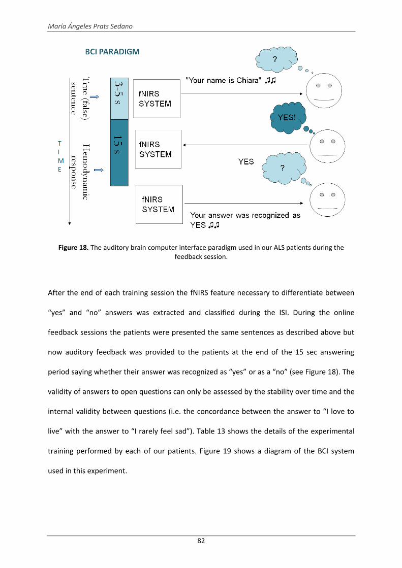
María Ángeles Prats Sedano
82
Figure 18. The auditory brain computer interface paradigm used in our ALS patients during the feedback session.
After the end of each training session the fNIRS feature necessary to differentiate between
“yes” and “no” answers was extracted and classified during the ISI. During the online
feedback sessions the patients were presented the same sentences as described above but
now auditory feedback was provided to the patients at the end of the 15 sec answering
period saying whether their answer was recognized as “yes” or as a “no” (see Figure 18). The
validity of answers to open questions can only be assessed by the stability over time and the
internal validity between questions (i.e. the concordance between the answer to “I love to
live” with the answer to “I rarely feel sad”). Table 13 shows the details of the experimental
training performed by each of our patients. Figure 19 shows a diagram of the BCI system
used in this experiment.

Cognitive processing and Brain communication in ALS
83
Table 13. Lists the total number of training, feedback, open question sessions performed by each patient.
Figure 19. The setup and flow diagram of the brain computer interface for communication in ALS
patients.
6.2.4 Data analysis
The fNIRS data acquired online throughout all the sessions was normalized. NIRS was filtered
using a bandpass filter of 0.0016 Hz – 0.3 Hz and processed using Modified Beer Lambert’s
law, as described in Cope et al., (205) and Chaudhary et al., (206), to calculate the relative
change in concentration of oxy (HbO2) and deoxy hemoglobin (HbR).

María Ángeles Prats Sedano
84
The relative change in HbO2 (with respect to the baseline), that was calculated online during
each training session was used to train a support vector machine (SVM) classifier model to
check the cross validation classification accuracy. In this study only the relative change in
HbO2 was used as because after the end of training sessions it was observed that O2Hb
provided stable and higher cross validation classification accuracy.
The offline classification procedure used the mean of relative change in HbO2 across each
channel as input feature to train a 5-fold linear support vector machine (SVM) classifier. The
SVM model interpolates the data corresponding to true and false sentences’ ISI in a two-
dimensional space such that the two categories are divided by a hyperplane and the gap
between them is as wide as possible. Firstly a model space is determined and the input
feature, extracted from the recorded and processed fNIRS signal, i.e., the relative change in
HbO2, is mapped onto the model space to determine the side of the hyperplane the input
feature falls on. For the fNIRS signal the mean of the relative change in HbO2 across all the
channels were used as input feature to map onto model space, while for EEG and EOG
signals common spatial pattern was used for feature extraction. The relative change in
HbO2, EEG and EOG data acquired during BCI sessions from our patients were processed off-
line separately for each patient to determine:
1) The statistical difference in the particular physiological signal (HbO2, EEG and EOG)
during the ISI of true (yes) and false (no) sentences.
2) Classification accuracy, using SVM as described above, of HbO2, EEG and EOG signals
across each session between the true and false sentences’ ISI.
Statistical analyses of the results were carried out using Matlab (The Mathworks Inc., USA).
The normal distribution of the data was assessed using the Kolmogorov-Smirnov test. The
assumption of normality was not violated in any of the data. Parametric t-tests for

Cognitive processing and Brain communication in ALS
85
independent samples were used to compare true and false sentences (using the averaged
ISI). The t-test analysis was performed across all the channels in a session and for all the
sessions of acquired HbO2, EEG and EOG signals. Furthermore, t-test was also performed
between the ISI of all the 10 true sentences and all the 10 false sentences across different
channels in a session averaged over many sessions varying slightly between patients. The
significance level for all statistics was set to 0.05.
6.3. Results
Results of the t-test (p < 0.05) performed between the average of all the true and false
sentences’ ISI using the relative change in HbO2, EEG and EOG from all the training sessions,
are summarized in Table 14. A significant difference was found between the true and false
sentences’ ISI across all patients. However, no significant differences were found in the EEG
and EOG data across all the training sessions showed no significant between the true and
false sentences’ ISI across each patient.
Table 14. Lists the total number of sessions averaged and degree of freedom used to perform t test analysis between the true and false sentence ISI corresponding to NIRS, EEG and EOG signal.

María Ángeles Prats Sedano
86
The relative change in HbO2 in five channels over the fronto-central brain region of our
patients during the true and false sentence ISI is shown in Figure 20. The figure illustrates
that the shape of the change in HbO2 during true sentence ISI is significantly different from
false sentence ISI. Figure 20 also illustrates that the shape of the change in HbO2 during a
true or a false sentence ISI is not consistent between the patients even though within each
patient the shape of the change in HbO2 is stable.
Figure 20. The averaged relative change in HbO2 across 5 out of 20 channels corresponding to YES and NO sentence interstimuli interval (ISI) in Patient F and B. In each subplot; five different colored
trace corresponds to relative change in HbO2 across five different channel, x-axis is time range in seconds and y-axis is the relative change in the HbO2.
Figure 21 illustrates the power spectrum density (PSD) of EEG oscillations, in the frequency
band 0 to 10 Hz, during the true and false sentence ISI from both patients. Only frequencies
between 0 and 10 Hz were used for classification and statistical testing because in ALS
patients in CLIS frequencies above 10 Hz are extremely rare10. The PSD of EEG signal shows
that there was no significant difference between the true and false sentences ISI across all

Cognitive processing and Brain communication in ALS
87
patients. The shapes of the relative change in HbO2 and EOG during ISI corresponding to
true and false sentences from all the sessions are also plotted in Figure 21. It illustrates that
there was no significant difference in the eye movements between the true and false
sentences ISI for all patients, confirmed by the t-test.
Figure 21. Power spectrum of the EEG and the EOG signal.
Figures 22 and 23 depict the SVM classification across all the sessions using the change in (a)
HbO2, (b) EEG and (c) EOG in Patient F and B respectively. A 65% cut off was used to define
whether the classification accuracy was above or below the acceptable level (chance level is
50%). The SVM results illustrate that highest classification accuracy was achieved using the
change in HbO2 for which more than 75% of the sessions yielded greater than 65%
classification accuracy for all the patients with an average classification accuracy of 70%.

María Ángeles Prats Sedano
88
While the SVM classification accuracy obtained using EEG and EOG data only few sessions
yielded greater than 65% classification accuracy across all patients.
Figure 22. Patient F: Linear support vector machine (SVM) classification accuracy across all sessions 1) Training (bar plot in spotted black) 2) Feedback (bar plot in solid green) and 3) Open question (bar
plot in solid red) obtained using a) Relative change in HbO2 b) EEG and c) EOG data. In each histogram plot x-axis is the number of sessions and y-axis is classification accuracy. The black
horizontal line represent the 65% classification accuracy.

Cognitive processing and Brain communication in ALS
89
Figure 23. Patient B: The description of Figure is same as described in Figure 15.

María Ángeles Prats Sedano
90
Classification results using fNIRS for open question in patients F and B using the criteria for
correctness described above in the Methods section ranged between 80-90% “correct”.
6.4. Discussion
All our 2 patients in the CLIS communicated with fronto-cortical oxygenation based BCI with
an average correct response rate of 70% over a period of several weeks. Correct response
rate for open questions as estimated exceeded even 80%. Patient F and B answered open
questions containing quality of life estimation (See Supplementary Material) with a yes/no
answers indicate a positive attitude towards the present situation and life in general as
found by other groups (22) in larger samples of ALS patients. Correct classification of
mentally “yes” and “no” responses through fNIRS surpassed classification of EEG oscillations
from 0-10 Hz (EEG frequencies in advanced ALS rarely show high frequencies) and vertical
and horizontal EOG classification. However, despite the absence of reliable eye
communication in all patients as the inclusion criteria in the study, EOG classification was at
some sessions above chance despite the inability of the social environment to perceive
them.
The results on two CLIS patients reported here allow the following conclusions:
a) Even after extended CLIS in ALS across months and years, reliable communication
is possible with NIRS-BCI using questions requiring a covert (cognitive) affirmative
(“yes”) or negative (“no”) answer. Moreover, we can conclude that fNIRS seems to
provide better classification of patients’ answers compared to oscillatory EEG
responses. We can propose that metabolic (vascular) brain changes permit superior
brain-self-control because of an existing feedback pathway between the vascular
system in the brain and the central nervous system, as it was previously proposed
(207).

Cognitive processing and Brain communication in ALS
91
b) Co-recording of NIRS and EEG oscillations from multiple sites might be important in
CLIS in order to monitor and eventually modify non-favorable vigilance changes to
improve NIRS classification.
Our results demonstrate that this novel approach of brain -communication is safe, reliable
and allowed so far the best communication possible for patients in completely locked-in
state.

María Ángeles Prats Sedano
92
DISCUSSION AND CONCLUSIONS
This thesis focused on the assessment of cognitive processing and brain communication in
patients suffering from ALS. Two main lines have been addressed along the thesis. Firstly,
the application of two novel approaches to assess cognitive functions in ALS (study one and
two). Secondly, the implementation of a functioning BCI-system for the completely
paralyzed locked-in patients due to ALS (CLIS), allowing brain communication based on
classical conditioning and the combined use of Functional Near-Infrared-Spectroscopy
(fNIRS) and EEG.
The first study has shown the feasibility of a novel approach, combining vibro-tactile P300s
and motor-independent neuropsychological tests for assessing cognitive functions in ALS.
The results of the neuropsychological and P300 assessment showed a mild impairment in
the attentional network with reduced P300 amplitudes in ALS group, and mild dysfunctions
in language processing reflected in the performance in oral comprehension tests.
The second study reported lower performance in neuropsychological tests related to the
executive system. Moreover, reduced amplitudes and delayed latencies of N200, P300, and
RON were found in ALS compared to controls. These results could be attributable to both an
alteration in change detection resulting in a reduction of the allocation and re-orientation of
attentional resources or a general slowing or reduction of neural processing efficiency in the
same system.
Taken together the results of the two first studies we can conclude that ALS is a
heterogeneous and complex disease, which affects extra-motor involvement of cognitive
functions and cognitive processing at different levels. Moreover these two studies provided
further evidence that (vibro-tactile and auditory) ERPs could represent a new and effective
tool for assessing cognitive and attentional processing in severely paralyzed ALS patients.

Cognitive processing and Brain communication in ALS
93
In the third study, we showed the feasibility of an fNIRS-based BCI system for
communication in the CLIS, which was reflected by higher accuracies in BCI performance of
our two CLIS patients (an average correct response rate of 70% over a period of several
weeks). We determined that even after extended CLIS in ALS across months and years,
reliable communication using questions that require a mental (imagined) affirmative (“yes”)
or a negative-rejecting (“no”) answer is possible with fNIRS-BCI. Moreover we observed that
fNIRS technology seems to provide better classification of patients’ answers than oscillatory
EEG responses. In conclusion, we strongly support the use of an fNIRS-based BCI to establish
communication with the CLIS.
The novelty of studies one and two is that we propose a neuropsychological paradigm that
does not require overt motor/verbal skills to assess cognitive functions in severely paralyzed
patients. This paradigm has first been described by Neumann and Kotchoubey (72).
However, they assessed a wide range of severely paralyzed patients, including individuals
with significant speech disabilities following brain damage (vegetative state, minimally
conscious state, aphasic patients etc.). In contrast, we decided to implement this innovative
paradigm to assess cognitive processing in patients with ALS. The rationale for this choice
was that even though some previous studies attempted to integrate neuropsychological
tests to assess the effects of ALS (152), these tests require minimal motor and/or speech
skills and are therefore not useful in severely paralyzed patients. Thus, our approach
responds to the need for cognitive evaluation of ALS patients also in later stages of the
disease (except CLIS).
The outcome of the neuropsychological assessment of the two first studies is in line with the
previous literature, reporting deficits in language and executive systems as the most affected
areas in ALS. From a classical point of view, executive dysfunctions have been seen as the

María Ángeles Prats Sedano
94
key feature of cognitive impairment in ALS. However, a growing body of evidence suggests
that language deficits in ALS are more common than previously thought. They may, in fact,
be just as frequent as executive dysfunctions (70,71).
Another novelty refers to the modalities that we used to assess cognitive and attentional
functioning in ALS using ERPs. The first one consisted of a vibro-tactile oddball paradigm that
aims at assessing attentional and cognitive processing in ALS. Previous studies suggest that
the somato-sensory system elicits robust ERPs (142,166). However, they were tested in
different populations (i.e., healthy subjects and spinal cord injured patients). In our study,
we used a novel technology consisting of a mechanical arm that was designed and
implemented by our group (118). This arm provided a vibration stimulus in the end-effector
of the device, which the patients had to grasp. The reason for using the somato-sensory
pathway to elicit ERPs was based on the observation that the somato-sensory pathways
seems to be intact until the terminal end of the disease. We further proposed that the
somato-sensory P300 could reflect cognitive changes in ALS.
The second study used an ERP approach using a distraction paradigm to investigate the
neural basis of selective attention in ALS. We introduced a novel modality compared to
classical ERPs oddball paradigms (54,93,112) by using 4 types of stimuli (long deviant, short
deviant, long frequent and short frequent). The implementation of this modified oddball
allowed us to assess selective attention more accurately, as this modified version required
more demanding attentional mechanisms due to the complexity of the task.
However, the most important contribution of this work lies in the combination of the
aforementioned motor-independent neuropsychological tests with the novel ERP
assessments, which allows for an exhaustive assessment of cognitive processing in ALS.

Cognitive processing and Brain communication in ALS
95
In contrast to the assessment of cognitive functions in the first two studies, the great impact
of our third study lies on the development of first reliable BCI system to reestablish
communication in CLIS patients following ALS. Previous attempts have been shown that
while brain-computer interfaces enabled LIS patients to communicate successfully (129), all
attempts to communicate with CLIS patients have failed regardless of the method that was
employed (e.g., invasive and non-invasive BCIs or any other assistive technology). This lack of
communication in CLIS left these patients in an unbearable “silence”. Prof. Birbaumer’s
group at the University of Tübingen has been working on the development of different BCI
systems during the past years with the intention to allow communication in CLIS. The first
successful attempts to reestablish communication with a CLIS patient have been reported in
a study by de Massari et al. (151), using an EEG-based BCI approach. Eventhough an
acceptable level of BCI performance accuracy was found in their CLIS patient in single
sessions, this accuracy was not be maintained across sessions. Gallegos et al. (137)
introduced a substantial change in the BCI strategy by introducing the hemodynamic brain
response (recorded by fNIRS) in combination with the semantical conditioning to ‘yes’ and
‘no’ questions. With this novel methodology they achieved an overall performance of
76.30% in one CLIS patient.
A further innovation was to combine the recording of hemodynamic and electrocortical
activity (using fNIRS and EEG respectively) in a sample of two CLIS patients using the “yes”
and “no” classical conditioning to known questions. Both patients achieved a higher accuracy
than the 70% that was found before using only fNIRS. However, the off-line classification of
the EEG-based BCI data did not show any good performance of our CLIS patients. This
suggests a greater efficacy of using the hemodynamic response to communicate in the late
stage of the disease. However, further investigation needs to be carried out to better

María Ángeles Prats Sedano
96
understand whether the low performance in the EEG based BCI approach is a consequence
of lower sensitivity of the EEG signal, whether EEG doesn’t differentiate sufficiently between
‘yes’ or ‘no’ answers, or whether the complexity of the signal demands better classifiers.
We can conclude that assessment of cognitive functions in the course of the ALS disease is
extremely important, as the patients are constantly facing decisions related to the choice of
clinical treatment and end-of-life management (i.e., nutritional and respiratory support,
including percutaneous endoscopic gastrostomy insertion, non-invasive ventilation, and
invasive mechanical ventilation). This makes cognitive impairment in ALS a sensitive topic,
because even though cognitive deficits may be significant, the may neither be dramatic.
Therefore, the reliable detection of cognitive deficits in ALS could provide important
information for health professionals in order to apply adaptive strategies that could
maximize the quality of life of both the patient and the caregiver. Likewise, we can
enormously improve the quality of life of patients in late stage of the disease (CLIS) by
reestablishing communication with them, thus breaking the “unbearable” silence.

Cognitive processing and Brain communication in ALS
97
SUPPLEMENTARY MATERIAL
Offene Fragen,Beispiele von Kategorien (German):
1. Gesundheit:
- Hast du Schmerzen?
- Sollen wir deine Augen befeuchten?
- Drückt dich etwas?
- Schläfst du viel?
- Kannst du mich/uns immer hören?
2. Familie/Freunde:
- Willst du, dass ... dich besuchen kommt?
- Sollen wir (Eltern etc) mehr Zeit mit dir verbringen?
- Willst du etwas mit deinen Freunden unternehmen?
- Willst du ... selber besuchen?
- Stört es dich, wenn wir da sind?
3. Laune/Zustand:
- Bist du gut gelaunt?
- Freust du dich auf ... (Person) ?
- Bist du öfters müde?
- Denkst du viel nach?
- Bist du traurig?
4. Wünsche:
- Willst du draußen sitzen?
- Willst du TV anschauen?
- Willst du Musik/Radio hören?

María Ángeles Prats Sedano
98
- Willst du in die Stadt/Park gehen?
- Sollen wir dir etwas vorlesen?
Open Questions, examples of categories (English Translation):
1. Health
- Do you have any pain?
- Should we humidify your eyes?
- Does anything squeezing you?
- Do you sleep a lot?
- Can you hear me/us?
2. Family/Friends:
- Do you wish that … will visit you?
- Should we spend more time with you?
- Do you want to organize … with your friends?
- Do you want to visit …?
- Is it disturbing you if we are near you?
3. State/Mood
- Are you in a good mood (now)?
- Are you looking forward to see …?
- Do you feel often tired?
- Do you think a lot?
- Are you sad?
4. Wishes
- Do you want to sit outside?

Cognitive processing and Brain communication in ALS
99
- Do you want to watch TV?
- Do you want to listen to music/radio?
- Do you want to go to the park?
- Should we read a book to you?

María Ángeles Prats Sedano
100
REFERENCES
1. Andrews J. Amyotrophic lateral sclerosis: clinical management and research update.
Curr Neurol Neurosci Rep (2009) 9:59–68
2. Strong M, Rosenfeld J. Amyotrophic lateral sclerosis: a review of current concepts.
Amyotroph Lateral Scler Other Motor Neuron Disord (2003) 4:136–43
3. Haverkamp LJ, Appel V, Appel SH. Natural history of amyotrophic lateral sclerosis in a
database population. Validation of a scoring system and a model for survival
prediction. Brain (1995)707–19
4. Wijesekera L, Leigh PN. Amyotrophic lateral sclerosis. Orphanet J Rare Dis (2009)
doi:10.1186/1750-1172-4-3
5. Logroscino G, Traynor BJ, Hardiman O, Chiò A, Mitchell D, Swingler RJ, Millul A, Benn
E, Beghi E. Incidence of amyotrophic lateral sclerosis in Europe. J Neurol Neurosurg
Psychiatry (2010) 81:385–90. doi:10.1136/jnnp.2009.183525
6. Chiò A, Mora G, Calvo A, Mazzini L, Bottacchi E, Mutani R. Epidemiology of ALS in Italy:
A 10-year prospective population-based study. Neurology (2009) 72:725–731.
doi:10.1212/01.wnl.0000343008.26874.d1
7. Ghatak NR, Campbell WW, Lippman RH, Hadfield MG. Anterior horn changes of motor
neuron disease associated with demyelinating radiculopathy. J Neuropathol Exp
Neurol (1986) 45:385–95
8. Hammer RP, Tomiyasu U, Scheibel AB. Degeneration of the human Betz cell due to

Cognitive processing and Brain communication in ALS
101
amyotrophic lateral sclerosis. Exp Neurol (1979) 63:336–346. doi:10.1016/0014-
4886(79)90129-8
9. Udaka F, Kameyama M, Tomonaga M. Degeneration of Betz cells in motor neuron
disease. A Golgi study. Acta Neuropathol (1986) 70:289–95
10. Murayama S, Inoue K, Kawakami H, Bouldin TW, Suzuki K. A unique pattern of
astrocytosis in the primary motor area in amyotrophic lateral sclerosis. Acta
Neuropathol (1991) 82:456–61
11. Pamphlett R, Kril J, Hng TM. Motor neuron disease: a primary disorder of
corticomotoneurons? Muscle Nerve (1995) 18:314–8. doi:10.1002/mus.880180308
12. Rosen DR, Sapp P, O’Regan J, McKenna-Yasek D, Schlumpf KS, Haines JL, Gusella JF,
Horvitz HR, Brown RH. Genetic linkage analysis of familial amyotrophic lateral sclerosis
using human chromosome 21 microsatellite DNA markers. Am J Med Genet (1994)
51:61–9. doi:10.1002/ajmg.1320510114
13. Gitcho MA, Baloh RH, Chakraverty S, Mayo K, Norton JB, Levitch D, Hatanpaa KJ,
White CL, Bigio EH, Caselli R, et al. TDP-43 A315T mutation in familial motor neuron
disease. Ann Neurol (2008) 63:535–8. doi:10.1002/ana.21344
14. Vance C, Rogelj B, Hortobágyi T, De Vos KJ, Nishimura AL, Sreedharan J, Hu X, Smith B,
Ruddy D, Wright P, et al. Mutations in FUS, an RNA processing protein, cause familial
amyotrophic lateral sclerosis type 6. Science (2009) 323:1208–11.
doi:10.1126/science.1165942
15. Kwiatkowski TJ, Bosco DA, Leclerc AL, Tamrazian E, Vanderburg CR, Russ C, Davis A,
Gilchrist J, Kasarskis EJ, Munsat T, et al. Mutations in the FUS/TLS gene on

María Ángeles Prats Sedano
102
chromosome 16 cause familial amyotrophic lateral sclerosis. Science (2009) 323:1205–
8. doi:10.1126/science.1166066
16. Renton AE, Majounie E, Waite A, Simón-Sánchez J, Rollinson S, Gibbs JR, Schymick JC,
Laaksovirta H, van Swieten JC, Myllykangas L, et al. A hexanucleotide repeat expansion
in C9ORF72 is the cause of chromosome 9p21-linked ALS-FTD. Neuron (2011) 72:257–
68. doi:10.1016/j.neuron.2011.09.010
17. Laferriere F, Polymenidou M. Advances and challenges in understanding the
multifaceted pathogenesis of amyotrophic lateral sclerosis. Swiss Med Wkly (2015)
145:w14054. doi:10.4414/smw.2015.14054
18. Brooks BR, Miller RG, Swash M, Munsat TL, World Federation of Neurology Research
Group on Motor Neuron Diseases. El Escorial revisited: revised criteria for the
diagnosis of amyotrophic lateral sclerosis. Amyotroph Lateral Scler Other Motor
Neuron Disord (2000) 1:293–9
19. Traynor BJ, Codd MB, Corr B, Forde C, Frost E, Hardiman OM, BR B, D C, M J, A E, et al.
Clinical Features of Amyotrophic Lateral Sclerosis According to the El Escorial and
Airlie House Diagnostic Criteria. Arch Neurol (2000) 57:1171.
doi:10.1001/archneur.57.8.1171
20. Bromberg MB. Assessing quality of life in ALS. J Clin Neuromuscul Dis (2007) 9:318–25.
doi:10.1097/CND.0b013e31815af9b7
21. Lulé D, Häcker S, Ludolph A, Birbaumer N, Kübler A. Depression and quality of life in
patients with amyotrophic lateral sclerosis. Dtsch Arztebl Int (2008) 105:397–403.
doi:10.3238/arztebl.2008.0397
Cognitive processing and Brain communication in ALS
103
22. Kubler A. Severity of depressive symptoms and quality of life in patients with
Amyotrophic Lateral Sclerosis. Neurorehabil Neural Repair (2005) 19:182–193.
doi:10.1177/1545968305276583
23. Kiebert GM, Green C, Murphy C, Mitchell JD, O’Brien M, Burrell A, Leigh PN. Patients’
health-related quality of life and utilities associated with different stages of
amyotrophic lateral sclerosis. J Neurol Sci (2001) 191:87–93
24. Robbins RA, Simmons Z, Bremer BA, Walsh SM, Fischer S. Quality of life in ALS is
maintained as physical function declines. Neurology (2001) 56:442–4
25. Pagnini F, Manzoni GM, Tagliaferri A, Gibbons CJ. Depression and disease progression
in amyotrophic lateral sclerosis: A comprehensive meta-regression analysis. J Health
Psychol (2015) 20:1107–28. doi:10.1177/1359105314530453
26. Ganzini L, Block S. Physician-Assisted Death — A Last Resort? N Engl J Med (2002)
346:1663–1665. doi:10.1056/NEJM200205233462113
27. Körner S, Kollewe K, Abdulla S, Zapf A, Dengler R, Petri S. Interaction of physical
function, quality of life and depression in Amyotrophic lateral sclerosis:
characterization of a large patient cohort. BMC Neurol (2015) 15:84.
doi:10.1186/s12883-015-0340-2
28. Hunter MD, Robinson IC, Neilson S. The functional and psychological status of patients
with amyotrophic lateral sclerosis: some implications for rehabilitation. Disabil Rehabil
15:119–26
29. Oh H, Sin M-K, Schepp KG, Choi-Kwon S. Depressive symptoms and functional
impairment among amyotrophic lateral sclerosis patients in South Korea. Rehabil Nurs

María Ángeles Prats Sedano
104
37:136–44. doi:10.1002/RNJ.00045
30. Simmons Z Robbins RA, et al. BBA. Quality of life in ALS depends on factors other than
strenght and physical function. Neurology (2000) 55:388–392.
31. Rabkin JG, Wagner GJ, Del Bene M. Resilience and distress among amyotrophic lateral
sclerosis patients and caregivers. Psychosom Med 62:271–9
32. Connolly S, Galvin M, Hardiman O. End-of-life management in patients with
amyotrophic lateral sclerosis. Lancet Neurol (2015) 14:435–442. doi:10.1016/S1474-
4422(14)70221-2
33. Simmons Z. Management strategies for patients with amyotrophic lateral sclerosis
from diagnosis through death. Neurologist (2005) 11:257–70
34. Miller RG, Mitchell JD, Moore DH. Riluzole for amyotrophic lateral sclerosis
(ALS)/motor neuron disease (MND). Cochrane database Syst Rev (2012) 3:CD001447.
doi:10.1002/14651858.CD001447.pub3
35. Bauer G, Gerstenbrand F, Rumpl E. Varieties of the locked-in syndrome. J Neurol
(1979) 221:77–91
36. Kübler A, Birbaumer N. Brain-computer interfaces and communication in paralysis:
extinction of goal directed thinking in completely paralysed patients? Clin
Neurophysiol (2008) 119:2658–66. doi:10.1016/j.clinph.2008.06.019
37. Birbaumer N, Murguialday AR, Cohen L. Brain-computer interface in paralysis. Curr
Opin Neurol (2008) 21:634–8. doi:10.1097/WCO.0b013e328315ee2d
38. Braunmühl A Von. Picksche krankheit und amyotrophische Lateralsklerose. Allg Z

Cognitive processing and Brain communication in ALS
105
Psychiat (1932)
39. Lomen-Hoerth C, Anderson T, Miller B. The overlap of amyotrophic lateral sclerosis
and frontotemporal dementia. Neurology (2002) 59:1077–1079.
doi:10.1097/WCO.0b013e3283168d1d
40. Ringholz GM, Appel SH, Bradshaw M, Cooke NA, Mosnik DM, Schulz PE. Prevalence
and patterns of cognitive impairment in sporadic ALS. Neurology (2005) 65:586–90.
doi:10.1212/01.wnl.0000172911.39167.b6
41. Lillo P, Savage S, Mioshi E, Kiernan MC, Hodges JR. Amyotrophic lateral sclerosis and
frontotemporal dementia: A behavioural and cognitive continuum. Amyotroph Lateral
Scler (2012) 13:102–9. doi:10.3109/17482968.2011.639376
42. Barulli MR, Fontana A, Panza F, Copetti M, Bruno S, Tursi M, Iurillo A, Tortelli R,
Capozzo R, Simone IL, et al. Frontal assessment battery for detecting executive
dysfunction in amyotrophic lateral sclerosis without dementia: a retrospective
observational study. BMJ Open (2015) 5:e007069. doi:10.1136/bmjopen-2014-007069
43. Seer C, Fürkötter S, Vogts M-B, Lange F, Abdulla S, Dengler R, Petri S, Kopp B.
Executive Dysfunctions and Event-Related Brain Potentials in Patients with
Amyotrophic Lateral Sclerosis. Front Aging Neurosci (2015) 7:225.
doi:10.3389/fnagi.2015.00225
44. Abrahams S, Leigh PN, Goldstein LH. Cognitive change in ALS: a prospective study.
Neurology (2005) 64:1222–1226. doi:10.1212/01.WNL.0000156519.41681.27
45. Ringholz GM, Greene SR. The relationship between amyotrophic lateral sclerosis and
frontotemporal dementia. Curr Neurol Neurosci Rep (2006) 6:387–92

María Ángeles Prats Sedano
106
46. Abrahams S, Goldstein LH, Al-Chalabi A, Pickering A, Morris RG, Passingham RE,
Brooks DJ, Leigh PN. Relation between cognitive dysfunction and pseudobulbar palsy
in amyotrophic lateral sclerosis. J Neurol Neurosurg Psychiatry (1997) 62:464–72
47. Moretti R, Torre P, Antonello RM, Carraro N, Cazzato G, Bava A. Complex cognitive
disruption in motor neuron disease. Dement Geriatr Cogn Disord (2002) 14:141–50.
doi:63600
48. Phukan J, Elamin M, Bede P, Jordan N, Gallagher L, Byrne S, Lynch C, Pender N,
Hardiman O. The syndrome of cognitive impairment in amyotrophic lateral sclerosis: a
population-based study. J Neurol Neurosurg Psychiatry (2012) 83:102–8.
doi:10.1136/jnnp-2011-300188
49. Rakowicz WP, Hodges JR. Dementia and aphasia in motor neuron disease: an
underrecognised association? J Neurol Neurosurg Psychiatry (1998) 65:881–9
50. Ludolph AC, Langen KJ, Regard M, Herzog H, Kemper B, Kuwert T, Böttger IG,
Feinendegen L. Frontal lobe function in amyotrophic lateral sclerosis: a
neuropsychologic and positron emission tomography study. Acta Neurol Scand (1992)
85:81–9
51. Abrahams S, Newton J, Niven E, Foley J, Bak TH. Screening for cognition and behaviour
changes in ALS. Amyotroph Lateral Scler Frontotemporal Degener (2014) 15:9–14.
doi:10.3109/21678421.2013.805784
52. Cohen MJ, Stanczak DE. On the Reliability, Validity, and Cognitive Structure of the
Thurstone Word Fluency Test. Arch Clin Neuropsychol (2000) 15:267–279.
doi:10.1016/S0887-6177(99)00017-7

Cognitive processing and Brain communication in ALS
107
53. Abrahams S, Goldstein LH, Simmons A, Brammer M, Williams SCR, Giampietro V, Leigh
PN. Word retrieval in amyotrophic lateral sclerosis: a functional magnetic resonance
imaging study. Brain (2004) 127:1507–17. doi:10.1093/brain/awh170
54. Volpato C, Piccione F, Silvoni S, Cavinato M, Palmieri A, Meneghello F, Birbaumer N.
Working memory in amyotrophic lateral sclerosis: auditory event-related potentials
and neuropsychological evidence. J Clin Neurophysiol (2010) 27:198–206.
doi:10.1097/WNP.0b013e3181e0aa14
55. Abrahams S, Goldstein LH, Kew JJM, Brooks DJ, Lloyd CM, Frith CD, Leigh PN. Frontal
lobe dysfunction in amyotrophic lateral sclerosis A PET study. (1996)2105–2120.
56. Gallassi R, Montagna P, Ciardulli C, Lorusso S, Mussuto V, Stracciari A. Cognitive
impairment in motor neuron disease. Acta Neurol Scand (1985) 71:480–4
57. Frank B, Haas J, Heinze HJ, Stark E, Münte TF. Relation of neuropsychological and
magnetic resonance findings in amyotrophic lateral sclerosis: evidence for subgroups.
Clin Neurol Neurosurg (1997) 99:79–86
58. Volpato C, Prats Sedano MA, Silvoni S, Segato N, Cavinato M, Merico A, Piccione F,
Palmieri A, Birbaumer N. Selective attention impairment in amyotrophic lateral
sclerosis. Amyotroph Lateral Scler Frontotemporal Degener (2016)1–9.
doi:10.3109/21678421.2016.1143514
59. Massman PJ, Sims J, Cooke N, Haverkamp LJ, Appel V, Appel SH. Prevalence and
correlates of neuropsychological deficits in amyotrophic lateral sclerosis. J Neurol
Neurosurg Psychiatry (1996) 61:450–5
60. Abrahams S, Leigh PN, Harvey A, Vythelingum GN, Grisé D, Goldstein LH. Verbal

María Ángeles Prats Sedano
108
fluency and executive dysfunction in amyotrophic lateral sclerosis (ALS).
Neuropsychologia (2000) 38:734–747. doi:10.1016/S0028-3932(99)00146-3
61. Hanagasi HA, Gurvit IH, Ermutlu N, Kaptanoglu G, Karamursel S, Idrisoglu HA, Emre M,
Demiralp T. Cognitive impairment in amyotrophic lateral sclerosis: evidence from
neuropsychological investigation and event-related potentials. Brain Res Cogn Brain
Res (2002) 14:234–44
62. Strong MJ, Grace GM, Orange JB, Leeper HA, Menon RS, Aere C. A prospective study
of cognitive impairment in ALS. Neurology (1999) 53:1665–1665.
doi:10.1212/WNL.53.8.1665
63. Raaphorst J, de Visser M, van Tol M-J, Linssen WHJP, van der Kooi AJ, de Haan RJ, van
den Berg LH, Schmand B. Cognitive dysfunction in lower motor neuron disease:
executive and memory deficits in progressive muscular atrophy. J Neurol Neurosurg
Psychiatry (2011) 82:170–5. doi:10.1136/jnnp.2009.204446
64. Bak TH, Hodges JR. Motor neurone disease, dementia and aphasia: coincidence, co-
occurrence or continuum? J Neurol (2001) 248:260–70
65. Bede P, Elamin M, Byrne S, McLaughlin RL, Kenna K, Vajda A, Pender N, Bradley DG,
Hardiman O. Basal ganglia involvement in amyotrophic lateral sclerosis. Neurology
(2013) 81:2107–15. doi:10.1212/01.wnl.0000437313.80913.2c
66. Brettschneider J, Del Tredici K, Toledo JB, Robinson JL, Irwin DJ, Grossman M, Suh E,
Van Deerlin VM, Wood EM, Baek Y, et al. Stages of pTDP-43 pathology in amyotrophic
lateral sclerosis. Ann Neurol (2013) 74:20–38. doi:10.1002/ana.23937
67. Machts J, Bittner V, Kasper E, Schuster C, Prudlo J, Abdulla S, Kollewe K, Petri S,

Cognitive processing and Brain communication in ALS
109
Dengler R, Heinze H-J, et al. Memory deficits in amyotrophic lateral sclerosis are not
exclusively caused by executive dysfunction: a comparative neuropsychological study
of amnestic mild cognitive impairment. BMC Neurosci (2014) 15:83.
doi:10.1186/1471-2202-15-83
68. Bak TH, O’Donovan DG, Xuereb JH, Boniface S, Hodges JR. Selective impairment of
verb processing associated with pathological changes in Brodmann areas 44 and 45 in
the motor neurone disease-dementia-aphasia syndrome. Brain (2001) 124:103–120.
doi:10.1093/brain/124.1.103
69. Grossman M, Anderson C, Khan A, Avants B, Elman L, McCluskey L. Impaired action
knowledge in amyotrophic lateral sclerosis. Neurology (2008) 71:1396–401.
doi:10.1212/01.wnl.0000319701.50168.8c
70. Taylor LJ, Brown RG, Tsermentseli S, Al-Chalabi A, Shaw CE, Ellis CM, Leigh PN,
Goldstein LH. Is language impairment more common than executive dysfunction in
amyotrophic lateral sclerosis? J Neurol Neurosurg Psychiatry (2013) 84:494–8.
doi:10.1136/jnnp-2012-303526
71. Abrahams S. Executive dysfunction in ALS is not the whole story. J Neurol Neurosurg
Psychiatry (2013) 84:474–475. doi:10.1136/jnnp-2012-303851
72. Neumann N, Kotchoubey B. Assessment of cognitive functions in severely paralysed
and severely brain-damaged patients: neuropsychological and electrophysiological
methods. Brain Res Brain Res Protoc (2004) 14:25–36.
doi:10.1016/j.brainresprot.2004.09.001
73. Lakerveld J, Kotchoubey B, Kübler A. Cognitive function in patients with late stage

María Ángeles Prats Sedano
110
amyotrophic lateral sclerosis. J Neurol Neurosurg Psychiatry (2008) 79:25–9.
doi:10.1136/jnnp.2007.116178
74. Mistridis P, Taylor KI, Kissler JM, Monsch AU, Kressig RW, Kivisaari SL. Distinct neural
systems underlying reduced emotional enhancement for positive and negative stimuli
in early Alzheimer’s disease. Front Hum Neurosci (2013) 7:939.
doi:10.3389/fnhum.2013.00939
75. Chainay H, Sava A, Michael GA, Landré L, Versace R, Krolak-Salmon P. Impaired
emotional memory enhancement on recognition of pictorial stimuli in Alzheimer’s
disease: No influence of the nature of encoding. Cortex (2014) 50:32–44.
doi:10.1016/j.cortex.2013.10.001
76. Cadieux NL, Greve KW. Emotion processing in Alzheimer’s disease. J Int Neuropsychol
Soc (1997) 3:411–9
77. Enrici I, Adenzato M, Ardito RB, Mitkova A, Cavallo M, Zibetti M, Lopiano L, Castelli L.
Emotion processing in Parkinson’s disease: a three-level study on recognition,
representation, and regulation. PLoS One (2015) 10:e0131470.
doi:10.1371/journal.pone.0131470
78. Xi C, Zhu Y, Mu Y, Chen B, Dong B, Cheng H, Hu P, Zhu C, Wang K. Theory of mind and
decision-making processes are impaired in Parkinson’s disease. Behav Brain Res
(2015) 279:226–33. doi:10.1016/j.bbr.2014.11.035
79. Garrido-Vásquez P, Pell MD, Paulmann S, Sehm B, Kotz SA. Impaired neural processing
of dynamic faces in left-onset Parkinson’s disease. Neuropsychologia (2016) 82:123–
33. doi:10.1016/j.neuropsychologia.2016.01.017
Cognitive processing and Brain communication in ALS
111
80. Beer JS, Ochsner KN. Social cognition: a multi level analysis. Brain Res (2006) 1079:98–
105. doi:10.1016/j.brainres.2006.01.002
81. Baron-Cohen S, Leslie AM, Frith U. Does the autistic child have a “theory of mind”?
Cognition (1985) 21:37–46
82. Zimmerman EK, Eslinger PJ, Simmons Z, Barrett AM. Emotional perception deficits in
amyotrophic lateral sclerosis. Cogn Behav Neurol (2007) 20:79–82.
doi:10.1097/WNN.0b013e31804c700b
83. Staios M, Fisher F, Lindell AK, Ong B, Howe J, Reardon K. Exploring sarcasm detection
in amyotrophic lateral sclerosis using ecologically valid measures. Front Hum Neurosci
(2013) 7:178. doi:10.3389/fnhum.2013.00178
84. Snowden JS, Austin NA, Sembi S, Thompson JC, Craufurd D, Neary D. Emotion
recognition in Huntington’s disease and frontotemporal dementia. Neuropsychologia
(2008) 46:2638–49. doi:10.1016/j.neuropsychologia.2008.04.018
85. Papps B, Abrahams S, Wicks P, Leigh PN, Goldstein LH. Changes in memory for
emotional material in amyotrophic lateral sclerosis (ALS). Neuropsychologia (2005)
43:1107–14. doi:10.1016/j.neuropsychologia.2004.11.027
86. Lulé D, Kurt A, Jürgens R, Kassubek J, Diekmann V, Kraft E, Neumann N, Ludolph AC,
Birbaumer N, Anders S. Emotional responding in amyotrophic lateral sclerosis. J
Neurol (2005) 252:1517–1524. doi:10.1007/s00415-005-0907-8
87. Lomen-Hoerth C, Murphy J, Langmore S, Kramer JH, Olney RK, Miller B. Are
amyotrophic lateral sclerosis patients cognitively normal? Neurology (2003) 60:1094–
7

María Ángeles Prats Sedano
112
88. Grossman AB, Woolley-Levine S, Bradley WG, Miller RG. Detecting neurobehavioral
changes in amyotrophic lateral sclerosis. Amyotroph Lateral Scler (2007) 8:56–61.
doi:10.1080/17482960601044106
89. Murphy J, Factor-Litvak P, Goetz R, Lomen-Hoerth C, Nagy PL, Hupf J, Singleton J,
Woolley S, Andrews H, Heitzman D, et al. Cognitive-behavioral screening reveals
prevalent impairment in a large multicenter ALS cohort. Neurology (2016)
doi:10.1212/WNL.0000000000002305
90. Girardi A, Macpherson SE, Abrahams S. Deficits in emotional and social cognition in
amyotrophic lateral sclerosis. Neuropsychology (2011) 25:53–65.
doi:10.1037/a0020357
91. Woolley SC, Zhang Y, Schuff N, Weiner MW, Katz JS. Neuroanatomical correlates of
apathy in ALS using 4 Tesla diffusion tensor MRI. Amyotroph Lateral Scler (2011)
12:52–8. doi:10.3109/17482968.2010.521842
92. Mouraux A, Iannetti GD. Across-trial averaging of event-related EEG responses and
beyond. Magn Reson Imaging (2008) 26:1041–54. doi:10.1016/j.mri.2008.01.011
93. Raggi A, Consonni M, Iannaccone S, Perani D, Zamboni M, Sferrazza B, Cappa SF.
Auditory event-related potentials in non-demented patients with sporadic
amyotrophic lateral sclerosis. Clin Neurophysiol (2008) 119:342–50.
doi:10.1016/j.clinph.2007.10.010
94. Goldstein LH, Abrahams S. Changes in cognition and behaviour in amyotrophic lateral
sclerosis: nature of impairment and implications for assessment. Lancet Neurol (2013)
12:368–80. doi:10.1016/S1474-4422(13)70026-7

Cognitive processing and Brain communication in ALS
113
95. Näätänen R, Picton T. The N1 wave of the human electric and magnetic response to
sound: a review and an analysis of the component structure. Psychophysiology (1987)
24:375–425
96. García-Larrea L, Lukaszewicz AC, Mauguière F. Revisiting the oddball paradigm. Non-
target vs neutral stimuli and the evaluation of ERP attentional effects.
Neuropsychologia (1992) 30:723–41
97. Näätänen R, Paavilainen P, Rinne T, Alho K. The mismatch negativity (MMN) in basic
research of central auditory processing: A review. Clin Neurophysiol (2007) 118:2544–
2590. doi:10.1016/j.clinph.2007.04.026
98. Näätänen R, Alho K. Mismatch negativity--the measure for central sound
representation accuracy. Audiol Neurootol 2:341–53
99. Näätänen R, Sussman ES, Salisbury D, Shafer VL. Mismatch negativity (MMN) as an
index of cognitive dysfunction. Brain Topogr (2014) 27:451–66. doi:10.1007/s10548-
014-0374-6
100. Folstein JR, Van Petten C. Influence of cognitive control and mismatch on the N2
component of the ERP: a review. Psychophysiology (2008) 45:152–70.
doi:10.1111/j.1469-8986.2007.00602.x
101. Sutton S, Braren M, Zubin J, John ER. Evoked-potential correlates of stimulus
uncertainty. Science (1965) 150:1187–8
102. Duncan CC, Barry RJ, Connolly JF, Fischer C, Michie PT, Näätänen R, Polich J, Reinvang
I, Van Petten C. Event-related potentials in clinical research: guidelines for eliciting,
recording, and quantifying mismatch negativity, P300, and N400. Clin Neurophysiol
María Ángeles Prats Sedano
114
(2009) 120:1883–908. doi:10.1016/j.clinph.2009.07.045
103. Polich J. Updating P300: an integrative theory of P3a and P3b. Clin Neurophysiol
(2007) 118:2128–48. doi:10.1016/j.clinph.2007.04.019
104. Picton TW, Bentin S, Berg P, Donchin E, Hillyard SA, Johnson R, Miller GA, Ritter W,
Ruchkin DS, Rugg MD, et al. Guidelines for using human event-related potentials to
study cognition: recording standards and publication criteria. Psychophysiology (2000)
37:127–52
105. Polich J, Kok A. Cognitive and biological determinants of P300: an integrative review.
Biol Psychol (1995) 41:103–46
106. Kutas M, Hillyard SA. Reading senseless sentences: brain potentials reflect semantic
incongruity. Science (1980) 207:203–5
107. Sitnikova T, Kuperberg G, Holcomb PJ. Semantic integration in videos of real-world
events: an electrophysiological investigation. Psychophysiology (2003) 40:160–4.
108. Kutas M, Federmeier KD. Thirty years and counting: finding meaning in the N400
component of the event-related brain potential (ERP). Annu Rev Psychol (2011)
62:621–47. doi:10.1146/annurev.psych.093008.131123
109. Kotchoubey B, Lang S, Bostanov V, Birbaumer N. Is there a mind? Electrophysiology of
unconscious patients. News Physiol Sci (2002) 17:38–42
110. Gil R, Neau JP, Dary-Auriol M, Agbo C, Tantot AM, Ingrand P. Event-related auditory
evoked potentials and amyotrophic lateral sclerosis. Arch Neurol (1995) 52:890–6.
111. Paulus KS, Magnano I, Piras MR, Solinas MA, Solinas G, Sau GF, Aiello I. Visual and

Cognitive processing and Brain communication in ALS
115
auditory event-related potentials in sporadic amyotrophic lateral sclerosis. Clin
Neurophysiol (2002) 113:853–61
112. Ogawa T, Tanaka H, Hirata K. Cognitive deficits in amyotrophic lateral sclerosis
evaluated by event-related potentials. Clin Neurophysiol (2009) 120:659–64.
doi:10.1016/j.clinph.2009.01.013
113. Münte TF, Tröger M, Nusser I, Wieringa BM, Matzke M, Johannes S, Dengler R.
Recognition memory deficits in amyotrophic lateral sclerosis assessed with event-
related brain potentials. Acta Neurol Scand (1998) 98:110–5
114. Silvoni S, Volpato C, Cavinato M, Marchetti M, Priftis K, Merico A, Tonin P, Koutsikos K,
Beverina F, Piccione F. P300-Based Brain-Computer Interface Communication:
Evaluation and Follow-up in Amyotrophic Lateral Sclerosis. Front Neurosci (2009) 3:60.
doi:10.3389/neuro.20.001.2009
115. Vieregge P, Wauschkuhn B, Heberlein I, Hagenah J, Verleger R. Selective attention is
impaired in amyotrophic lateral sclerosis--a study of event-related EEG potentials.
Brain Res Cogn Brain Res (1999) 8:27–35
116. Kotchoubey B, Lang S, Winter S, Birbaumer N. Cognitive processing in completely
paralyzed patients with amyotrophic lateral sclerosis. Eur J Neurol (2003) 10:551–8.
117. Pinkhardt EH, Jürgens R, Becker W, Mölle M, Born J, Ludolph AC, Schreiber H. Signs of
impaired selective attention in patients with amyotrophic lateral sclerosis. J Neurol
(2008) 255:532–8. doi:10.1007/s00415-008-0734-9
118. Silvoni S, Konicar L, Prats-Sedano MA, Garcia-Cossio E, Genna C, Volpato C, Cavinato
M, Paggiaro A, Veser S, De Massari D, et al. Tactile event-related potentials in

María Ángeles Prats Sedano
116
amyotrophic lateral sclerosis (ALS): Implications for brain-computer interface. Clin
Neurophysiol (2015) doi:10.1016/j.clinph.2015.06.029
119. Birbaumer N. Breaking the silence: brain-computer interfaces (BCI) for communication
and motor control. Psychophysiology (2006) 43:517–32. doi:10.1111/j.1469-
8986.2006.00456.x
120. Vidal JJ. Toward Direct Brain-Computer Communication. Annu Rev Biophys Bioeng
(1973) 2:157–180. doi:10.1146/annurev.bb.02.060173.001105
121. Bansal AK, Truccolo W, Vargas-Irwin CE, Donoghue JP. Decoding 3D reach and grasp
from hybrid signals in motor and premotor cortices: spikes, multiunit activity, and
local field potentials. J Neurophysiol (2012) 107:1337–55. doi:10.1152/jn.00781.2011
122. So K, Dangi S, Orsborn AL, Gastpar MC, Carmena JM. Subject-specific modulation of
local field potential spectral power during brain-machine interface control in
primates. J Neural Eng (2014) 11:026002. doi:10.1088/1741-2560/11/2/026002
123. Chapin JK, Moxon KA, Markowitz RS, Nicolelis MA. Real-time control of a robot arm
using simultaneously recorded neurons in the motor cortex. Nat Neurosci (1999)
2:664–70. doi:10.1038/10223
124. Carmena JM, Lebedev MA, Crist RE, O’Doherty JE, Santucci DM, Dimitrov DF, Patil PG,
Henriquez CS, Nicolelis MAL. Learning to control a brain-machine interface for
reaching and grasping by primates. PLoS Biol (2003) 1:E42.
doi:10.1371/journal.pbio.0000042
125. Serruya MD, Hatsopoulos NG, Paninski L, Fellows MR, Donoghue JP. Instant neural
control of a movement signal. Nature (2002) 416:141–2. doi:10.1038/416141a

Cognitive processing and Brain communication in ALS
117
126. Leuthardt EC, Schalk G, Wolpaw JR, Ojemann JG, Moran DW. A brain-computer
interface using electrocorticographic signals in humans. J Neural Eng (2004) 1:63–71.
doi:10.1088/1741-2560/1/2/001
127. Felton EA, Wilson JA, Williams JC, Garell PC. Electrocorticographically controlled brain-
computer interfaces using motor and sensory imagery in patients with temporary
subdural electrode implants. Report of four cases. J Neurosurg (2007) 106:495–500.
doi:10.3171/jns.2007.106.3.495
128. Clancy KB, Koralek AC, Costa RM, Feldman DE, Carmena JM. Volitional modulation of
optically recorded calcium signals during neuroprosthetic learning. Nat Neurosci
(2014) 17:807–9. doi:10.1038/nn.3712
129. Birbaumer N, Ghanayim N, Hinterberger T, Iversen I, Kotchoubey B, Kübler A,
Perelmouter J, Taub E, Flor H. A spelling device for the paralysed. Nature (1999)
398:297–8. doi:10.1038/18581
130. Kübler A, Neumann N, Kaiser J, Kotchoubey B, Hinterberger T, Birbaumer NP. Brain-
computer communication: self-regulation of slow cortical potentials for verbal
communication. Arch Phys Med Rehabil (2001) 82:1533–9
131. Wolpaw JR, Birbaumer N, McFarland DJ, Pfurtscheller G, Vaughan TM. Brain–
computer interfaces for communication and control. Clin Neurophysiol (2002)
113:767–791. doi:10.1016/S1388-2457(02)00057-3
132. Farwell LA, Donchin E. Talking off the top of your head: toward a mental prosthesis
utilizing event-related brain potentials. Electroencephalogr Clin Neurophysiol (1988)
70:510–523. doi:10.1016/0013-4694(88)90149-6

María Ángeles Prats Sedano
118
133. Yoo S-S, Kim H, Filandrianos E, Taghados SJ, Park S. Non-invasive brain-to-brain
interface (BBI): establishing functional links between two brains. PLoS One (2013)
8:e60410. doi:10.1371/journal.pone.0060410
134. Zhu D, Bieger J, Garcia Molina G, Aarts RM. A survey of stimulation methods used in
SSVEP-based BCIs. Comput Intell Neurosci (2010)702357. doi:10.1155/2010/702357
135. Sitaram R, Caria A, Veit R, Gaber T, Rota G, Kuebler A, Birbaumer N. FMRI brain-
computer interface: a tool for neuroscientific research and treatment. Comput Intell
Neurosci (2007)25487. doi:10.1155/2007/25487
136. Sitaram R, Caria A, Birbaumer N. Hemodynamic brain-computer interfaces for
communication and rehabilitation. Neural Netw (2009) 22:1320–8.
doi:10.1016/j.neunet.2009.05.009
137. Gallegos-Ayala G, Furdea A, Takano K, Ruf CA, Flor H, Birbaumer N. Brain
communication in a completely locked-in patient using bedside near-infrared
spectroscopy. Neurology (2014) 82:1930–2. doi:10.1212/WNL.0000000000000449
138. Chaudhary U, Birbaumer N, Ramos-Murguialday A. Brain–computer interfaces for
communication and rehabilitation. Nat Rev Neurol (2016) 12:513–525.
doi:10.1038/nrneurol.2016.113
139. Piccione F, Giorgi F, Tonin P, Priftis K, Giove S, Silvoni S, Palmas G, Beverina F. P300-
based brain computer interface: Reliability and performance in healthy and paralysed
participants. Clin Neurophysiol (2006) 117:531–537. doi:10.1016/j.clinph.2005.07.024
140. Furdea A, Halder S, Krusienski DJ, Bross D, Nijboer F, Birbaumer N, Kübler A. An
auditory oddball (P300) spelling system for brain-computer interfaces.

Cognitive processing and Brain communication in ALS
119
Psychophysiology (2009) 46:617–625. doi:10.1111/j.1469-8986.2008.00783.x
141. Halder S, Hammer EM, Kleih SC, Bogdan M, Rosenstiel W, Birbaumer N, Kübler A.
Prediction of auditory and visual p300 brain-computer interface aptitude. PLoS One
(2013) 8:e53513. doi:10.1371/journal.pone.0053513
142. Brouwer A-M, van Erp JBF. A tactile P300 brain-computer interface. Front Neurosci
(2010) 4:19. doi:10.3389/fnins.2010.00019
143. Kübler A, Kotchoubey B. Brain-computer interfaces in the continuum of
consciousness. Curr Opin Neurol (2007) 20:643–9.
doi:10.1097/WCO.0b013e3282f14782
144. Kennedy PR, Bakay RA, Moore MM, Adams K, Goldwaithe J. Direct control of a
computer from the human central nervous system. IEEE Trans Rehabil Eng (2000)
8:198–202
145. Kennedy P, Andreasen D, Ehirim P, King B, Kirby T, Mao H, Moore M. Using human
extra-cortical local field potentials to control a switch. J Neural Eng (2004) 1:72–7.
doi:10.1088/1741-2560/1/2/002
146. Gilja V, Pandarinath C, Blabe CH, Nuyujukian P, Simeral JD, Sarma AA, Sorice BL, Perge
JA, Jarosiewicz B, Hochberg LR, et al. Clinical translation of a high-performance neural
prosthesis. Nat Med (2015) 21:1142–5. doi:10.1038/nm.3953
147. Kübler A, Birbaumer N. Brain-computer interfaces and communication in paralysis:
extinction of goal directed thinking in completely paralysed patients? Clin
Neurophysiol (2008) 119:2658–66. doi:10.1016/j.clinph.2008.06.019
148. Sellers EW, Vaughan TM, Wolpaw JR. A brain-computer interface for long-term

María Ángeles Prats Sedano
120
independent home use. Amyotroph Lateral Scler (2010) 11:449–55.
doi:10.3109/17482961003777470
149. Kuebler A, Kotchoubey B, Salzmann HP, Ghanayim N, Perelmouter J, Hömberg V,
Birbaumer N. Self-regulation of slow cortical potentials in completely paralyzed
human patients. Neurosci Lett (1998) 252:171–4
150. Sellers EW, Ryan DB, Hauser CK. Noninvasive brain-computer interface enables
communication after brainstem stroke. Sci Transl Med (2014) 6:257re7.
doi:10.1126/scitranslmed.3007801
151. De Massari D, Ruf CA, Furdea A, Matuz T, van der Heiden L, Halder S, Silvoni S,
Birbaumer N. Brain communication in the locked-in state. Brain (2013) 136:1989–
2000. doi:10.1093/brain/awt102
152. Niven E, Newton J, Foley J, Colville S, Swingler R, Chandran S, Bak TH, Abrahams S.
Validation of the Edinburgh Cognitive and Behavioural Amyotrophic Lateral Sclerosis
Screen (ECAS): A cognitive tool for motor disorders. Amyotroph Lateral Scler
Frontotemporal Degener (2015) 16:172–9. doi:10.3109/21678421.2015.1030430
153. David AS, Gillham RA. Neuropsychological study of motor neuron disease.
Psychosomatics (1986) 27:441–5. doi:10.1016/S0033-3182(86)72673-X
154. Kew JJM, Goldstein LH, Leigh PN, Abrahams S, Cosgrave N, Passingham RE, Frackowiak
RSJ, Brooks DJ. The relationship between abnormalities of cognitive function and
cerebral activation in amyotrophic lateral sclerosis: A neuropsychological and positron
emission tomography study. Brain (1993) 116:1399–1423.
doi:10.1093/brain/116.6.1399

Cognitive processing and Brain communication in ALS
121
155. Birbaumer N. Brain-computer-interface research: coming of age. Clin Neurophysiol
(2006) 117:479–83. doi:10.1016/j.clinph.2005.11.002
156. Hedges D, Janis R, Mickelson S, Keith C, Bennett D, Brown BL. P300 Amplitude in
Alzheimer’s Disease: A Meta-Analysis and Meta-Regression. Clin EEG Neurosci (2016)
47:48–55. doi:10.1177/1550059414550567
157. Lee M-S, Lee S-H, Moon E-O, Moon Y-J, Kim S, Kim S-H, Jung I-K. Neuropsychological
correlates of the P300 in patients with Alzheimer’s disease. Prog
Neuropsychopharmacol Biol Psychiatry (2013) 40:62–9.
doi:10.1016/j.pnpbp.2012.08.009
158. Parra MA, Ascencio LL, Urquina HF, Manes F, Ibáñez AM. P300 and neuropsychological
assessment in mild cognitive impairment and Alzheimer dementia. Front Neurol
(2012) 3:172. doi:10.3389/fneur.2012.00172
159. Silva Lopes M da, Souza Melo A de, Nóbrega AC. Delayed latencies of auditory evoked
potential P300 are associated with the severity of Parkinson’s disease in older
patients. Arq Neuropsiquiatr (2014) 72:296–300.
160. Solís-Vivanco R, Rodríguez-Violante M, Rodríguez-Agudelo Y, Schilmann A, Rodríguez-
Ortiz U, Ricardo-Garcell J. The P3a wave: A reliable neurophysiological measure of
Parkinson’s disease duration and severity. Clin Neurophysiol (2015) 126:2142–9.
doi:10.1016/j.clinph.2014.12.024
161. Ivica N, Titlic M, Pavelin S. P300 wave changes in patients with multiple sclerosis. Acta
Inform medica AIM J Soc Med Informatics Bosnia Herzegovina casopis Drustva za
Med Inform BiH (2013) 21:205–7. doi:10.5455/aim.2013.21.205-207

María Ángeles Prats Sedano
122
162. Sundgren M, Wahlin Å, Maurex L, Brismar T. Event related potential and response
time give evidence for a physiological reserve in cognitive functioning in relapsing-
remitting multiple sclerosis. J Neurol Sci (2015) 356:107–12.
doi:10.1016/j.jns.2015.06.025
163. Murguialday AR, Hill J, Bensch M, Martens S, Halder S, Nijboer F, Schoelkopf B,
Birbaumer N, Gharabaghi A. Transition from the locked in to the completely locked-in
state: a physiological analysis. Clin Neurophysiol (2011) 122:925–33.
doi:10.1016/j.clinph.2010.08.019
164. Nakajima Y, Imamura N. Relationships between attention effects and intensity effects
on the cognitive N140 and P300 components of somatosensory ERPs. Clin
Neurophysiol (2000) 111:1711–8
165. Satomi K, Horai T, Kinoshita Y, Wakazono A. Hemispheric asymmetry of event-related
potentials in a patient with callosal disconnection syndrome: a comparison of
auditory, visual and somatosensory modalities. Electroencephalogr Clin Neurophysiol
(1995) 94:440–9
166. Lugo ZR, Rodriguez J, Lechner A, Ortner R, Gantner IS, Laureys S, Noirhomme Q, Guger
C. A vibrotactile p300-based brain-computer interface for consciousness detection
and communication. Clin EEG Neurosci (2014) 45:14–21.
doi:10.1177/1550059413505533
167. Kaufmann T, Holz EM, Kübler A. Comparison of tactile, auditory, and visual modality
for brain-computer interface use: a case study with a patient in the locked-in state.
Front Neurosci (2013) 7:129. doi:10.3389/fnins.2013.00129

Cognitive processing and Brain communication in ALS
123
168. Ludolph AC, Brettschneider J, Weishaupt JH. Amyotrophic lateral sclerosis. Curr Opin
Neurol (2012) 25:530–5. doi:10.1097/WCO.0b013e328356d328
169. Ross MA, Miller RG, Berchert L, Parry G, Barohn RJ, Armon C, Bryan WW, Petajan J,
Stromatt S, Goodpasture J, et al. Toward earlier diagnosis of amyotrophic lateral
sclerosis: revised criteria. rhCNTF ALS Study Group. Neurology (1998) 50:768–72.
170. Cedarbaum JM, Stambler N, Malta E, Fuller C, Hilt D, Thurmond B, Nakanishi A. The
ALSFRS-R: a revised ALS functional rating scale that incorporates assessments of
respiratory function. BDNF ALS Study Group (Phase III). J Neurol Sci (1999) 169:13–21.
171. American Psychiatric Association. Diagnostic and statistical manual of mental
disorders: DSM-5 (5th ed.). Arlington, VA: American Psychiatric Publishing (2013).
172. Raven JC. Guide to using the coloured progressive matrice: Sets A. Ab. B.1. London: H.
K. Lewis (1965).
173. SanioFancello G CC. Modified Card Sorting Test (MCST): Adattamento Italiano per
L’età Evolutiva del Wisconsin Card Sorting Test. Firenze: Organizzazioni Speciali
174. Wilson B, Cockburn J, Baddeley A. The Rivermead Behavioural Memory Test. (1985).
doi:10.1177/107319119600300410
175. Luzzatti C, Willmes K DBR. Aachener Aphasie Test. Versione i. Florence: Organizzazioni
Speciali (1991).
176. Schalk G, McFarland DJ, Hinterberger T, Birbaumer N, Wolpaw JR. BCI2000: a general-
purpose brain-computer interface (BCI) system. IEEE Trans Biomed Eng (2004)
51:1034–43. doi:10.1109/TBME.2004.827072

María Ángeles Prats Sedano
124
177. Wilson JA, Mellinger J, Schalk G, Williams J. A procedure for measuring latencies in
brain-computer interfaces. IEEE Trans Biomed Eng (2010) 57:1785–97.
doi:10.1109/TBME.2010.2047259
178. Holm A, Ranta-aho PO, Sallinen M, Karjalainen PA, Müller K. Relationship of P300
single-trial responses with reaction time and preceding stimulus sequence. Int J
Psychophysiol (2006) 61:244–52. doi:10.1016/j.ijpsycho.2005.10.015
179. Schreuder M, Blankertz B, Tangermann M. A new auditory multi-class brain-computer
interface paradigm: spatial hearing as an informative cue. PLoS One (2010) 5:e9813.
doi:10.1371/journal.pone.0009813
180. Veit R, Konicar L, Klinzing JG, Barth B, Yilmaz O, Birbaumer N. Deficient fear
conditioning in psychopathy as a function of interpersonal and affective disturbances.
Front Hum Neurosci (2013) 7:706. doi:10.3389/fnhum.2013.00706
181. Spinnler H TG. Standardizzazione taratura italiana dei test neuropsicologici. Ital J
Neurol Sci (1987) Suppl. 8:1–120.
182. Mantovan MC, Baggio L, Dalla Barba G, Smith P, Pegoraro E, Soraru’ G, Bonometto P,
Angelini C. Memory deficits and retrieval processes in ALS. Eur J Neurol (2003)
10:221–7.
183. Kärgel C, Sartory G, Kariofillis D, Wiltfang J, Müller BW. Mismatch negativity latency
and cognitive function in schizophrenia. PLoS One (2014) 9:e84536.
doi:10.1371/journal.pone.0084536
184. Appel SH, Zhao W, Beers DR, Henkel JS. The microglial-motoneuron dialogue in ALS.
Acta Myol myopathies cardiomyopathies Off J Mediterr Soc Myol (2011) 30:4–8.

Cognitive processing and Brain communication in ALS
125
185. Strong MJ, Grace GM, Freedman M, Lomen-Hoerth C, Woolley S, Goldstein LH,
Murphy J, Shoesmith C, Rosenfeld J, Leigh PN, et al. Consensus criteria for the
diagnosis of frontotemporal cognitive and behavioural syndromes in amyotrophic
lateral sclerosis. Amyotroph Lateral Scler (2009) 10:131–146. doi:Pii 907462414\rDoi
10.1080/17482960802654364
186. Raggi A, Iannaccone S, Cappa SF. Event-related brain potentials in amyotrophic lateral
sclerosis: A review of the international literature. Amyotroph Lateral Scler (2010)
11:16–26. doi:10.3109/17482960902912399
187. Hammer A, Vielhaber S, Rodriguez-Fornells A, Mohammadi B, Münte TF. A
neurophysiological analysis of working memory in amyotrophic lateral sclerosis. Brain
Res (2011) 1421:90–9. doi:10.1016/j.brainres.2011.09.010
188. Westphal KP, Heinemann HA, Grözinger B, Kotchoubey BJ, Diekmann V, Becker W,
Kornhuber HH. Bereitschaftspotential in amyotrophic lateral sclerosis (ALS): lower
amplitudes in patients with hyperreflexia (spasticity). Acta Neurol Scand (1998) 98:15–
21
189. Mannarelli D, Pauletti C, Locuratolo N, Vanacore N, Frasca V, Trebbastoni A, Inghilleri
M, Fattapposta F. Attentional processing in bulbar- and spinal-onset amyotrophic
lateral sclerosis: Insights from event-related potentials. Amyotroph Lateral Scler Front
Degener (2014) 15:30–38. doi:10.3109/21678421.2013.787628
190. Escera C, Alho K, Winkler I, Näätänen R. Neural Mechanisms of Involuntary Attention
to Acoustic Novelty and Change. J Cogn Neurosci (1998) 10:590–604.
doi:10.1162/089892998562997

María Ángeles Prats Sedano
126
191. Näätänen R, Michie PT. Early selective-attention effects on the evoked potential: A
critical review and reinterpretation. Biol Psychol (1979) 8:81–136. doi:10.1016/0301-
0511(79)90053-X
192. Donchin E, Coles MGH. Is the P300 component a manifestation of context updating?
Behav Brain Sci (1988) 11:355–425. doi:10.1017/S0140525X00058027
193. Schröger E, Giard MH, Wolff C. Auditory distraction: event-related potential and
behavioral indices. Clin Neurophysiol (2000) 111:1450–60.
194. Nasreddine ZS, Phillips NA, Bédirian V, Charbonneau S, Whitehead V, Collin I,
Cummings JL, Chertkow H. The Montreal Cognitive Assessment, MoCA: A Brief
Screening Tool For Mild Cognitive Impairment. J Am Geriatr Soc (2005) 53:695–699.
doi:10.1111/j.1532-5415.2005.53221.x
195. Berti S, Schröger E. A comparison of auditory and visual distraction effects: behavioral
and event-related indices. Cogn Brain Res (2001) 10:265–273. doi:10.1016/S0926-
6410(00)00044-6
196. Berti S. Cognitive control after distraction: Event-related brain potentials (ERPs)
dissociate between different processes of attentional allocation. Psychophysiology
(2008) 45:608–620. doi:10.1111/j.1469-8986.2008.00660.x
197. Delorme A, Makeig S. EEGLAB: an open source toolbox for analysis of single-trial EEG
dynamics including independent component analysis. J Neurosci Methods (2004)
134:9–21. doi:10.1016/j.jneumeth.2003.10.009
198. Neary D, Snowden J., Mann DM. Cognitive change in motor neurone
disease/amyotrophic lateral sclerosis (MND/ALS). J Neurol Sci (2000) 180:15–20.

Cognitive processing and Brain communication in ALS
127
doi:10.1016/S0022-510X(00)00425-1
199. Sams M, Alho K, Näätänen R. Sequential effects on the ERP in discriminating two
stimuli. Biol Psychol (1983) 17:41–58. doi:10.1016/0301-0511(83)90065-0
200. Horváth J, Czigler I, Birkás E, Winkler I, Gervai J. Age-related differences in distraction
and reorientation in an auditory task. Neurobiol Aging (2009) 30:1157–1172.
doi:10.1016/j.neurobiolaging.2007.10.003
201. Gu Y, Farina D, Murguialday AR, Dremstrup K, Birbaumer N. Comparison of movement
related cortical potential in healthy people and amyotrophic lateral sclerosis patients.
Front Neurosci (2013) 7:65. doi:10.3389/fnins.2013.00065
202. Jacobsen T, Schröger E. Measuring duration mismatch negativity. Clin Neurophysiol
(2003) 114:1133–1143. doi:10.1016/S1388-2457(03)00043-9
203. Birbaumer N, Murguialday AR, Cohen L. Brain-computer interface in paralysis. Curr
Opin Neurol (2008) 21:634–8. doi:10.1097/WCO.0b013e328315ee2d
204. De Massari D, Ruf CA, Furdea A, Matuz T, van der Heiden L, Halder S, Silvoni S,
Birbaumer N. Brain communication in the locked-in state. Brain (2013) 136:1989–
2000. doi:10.1093/brain/awt102
205. Cope M, Delpy DT, Reynolds EO, Wray S, Wyatt J, van der Zee P. Methods of
quantitating cerebral near infrared spectroscopy data. Adv Exp Med Biol (1988)
222:183–9.
206. Chaudhary U, Hall M, DeCerce J, Rey G, Godavarty A. Frontal activation and
connectivity using near-infrared spectroscopy: verbal fluency language study. Brain
Res Bull (2011) 84:197–205. doi:10.1016/j.brainresbull.2011.01.002

María Ángeles Prats Sedano
128
207. Birbaumer N, Ruiz S, Sitaram R. Learned regulation of brain metabolism. Trends Cogn
Sci (2013) 17:295–302. doi:10.1016/j.tics.2013.04.009