Embed Size (px)
Citation preview
Cognitive NeuroscienceNEUR 3860
Review Session
October 19, 2009Megan Metzler
Outline
• History of neuroscience• Techniques of cognitive neuroscience• Vision
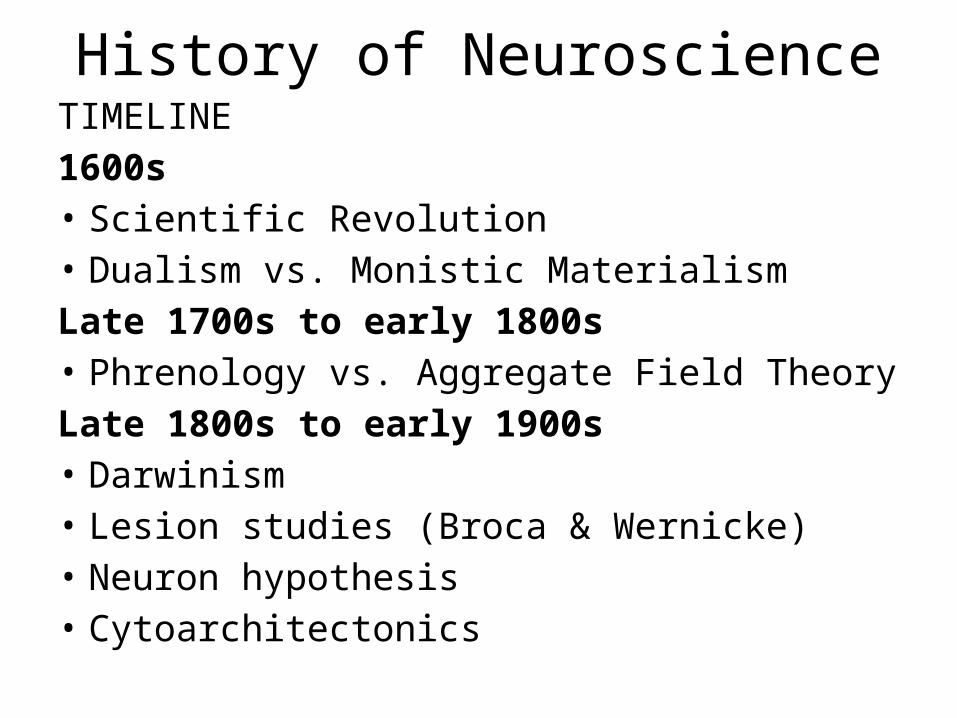
History of NeuroscienceTIMELINE1600s• Scientific Revolution• Dualism vs. Monistic MaterialismLate 1700s to early 1800s• Phrenology vs. Aggregate Field TheoryLate 1800s to early 1900s• Darwinism• Lesion studies (Broca & Wernicke)• Neuron hypothesis • Cytoarchitectonics
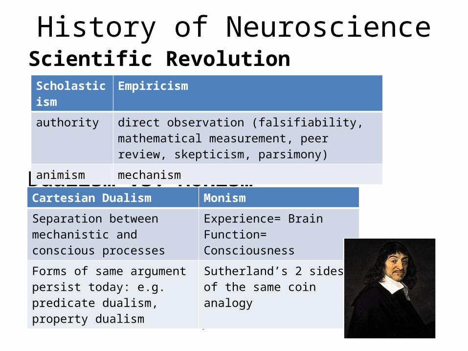
History of NeuroscienceScientific Revolution
Dualism vs. Monism
• Rene Descartes, Galileo
Scholasticism Empiricism
authority direct observation (falsifiability, mathematical measurement, peer review, skepticism, parsimony)
animism mechanism
Cartesian Dualism Monism
Separation between mechanistic and conscious processes
Experience= Brain Function= Consciousness
Forms of same argument persist today: e.g. predicate dualism, property dualism
Sutherland’s 2 sides of the same coin analogy
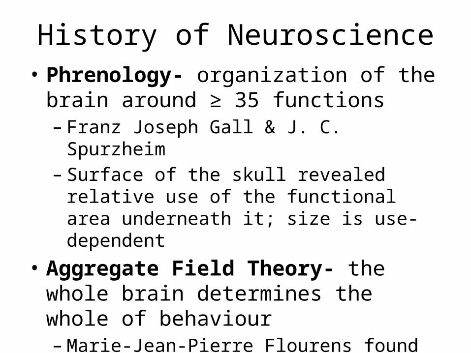
History of Neuroscience• Phrenology- organization of the brain around
≥ 35 functions – Franz Joseph Gall & J. C. Spurzheim– Surface of the skull revealed relative use of the
functional area underneath it; size is use-dependent
• Aggregate Field Theory- the whole brain determines the whole of behaviour– Marie-Jean-Pierre Flourens found that regardless
of where a bird’s brain was lesioned, it recovered.
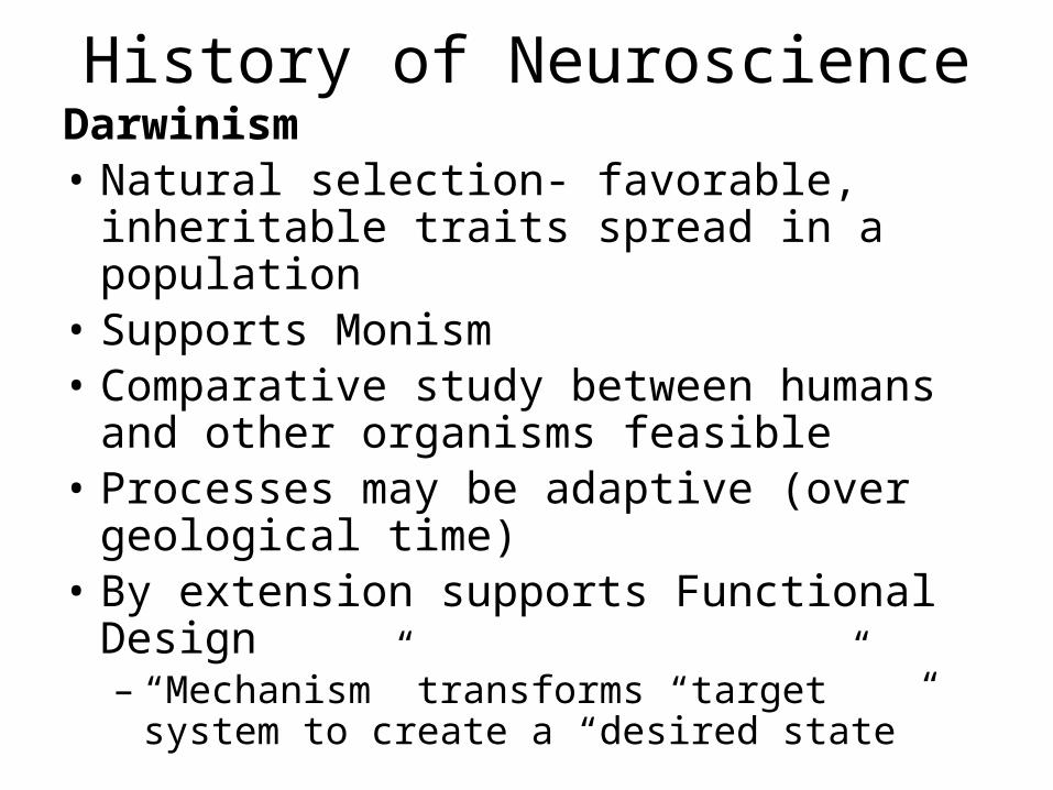
History of NeuroscienceDarwinism• Natural selection- favorable, inheritable traits
spread in a population• Supports Monism• Comparative study between humans and
other organisms feasible• Processes may be adaptive (over geological
time)• By extension supports Functional Design– “Mechanism” transforms “target” system to
create a “desired state”
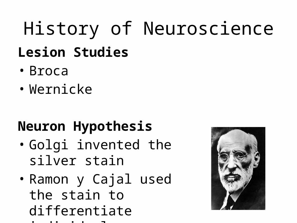
History of NeuroscienceLesion Studies• Broca• Wernicke
Neuron Hypothesis• Golgi invented the silver stain • Ramon y Cajal used the stain to
differentiate individual neurons
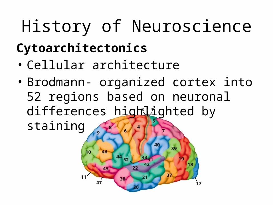
History of NeuroscienceCytoarchitectonics• Cellular architecture• Brodmann- organized cortex into 52 regions
based on neuronal differences highlighted by staining
History of NeuroscienceQUESTIONS
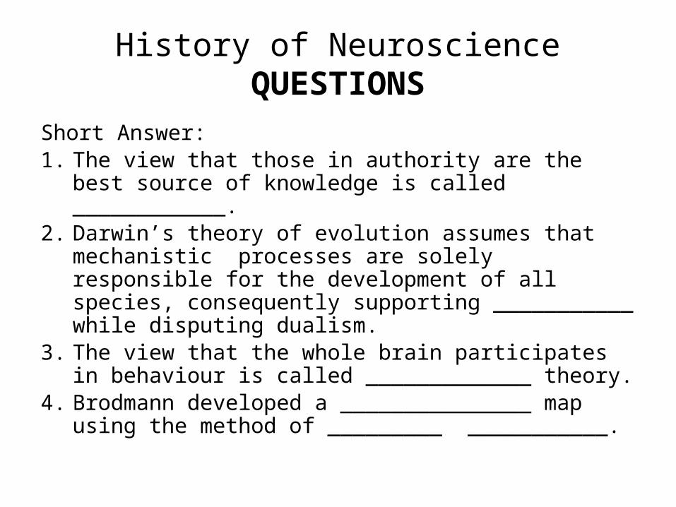
Short Answer:1. The view that those in authority are the best source
of knowledge is called ____________.2. Darwin’s theory of evolution assumes that
mechanistic processes are solely responsible for the development of all species, consequently supporting ___________ while disputing dualism.
3. The view that the whole brain participates in behaviour is called _____________ theory.
4. Brodmann developed a _______________ map using the method of _________ ___________.
History of NeuroscienceQUESTIONS
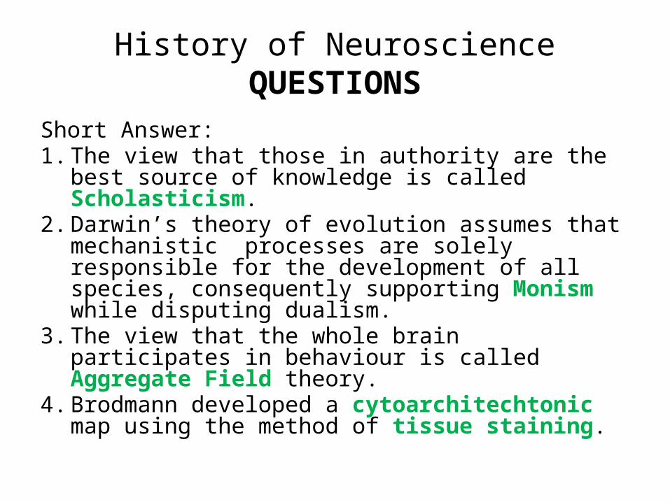
Short Answer:1. The view that those in authority are the best
source of knowledge is called Scholasticism.2. Darwin’s theory of evolution assumes that
mechanistic processes are solely responsible for the development of all species, consequently supporting Monism while disputing dualism.
3. The view that the whole brain participates in behaviour is called Aggregate Field theory.
4. Brodmann developed a cytoarchitechtonic map using the method of tissue staining.
History of NeuroscienceQUESTIONS
Multiple Choice:5. According to Cartesian dualism
a) The processes that underlie consciousness are mechanistic.b) Human consciousness cannot be understood by mechanistic processes because it is not mechanistic.c) Spirit-like and mechanistic properties coexist in all matter.d) Empirical and authoritative perspectives are complementary.
History of NeuroscienceQUESTIONS
6. The most significant contribution to neurology in the 19th century was:a) Phrenology and resultant methods to identify varying development of specific cognitive abilities across individuals.b) Animal research with pigeons which studied the behavioural effects of localized lesions.c) Use of the “the black reaction” to differentiate individual neurons.d) Detailed anatomical drawings of the human brain.
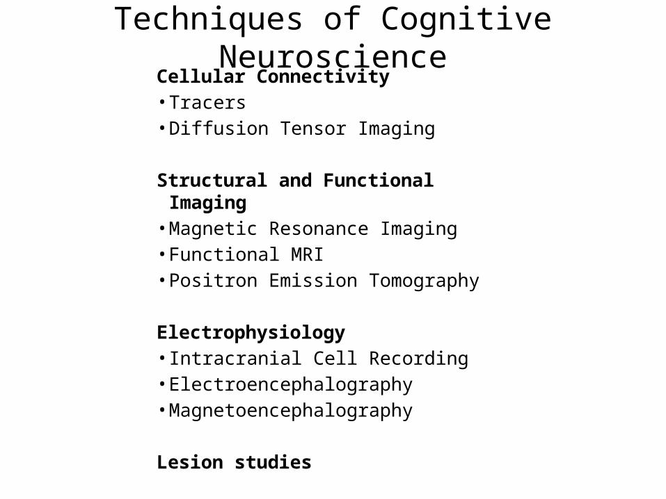
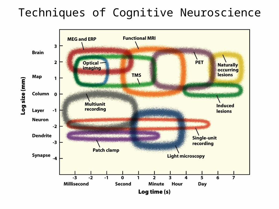
Techniques of Cognitive NeuroscienceCellular Connectivity• Tracers• Diffusion Tensor Imaging
Structural and Functional Imaging• Magnetic Resonance Imaging• Functional MRI• Positron Emission Tomography
Electrophysiology• Intracranial Cell Recording• Electroencephalography• Magnetoencephalography
Lesion studies
Cellular ConnectivityGeneral Principles• High degree of interconnections in the brain
which is likely responsible for cognition• Prime example: in the visual system there is
estimated to be 10 descending projections for each ascending projection also highlights the limitations associated with structural assessment techniques
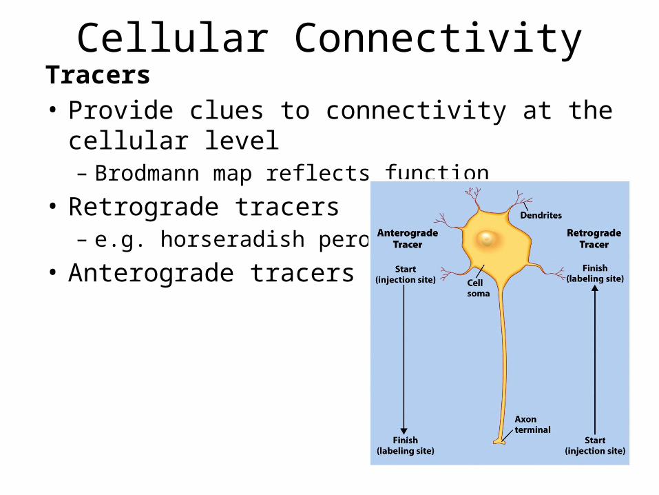
Cellular ConnectivityTracers• Provide clues to connectivity at the cellular level– Brodmann map reflects function
• Retrograde tracers– e.g. horseradish peroxidase
• Anterograde tracers
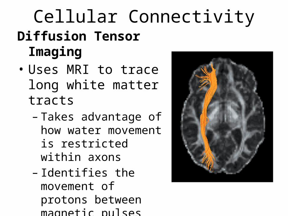
Cellular ConnectivityDiffusion Tensor Imaging• Uses MRI to trace long
white matter tracts– Takes advantage of how
water movement is restricted within axons
– Identifies the movement of protons between magnetic pulses and identifies non-random patterns
Structural and Functional ImagingGeneral Principles• Importance of co-registering structural and
functional images– Resolution of structural techniques combined with
change detected by functional technique• Functional images relay information about
electric fields, magnetic fields, oxygenated blood• Correlation does not determine causation• Common language of describing location in the
brain– Reported as voxels (volumes, slices, pixels)– Talairach Coordinate System– Montreal Neurological Institute (MNI) Template – MNI “Representative” Brain
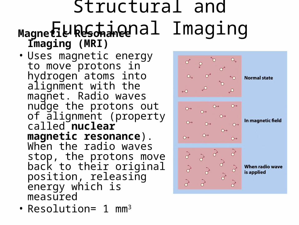
Structural and Functional ImagingMagnetic Resonance Imaging
(MRI)• Uses magnetic energy to
move protons in hydrogen atoms into alignment with the magnet. Radio waves nudge the protons out of alignment (property called nuclear magnetic resonance). When the radio waves stop, the protons move back to their original position, releasing energy which is measured
• Resolution= 1 mm3

Structural and Functional Imagingfunctional MRI• Blood oxygenation level-dependent (BOLD) effect:
change in the ratio of oxygenated to deoxygenated hemoglobin
• Resolution= 3mm3
Positron Emission Tomography (PET)• Inject a radioisotope (usually of oxygen) and monitor
concentration of decaying tracer (gives off gamma rays in a predictable pattern)
• Resolution of 5-10 mm3
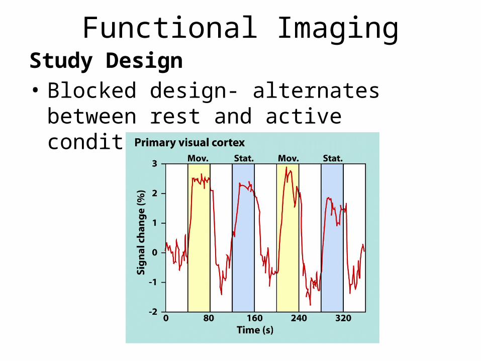
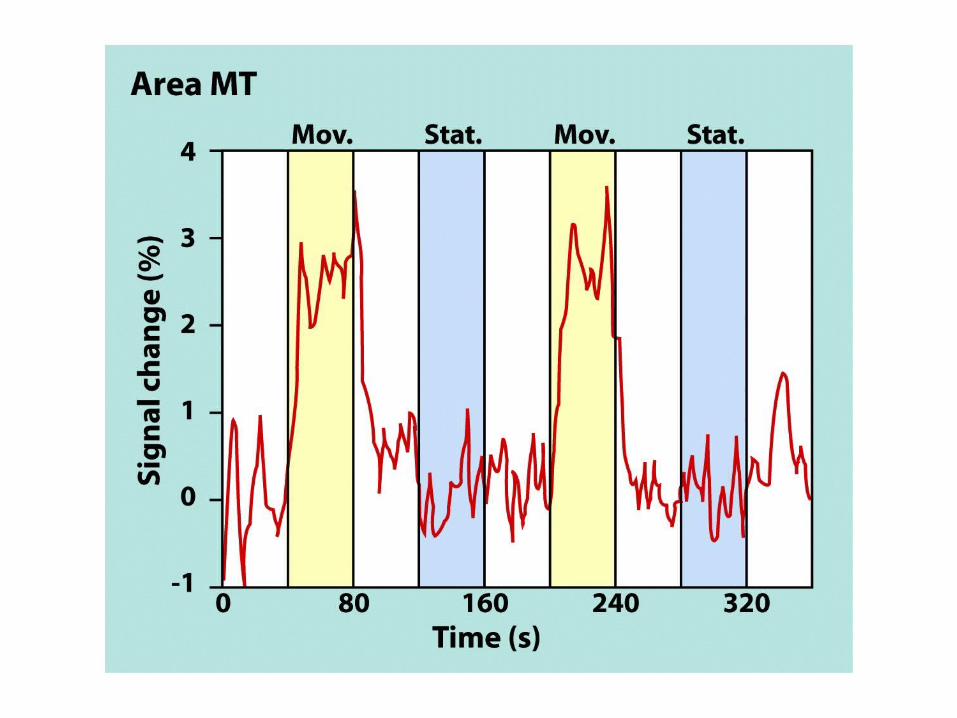
Functional ImagingStudy Design• Blocked design- alternates between rest and
active conditions
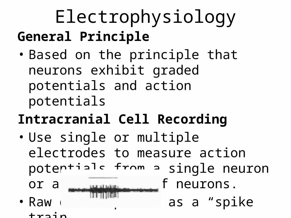
ElectrophysiologyGeneral Principle• Based on the principle that neurons exhibit
graded potentials and action potentialsIntracranial Cell Recording• Use single or multiple electrodes to measure
action potentials from a single neuron or a population of neurons.
• Raw data depicted as a “spike train.”
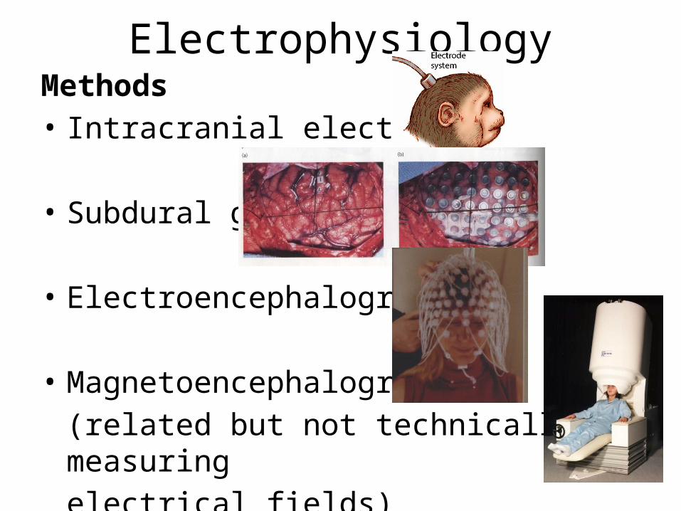
ElectrophysiologyMethods• Intracranial electrodes
• Subdural grid
• Electroencephalography
• Magnetoencephalography(related but not technically measuringelectrical fields)
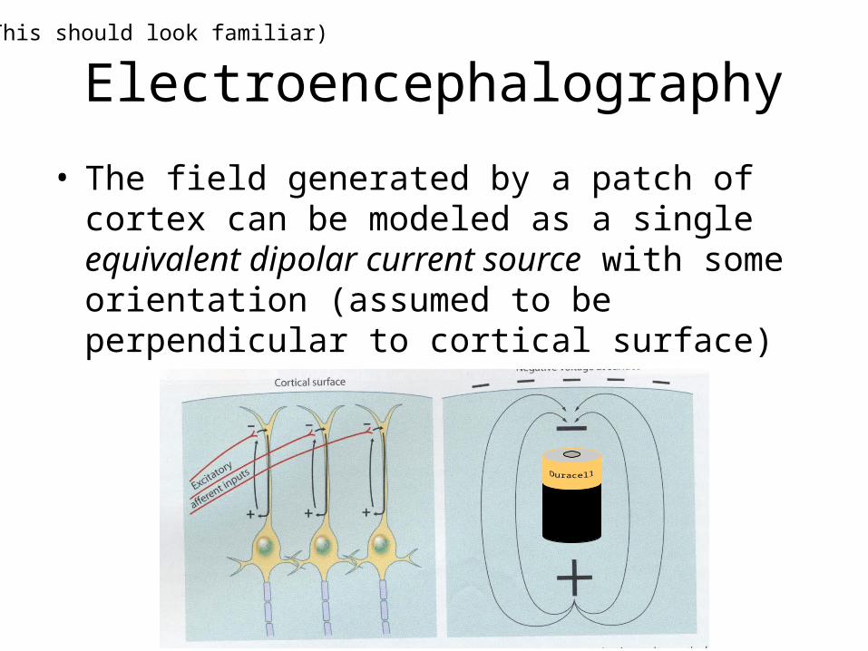
Electroencephalography
• The field generated by a patch of cortex can be modeled as a single equivalent dipolar current source with some orientation (assumed to be perpendicular to cortical surface)
(This should look familiar)
Magnetoencephalography
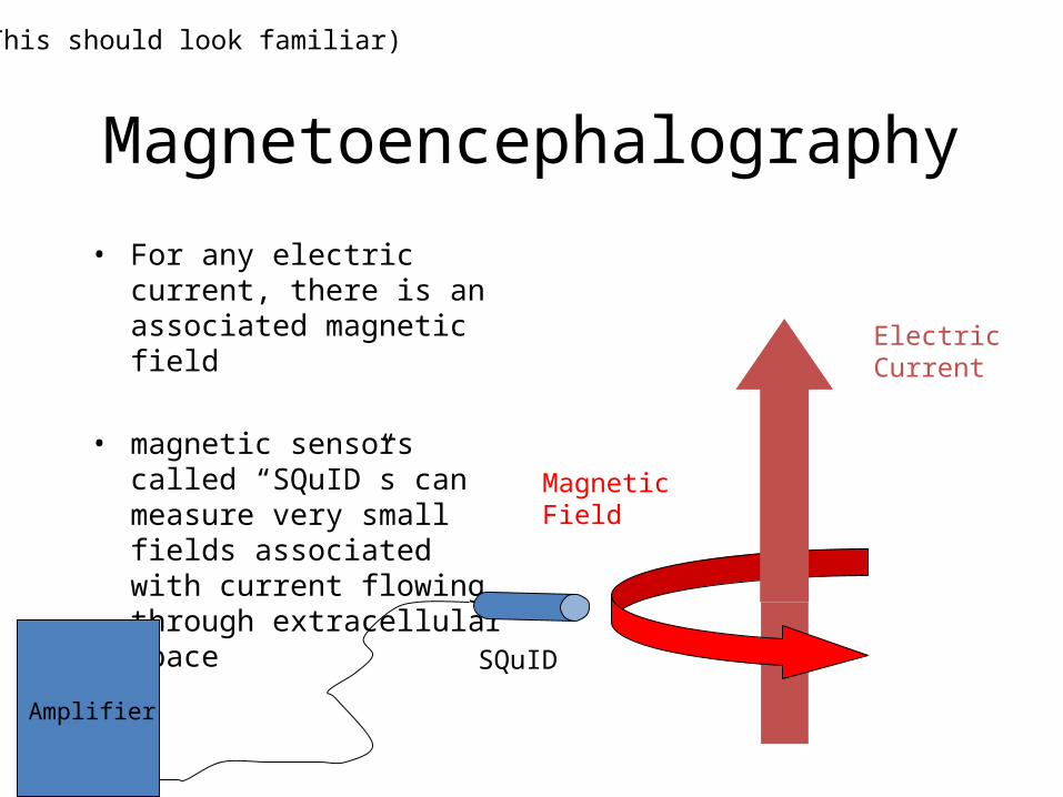
• For any electric current, there is an associated magnetic field
• magnetic sensors called “SQuID”s can measure very small fields associated with current flowing through extracellular space
Magnetic Field
Electric Current
SQuID
Amplifier
(This should look familiar)
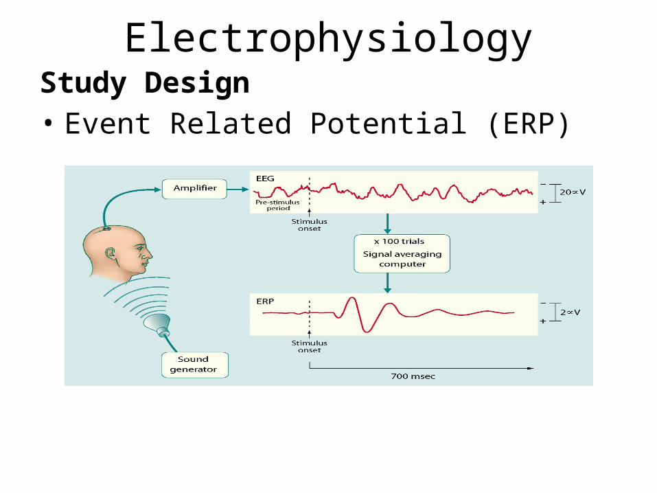
ElectrophysiologyStudy Design• Event Related Potential (ERP)
ElectrophysiologyLocalization• Few methods that apply to EEG and MEG– Plot oscillations across sensors as a 2D map, called
an isopotential map– Brain Electrical Source Analysis• Identify 3D generator source through modelling neural
activity as one or more equivalent current dipoles
– Beamforming• Signal processing technique that adjusts the signal
recorded at each sensor to tune the array to a single voxel at a time
– Possible to coregister with structural image (MRI)
Lesion StudiesMethods- Animals– Aspiration– Electrolytic– Vascular lesions (e.g. endothelin 1- stroke)– Reversible– Selective pathways (e.g. MPTP- Parkinsons)– Transgenic animals (Gene knock-Out, Knock-In)• Selectively block specific receptors
• ASSUMES that behavioural change after a lesion reveals function of the damaged area.– Issue of interconnectivity
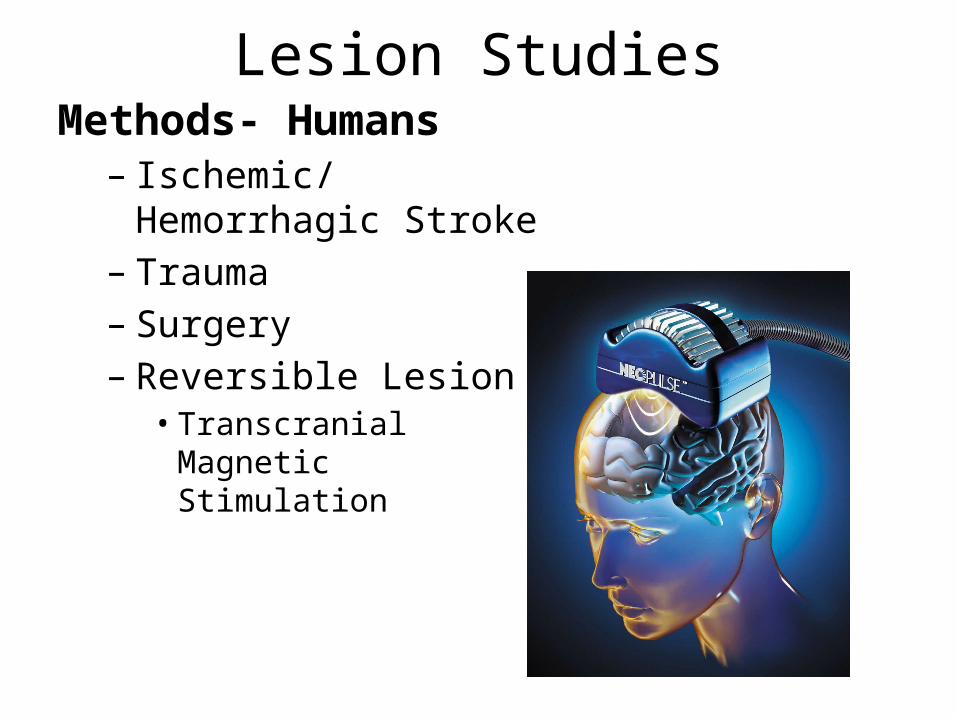
Lesion StudiesMethods- Humans– Ischemic/Hemorrhagic
Stroke– Trauma– Surgery – Reversible Lesion • Transcranial Magnetic
Stimulation
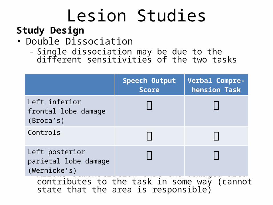
Lesion StudiesStudy Design• Double Dissociation– Single dissociation may be due to the different sensitivities
of the two tasks
– Allows demonstration that the damaged area contributes to the task in some way (cannot state that the area is responsible)
Speech Output Score Verbal Compre-hension Task
Left inferior frontal lobe damage (Broca’s) Controls Left posterior parietal lobe damage (Wernicke’s)
Techniques of Cognitive Neuroscience
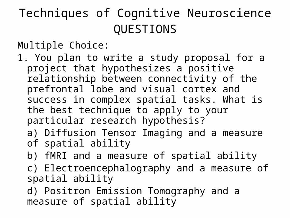
Techniques of Cognitive NeuroscienceQUESTIONS
Multiple Choice:1. You plan to write a study proposal for a project that
hypothesizes a positive relationship between connectivity of the prefrontal lobe and visual cortex and success in complex spatial tasks. What is the best technique to apply to your particular research hypothesis?a) Diffusion Tensor Imaging and a measure of spatial abilityb) fMRI and a measure of spatial abilityc) Electroencephalography and a measure of spatial abilityd) Positron Emission Tomography and a measure of spatial ability
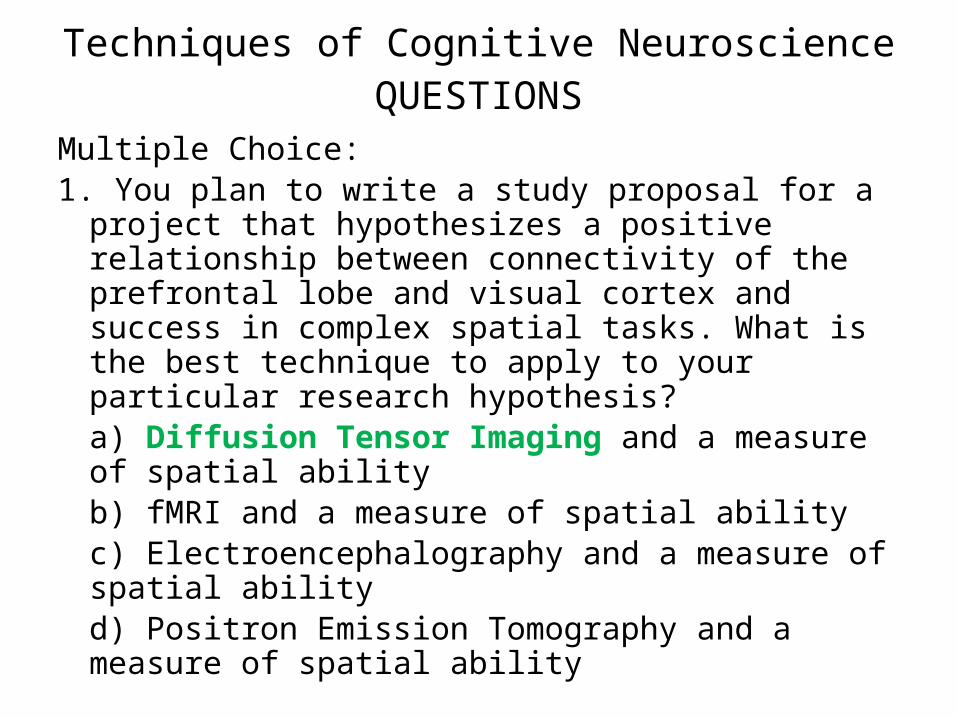
Techniques of Cognitive NeuroscienceQUESTIONS
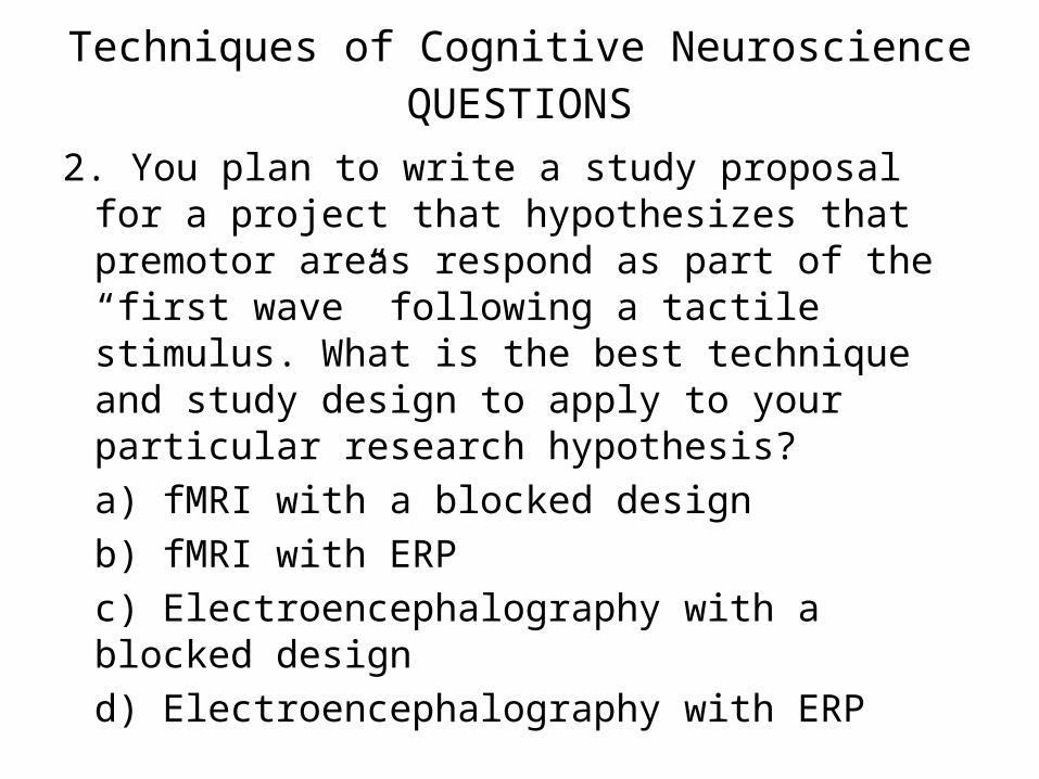
2. You plan to write a study proposal for a project that hypothesizes that premotor areas respond as part of the “first wave” following a tactile stimulus. What is the best technique and study design to apply to your particular research hypothesis?a) fMRI with a blocked designb) fMRI with ERPc) Electroencephalography with a blocked designd) Electroencephalography with ERP
Techniques of Cognitive NeuroscienceQUESTIONS
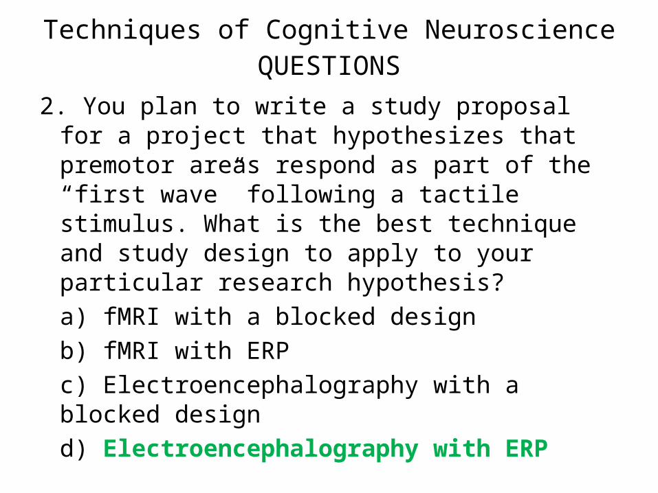
2. You plan to write a study proposal for a project that hypothesizes that premotor areas respond as part of the “first wave” following a tactile stimulus. What is the best technique and study design to apply to your particular research hypothesis?a) fMRI with a blocked designb) fMRI with ERPc) Electroencephalography with a blocked designd) Electroencephalography with ERP