Embed Size (px)
Citation preview
UNIVERSITÄTSKLINIKUM HAMBURG-EPPENDORF
Aus dem Institut für experimentelle Immunologie und Hepatologie des Zentrums für
Innere Medizin des Universitätsklinikum Hamburg-Eppendorf
Direktorin: Prof. Dr. Gisa Tiegs
Coffee interferes with HCV replication by inhibition of wnt
signaling pathway
Dissertation
zur Erlangung des Grades eines Doktors der Medizin
der Medizinischen Fakultät der Universität Hamburg
vorgelegt von
Amelie Dorothea Keller
aus
Mainz am Rhein
Hamburg 2014

2
Angenommen von der Medizinischen Fakultät der Universität Hamburg am: 20.02.2015 Veröffentlicht mit Genehmigung der Medizinische Fakultät der Universität Hamburg Prüfungsausschuss, der/die Vorsitzende: Prof. Dr. Gisa Tiegs Prüfungsausschuss, zweite/r Gutachter/in: PD Uwe Kordes Prüfungsausschuss, dritte/r Gutachter/in:

3
Für meine Eltern

4
Index
1. Introduction
1.1 Projektskizze/ Aim of Study
1.2 Introduction
2. Experimental Procedures
2.1 Material
2.1.1 Technical Equipment
2.1.2 Consumables
2.1.3 Reagents and Kits
2.1.4 Buffers and Solutions
2.1.5 Oligonucleotides
2.1.6 Plasmids
2.1.7 Antibodies
2.1.8 Software
2.2 Methods
2.2.1 Cell lines
2.2.2 Isolation of primary murine hepatocytes
2.2.3 Reagents’ preparation
2.2.4 Analysis of cell viability
2.2.5 Luciferase reporter assay
2.2.6 Beta-catenin-regulated transcription (CRT)
2.2.7 Detection of mRNA by real-time RT-PCR

5
2.2.8 Infection with virus particles
2.2.9 Immunofluorescence
2.2.10 TCID50
2.2.11 Statistical analysis
3. Results
3.1 Coffee reduces HCV replication of genotype 1b, replicon system
3.2 Coffee reduces HCV replication of genotype 2a, subgenomic replicon system
3.3 Coffee reduces HCV replication and infectivity of genotype 2a, infectious
system
3.4 Coffee interferes with viral entry by HCV receptor down-regulation
3.5 Caffeine interferes with replication of HCV genotype 1b, replicon system
3.6 Chlorogenic acid and genistein do not interfere with HCV replication
3.7 Cafestol does not interfere with HCV replication
3.8 Coffee and caffeine increase interferon effects on HCV replication
3.9 Coffee, but not caffeine, decreases wnt signaling activity
4. Discussion
5. Abstract
5.1 Abstract
5.2 Zusammenfassung
6. Abbreviations

6
7. References
7.1 Journals
7.2 Internet
8. Acknowledgements
9. Curriculum vitae
10. Eidesstattliche Erklärung

7
1. Introduction
1.1. Projektskizze/ Aim of Study
Die chronische Infektion mit Hepatitis C Virus (HCV) ist durch neue Medikamente
mittlerweile gut beherrschbar. Dennoch ist die Entstehung einer Leberzirrhose und
möglicherweise eines hepatozelluläres Karzinom (HCC) weiterhin eine Folge der
chronischen Hepatitis C. Noch vor einigen Jahren hielt sich die Meinung, dass
Kaffeekonsum schädlich für den menschlichen Körper sei. Es konnten jedoch in den letzten
Jahren verschiedene positive Wirkungen von Kaffeekonsum nachgewiesen werden. Neben
positiven Effekten bei Krankheiten wie Alzheimer oder Parkinson, konnte auch eine
Wirkung des Kaffees auf verschiedene Viren (HBV, HIV, HSV) festgestellt werden. Bislang
wurden Kaffeewirkungen auf das Hepatitis C Virus noch nicht untersucht. Die Effekte bei
anderen Virusinfektionen werfen nun die Frage auf, inwieweit Kaffeekonsum auch den
Verlauf einer HCV Infektion günstig beeinflussen könnte.
Zur Untersuchung der HCV Replikation stehen in unserem Labor die Replikonzelllinien
HUH5-15 und Luc-Ubi-Neo/ET zur Verfügung. Es handelt sich dabei um Zelllinien, welche
auf der humanen Hepatomzelllinie HUH7 basieren, die die HCV Proteine NS3 bis NS5B
stabil unter der Kontrolle der HCV UTRs exprimiert. Im Fall der Linie Luc-Ubi-Neo/ET
dient ein zusätzlich vorhandenes Luziferase-Reportergen zur einfachen Quantifizierung der
HCV Replikation. Als primäre Messgröße verwenden wir dabei die durch real-time RT-PCR
zu messende Expression der Gene für NS3 bis NS5B, die mit der HCV Replikation
korrelieren. In dieser Arbeit sollen die Effekte von koffeinhaltigem und entkoffeiniertem
Kaffee, Koffein und verschiedenen Kaffeeinhaltsstoffen und Abbauprodukten auf die HCV
Replikation gestestet werden.

8
1.2. Introduction
Coffee
Coffee is the most frequently consumed legal drug all over the world. It is a beverage
derived from roasted fruits of the coffee tree, known as coffee cherries. Historically, coffee
originates from the region Caffa in Ethiopia and was first described in the 9th century. First
reports of coffee in Europe are from the 15th-century and the first German coffeehouse was
built in 16th century. The most common species of coffee beans are Coffea arabica
(„Arabica“) and Coffea canephora („Robusta“). After roasting and grinding, the coffee
flour is brewed to coffee using different techniques, e.g. boiling coffee in hot water in
Turkey or using the French press or a coffee percelerator, which is common in Europe and
the United States. Coffee is consumed as a hot or cold beverage, with cream and sugar or
just as black coffee. Enhanced concentration and less fatigue are the reasons for many
people to consume high amounts of coffee.
Bad Coffee?
During the long tradition of coffee drinking, coffee effects were discussed controversially.
Despite its well-known effects on fatigue and concentration, coffee was accused to have
bad side effects on human health. Therefore, coffee intake was suspected to cause and
aggravate chronic diseases. In particular, damages of the gastrointestinal tract and the
cardiovascular system have been discussed to be caused by coffee consumption. In fact, it
has been shown that coffee activates gastric acid secretion and thereby can promote gastro-
oesophageal reflux. Nevertheless an association between coffee-consumption and
dyspepsia could not be confirmed (Boekema PJ et al. 1999). Another study showed that
caffeine reduces exercise-induced myocardial blood flow especially in patients with

9
coronary heart diseases or arteriosclerosis (Namdar M et al. 2006), and thereby raises the
question whether caffeine consume is safe for patients with coronary artery diseases.
Healthy Coffee!
In the last years, many studies investigated the effects of coffee on human health and it
turned out that coffee seems to have overly beneficial effects (Butt MS, Sultan MT 2011).
Clinical studies revealed that coffee consumption reduces general mortality, especially
mortality due to cardiovascular diseases (Lopez-Garcia E et al. 2008; Liu J et al. 2013).
The reduced risk of heart diseases, especially in women, (Wu JN et al. 2009) and stroke
(Lopez-Garcia E et al. 2009) might be caused by antioxidants contained in coffee. The risk
to develop a metabolic syndrome or diabetes typ-2 (van Dam RM, Hu FB 2005) was found
to be reduced among coffee consumers. This might also be due to anti-oxidative
components in coffee, which enhance insulin sensitivity. Likewise, the risk of Alzheimer’s
disease (Eskelinen MH, Kivipelto M 2010) or Parkinson’s disease (Costa J et al. 2010) was
found reduced via the antioxidant defense by coffee. Recently it has been shown that coffee
consumption decreases the risk of liver fibrosis formation (Modi AA et al. 2010).
Furthermore, coffee has been shown to reduce the risk to develop colorectal (Galeone C et
al. 2010), liver (Larsson SC, Wolk A 2007) and breast cancer (Lowcock EC et al. 2013).
Coffee ingredients
Coffee contains more than 2000 components (Tuomilehto J. 2013), of which many are not
yet characterized. Those substances can be divided into groups, e.g. carbohydrates, lipids,
proteins, acids, alkaloids, minerals and aromatic compounds. During roasting, grinding and
brewing, concentrations of all these substances change.
The most commonly known substance in regular coffee is caffeine. In the human body,
caffeine is metabolized in the liver into its main degradation product paraxanthine (81%) as
well as theofylline (11%) and theobromine (5%) by cytochrome oxidases (Gu L et al. 1992;

10
Orrú M et al. 2013). As a clinical study has shown, caffeine consumption is associated with
reduced liver fibrosis formation (Modi AA et al. 2010) and reduced histological activity in
patients with chronic hepatitis C (Costentin CE et al. 2011). The caffeine degradation
product paraxanthine has been shown to reduce intercellular collagen deposition in the
cholestatic rat liver (Klemmer I et al. 2011) and to be an inhibitor of CTGF (connective
tissue growth factor) expression in liver parenchymal cells (Gressner OA et al. 2009).
Paraxanthine, theofylline and theobromine act as phosphodiesterase (PDE) inhibitors. The
PDE-inhibitor pentoxifylline has been shown to have hepatoprotective properties and
antiviral effects in HCV patients (Gutierrez-Reyes G et al. 2006). Furthermore,
supplementation of HCV treatment (PEG-IFN/ribavirin) with pentoxifylline improved the
sustained virological response (SVR) (Jiménez-Luévano MÁ et al. 2013). Per definition
SVR is the absence of detectable HCV RNA in blood serum up to six months after therapy.
Other coffee ingredients have been shown to have positive effects on different types of
cancer as well. Cafestol and kahweol are diterpens belonging to the coffee lipids and are
mostly consumed with unfiltered coffee. Both have been reported to exhibit anti-
carcinogenic activity in hepatic, intestinal and kidney tissue (Cavin C et al. 2002).
Chlorogenic acid is part of the coffee acids. It is the ester of caffeic acid and has been
reported to affect colon cancer (Kang NJ et al. 2011). Genistein belongs to the group of
isoflavones, which are flavonoides belonging to the secondary plant compounds.
Secondary plant compounds are nonessential plant compounds, which do not directly affect
growth and development of an organism. Genistein has been shown to inhibit the wnt/beta-
catenin signaling pathway (Zhang Y, Chen H 2011), which is linked to cancer
development.
11
Hepatitis C Virus – HCV
The acute infection with HCV displays no specific symptoms and therefore remains mostly
undetected and untreated. As a result, almost 70% of all acute HCV infections become
chronic and about 20% of chronically infected patients develop liver cirrhosis. Chronic
HCV infection currently affects about 3% of the world’s population (Tang H, Grisé H
2009) and is a major cause for the development of hepatic cancer (Blonski W, Reddy KR
2008). In Germany, HCV prevalence is about 0.5%, while in high-risk areas in Africa the
prevalence is up to 14%. HCV, which is a small RNA virus and belongs to the family of
Flaviviridae, is transmitted parenterally. Up to now, seven major genotypes have been
identified, which vary in their prevalence between the continents. In Germany, genotype 1
still is the major genotype with the highest prevalence, while the percentage of patients
with genotype 3 infections is increasing (Cornberg M et al. 2011). Because of the high
mutation rate of the virus and the thereby fast increasing number of subtypes, it has not
been possible to develop a vaccine so far. Since the most common way of transmission is
via blood and blood products, there is a higher risk of HCV infection in dialysis patients,
intravenous drug users and after blood transfusion. Target cell of HCV is the hepatocyte,
which is entered by the virus via cell surface receptors. So far CD81, CLDN1, OCLN
(Fraquhar MJ et al. 2012) and LDL-R (Albecka A et al. 2012) have been identified as
essential HCV receptors.
HCV therapy
Until very recently, treatment of acute and chronic hepatitis C infection has predominantly
been performed by a combination of pegylated interferon alpha2 (PEG-IFN) with the
nucleoside analogue ribavirin. IFN alpha activates the JAK-STAT-signaling pathway,
which at least leads to inhibition of viral proteinsynthesis and as well cause degradation of
viral mRNA (Bandurska K et al. 2013). Ribavirin is a nucleoside analogue, which has been

12
shown to have antiviral activity via direct and indirect mechanisms e.g. inhibition of
inosine-monophosphate-dehydrogenase and immune modulation. With this combined
treatment, a sustained virological response (SVR) could be achieved. Due to PEG-
IFN/ribavirin therapy only 45% of genotype 1 patients achieve SVR in contrast to 80% of
genotype 2 or 3 infected patients (Di Bisceglie AM, Hoofnagle JH 2002). The recent
approval of the HCV NS3-4A protease inhibitors Telaprevir and Boceprevir raises hopes
for an improvement of therapy (Vermehren J, Sarrazin C 2011). In fact, a combination of
protease inhibitors with PEG-IFN/ribavirin enhanced SVR rates in patients infected with
HCV genotype 1 (Vagu C et al. 2013). In phase III clinical studies, Daclatasvir, an NS5A
inhibitor, has been shown to effectively interfere with HCV genotype 1a and especially
HCV genotype 1b in patients (Herbst DA, Reddy KR 2013).
Coffee and HCV – what this work is about
In 2011 a clinical study reported that coffee consumption improved the effects of PEG-
IFN/ribavirin therapy in HCV patients (Freedman ND et al. 2011). Therefore, this thesis
was conducted to investigate, if coffee in fact has anti-HCV activity and to elucidate
mechanisms and mediators in vitro. We decided to incubate hepatocytes and HCV
replicating human cell lines with concentrations of coffee corresponding to up to 3 cups of
coffee (200 mL per cup), an amount easily consumed during a day. Cell lines were also
incubated with coffee ingredients and degradation products to break down effects of coffee
to single substances.
13
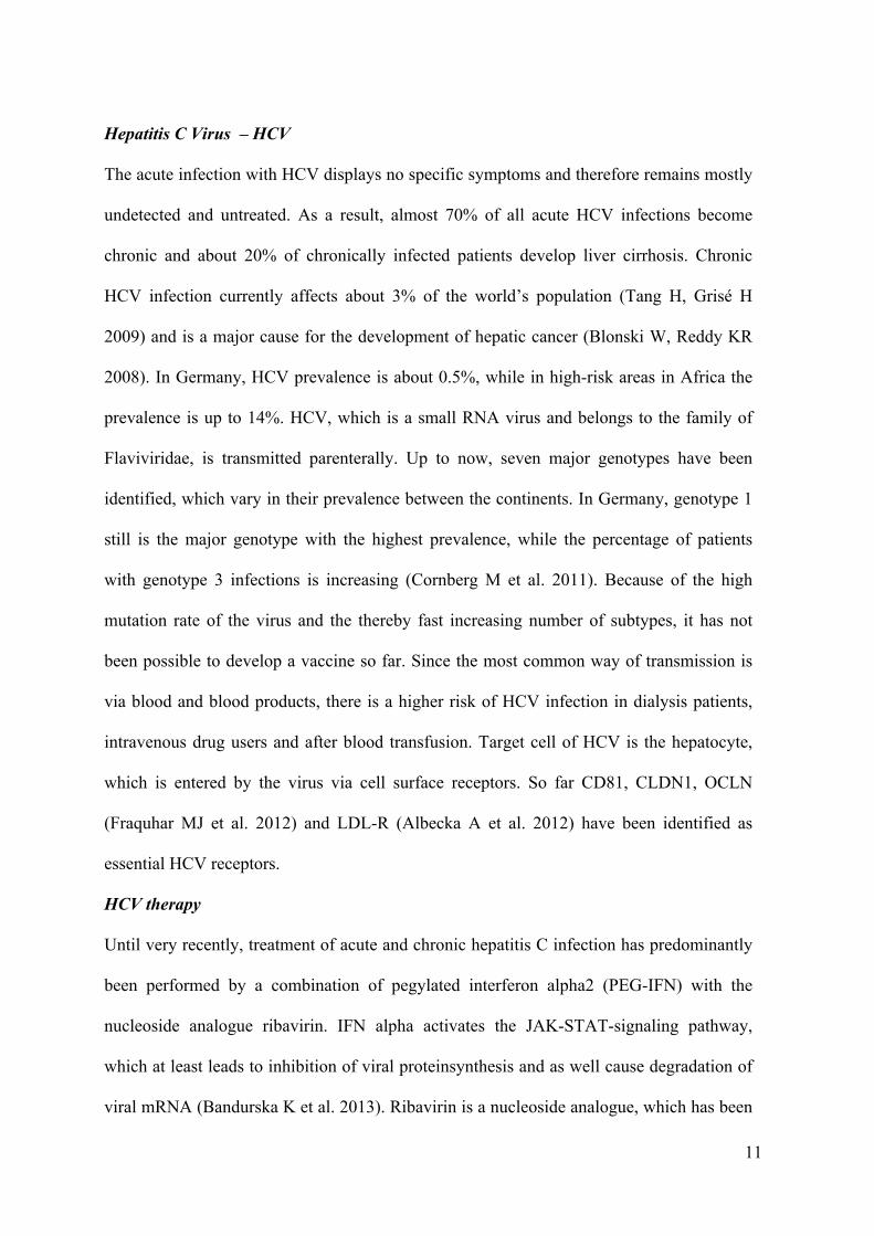
2. Materials and Methods
2.1 Materials
2.1.1 Technical Equipment
Equipment Supplier
Casy Cell Counter Roche Innovatis AG, Reutlingen, D
Centrifuge 5417 R Eppendorf, Hamburg, D
Centrifuge 5810 R Eppendorf, Hamburg, D
CFXTM Real-Time sytem Bio-Rad, München, D
CK40 Microscope Olympus, Hamburg, D
Electrophoresis Unit Bio-Rad, München, D
Eppendorf Research® Plus Pipettes Eppendorf, Hamburg, D
Incubator HERAcell 240 CO2 Thermo Fisher, Langenselbold, D
LCachN/20X/0.40 Phc/1/FN22 UIS objective
Light Cycler, CFX96 Real-Time System Bio-Rad, München, D
Neubauer Improved Chamber Marienfeld GmbH, Lauda-
Königshofen, D
Pipetboy acu Integra Biosciences GmbH,
Fernwald, D
Precision balance TE1245 Sartorius, Göttingen, D
Peristaltic pump Medorex, Nörten-Hardenberg, D
Sterilbench HERAsafe Thermo Fisher, Langenselbold, D
Tecan® infinite M2000 Tecan Group Ltd., Männedorf, CH

14
Thermal Cycler Primus 96 plus Eurofins MWG Operon, Ebersberg, D
VersaDoc 4000MP Bio-Rad, München, D
Vortex, Reax200 Heidolph Instruments, Schwabach, D
Water bath Lauda GmbH&Co , Lauda-
Königshofen, D
2.1.2 Consumables
Consumable Supplier
Cell culture bottle Cellstar® Greiner Bio-One GmbH,
Frickenhausen, D
Cell culture plates, 24-well, 96-well Thermo Fisher, Roskilde, DK
Centrifuge tubes Sarstedt, Nürnbrecht, D
Cuvette (single-use) Roth, Karlsruhe, D
Mµltiguard®-Tips Roth, Karlsruhe, D
PCR-Tubes ABgene, Thermo Fisher, Hamburg, D
Pipets Research® variabel Eppendorf, Hamburg, D
Pipets (single-use) Sarstedt, Nürnbrecht, D
Pipet Tips Plastibrand® Brand, Wertheim, D
Quali-PCR-Tube Kisker GbR, Steinfurt, D
Tubes, 1,5ml, 2ml Roth, Karlsruhe, D
2.1.3 Reagents and Kits
Reagent Supplier
(3-(4,5-Dimethylthiazol-2-yl)-2,5-diphenyl- Sigma-Aldrich, St Louis, USA

15
Tetrazolium bromide (MTT)
4,5,6,7-tetrabromo-2-azabenzimidazole (TBB) Sigma Aldrich, St Louis, USA
Bradford Bio-Rad,München, D
BSA Roth, Karlsruhe, D
Cafestol Sigma Aldrich, St Louis, USA
Caffeine Sigma Aldrich, St Louis, USA
Chlorogenic acid Sigma Aldrich, St Louis, USA
Coffee regular/ decaffeinated (100% Arabica)
Dimethyl sulfoxide (DMSO) Sigma-Aldrich, St Louis, USA
Distilled water, RNase free Thermo Fisher Scientific, Waltham,
USA
DMEM (Dulbecco´s Modified Eagle´s Medium) Gibco®, Invitrogen, Darmstadt, D
+ 4,5 g/l Glucose, + L-glutamin, - Pyruvate
Fetal calf serum (FCS) Invitrogen, Darmstadt, D
Genistein Sigma Aldrich, St Louis, USA
IFN alpha
L-glutamine Invitrogen, Darmstadt, D
LiChrosolv Water Merck, Darmstadt, D
LipofectamineTM 2000 Invitrogen GmbH, Karlsruhe, D
Luciferase Assay System Promega, Mannheim, D
Master mix (2x) Thermo Fisher Scientific, Waltham,
USA
Nicotinic acid Sigma Aldrich, St Louis, USA
Optimem (1X) Gibco®, Invitrogen, Darmstadt, D
Paraxanthine Sigma Aldrich, St Louis, USA

16
Percoll GE Healthcare, Chalfont St. Giles,
UK
PFA Roth, Karlsruhe, D
Quercetin hydrate Fisher Scientific, Schwerte, D
rDNase Kit Macherey-Nagel, Düren, D
Sodium chloride (NaCl) AppliChem, Darmstadt, D
Sodium dodecyl sulfate (SDS) AppliChem, Darmstadt, D
Sodium hydrogen carbonate (NaHCO3) Roth, Karlsruhe, D
Sodium hydroxide (NaOH) Roth, Karlsruhe, D
Theofylline Sigma Aldrich, St Louis, USA
Theobromine Sigma Aldrich, St Louis, USA
Triton Roth, Karlsruhe, D
Trizol Reagent Invitrogen, Karlsruhe, D
Trypan blue Sigma-Aldrich, St Louis, USA
VersoTM cDNA Kit Thermo Fisher Scientific, Waltham, USA
William’s medium E (1X) + GlutaMAXTM-I Gibco®, Invitrogen, Darmstadt, D
2.1.4 Buffers and Solutions
PBS · 137.9 mM NaCl
· 6.5 mM Na2HPO4 x 2 H2O
· 1.5 mM KH2PO4
· 2.7 mM KCl
· pH 7.4 (NaOH)

17
SDS-lysis-buffer 10 g SDS
ad to 50ml PBS
100 µl 10 M HCl
2.1.5 Oligonucleotides
Oligonucleotides for subsequent PCR-reactions were obtained from Metabion International
AG (Martinsried, Germany) (Table 1).

18
Table 1: Oligonucleotides and Sequences
Oligonucleotide Sequence 5´-3´
5’GAPDH 5´-ACCCAGAAGACTGTGGATGG -3´
3’GAPDH 5´-TTCTAGACGGCAGGTCAGGT-3´
5’ATPsy 5’-GCCCACTTCTTACCACAAGG-3’
3’ATPsy 5’-GCGACAGCGATTTCTAGGAT-3’
5' HCV214 5´-TGCGGAACCGGTGAGTACA-3´
3' HCV214 5´-AGGTTTAGGATTCGTGCTCAT-3´
5’ Conductin 5’- AGGGAGAAATGCGTGGATAC-3’
3’ Conductin 5’-TGGAATCAATCTGCTGCTTC-3’
5’ Beta-catenin 5’-AAAGCGGCTGTTAGTCACTGG-3’
3’ Beta-catenin 5’-CGAGTCATTGCATACTGTCCAT-3’
5’ CK2alpha 5’-GAACGCTTTGTCCACCGTG -3’
3’ CK2alpha 5’-GTTGGCAGCAGCAATCACTG -3’
5’ CK2alphaprime 5´-CTTGTTCGCATTGCCAAGGTTC-3´
3’ CK2alphaprime 5´-CACTGGAAAGCACAGCATTGTC-3´
5’ LDLR 5’-GTGCTCCTCGTCTTCCTTTG-3’
3’ LDLR 5’-TAGCTGTAGCCGTCCTGGTT-3’
5’ Claudin-1 5´-CCGTTGGCATGAAGTGTATG -3´
3’ Claudin-1 5´-CCAGTCAAGAGAGCCTGACC-3´

19
2.1.6 Plasmids
TOPFlash plasmid (M50) kind gift from Dr. Wege, UKE
FOPFlash (M51) kind gift from Dr. Wege, UKE
2.1.7 Antibodies
anti-NS3 Biofront Technologies; 1:3000
2E3 Biofront Technologies; 1:3000
anti-mouse-HRP Sigma; A4416; 1:10000
HCV-2E3 BioFront Technologies
NS3 BioFront Technologies
A3R3 gift of Mansun Law, The Scripps Research Institute
E2 gift of Mansun Law, The Scripps Research Institute
Alexa-594 Molecular Probes
Alexa-488 Molecular Probes
2.1.8 Software
MS Office 2003 Microsoft, Redmond, USA
GraphPad Prism 5 GraphPad Software, La Jolla, USA
Bio-Rad CFX Manager 2.0 Bio-Rad, Hercules, USA
iControl 5.0 Tecan, Crailshaim, D
20
2.2 Methods
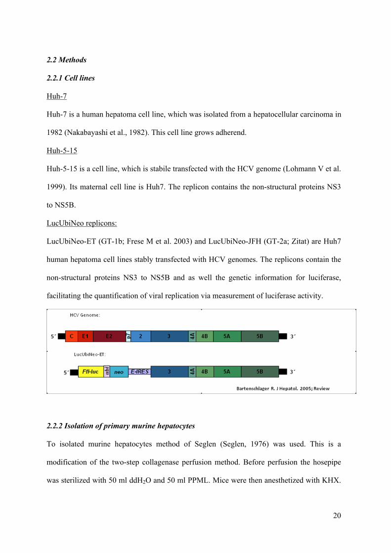
2.2.1 Cell lines
Huh-7
Huh-7 is a human hepatoma cell line, which was isolated from a hepatocellular carcinoma in
1982 (Nakabayashi et al., 1982). This cell line grows adherend.
Huh-5-15
Huh-5-15 is a cell line, which is stabile transfected with the HCV genome (Lohmann V et al.
1999). Its maternal cell line is Huh7. The replicon contains the non-structural proteins NS3
to NS5B.
LucUbiNeo replicons:
LucUbiNeo-ET (GT-1b; Frese M et al. 2003) and LucUbiNeo-JFH (GT-2a; Zitat) are Huh7
human hepatoma cell lines stably transfected with HCV genomes. The replicons contain the
non-structural proteins NS3 to NS5B and as well the genetic information for luciferase,
facilitating the quantification of viral replication via measurement of luciferase activity.
2.2.2 Isolation of primary murine hepatocytes
To isolated murine hepatocytes method of Seglen (Seglen, 1976) was used. This is a
modification of the two-step collagenase perfusion method. Before perfusion the hosepipe
was sterilized with 50 ml ddH2O and 50 ml PPML. Mice were then anesthetized with KHX.

21
After opening the abdomen, the upper part of the vena cava superior was ligated. 10 ml
PPML, followed by 25 ml PM (42 °C), containing 1 mg Liberase (Roche, Basel, CH), were
used to perfuse the portal vein at a flow-rate of 1.8 ml/min. In the next step the liver was
perfused with 25 ml PM. After excising the liver it was transferred to a petri-dish with 25 ml
PM (42 °C), where it was homogenized with tweezers by gentle shaking. This was followed
by sterile filteration through a sterile cell strainer (100 µm) into a sterile 50 ml tube. All
following steps were carried out under sterile conditions inside a clean bench. In the next 15-
20 minutes the hepatocytes were incubated at room temperature for sedimentation, after this
step 25 ml of the supernatant were discarded. Isolated hepatocytes were gently resuspended
by pipetting up and down and transferred to 25 ml of a 4 °C Percoll-gradient containing 90%
Percoll (GE-Healthcare, Chalfont St. Giles, UK) and 10% 10x DPBS (Gibco®, Invitrogen,
Darmstadt, D). The mixture was inverted and centrifugated at 72 g and 4 °C for 10 min.
After discharging the supernatant the hepatocyte pellet was washed in 30 ml
William’s_E_GlutaMAXTM-I medium (Gibco®, Invitrogen, Darmstadt, D), supplemented
with 10% fetal bovine serum (Invitrogen, Darmstadt, D), 1% penicillin/streptomycin
(Gibco®, Invitrogen, Darmstadt) and 1% L-glutamine (Invitrogen, Darmstadt) and
centrifugated at 72 g and 4 °C for 5 min. Again supernatant was discarded and the pellet was
resuspended in William’s medium supplemented as above. The hepatocytes were stained
with trypan blue to determine cell viability. Cells were counted with a Neubauer counting
cell chamber. Cells were seed in concentrations of 1,5 x 10⁵ cells per well in 24-well plates
and 3 x 10⁴ cells in 96-well plates and incubated in a 40% O2 and 5% CO2 humidified
atmosphere at 37 °C. William’s medium was exchanged 4h after isolation.

22
2.2.3 Reagents’ preparation
1 g of regular or decaffeinated coffee (100% Arabica) was extracted in 5 ml of LiChrosolv
Water. Following centrifugation at 4000 rpm for 5 minutes, supernatants were sterile filtered
(0.2 µM), aliquoted and stored at -20°C until use. Control cells were incubated with water as
used for coffee preparations. Caffeine, theofylline, theobromine, paraxanthine, chlorogenic
acid and genistein (all: Sigma Aldrich GmbH, Steinheim, Germany) were dissolved in aqua
destillata and stored at -20°C until use. Quercetin hydrate (Acros Organics, part of Thermo
Fisher Scientific, Waltham, MA, USA), 4,5,6,7-tetrabromo-2-azabenzimidazole (TBB) and
cafestol (both: Sigma Aldrich GmbH, Steinheim, Germany) were dissolved in DMSO.
Vehicle control (DMSO) was dissolved in cell culture medium to concentrations used on
incubated cells and measured in parallel as indicated in the Figures and Figure Legends.
2.2.4 Analysis of cell viability
Cell viability was measured by using (3-4, 5-Dimethylthiazol-2-yl)-2, 5-diphenyltetrazolium
bromide (MTT; Sigma Aldrich GmbH, Steinheim, Germany) according to the
manufacturer’s instructions. Viability of cells in immunohistochemistry was visualized by
bright field microscopy.
2.2.5 Luciferase reporter assay
Luc-Ubi-Neo-ET cells were seeded in 24-well plates and allowed to adhere over night.
Treatment with different substances was performed for 24 hours. After treatment the
medium was discarded and cells were washed with PBS. After washing cells were incubated
with 100 µl 1 x cell culture lysis-buffer (Promega, Madison, USA) at room temperature for 5
min, followed by scratching of the cells with a pipette-tip. The cell-lysates were then
transferred into a 1.5 ml tube and centrifugated at 20,800 g and 4 °C for 1 min. After this
23
step the lysates were kept on ice. To measure luciferase-activity, 70 µl of the lysate
supernatant were transferred to a white 96-well plate (Nunc A/S, Roskilde, DK) and 50 µl
luciferase assay reagent (Promega, Madison, USA) were added. Luciferaseluminescence was
measured photometrically with the Infinite M200 Photometer (TECAN, Crailshaim, D)
according to the manufacturer’s instructions. Protein concentrations were quantified via
Bradford assay (Bradford 1976). Luciferase activity was normalized to the protein
concentrations of the cells.
2.2.6 Beta-catenin-regulated transcription (CRT)
CRT was monitored using a luciferase reporter system consisting of Super8xTOPFlash and
Super8xFOPFlash. The TOPFlash plasmid (M50) contains 8 TCF/LEF binding sites
enhancing firefly luciferase expression. The control plasmid FOPFlash (M51) carries mutant
TCF/LEF binding sites. 24h after transfection cells were assayed using a Reporter Assay
Systems (Promega GmbH, Mannheim, Germany).
2.2.7 Detection of mRNA by real-time RT-PCR
RNA isolation was performed using Trizol Reagent (Invitrogen, Darmstadt, D) according to
the manufacturer’s’ instructions. Cells were cultured on 24-well plates for 24 hours. After 24
hours the culture medium was discarded and cells were washed with PBS. Cells were
homogenized with 500 µl Trizol for 5 min at RT after washing. Lysates were then
transferred into sterile, RNase-free 1.5 ml tubes and 100 µl chloroform were added. The
samples were shaken for 15 s, followed by 15 min centrifugation at 12,000 g at 4 °C to
separate phases. The upper phase, containing the RNA was transferred into a new sterile,
RNase-free tube after centrifugation, mixed with 250 µl isopropanol and stored at -20 °C for
20-30 min. This was followed by 10 min centrifugation at 12,000 g and 4 °C. After

24
discarding the supernatant 500 µl of 70% ethanol solved in DEPC-treated ddH2O were added
to the tubes, followed by 5 min centrifugation at 7,500 g and 4 °C. The RNA pellets were
dried in a heat block for 10 min at 55 °C after discarding the ethanol, to ensure that no
ethanol remained. Afterwards RNA pellets were resolved in 40 µl RNase-free ddH2O by
incubation at 55 °C for 10 min in a heat block. mRNA was transcribed into cDNA by
reverse-transcriptase, to analyse altered gene expression. PCR was performed by using the
VersoTM cDNA Kit (Thermo Fisher Scientific, Waltham, MA, USA) according to the
manufacturer’s instructions. Oligonucleotides for PCR-reactions were obtained from
Metabion International AG (Martinsried, Germany). Oligonucleotide pairs used for real-time
PCR are summarized in section 2.1.5. CFXTM Real-Time system (BIO-RAD, Munich,
Germany) and reagents from ABgene® (Epsom, UK) were used to performe real-time RT-
PCR. To confirm amplification specificity PCR products were subjected to a melting curve
analysis. For normalization Glyceraldehyde 3-phosphate dehydrogenase (GAPDH) and
Actin were used as a housekeeping-genes.
2.2.8 Infection with virus particles
Huh7.5 cells were seeded into 12-well plates at 4×10⁴ cells/well 24h prior to infection. For
infection experiments cells were pre-incubated with coffee for 2 hours, followed by washing.
Infection was carried out for 24 hours before cells were washed and incubated with coffee
for 24 hours. Infection was carried out using HCV genotype 2a strain JC1 (Pietschmann T et
al. 2006) at an MOI of 0.5.
2.2.9 Immunofluorescence
Mouse monoclonal antibody HCV-2E3 against NS3 (BioFront Technologies), human
monoclonal antibody A3R3 against E2 (a kind gift of Mansun Law, The Scripps Research

25
Institute, La Jolla, CA, USA), goat anti-mouse Alexa-594 and chicken anti-human Alexa-
488-conjugated antibodies from Molecular Probes were used for staining. To visualize HCV
infection, NS3 and E2 proteins were stained. The procedure included fixation (4% PFA; 20
min. at RT), permeabilization (0.1% Triton X100; 4°C for 10 min) and blocking (5% BSA;
20 min. at RT). Pictures were taken in an inverted microscope CKX41 (Olympus) with an
LCachN/20X/0.40 Phc/1/FN22 UIS objective.
2.2.10 TCID50
Huh7.5 cells were seeded 24h prior to infection at 1.1x10⁴ in a 96 well plate. Supernatants
from JC1 infected cells were serially diluted starting from 1:5, 6 wells per dilution were
incubated. 72h post infection cells were fixed (4% of PFA; 20 min. at RT), permeabilized
(0.1% Triton X100; 10 min. at 4°C) and blocked (5% BSA; 20 min. at RT). After incubation
with the first antibody (anti-NS3, 2E3, Biofront Technologies; 1:3000) for 1h at RT, cells
were incubated with the second antibody (anti-mouse-HRP; Sigma; A4416; 1:10000) for 1h
at RT. Washing consisted of three rinses with PBS and bound peroxidase was detected.
TCID50 was calculated based on as described previously (Muench H 1938).
2.2.11 Statistical analysis
Results were analyzed using Student’s t test, if two groups were compared and the Dunnett’s
test if more groups were tested against a control group. If variances were inhomogeneous in
the Student’s t test, the results were analyzed using the Welsh test. All data in this study are
expressed as a mean ± SEM. P ≤ 0.05 was considered significant.

26
3. Results
3.1 Coffee reduces HCV replication of genotype 1b, replicon system
Coffee consumption has been shown to interfere with fibrosis formation (Modi AA et al.
2010) and to improve the effects of interferon alpha and ribavirin treatment in HCV patients
(Freedman ND et al. 2011). Using the sub-genomic replicon system for genotype 1b we now
investigated effects of coffee on HCV replication in vitro. HCV replication was detected by
measurement of luciferase activity, which in this reporter assay is directly linked to HCV
replication. Our results show that both, regular or decaffeinated coffee, were able to dose-
dependently reduce HCV replication (Figure 1A). These effects were not due to cytotoxicity
for the replicon cell culture system, since cell viability was not significantly altered within an
incubation period of 72 hours (Figure 1B).
A B
B
Figure 1: Coffee reduces HCV replication after 72h treatment in subgenomic replicon for genotype 1b LucUbiNeo-ET replicon cells were incubated with regular or decaffeinated coffee at concentrations indicated for 72 hours (A+B). Viral replication was measured by luciferase reporter assay (A). Cell viability was measured by MTT assay (B). * p ≤ 0.05; ** p ≤ 0.01; *** p ≤ 0.001.
27
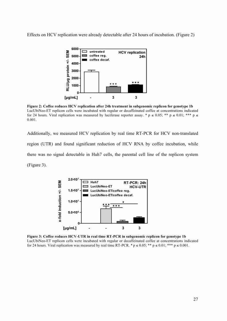
Effects on HCV replication were already detectable after 24 hours of incubation. (Figure 2)
B
Figure 2: Coffee reduces HCV replication after 24h treatment in subgenomic replicon for genotype 1b LucUbiNeo-ET replicon cells were incubated with regular or decaffeinated coffee at concentrations indicated for 24 hours. Viral replication was measured by luciferase reporter assay. * p ≤ 0.05; ** p ≤ 0.01; *** p ≤ 0.001.
Additionally, we measured HCV replication by real time RT-PCR for HCV non-translated
region (UTR) and found significant reduction of HCV RNA by coffee incubation, while
there was no signal detectable in Huh7 cells, the parental cell line of the replicon system
(Figure 3).
Figure 3: Coffee reduces HCV-UTR in real time RT-PCR in subgenomic replicon for genotype 1b LucUbiNeo-ET replicon cells were incubated with regular or decaffeinated coffee at concentrations indicated for 24 hours. Viral replication was measured by real time RT-PCR. * p ≤ 0.05; ** p ≤ 0.01; *** p ≤ 0.001.
28
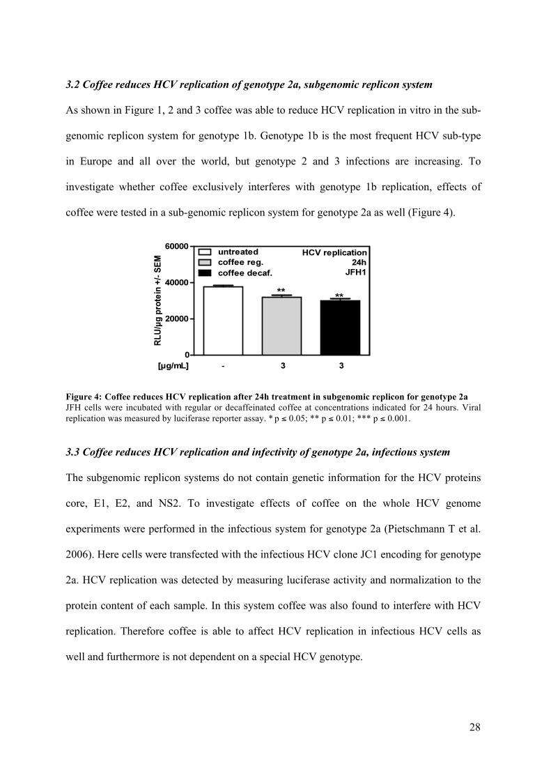
3.2 Coffee reduces HCV replication of genotype 2a, subgenomic replicon system
As shown in Figure 1, 2 and 3 coffee was able to reduce HCV replication in vitro in the sub-
genomic replicon system for genotype 1b. Genotype 1b is the most frequent HCV sub-type
in Europe and all over the world, but genotype 2 and 3 infections are increasing. To
investigate whether coffee exclusively interferes with genotype 1b replication, effects of
coffee were tested in a sub-genomic replicon system for genotype 2a as well (Figure 4).
Figure 4: Coffee reduces HCV replication after 24h treatment in subgenomic replicon for genotype 2a JFH cells were incubated with regular or decaffeinated coffee at concentrations indicated for 24 hours. Viral replication was measured by luciferase reporter assay. * p ≤ 0.05; ** p ≤ 0.01; *** p ≤ 0.001.
3.3 Coffee reduces HCV replication and infectivity of genotype 2a, infectious system
The subgenomic replicon systems do not contain genetic information for the HCV proteins
core, E1, E2, and NS2. To investigate effects of coffee on the whole HCV genome
experiments were performed in the infectious system for genotype 2a (Pietschmann T et al.
2006). Here cells were transfected with the infectious HCV clone JC1 encoding for genotype
2a. HCV replication was detected by measuring luciferase activity and normalization to the
protein content of each sample. In this system coffee was also found to interfere with HCV
replication. Therefore coffee is able to affect HCV replication in infectious HCV cells as
well and furthermore is not dependent on a special HCV genotype.
29
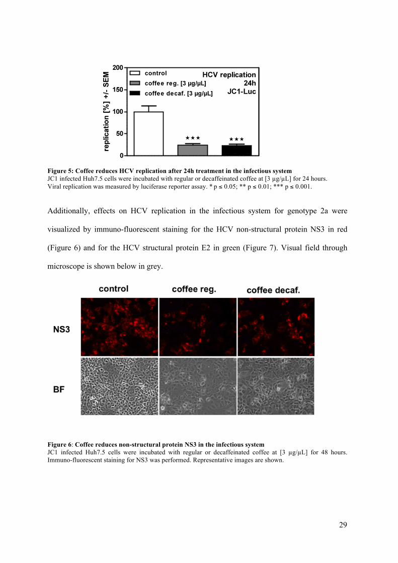
Figure 5: Coffee reduces HCV replication after 24h treatment in the infectious system JC1 infected Huh7.5 cells were incubated with regular or decaffeinated coffee at [3 µg/µL] for 24 hours. Viral replication was measured by luciferase reporter assay. * p ≤ 0.05; ** p ≤ 0.01; *** p ≤ 0.001.
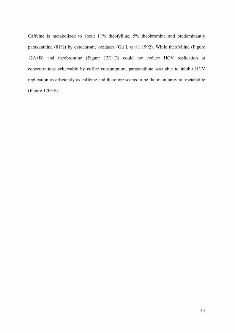
Additionally, effects on HCV replication in the infectious system for genotype 2a were
visualized by immuno-fluorescent staining for the HCV non-structural protein NS3 in red
(Figure 6) and for the HCV structural protein E2 in green (Figure 7). Visual field through
microscope is shown below in grey.
Figure 6: Coffee reduces non-structural protein NS3 in the infectious system JC1 infected Huh7.5 cells were incubated with regular or decaffeinated coffee at [3 µg/µL] for 48 hours. Immuno-fluorescent staining for NS3 was performed. Representative images are shown.

30
Figure 7: Coffee reduces structural protein E2 in the infectious system JC1 infected Huh7.5 cells were incubated with regular or decaffeinated coffee at [3 µg/µL] for 48 hours. Immuno-fluorescent staining for E2 was performed. Representative images are shown.
Coffee is not only able to reduce expression of HCV non-structural protein NS3 and HCV
structural protein E2. Moreover, coffee interfered with infectivity of infectious viral particles
(Figure 8). So incubation with coffee was able to reduce cell infection with HCV. Infectivity
was measured by TCID50 titration of infectious viral particles in supernatants.
Figure 8: Coffee reduces HCV infectivity in the infectious system, genotype 2a JC1 infected Huh7.5 cells were incubated with regular or decaffeinated coffee at [3 µg/µL] for 24 hours. Viral infectivity was measured by titration of the TCID50. * p ≤ 0.05; ** p ≤ 0.01; *** p ≤ 0.001.

31
3.4 Coffee interferes with viral entry by HCV receptor down-regulation
To investigate effects of coffee on HCV entry, Huh 7.5 cells were pre-incubated with coffee
for 2 hours before 2 hours of infection with the HCV clone JC1 encoding genotype 2a.
Experiments were evaluated after an incubation period of 48 hours. Pre-incubation with
coffee interfered with HCV infection, as detected by immuno-fluorescent staining for non-
structural protein NS3 (Figure 9). Visual field through microscope is shown below in grey.
Figure 9: Coffee pre-incubation reduces non-structural protein NS3 in the infectious system Huh7.5 cells were incubated with regular or decaffeinated coffee [3 µg/µL] for 2 hours. After pre-incubation cells were transfected with JC1 for 2 hours. After 48 hours immuno-fluorescent staining for NS3 was performed. Representative images are shown.
This observation prompted us to investigate effects of coffee on essential HCV entry
receptors, of which tetraspanin CD81, scavenger receptor class B member I and tight
junction proteins claudin-1 and occludin have been described (Fraquhar MJ 2012) (Albecka
A et al. 2012). Human hepatocytes were incubated with regular coffe for 2 and 24 hours.
Receptor expression was measured by real-time RT-PCR. Our results show that LDL-
receptor was already down regulated after 2 hours of coffee incubation, while claudin-1 was
down regulated after 24 hours (Figure 10).

32
Figure 10: Coffee leeds to HCV entry receptor downregulation Human hepatocytes were incubated with 3µg/µl regular coffee for 2 and 24 hours. Receptor expression was measured by real-time RT-PCR * p ≤ 0.05; ** p ≤ 0.01; *** p ≤ 0.001
3.5 Caffeine interferes with replication of HCV genotype 1b, replicon system
Although decaffeinated coffee was able to reduce HCV replication effects of caffeine-
containing coffee were more pronounced. This prompted us to investigate effects of caffeine
on HCV replication. Our results show that caffeine decreased HCV replication dose-
dependently (Figure 11A) without decreasing cell viability (Figure 11B).
A B
B
Figure 11: Caffeine reduces HCV replication in subgenomic replicon system, genotype 1b LucUbiNeo-ET replicon cells were incubated with Caffeine at concentrations indicated for 24 hours (A+B). Viral replication was measured by luciferase reporter assay (A). Cell viability was measured by MTT assay (B). * p ≤ 0.05; ** p ≤ 0.01; *** p ≤ 0.001
33
Caffeine is metabolized to about 11% theofylline, 5% theobromine and predominantly
paraxanthine (81%) by cytochrome oxidases (Gu L et al. 1992). While theofylline (Figure
12A+B) and theobromine (Figure 12C+D) could not reduce HCV replication at
concentrations achievable by coffee consumption, paraxanthine was able to inhibit HCV
replication as efficiently as caffeine and therefore seems to be the main antiviral metabolite
(Figure 12E+F).

34
A B
C D
E F
Figure 12: Caffeine metabolites interfere with HCV replication LucUbiNeo-ET replicon cells were incubated with theofylline (A+B), theobromine (C+D) or paraxanthine (E+F) at concentrations indicated for 24 hours. Viral replication was measured by luciferase reporter assay (A+C+E). Cell viability was measured by MTT assay (B+D+F) * p ≤ 0.05; ** p ≤ 0.01; *** p ≤ 0.001
35
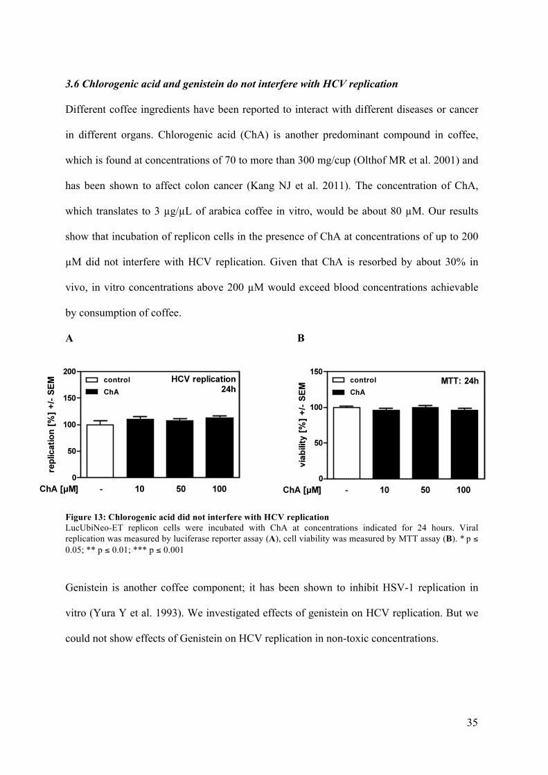
3.6 Chlorogenic acid and genistein do not interfere with HCV replication
Different coffee ingredients have been reported to interact with different diseases or cancer
in different organs. Chlorogenic acid (ChA) is another predominant compound in coffee,
which is found at concentrations of 70 to more than 300 mg/cup (Olthof MR et al. 2001) and
has been shown to affect colon cancer (Kang NJ et al. 2011). The concentration of ChA,
which translates to 3 µg/µL of arabica coffee in vitro, would be about 80 µM. Our results
show that incubation of replicon cells in the presence of ChA at concentrations of up to 200
µM did not interfere with HCV replication. Given that ChA is resorbed by about 30% in
vivo, in vitro concentrations above 200 µM would exceed blood concentrations achievable
by consumption of coffee.
A B
Figure 13: Chlorogenic acid did not interfere with HCV replication LucUbiNeo-ET replicon cells were incubated with ChA at concentrations indicated for 24 hours. Viral replication was measured by luciferase reporter assay (A), cell viability was measured by MTT assay (B). * p ≤ 0.05; ** p ≤ 0.01; *** p ≤ 0.001
Genistein is another coffee component; it has been shown to inhibit HSV-1 replication in
vitro (Yura Y et al. 1993). We investigated effects of genistein on HCV replication. But we
could not show effects of Genistein on HCV replication in non-toxic concentrations.

36
A B
Figure 14: Genistein did not interfere with HCV replication LucUbiNeo-ET replicon cells were incubated with Genistein at concentrations indicated for 24 hours. Viral replication was measured by luciferase reporter assay (A), cell viability was measured by MTT assay (B). * p ≤ 0.05; ** p ≤ 0.01; *** p ≤ 0.001
3.7 Cafestol does not interfere with HCV replication
Coffee beans also contain lipids, mostly triglycerides and diterpenesters. The diterpenes
cafestol and kahweol have been shown to exert anti-carcinogenic activity in vitro (Cavin C et
al. 2002). This prompts us to investigate effects of cafestol on HCV replication. Our results
show that cafestol did not interfere with HCV replication in concentrations used.
Furthermore cafestol concentration of 50 µM reduced cell viability.
A B
Figure 15: Cafestol did not interfere with HCV replication LucUbiNeo-ET replicon cells were incubated with cafestol at concentrations indicated for 24 hours. Viral replication was measured by luciferase reporter assay (A), cell viability was messured by MTT assay (B). * p ≤ 0.05; ** p ≤ 0.01; *** p ≤ 0.001

37
3.8 Coffee and caffeine increase interferon effects on HCV replication
It has been reported that coffee increases effects of interferon alpha and ribavirin therapy in
HCV patients (Freedman ND et al. 2011). Our results confirm this finding in vitro, since in
the HCV replicon system for genotype 1b regular coffee (Figure 16A) as well as caffeine
(Figure 16B) significantly increased antiviral effects of interferon. Regular coffee had a more
pronounced effect compared to decaffeinated coffee, which in multiple experiments did not
increase interferon effects significantly (Figure 16A). Therefore, caffeine seems to play a
pivotal role in increasing interferon effects.
A B
B
Figure 16: Coffee and caffeine increase interferon effects on HCV replication LucUbiNeo-ET replicon cells were co-incubated with interferon and regular or decaffeinated coffee (A) or caffeine (B) at concentrations indicated for 24 hours. Viral replication was measured by luciferase reporter assay. * p ≤ 0.05; ** p ≤ 0.01; *** p ≤ 0.001
3.9 Coffee, but not caffeine, decreases wnt signaling activity
Interferon alpha exhibit its antiviral potential by anti-proliferative effects on the viral cells
and and immunsystem activating effects in the human body. But it has also been reported to
inhibit the wnt signaling pathway (Thompson MD et al. 2011). IFN alpha is able to decrease
wnt pathway activity factor beta-catenin through increasing beta-catenin nuclear export
factor RanBP3 (Thompson MD et al. 2011). It has been described that beta-catenin

38
expression is elevated in patients with chronic HCV infection in dependence on the HCV
non-structural protein NS5A (Park CY et al. 2009), implicating that the wnt signaling
pathway might affect HCV replication. Comparing our replicon cell lines to their parental
cell line Huh7 we observed significantly enhanced wnt activity measured by a reporter assay
for beta-catenin driven transcription (CRT). CRT was significantly decreased in the presence
of regular or decaffeinated coffee, but not in the presence of caffeine alone.
Figure 17: Coffee, but not caffeine, decreases wnt signaling activity Parental cell line Huh7 and HCV replicating Huh5-15 cells were transfected with M50 and M51 plasmids for 24 hours. After transfection cells were incubated with regular or decaffeinated coffee or caffeine at concentrations indicated for another 24 hours. Viral replication was measured by CRT assay. * p ≤ 0.05; ** p ≤ 0.01; *** p ≤ 0.001
Increased wnt signaling activity in HCV replicating cells was also indicated by enhanced
expression of the wnt activation markers conductin and beta-catenin as well as the beta-
catenin stabilizing kinase CK2 catalytic subunits alpha (CK2a) and alpha’ (CK2ap). As
observed for CRT, incubation with coffee, but not caffeine significantly reduced expression
of conductin, beta-catenin and CK2 subunit alpha.

39
A B
Figure 18: Coffee, but not caffeine, decreases wnt activation markers Huh7 and Huh-5-15 cells were incubated with regular or decaffeinated coffee or caffeine at concentrations indicated for 24 hours. Different wnt activation markers were measured by real time RT-PCR. * p ≤ 0.05; ** p ≤ 0.01; *** p ≤ 0.001.
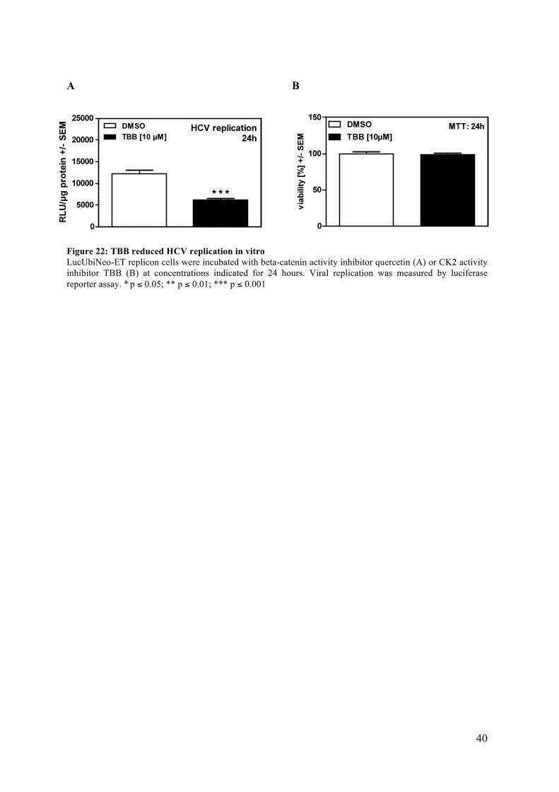
Using inhibitors of beta-catenin activity (Quercetin, Figure 19) or CK2 activity (TBB, Figure
20) we found that HCV replication was significantly reduced. Our results show that wnt
signaling promotes HCV replication and that coffee interferes with this mechanism.
A B
Figure 19: Quercetin reduced HCV replication in vitro LucUbiNeo-ET replicon cells were incubated with beta-catenin activity inhibitor quercetin (A) or CK2 activity inhibitor TBB (B) at concentrations indicated for 24 hours. Viral replication was measured by luciferase reporter assay. * p ≤ 0.05; ** p ≤ 0.01; *** p ≤ 0.001
40
A B
Figure 22: TBB reduced HCV replication in vitro LucUbiNeo-ET replicon cells were incubated with beta-catenin activity inhibitor quercetin (A) or CK2 activity inhibitor TBB (B) at concentrations indicated for 24 hours. Viral replication was measured by luciferase reporter assay. * p ≤ 0.05; ** p ≤ 0.01; *** p ≤ 0.001

41
4. Discussion
Coffee is the most favourite beverage in Germany. With 150 litres per person per year the
Germans consume more coffee than water or beer (Deutscher Kaffeeverband e.V.,
www.kaffeeverband.de). Regular coffee increases levels of alertness and concentration via
competitive antagonism at adenosine-receptors (Chou CC, Vickroy TW 2003), and
therefore, it is the worldwide most frequently used legal psychoactive substance (Dórea JG,
Da Costa THM 2005).
Coffee has been discredited for a long time. Even the children song “C-A-F-F-E-E “ (Carl
Gottlieb Hering, 1766-1853) warns of higher coffee consumption. But in recent years, many
publications have shown that coffee consumption is associated with multiple positive effects
on the human organism. Coffee contains phytoprotectants, of which coffee acids, alkaloids,
melanoids, flavinoids and polyphenols exhibit anti-oxidative properties, at least in vitro
(Svilaas A et al. 2004). Since free radicals seem to play a role in cardiovascular and
metabolic diseases as well in tumor development (Davì G et al. 2005), this might serve as an
explanation for the ability of coffee to reduce general mortality (Lopez-Garcia E et al. 2008).
This is in line with findings that coffee drinking decreases the risk to develop e.g. heart
diseases (Wu JN et al. 2009), stroke (Lopez-Garcia E et al. 2009), diabetes typ-2 (van Dam
RM, Hu FB 2005), Alzheimer’s (Eskelinen MH, Kivipelto M 2010), and Parkinson’s disease
(Costa J et al. 2010), or gastrointestinal cancers (Galeone C et al. 2010), (Larsson SC, Wolk
A 2007) and breast cancer (Lowcock EC et al. 2013).
Coffee contains over 2000 components (Tuomilehto J. 2013), which makes it difficult to
break down the mechanisms for all those described effects to single substances. The most
commonly known substance in regular coffee is caffeine. But in all these clinical studies, the
effects of coffee could not be clearly ascribed to the caffeine in regular coffee. Decaffeinated

42
coffee showed the same reductive effects as regular coffee, but not as strong as caffeine-
containing coffee. Nevertheless, part of this effect could be attributed to caffeine
(Utsunomiya H et al. 2008).
The idea that coffee might interfere with viral replication is not new. In 1998, Dorothy Bonn
suggested that green coffee beans might contain substances able to interfere with viral
infection, showing inhibition of HIV integrase (Bonn D 1998). It has also been reported that
coffee extracts are able to decrease HSV type 1 and poliovirus multiplication, possibly due
to interference with infectivity and virus particle formation (Utsunomiya H et al. 2008). With
regard to hepatitis virus infections, regular and decaffeinated coffees have been shown to
interfere with HBV replication due to the coffee ingredients chlorogenic acid and caffeic
acid (Wang GF et al. 2009). This observation could be broadened, showing that HCC due to
HBV infection and was less pronounced in coffee drinkers, while there was no reduction
found in consummates of green tea (Inoue M et al. 2009).
Epidemiological studies now provide evidence for effects of coffee on HCV. A reduced risk
for coffee drinking patients with chronic HCV infection to develop or die from HCC has
been reported (Ohfuji S et al. 2006; Wakai K et al. 2007). Although a lot of evidence points
to direct effects of coffee on HCV replication, this has not been investigated in vitro to
define active substances and molecular mechanisms of coffee on viral replication. Our
results show that concentrations of coffee, which are achievable by consumption of 1-3 cups
(200 mL per cup) per day are able to significantly and dose-dependently reduce HCV
replication in vitro without exerting toxic effects on the cell culture system.
We tested coffee effects on replication in the replicon system for genotyp 1b and in the
infectious system for genotype 2a and could show comparable results. Coffee did not only
exhibit antiviral effects on replication, but also coffee incubation decreased infectivity of
viral particles. This is new for HCV, but decrease in multiplication by coffee extracts has
43
already been shown for HSV type 1 and poliovirus (Utsunomiya H et al. 2008). Incubation
of uninfected cells with coffee prior to infection resulted in reduced infection compared to
control pre-incubated cells, which could be due to a reduction in the expression of HCV
entry receptors. LDL-receptor (Albecka A et al. 2012) and Claudin-1 (Fraquhar MJ 2012)
are described to act as HCV entry receptors and our results show significant reduction for
LDL-receptor and Claudin-1 expression on human primary hepatocytes after treatment with
regular coffee. Therefore, coffee incubation seems to have several target points of antiviral
activity, interfering with viral replication, decreasing infectivity of viral particles and directly
protecting host cells from infection.
We performed experiments with regular and decaffeinated coffee, which both reduced HCV
replication, although effects of regular coffee turned out to be stronger. This prompted us to
investigate effects of caffeine in the HCV replicon system. Higher caffeine consumption is
associated with less liver cirrhosis, among others caused by chronic HCV infection (Modi
AA et al. 2010). Our results show that concentrations of caffeine corresponding to 3-4 cups
of coffee (200 mL per cup) were sufficient to significantly impair HCV replication in vitro
without having toxic effects on the cell culture system. The alkaloid caffeine and its
metabolites paraxanthine, theobromine and theofylline are known to act as competitive
adeonsine receptor antagonists in the brain (Fisone G et al. 2004), (Chou CC, Vickroy TW
2003). Adenosine is a purine nucleoside, which promotes sleep by inhibition of release of
activating neurotransmitters. So inhibition of adenosine receptors by caffeine is primarily
used to avoid fatigue. Beside its sleep promoting effect, adenosine is also known for
promoting wound healing and matrix production by activation of its receptors (Macedo L
2007). Furthermore adenosine has been reported to promote fibrosis in skin and lungs as
well as in the liver via excess matrix production (Cronstein BN 2011). This might imply that
inhibition of adenosine receptors by caffeine could inhibit fibrosis development, which
44
might be another effect besides inhibition of HCV replication, why coffee is able to slow
down hepatic fibrosis development in HCV infected patients (Modi AA et al. 2010). Of the
three caffeine degradation products paraxanthine, theobromine and theofylline, we found
inhibition of HCV replication only by the main degradation product paraxanthine. Besides
the effect on adenosine receptors, all three degradation products are phosphodiesterase
inhibitors as well, but just paraxanthine is responsible for the lipolytic effects of caffeine
(Jiang M et al. 1998). So it seems that caffeine is able to inhibit HCV replication via its
degradation product paraxanthine.
Since decaffeinated coffee is also able to interfere with HCV replication we tried to identify
other antivirally active substances in coffee. Besides caffeine, coffee beans contain about
2000 other substances: Coffee beans contain about 17% of lipids, mostly triglycerides and
diterpenesters. The diterpens cafestol and kahweol have been shown to exert anti-
carcinogenic activity in vitro (Cavin C et al. 2002). In our experiments, we did not observe
decreased HCV replication at non-toxic concentrations of cafestol in replicon cell lines. The
polyphenols caffeic acid and chlorogenic acid exhibit antioxidative effects and have been
shown to affect colon cancer (Kang NJ et al. 2011). Incubation of HCV replicating cells with
chlorogenic acid did not reduce HCV replication. The isoflavone genistein has been shown
to inhibit HSV-1 replication in vitro (Yura Y et al. 1993), but did not inhibit HCV
replication in vitro. Therefore, we were not able to identify another substance contained in
coffee, besides caffeine, which was able to reduce HCV replication at non-toxic
concentrations. On the other hand, it might also be a combination of substances, which in a
concerted action are able to inhibit HCV replication.
Concerning the mechanism by which coffee is able to interfere with viral replication, we
found hints to an involvement of the wnt signaling pathway. This pathway transduces signals
from the outside of the cell via surface receptors to the inside of the cell. The cascade is

45
started by the signal protein ligand “wnt”, which binds to the receptor “frizzled“ and leads to
inhibition of GSK3beta and thereby inhibits beta-catenin degradation. Concomitantly,
activation of the wnt signaling pathway leads to beta-catenin accumulation in the cytoplasm,
causing stabilized beta-catenin to act as a co-activator of transcription factors and leads to
changes in target gene transcription (Nusse R, Varmus HE. 1992). Recently beta-catenin
activity was found to be enhanced in HCV patients by direct interaction between the non-
structural HCV protein NS5A and components of the wnt signaling pathway (Park CY et al.
2009); furthermore, wnt signaling pathway activity has also been found to be enhanced by
HCV core protein (Liu J et al. 2011). This met our observations that wnt signaling activity
was significantly enhanced in the HCV replicon system in comparison to its parental cell
line not containing the replicon. A connection from wnt pathway activity to enhanced HCV
replication could be made by showing that interference with wnt signaling, either by use of
the beta-catenin inhibitor quercetin or a knockdown of beta-catenin expression, resulted in a
direct inhibitory effect on HCV replication.
In fact we found that both regular and decaffeinated coffee, but not caffeine, were able to
interfere with wnt signaling pathway activity in replicon cells. HCV seems to up-regulate
wnt pathway inducers while down-regulating wnt pathway inhibitors. This results in higher
expression of wnt target genes like conductin, myc or claudin-1, implicating that HCV might
force infected cells towards higher HCV expression but also towards cancer development, as
recently shown for the development of colon (Jeong WJ et al. 2012) and breast (Zhang H et
al. 2012) cancer. Higher wnt pathway activity has recently been described to be associated
with hepatocellular carcinoma (Li ZQ et al. 2012).
Until very recently, treatment of chronic hepatitis C infection has predominantly been
performed by a combination of pegylated interferon alpha2 with the nucleoside analogue
ribavirin. Interferon alpha exhibits its antiviral potential by anti-proliferative effects on the
46
viral cells and immunsystem activating effects in the human body. But it has also been
reported to inhibit the wnt signaling pathway (Thompson MD et al. 2011). Interestingly, also
the response to interferon/ribavirin treatment of HCV patients was found enhanced by coffee
drinking (Freedman ND et al. 2011). We were able to show that inhibition of HCV
replication in vitro increased when interferon alpha was co-incubated with regular coffee or
caffeine. Decaffeinated coffee did not significantly increase interferon effects on HCV
replication. Therefore, this effect seems to be due to caffeine. Further research is needed to
determine possible positive effects of caffeine supplementation of HCV therapy.
So there might be two different mechanisms why coffee is able to reduce HCV replication
and increase IFN alpha therapy effects. While inhibition of HCV replication could be
achieved by regular and decaffeinated coffee and caffeine itself, decaffeinated coffee could
not increase IFN alpha effects on HCV replication. Accordingly, this is in line with our
suspicion that caffeine is not the only antiviral effective substance in coffee and the coffee
effects on HCV replication and HCV therapy might be due to combination of substances.
This thesis is furthermore provided by the finding that other caffeine-containing beverages
besides coffee, were not able to reduce liver fibrosis formation in HCV infected patients
(Modi AA et al. 2010).
In this work, we could show antiviral properties of regular and decaffeinated coffee, caffeine
and the caffeine degradation product paraxanthine on HCV replication in vitro. Additionally,
coffee had effects on HCV entry and HCV infection in vitro, and did not show restriction to
a special genotype. HCV therapy with IFN/ribavirin was shown to be improved by regular
and decaffeinated coffee, but not by caffeine. Our data indicate that wnt signaling pathway
inhibition significantly contributes to the antiviral effects of coffee, but not caffeine. So non-
caffeine induced effects of coffee on HCV replication seems to be due to interference with
wnt signaling and might improve IFN/ribavirin therapy. Our results also show that caffeine

47
is not the only antivirally active substance in coffee, but all other substances contained in
coffee, which have been part of our experiments could not be identified to have effects on
HCV replication in vitro. Further experiments need to be done to identify a combination of
substances or a single substance, which is responsible for the effects of coffee on the
Hepatitis C Virus.
In conclusion, coffee consumption of about 3 cups per day might interfere with HCV
replication and support HCV therapy and also slow down development of liver cirrhosis and
hepatocellular carcinoma (HCC).

48
5. Abstract
5.1 Abstract
Background & Aims: Coffee is the most frequently consumed legal drug all over the world.
Coffee consumption has been shown to decrease the risk of fibrosis formation and to
improve the effects of interferon treatment in HCV patients. We investigated effects of
regular and decaffeinated coffee and coffee ingredients on HCV replication and infection in
vitro.
Methods: To investigate HCV replication, infectious as well as replicon systems for
genotypes 1b and 2a were used. Cells were incubated in the presence of regular or
decaffeinated coffee, coffee ingredients, caffeine and caffeine metabolites. Effects on wnt
signaling pathway activity and antiviral effects in co-incubation with interferon alpha were
measured. Furthermore effects of coffee on viral entry have been tested in the infectious
system.
Results: Both, regular and decaffeinated coffee, were able to reduce HCV infection and
replication significantly with regular coffee having a more pronounced effect. Caffeine as
well as its main metabolite paraxanthine significantly reduced HCV replication. While
caffeine and its metabolite paraxanthine did not; both, regular and decaffeinated coffee, were
able to reduce wnt pathway activity, which has been found markedly increased by HCV
replication. Likewise, inhibition of wnt signaling by the beta-catenin inhibitor quercetin,
siRNA directly against beta-catenin or inhibition of the wnt pathway inducer CK2 by its
inhibitor 4,5,6,7-tetrabromo-2-azabenzimidazole (TBB) significantly interfered with HCV
replication. Furthermore coffee as well as caffeine was able to increase inhibitory effects of
interferon alpha on HCV replication. Nevertheless coffee pre-incubated cells show
significantly less HCV infection compared with water pre-incubated cells. Furthermore
49
coffee incubated uninfected hepatocytes showed significant down-regulation of essential
HCV entry receptors.
Conclusions: Coffee effects in HCV infected patients, which have been described by
clinical studies are due effects on HCV replication level. Coffee, but not caffeine, seems to
exhibit antiviral activity by inhibition of the wnt signaling pathway, which might represent a
novel target for HCV therapy. Furthermore coffee increases effects of interferon alpha
therapy in vitro and therefore could take place in therapy guidelines. Nevertheless coffee
pre-incubation decreases essential HCV receptors and thereby decreases HCV infection and
could act as a preventive substance.

50
5.2 Zusammenfassung
Hintergrund: Kaffee ist die am häufigsten konsumierte legale Droge der Welt. Studien
belegen, dass regelmäßiger Kaffeekonsum das Risiko einer Leberfibrose signifikant senken
und die Effekte der Interferon Therapie bei HCV Patienten verbessern kann. Wir haben in
dieser Arbeit die Effekte von koffeinhaltigem und entkoffeiniertem Kaffee auf die HCV
Replikation und die HCV Infektion in vitro getestet
Methoden: Für die Experimente wurden sowohl ein infektiöses System für Genotyp 2a, als
auch Replikonzelllinien für die Genotypen 1b und 2a verwendet. Die Zellen wurden mit
koffeinhaltigem und entkoffeiniertem Kaffee, Kaffeeinhaltsstoffen, Koffein und
Koffeinabbauprodukten behandelt. Es wurden die Effekte auf die Aktivität des wnt signaling
pathway und antivirale Effekte von der Kombinationsbehandlung mit Interferon gemessen.
In weiteren Versuchen im infektiösen System wurden die Effekte von Kaffee auf den
Vireneintritt in die Zelle gemessen.
Ergebnisse: Sowohl koffeinhaltiger als auch entkoffeinierter Kaffee konnten die HCV
Infektion und die HCV Replikation signifikant reduzieren, wobei der koffeinhaltige Kaffee
einen etwas ausgeprägteren Effekt hatte. Koffein und das Hauptabbauprodukt von Koffein,
Paraxanthin konnten die HCV Replikation signifikant reduzieren. Es konnte eine deutlich
erhöhte Aktivität des wnt signaling pathways in HCV replizierenden Zellen gezeigt werden.
Koffeinhaltiger als auch entkoffeinierter Kaffee reduzierten die Aktivität des wnt signaling
pathway, während Koffein und Paraxanthin keinen Effekt auf den wnt signaling pathway
hatten. Dies deckt sich mit den Beobachtungen, dass eine Hemmung des wnt signaling
pathways durch den beta-catenin Inhibitor Quercetin, eine siRNA gegen beta-catenin oder
einen Inhibitor des wnt pathway Induktors CK2 4,5,6,7-tetrabromo-2-azabenzimidazole
(TBB) die HCV Replikation signifikant senken konnten. Es konnte zusätzlich gezeigt
werden, dass koffeinhaltiger Kaffee und Koffein in der Lage waren die Effekte von

51
Interferon auf die HCV Replikation zu verstärken. Eine Vorbehandlung der Zellen mit
Kaffee vor der Infektion mit dem HCV Virus im infektiösen System, zeigte eine signifikant
reduzierte Infektionsrate im Vergleich mit Zellen, welche mit Wasser vorbehandelt wurden.
Gesunde Hepatozyten zeigten nach Behandlung mit Kaffee eine deutliche reduzierte
Expression von essentiellen Rezeptoren für den HCV Eintritt in die Zelle.
Schlussfolgerungen: Die in Studien beschriebenen Kaffeewirkungen bei HCV infizierten
Patienten sind auf Effekte des Kaffees auf die HCV Replikation zurückzuführen. Kaffee
selbst wirkt antiviral über die Hemmung des wnt signaling pathways, für Koffein konnte
dies nicht gezeigt werden. Die Hemmung des wnt signaling pathways könnte ein neues
Angriffsziel von Therapien für HCV sein. Es konnte zusätzlich eine Verstärkung der
Wirkung von Interferon alpha durch Kaffee gezeigt werden, eine entsprechende Empfehlung
könnte in die Therapieleitlinien für HCV aufgenommen werden. Außerdem konnte eine
Vorbehandlung von gesunden Zellen die Eintrittsrezeptoren für HCV vermindern und
scheint somit auch einer HCV Infektionsprävention zu dienen.

52
6. Abbreviations
CD81: Cluster of Differentiation 81
CLDN1: Claudin-1
CRT: Beta-catenin-regulated transcription
CTGF: connective tissue growth factor
GSK3beta: Glycogen synthase kinase 3 beta
HBV: Hepatitis B Virus
HCC: Hepatocellular carcinoma
HCV: Hepatitis C Virus
HIV: Human immunodeficiency virus
HSV: Herpes simplex Virus
OCLN: Occludin
PDE: Phosphodiesterase
PEG-IFN: pegylated interferon alpha
RT-PCR: real-time Polymerase chain reaction
TCID50: Tissue Culture Infection Dose 50
UTR: un-translated region

53
7. References
7.1 Journals
1. Albecka A, Belouzard S, Op de Beeck A, Descamps V, Goueslain L, Bertrand-Michel J,
et al. Role of low-density lipoprotein receptor in the hepatitis C virus life cycle.
Hepatology 2012, Apr;55(4):998-1007.
2. Bandurska K, Król I, Myga-Nowak M. Interferons: Between structure and function.
Postepy Higieny I Medycyny Doswiadczalnej (Online) 2013;68:428-40.
3. Blonski W, Reddy KR. Hepatitis C virus infection and hepatocellular carcinoma. Clinics
in Liver Disease 2008;12(3):661-74.
4. Boekema PJ, Samsom M, van Berge Henegouwen GP, Smout AJ. Coffee and
gastrointestinal function: Facts and fiction. A review. Scand J Gastroenterol Suppl
1999;230:35-9.
5. Bonn D. Green coffee beans may yield new class of anti-hiv-1 agents. The Lancet
1998;352(9133):1039.
6. Butt MS, Sultan MT. Coffee and its consumption: Benefits and risks. Critical Reviews in
Food Science and Nutrition 2011;51(4):363-73.
7. Cavin C, Holzhaeuser D, Scharf G, Constable A, Huber WW, Schilter B. Cafestol and
kahweol, two coffee specific diterpenes with anticarcinogenic activity. Food Chem
Toxicol 2002, Aug;40(8):1155-63.
8. Chou CC, Vickroy TW. Antagonism of adenosine receptors by caffeine and caffeine
metabolites in equine forebrain tissues. Am J Vet Res 2003, Feb;64(2):216-24.
9. Cornberg M, Razavi HA, Alberti A, Bernasconi E, Buti M, Cooper C, et al. A systematic
review of hepatitis C virus epidemiology in europe, canada and israel. Liver Int 2011,

54
Jul;31 Suppl 2:30-60.
10. Costa J, Lunet N, Santos C, Santos J, Vaz-Carneiro A. Caffeine exposure and the risk of
parkinson's disease: A systematic review and meta-analysis of observational studies. J
Alzheimers Dis 2010;20 Suppl 1:S221-38.
11. Costentin CE, Roudot-Thoraval F, Zafrani ES, Medkour F, Pawlotsky JM, Mallat A,
Hézode C. Association of caffeine intake and histological features of chronic hepatitis
C. J Hepatol 2011, Jun;54(6):1123-9.
12. Cronstein BN. Adenosine receptors and fibrosis: A translational review. F1000 Biol Rep
2011;3:21.
13. Davì G, Falco A, Patrono C. Lipid peroxidation in diabetes mellitus. Antioxidants &
Redox Signaling 2005;7(1-2):256-68.
14. Di Bisceglie AM, Hoofnagle JH. Optimal therapy of hepatitis C. Hepatology
2002;36(5B):s121-7.
15. Dórea JG, Da Costa THM. Is coffee a functional food? British Journal of Nutrition
2005;93(6):773.
16. Eskelinen MH, Kivipelto M. Caffeine as a protective factor in dementia and alzheimer's
disease. J Alzheimers Dis 2010;20 Suppl 1:S167-74.
17. Farquhar MJ, Hu K, Harris HJ, Davis C, Brimacombe CL, Fletcher SJ, et al. Hepatitis C
virus induces CD81 and claudin-1 endocytosis. J Virol 2012, Apr;86(8):4305-16.
18. Fisone G, Borgkvist A, Usiello A. Caffeine as a psychomotor stimulant: Mechanism of
action. Cellular and Molecular Life Sciences 2004;61(7):857-72.
19. Freedman ND, Curto TM, Lindsay KL, Wright EC, Sinha R, Everhart JE, HALT-C
TRIAL GROUP. Coffee consumption is associated with response to peginterferon and

55
ribavirin therapy in patients with chronic hepatitis C. Gastroenterology 2011,
Jun;140(7):1961-9.
20. Frese M, Barth K, Kaul A, Lohmann V, Schwärzle V, Bartenschlager R. Hepatitis C
virus RNA replication is resistant to tumour necrosis factor-alpha. J Gen Virol 2003,
May;84(Pt 5):1253-9.
21. Galeone C, Turati F, La Vecchia C, Tavani A. Coffee consumption and risk of colorectal
cancer: A meta-analysis of case-control studies. Cancer Causes Control 2010,
Nov;21(11):1949-59.
22. Gressner OA, Lahme B, Siluschek M, Gressner AM. Identification of paraxanthine as
the most potent caffeine-derived inhibitor of connective tissue growth factor
expression in liver parenchymal cells. Liver Int 2009, Jul;29(6):886-97.
23. Gu L, Gonzalez FJ, Kalow W, Tang BK. Biotransformation of caffeine, paraxanthine,
theobromine and theophylline by cdna-expressed human CYP1A2 and CYP2E1.
Pharmacogenetics 1992, Apr;2(2):73-7.
24. Gutierrez-Reyes G, Lopez-Ortal P, Sixtos S, Cruz S, Ramirez-Iglesias MT, Gutierrez-
Ruiz MC, et al. Effect of pentoxifylline on levels of pro-inflammatory cytokines
during chronic hepatitis C. Scand J Immunol 2006, Jun;63(6):461-7.
25. Herbst DA, Reddy KR. NS5A inhibitor, daclatasvir, for the treatment of chronic hepatitis
C virus infection. Expert Opinion on Investigational Drugs 2013;22(10):1337-46.
26. Inoue M, Kurahashi N, Iwasaki M, Shimazu T, Tanaka Y, Mizokami M, et al. Effect of
coffee and green tea consumption on the risk of liver cancer: Cohort analysis by
hepatitis virus infection status. Cancer Epidemiol Biomarkers Prev 2009,
Jun;18(6):1746-53.

56
27. Jeong WJ, Yoon J, Park JC, Lee SH, Lee SH, Kaduwal S, et al. Ras stabilization through
aberrant activation of wnt/β-catenin signaling promotes intestinal tumorigenesis. Sci
Signal 2012;5(219):ra30.
28. Jiang M, Kameda K, Han L-K, Kimura Y, Okuda H. Isolation of lipolytic substances
caffeine and 1, 7-dimethylxanthine from the stem and rhizome of< EM EMTYPE=.
Planta Med1998;64(04):375-7.
29. Jiménez-Luévano MÁ, Lerma-Díaz JM, Hernández-Flores G, Jiménez-Partida MÁ,
Bravo-Cuellar A. Addition of pentoxifylline to pegylated interferon-alpha-2a and
ribavirin improves sustained virological response to chronic hepatitis C virus: A
randomized clinical trial. Ann Hepatol 2013;12(2):248-55.
30. Kang NJ, Lee KW, Kim BH, Bode AM, Lee HJ, Heo YS, et al. Coffee phenolic
phytochemicals suppress colon cancer metastasis by targeting MEK and TOPK.
Carcinogenesis 2011, Jun;32(6):921-8.
31. Klemmer I, Yagi S, Gressner OA. Oral application of 1,7-dimethylxanthine
(paraxanthine) attenuates the formation of experimental cholestatic liver fibrosis.
Hepatol Res 2011, Nov;41(11):1094-109.
32. Larsson SC, Wolk A. Coffee consumption and risk of liver cancer: A meta-analysis.
Gastroenterology 2007, May;132(5):1740-5.
33. Li ZQ, Ding W, Sun SJ, Li J, Pan J, Zhao C, et al. Cyr61/CCN1 is regulated by wnt/β-
catenin signaling and plays an important role in the progression of hepatocellular
carcinoma. PLoS One 2012;7(4):e35754.
34. Liu J, Wang Z, Tang J, Tang R, Shan X, Zhang W, et al. Hepatitis C virus core protein
activates wnt/β-catenin signaling through multiple regulation of upstream molecules in
the SMMC-7721 cell line. Arch Virol 2011, Jun;156(6):1013-23.

57
35. Liu J, Sui X, Lavie CJ, Hebert JR, Earnest CP, Zhang J, Blair SN. Association of coffee
consumption with all-cause and cardiovascular disease mortality. Mayo Clin Proc
2013, Oct;88(10):1066-74.
36. Lohmann V, Körner F, Koch JO, Herian U, Theilmann L, Bartenschlager R. Replication
of subgenomic hepatitis C virus rnas in a hepatoma cell line. Science
1999;285(5424):110-3.
37. Lopez-Garcia E, van Dam RM, Li TY, Rodriguez-Artalejo F, Hu FB. The relationship of
coffee consumption with mortality. Annals of Internal Medicine 2008;148(12):904-14.
38. Lopez-Garcia E, Rodriguez-Artalejo F, Rexrode KM, Logroscino G, Hu FB, van Dam
RM. Coffee consumption and risk of stroke in women. Circulation 2009;119(8):1116-
23.
39. Lowcock EC, Cotterchio M, Anderson LN, Boucher BA, El-Sohemy A. High coffee
intake, but not caffeine, is associated with reduced estrogen receptor negative and
postmenopausal breast cancer risk with no effect modification by CYP1A2 genotype.
Nutrition and Cancer 2013;65(3):398-409.
40. Macedo L, Pinhal-Enfield G, Alshits V, Elson G, Cronstein BN, Leibovich SJ. Wound
healing is impaired in myd88-deficient mice: A role for myd88 in the regulation of
wound healing by adenosine A2A receptors. Am J Pathol 2007, Dec;171(6):1774-88.
41. Modi AA, Feld JJ, Park Y, Kleiner DE, Everhart JE, Liang TJ, Hoofnagle JH. Increased
caffeine consumption is associated with reduced hepatic fibrosis. Hepatology 2010,
Jan;51(1):201-9.
42. Muench H. A simple method of estimating fifty percent endpoints. American. J. Hyg
1938;27:493-7.

58
43. Namdar M, Koepfli P, Grathwohl R, Siegrist PT, Klainguti M, Schepis T, et al. Caffeine
decreases exercise-induced myocardial flow reserve. J Am Coll Cardiol 2006, Jan
17;47(2):405-10.
44. Nusse R, Varmus HE. Wnt genes. Cell 1992, Jun 26;69(7):1073-87.
45. Ohfuji S, Fukushima W, Tanaka T, Habu D, Tamori A, Sakaguchi H, et al. Coffee
consumption and reduced risk of hepatocellular carcinoma among patients with
chronic type C liver disease: A case-control study. Hepatol Res 2006, Nov;36(3):201-
8.
46. Olthof MR, Hollman PCH, Katan MB. Chlorogenic acid and caffeic acid are absorbed in
humans. J Nutr 2001;131(1):66-71.
47. Orrú M, Guitart X, Karcz-Kubicha M, Solinas M, Justinova Z, Barodia SK, et al.
Psychostimulant pharmacological profile of paraxanthine, the main metabolite of
caffeine in humans. Neuropharmacology 2013, Apr;67:476-84.
48. Park CY, Choi SH, Kang SM, Kang JI, Ahn BY, Kim H, et al. Nonstructural 5A protein
activates beta-catenin signaling cascades: Implication of hepatitis C virus-induced liver
pathogenesis. J Hepatol 2009, Nov;51(5):853-64.
49. Pietschmann T, Kaul A, Koutsoudakis G, Shavinskaya A, Kallis S, Steinmann E, et al.
Construction and characterization of infectious intragenotypic and intergenotypic
hepatitis C virus chimeras. Proc Natl Acad Sci U S A 2006, May 9;103(19):7408-13.
50. Svilaas A, Sakhi AK, Andersen LF, Svilaas T, Ström EC, Jacobs DR, et al. Intakes of
antioxidants in coffee, wine, and vegetables are correlated with plasma carotenoids in
humans. J Nutr 2004, Mar;134(3):562-7.
51. Tang H, Grisé H. Cellular and molecular biology of HCV infection and hepatitis. Clin
59
Sci (Lond) 2009, Jul;117(2):49-65.
52. Thompson MD, Dar MJ, Monga SP. Pegylated interferon alpha targets wnt signaling by
inducing nuclear export of β-catenin. J Hepatol 2011, Mar;54(3):506-12.
53. Tuomilehto J. [Coffee and health]. Duodecim 2013;129(13):1398-405.
54. Utsunomiya H, Ichinose M, Uozaki M, Tsujimoto K, Yamasaki H, Koyama AH.
Antiviral activities of coffee extracts in vitro. Food Chem Toxicol 2008,
Jun;46(6):1919-24.
55. Vagu C, Sultana C, Ruţă S. Understanding the molecular targets for new therapeutical
agents in hepatitis c infection. Roum Arch Microbiol Immunol 2013;72(1):5-24.
56. Van Dam RM, Hu FB. Coffee consumption and risk of type 2 diabetes. JAMA: The
Journal of the American Medical Association 2005;294(1):97-104.
57. Vermehren J, Sarrazin C. New hepatitis C therapies in clinical development. European
Journal of Medical Research 2011;16(7):303.
58. Wakai K, Kurozawa Y, Shibata A, Fujita Y, Kotani K, Ogimoto I, et al. Liver cancer
risk, coffee, and hepatitis C virus infection: A nested case-control study in japan. Br J
Cancer 2007, Aug 6;97(3):426-8.
59. Wang GF, Shi LP, Ren YD, Liu QF, Liu HF, Zhang RJ, et al. Anti-hepatitis B virus
activity of chlorogenic acid, quinic acid and caffeic acid in vivo and in vitro. Antiviral
Res 2009, Aug;83(2):186-90.
60. Wu JN, Ho SC, Zhou C, Ling WH, Chen WQ, Wang CL, Chen YM. Coffee
consumption and risk of coronary heart diseases: A meta-analysis of 21 prospective
cohort studies. Int J Cardiol 2009, Nov 12;137(3):216-25.
61. Yura Y, Yoshida H, Sato M. Inhibition of herpes simplex virus replication by genistein,

60
an inhibitor of protein-tyrosine kinase. Arch Virol 1993;132(3-4):451-61.
62. Zhang H, Zhang X, Wu X, Li W, Su P, Cheng H, et al. Interference of frizzled 1 (FZD1)
reverses multidrug resistance in breast cancer cells through the wnt/β-catenin pathway.
Cancer Lett 2012, Apr 4.
63. Zhang Y, Chen H. Genistein attenuates WNT signaling by up-regulating sfrp2 in a
human colon cancer cell line. Experimental Biology and Medicine 2011;236(6):714-
22.

61
7.2 Internet
1. Deutscher Kaffeeverband e.V.
http://www.kaffeeverband.de/kaffeewissen/kaffeekultur/geschichte/kaffee-in-
deutschland, (Stand: 26.09.2014, 14:30).
62
8. Acknowledgements
The author thanks Dr. Henning Wege (Hamburg, Germany) for providing Super8xTOPFlash
and Super8xFOPFlash for CRT reporter assays. Thanks to Dr. Katrin Singethan (Munich,
Germany) for assistance in handling of infectious system for genotype 2a. Expert technical
assistance by Elena Tasika, Lisa-Marie Böttcher and Carsten Rothkegel is gratefully
acknowledged. Thanks to Prof. Dr. Ansgar Lohse and Prof. Dr. Gisa Tiegs for mentoring the
Graduiertenkolleg “Entzündung und Regeneration”.
Thanks to Prof. Dr. Gabriele Sass for telling me there is always a reason why something
happens, that simply breathing might help a little and at least always having a coffee ready
which was not stored at -20°C.
Special thanks to Prof. Dr. Gisa Tiegs for her support to finish this work.
“Everything will be okay in the end. If it is not okay, it is not the end”
John Lennon

63
9. Curriculum vitae entfällt aus datenschutzrechtlichen Gründen

64
10. Eidesstattliche Versicherung
Ich versichere ausdrücklich, dass ich die Arbeit selbständig und ohne fremde Hilfe verfasst,
andere als die von mir angegebenen Quellen und Hilfsmittel nicht benutzt und die aus den
benutzten Werken wörtlich oder inhaltlich entnommenen Stellen einzeln nach Ausgabe
(Auflage und Jahr des Erscheinens), Band und Seite des benutzten Werkes kenntlich
gemacht habe.
Ferner versichere ich, dass ich die Dissertation bisher nicht einem Fachvertreter an einer
anderen Hochschule zur Überprüfung vorgelegt oder mich anderweitig um Zulassung zur
Promotion beworben habe.
Ich erkläre mich einverstanden, dass meine Dissertation vom Dekanat der Medizinischen
Fakultät mit einer gängigen Software zur Erkennung von Plagiaten überprüft werden kann.
Unterschrift: ......................................................................

65


![Elizabeth Sherman, PharmD, AAHIVPhivaidsinstitute.med.miami.edu/documents/...HIV-HCV...• SVR rates similar to HCV monoinfected [1,2] • In HCV/HIV coinfection, treat HCV as though](https://img.pdfslide.us/doc/110x75/5fbc30e57653e03e261e9924/elizabeth-sherman-pharmd-aa-a-svr-rates-similar-to-hcv-monoinfected-12.jpg)