Embed Size (px)
Citation preview
3
Chapter 1
Coagulation Pathway and PhysiologyRussell A. Higgins, MD
IntroductionOur understanding of blood clotting is intimately tied to the history of civilization. With the advent of writ-ing 5000 years ago, it could be argued that the first symbols used for blood, bleeding, or clotting repre-sented the first published coagulation pathway. The ancient peoples of the world always held blood in the utmost mystical esteem. Through the ages, this esteem has been transmitted to modern times in the many ex-pressions that use the word blood, such as “blood is thicker than water,” “blood of our fathers,” and others.
Mysticism aside, the study of blood clotting and the development of laboratory tests for blood clot-ting abnormalities are historically inseparable. The workhorse tests of the modern coagulation laboratory, the prothrombin time (PT) and the activated partial thromboplastin time (aPTT), are the basis for the pub-lished extrinsic and intrinsic coagulation pathways, even though it is now known that these pathways do not accurately reflect the function of blood clotting in a living organism. In this chapter, and, ultimately, in this textbook, the many authors hope to present a clear explanation of coagulation testing and its impor-tant place in the medical armamentarium for diagnos-ing and treating disease.
Constituents of the Hemostatic SystemWith the evolution of vertebrates and their pressur-ized circulatory system, there had to arise some meth-od to seal the system if injured—hence, the hemostatic system. It is interesting to note that there is nothing quite comparable to the vertebrate hemostatic system in invertebrate species. In all vertebrates studied, the basic constituents of the hemostatic system appear to be conserved.
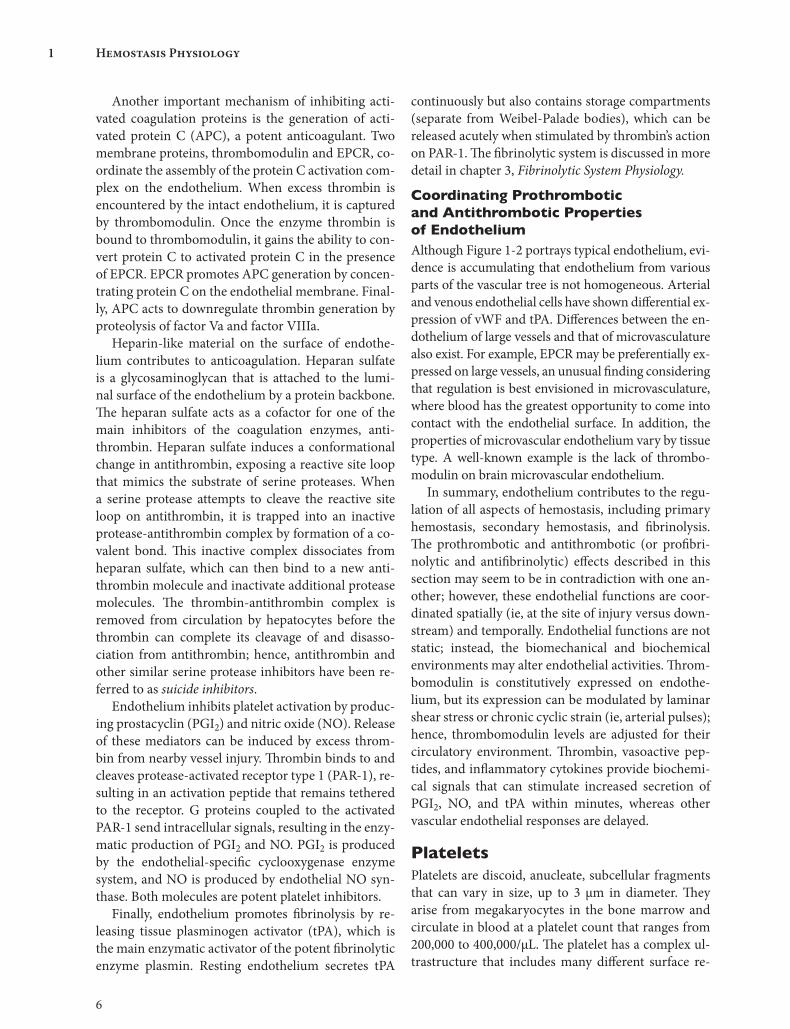
Figure 1-1 illustrates the three major constituents of the hemostatic system—coagulation proteins, plate-lets, and endothelium—and how they are interrelated. Each element of the hemostatic system occupies a site at the vertex of an equilateral triangle. This represen-tation implies that each system constituent interacts
Coagulation Proteins
Platelets Endothelium
Figure 1-1. Basic representation of the elements of hemostasis.
with and influences all other constituents. In the nor-mal resting state, these interactions maintain the flu-idity of the blood to ensure survival of the organism. Normally, only at the site of an injury will the fluidity of the blood be altered and a blood clot form.
When a blood vessel is injured, platelets recognize the exposed subendothelial matrix of vessels via their surface receptors and adhere to the injury site. Ad-ditional platelets are recruited and aggregate to one another, ultimately resulting in a platelet plug. For-mation of the platelet plug is referred to as primary hemostasis. The activation of coagulation proteins is referred to as secondary hemostasis, which results in the production of an insoluble fibrin meshwork that forms within and around the platelet plug, thus stabi-lizing the clot. Finally, as healing occurs, the fibrin clot may be removed enzymatically by the process called fibrinolysis. Endothelium prevents premature fibrino-lysis near the injury, while downstream endothelium has mechanisms to prevent inappropriate clot forma-tion away from the injury site. All three constituents of the hemostatic system tightly regulate primary hemo-stasis, secondary hemostasis, and fibrinolysis.
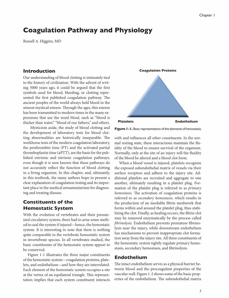
EndotheliumThe intact endothelium serves as a physical barrier be-tween blood and the procoagulant properties of the vascular wall. Figure 1-2 shows some of the basic prop-erties of the endothelium. The subendothelial matrix
1 Hemostasis Physiology
4
is composed of extracellular matrix proteins such as fibronectin, collagen, thrombospondin, among others. When exposed to the flowing blood after endothelial injury, the subendothelial matrix induces platelet ad-hesion. The exposed vascular wall’s adventitial cells express tissue factor, which activates coagulation. Thus, the adventia of the vascular wall forms the so-called hemostatic envelope that ensures clot formation when blood escapes past the injured endothelium.
Tissue factor is a glycosylated intrinsic membrane protein that is constitutively expressed on the plasma membrane of adventitial vascular wall cells and is exposed to flowing blood during vascular injury or endothelial denudation. Tissue factor, when bound to factor VIIa in the presence of membrane phospho-lipid, is the major activator of the extrinsic pathway of coagulation. Although extravascular tissues clearly express tissue factor, the source of circulating or intra-vascular tissue factor is still debated. Contrary to pre-
Figure 1-2. A stylized view of endothelium and vascular wall functions related to hemostasis. Vessel injury is represented in the middle section, with platelet adhesion and tissue factor–induced coagulation. Excess thrombin generated from the injury site interacts with endothelium in many ways. Procoagulant and antifibrinolytic functions are represented on the left side of the figure. Anticoagulant and fibrinolytic functions are represented on the right side of the figure. Abbreviations: APC, activated protein C; AT, antithrombin; EPCR, endothelial protein C receptor; PAR-1, protease-activated receptor type 1; PGI2, prostacyclin; NO, nitric oxide; TAFI, thrombin-activatable fibrinolysis inhibitor; TAT, thrombin-antithrombin (complexes); TFPI, tissue factor pathway inhibitor; TM, thrombomodulin.
vious belief, endothelial cells do not normally express functional tissue factor. Instead, endothelial cells con-tain nonfunctional, cryptic tissue factor, which may be converted to functional tissue factor after endothelial injury.
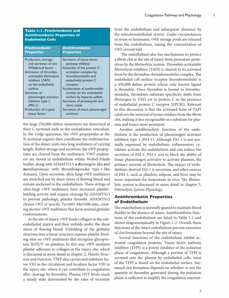
Prothrombotic Properties of EndotheliumProthrombotic functions of the endothelium are listed in Table 1-1. Endothelium is the major synthetic and storage site for von Willebrand factor. Von Willebrand factor (vWF) has a major role in platelet function but also acts as a carrier protein for factor VIII (antihemo-philic factor), preventing its premature clearance from plasma. VWF is secreted from the endothelial cell both into the plasma and also abluminally into the suben-dothelial matrix. It is a large multimeric protein that acts as the intercellular glue binding platelets to one another and also to the subendothelial matrix at an in-jury site. During vWF synthesis by the endothelial cell,
von Willebrand factorPlasminogen activator inhibitor-1 (PAI-1)
Fibrinogen
Activated TAFI TAFI
Thrombin
TFPI(-) Fibrin
Inactive TAT complex
Tissue plasminogen activator (tPA)
Heparan sulfate
Protein C
APC
EPCRPAR-1
NO and PGI2
Platelet
Platelets
Coagulation
Collagen
Tissue factorTissue factor initiates coagulation cascade
Collagen and von Willebrand factor mediate platelet adhesion
Endothelial cell Vascular wall adventitial fibroblast
(-)
Upstream Endothelium
Injury Site
Downstream Endothelium
Blood Flow
Vascular Wall (Hemostatic Envelope)
ATTM TM
5
Coagulation Pathway and Physiology 1
Table 1-1. Prothrombotic and Antithrombotic Properties of Endothelial Cells
Prothrombotic Properties
Antithrombotic Properties
Production, storage, and secretion of von Willebrand factor
Activation of thrombin-activatable fibrinolysis inhibitor (TAFI) on the endothelial surface
Secretion of plasminogen activator inhibitor type 1 (PAI-1)
Production of cryptic tissue factor
Secretion of tissue factor pathway inhibitor
Assembly of the protein C activation complex by thrombomodulin and endothelial protein C receptor
Acceleration of antithrombin activity on the endothelial surface by heparan sulfate
Secretion of prostacyclin and nitric oxide
Secretion of tissue plasminogen activator
the large 250,000-dalton monomers are dimerized at their C-terminal ends in the endoplasmic reticulum. In the Golgi apparatus, the vWF propeptides at the N-terminal regions then coordinate the multimeriza-tion of the dimer units into long multimers of varying length. Before storage and secretion, the vWF propep-tides are cleaved from the multimers. VWF multim-ers are stored in endothelium within Weibel-Palade bodies, along with ADAMTS13, a disintegrin-like and metalloprotease with thrombospondin type-1-like domains. Upon secretion, ultra-large vWF multimers are stretched out by shear stress of flowing blood and remain anchored to the endothelium. These strings of ultra-large vWF multimers have increased platelet-binding activity and require cleavage by ADAMTS13 to prevent pathologic platelet thrombi. ADAMTS13 cleaves vWF at specific Tyr1605-Met1606 sites, creat-ing shorter vWF multimers that form normal globular conformations.
At the site of injury, vWF binds collagen in the sub-endothelial matrix and then unfolds under the shear stress of flowing blood. Unfolding of the globular structure into a linear structure exposes platelet-bind-ing sites on vWF multimers that recognize glycopro-tein Ib/IX/V on platelets. In this way, vWF mediates platelet adhesion to collagen at the injury site, which is discussed in more detail in chapter 2, Platelet Struc-ture and Function. VWF also carries and stabilizes fac-tor VIII in the circulation and localizes factor VIII to the injury site, where it can contribute to coagulation after cleavage by thrombin. Plasma vWF levels reach a steady state determined by the rates of secretion
from the endothelium and subsequent clearance by the reticuloendothelial system. Under circumstances of stress or hormones, vWF storage pools are released from the endothelium, raising the concentration of vWF several fold.
The endothelium also has mechanisms to protect a fibrin clot at the site of injury from premature prote-olysis by the fibrinolytic system. Thrombin-activatable fibrinolysis inhibitor (TAFI) is cleaved to its activated form by the thrombin-thrombomodulin complex. The endothelial cell surface receptor thrombomodulin is a 450,000-dalton protein whose only known ligand is thrombin. Once thrombin is bound to thrombo-modulin, thrombin’s substrate specificity shifts from fibrinogen to TAFI (or to protein C in the presence of endothelial protein C receptor [EPCR]). Relevant to this discussion is that the activated form of TAFI catalyzes the removal of lysine residues from the fibrin clot, making it less recognizable as a substrate for plas-min and hence more persistent.
Another antifibrinolytic function of the endo-thelium is the production of plasminogen activator inhibitor type 1 (PAI-1). Although PAI-1 is not nor-mally expressed by endothelium, inflammatory cy-tokines activate the endothelium and can induce the secretion of PAI-1. PAI-1 acts to block the ability of tissue plasminogen activator to activate plasmin, the primary enzyme of fibrinolysis. The impact of endo-thelium-derived PAI-1 is uncertain, and other sources of PAI-1, such as platelets, adipose, and liver, may be more important for hemostasis in vivo. The fibrino-lytic system is discussed in more detail in chapter 3, Fibrinolytic System Physiology.
Antithrombotic Properties of EndotheliumThe endothelium is normally geared to maintain blood fluidity in the absence of injury. Antithrombotic func-tions of the endothelium are listed in Table 1-1 and shown diagrammatically in Figure 1-2. Overall, these functions of the intact endothelium prevent extension of clot formation beyond the site of injury.
Several functions of the endothelium inhibit ac-tivated coagulation proteins. Tissue factor pathway inhibitor (TFPI) is a potent inhibitor of the initiation phase of coagulation. Although a portion of TFPI is secreted into the plasma by endothelial cells, most of the TFPI is found on the endothelial surface. Sus-tained clot formation depends on whether or not the quantity of thrombin generated during the initiation phase is sufficient to amplify the coagulation reaction.
1 Hemostasis Physiology
6
Another important mechanism of inhibiting acti-vated coagulation proteins is the generation of acti-vated protein C (APC), a potent anticoagulant. Two membrane proteins, thrombomodulin and EPCR, co-ordinate the assembly of the protein C activation com-plex on the endothelium. When excess thrombin is encountered by the intact endothelium, it is captured by thrombomodulin. Once the enzyme thrombin is bound to thrombomodulin, it gains the ability to con-vert protein C to activated protein C in the presence of EPCR. EPCR promotes APC generation by concen-trating protein C on the endothelial membrane. Final-ly, APC acts to downregulate thrombin generation by proteolysis of factor Va and factor VIIIa.
Heparin-like material on the surface of endothe-lium contributes to anticoagulation. Heparan sulfate is a glycosaminoglycan that is attached to the lumi-nal surface of the endothelium by a protein backbone. The heparan sulfate acts as a cofactor for one of the main inhibitors of the coagulation enzymes, anti-thrombin. Heparan sulfate induces a conformational change in antithrombin, exposing a reactive site loop that mimics the substrate of serine proteases. When a serine protease attempts to cleave the reactive site loop on antithrombin, it is trapped into an inactive protease-antithrombin complex by formation of a co-valent bond. This inactive complex dissociates from heparan sulfate, which can then bind to a new anti-thrombin molecule and inactivate additional protease molecules. The thrombin-antithrombin complex is removed from circulation by hepatocytes before the thrombin can complete its cleavage of and disasso-ciation from antithrombin; hence, antithrombin and other similar serine protease inhibitors have been re-ferred to as suicide inhibitors.
Endothelium inhibits platelet activation by produc-ing prostacyclin (PGI2) and nitric oxide (NO). Release of these mediators can be induced by excess throm-bin from nearby vessel injury. Thrombin binds to and cleaves protease-activated receptor type 1 (PAR-1), re-sulting in an activation peptide that remains tethered to the receptor. G proteins coupled to the activated PAR-1 send intracellular signals, resulting in the enzy-matic production of PGI2 and NO. PGI2 is produced by the endothelial-specific cyclooxygenase enzyme system, and NO is produced by endothelial NO syn-thase. Both molecules are potent platelet inhibitors.
Finally, endothelium promotes fibrinolysis by re-leasing tissue plasminogen activator (tPA), which is the main enzymatic activator of the potent fibrinolytic enzyme plasmin. Resting endothelium secretes tPA
continuously but also contains storage compartments (separate from Weibel-Palade bodies), which can be released acutely when stimulated by thrombin’s action on PAR-1. The fibrinolytic system is discussed in more detail in chapter 3, Fibrinolytic System Physiology.
Coordinating Prothrombotic and Antithrombotic Properties of EndotheliumAlthough Figure 1-2 portrays typical endothelium, evi-dence is accumulating that endothelium from various parts of the vascular tree is not homogeneous. Arterial and venous endothelial cells have shown differential ex-pression of vWF and tPA. Differences between the en-dothelium of large vessels and that of microvasculature also exist. For example, EPCR may be preferentially ex-pressed on large vessels, an unusual finding considering that regulation is best envisioned in microvasculature, where blood has the greatest opportunity to come into contact with the endothelial surface. In addition, the properties of microvascular endothelium vary by tissue type. A well-known example is the lack of thrombo-modulin on brain microvascular endothelium.
In summary, endothelium contributes to the regu-lation of all aspects of hemostasis, including primary hemostasis, secondary hemostasis, and fibrinolysis. The prothrombotic and antithrombotic (or profibri-nolytic and antifibrinolytic) effects described in this section may seem to be in contradiction with one an-other; however, these endothelial functions are coor-dinated spatially (ie, at the site of injury versus down-stream) and temporally. Endothelial functions are not static; instead, the biomechanical and biochemical environments may alter endothelial activities. Throm-bomodulin is constitutively expressed on endothe-lium, but its expression can be modulated by laminar shear stress or chronic cyclic strain (ie, arterial pulses); hence, thrombomodulin levels are adjusted for their circulatory environment. Thrombin, vasoactive pep-tides, and inflammatory cytokines provide biochemi-cal signals that can stimulate increased secretion of PGI2, NO, and tPA within minutes, whereas other vascular endothelial responses are delayed.
PlateletsPlatelets are discoid, anucleate, subcellular fragments that can vary in size, up to 3 µm in diameter. They arise from megakaryocytes in the bone marrow and circulate in blood at a platelet count that ranges from 200,000 to 400,000/µL. The platelet has a complex ul-trastructure that includes many different surface re-
7
Coagulation Pathway and Physiology 1
ceptors, several types of storage granules, and a net-work of actin and myosin filaments. At the site of an injury, the platelets contact extracellular matrix com-ponents, causing a series of metabolic changes that result in the formation of a platelet plug. These meta-bolic changes are usually termed aggregation. The role of platelets in limiting clot formation to the injury site cannot be overlooked. Platelet regulatory functions overlap with the endothelial functions described, such as secretion of vWF and expression of TFPI. Platelet structure and function are described in more detail in chapter 2, Platelet Structure and Function.
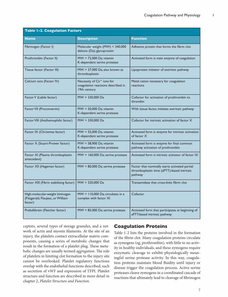
Coagulation ProteinsTable 1-2 lists the proteins involved in the formation of the fibrin clot. Many coagulation proteins circulate as zymogens (eg, prothrombin), with little to no activ-ity in healthy individuals, and these zymogens require enzymatic cleavage to exhibit physiologically mean-ingful serine protease activity. In this way, coagula-tion proteins maintain blood fluidity until injury or disease trigger the coagulation process. Active serine proteases cleave zymogens in a coordinated cascade of reactions that ultimately lead to cleavage of fibrinogen
Table 1-2. Coagulation Factors
Name Description Function
Fibrinogen (Factor I) Molecular weight (MW) = 340,000 daltons (Da); glycoprotein
Adhesive protein that forms the fibrin clot
Prothrombin (Factor II) MW = 72,000 Da; vitamin K-dependent serine protease
Activated form is main enzyme of coagulation
Tissue factor (Factor III) MW = 37,000 Da; also known as thromboplastin
Lipoprotein initiator of extrinsic pathway
Calcium ions (Factor IV) Necessity of Ca++ ions for coagulation reactions described in 19th century
Metal cation necessary for coagulation reactions
Factor V (Labile factor) MW = 330,000 Da Cofactor for activation of prothrombin to thrombin
Factor VII (Proconvertin) MW = 50,000 Da; vitamin K-dependent serine protease
With tissue factor, initiates extrinsic pathway
Factor VIII (Antihemophilic factor) MW = 330,000 Da Cofactor for intrinsic activation of factor X
Factor IX (Christmas factor) MW = 55,000 Da; vitamin K-dependent serine protease
Activated form is enzyme for intrinsic activation of factor X
Factor X (Stuart-Prower factor) MW = 58,900 Da; vitamin K-dependent serine protease
Activated form is enzyme for final common pathway activation of prothrombin
Factor XI (Plasma thromboplastin antecedent)
MW = 160,000 Da; serine protease Activated form is intrinsic activator of factor IX
Factor XII (Hageman factor) MW = 80,000 Da; serine protease Factor that nominally starts activated partial thromboplastin time (aPTT)-based intrinsic pathway
Factor XIII (Fibrin stabilizing factor) MW = 320,000 Da Transamidase that cross-links fibrin clot
High-molecular-weight kininogen (Fitzgerald, Flaujeac, or William factor)
MW = 110,000 Da; circulates in a complex with factor XI
Cofactor
Prekallikrein (Fletcher factor) MW = 85,000 Da; serine protease Activated form that participates at beginning of aPTT-based intrinsic pathway
1 Hemostasis Physiology
8
into fibrin. Serine proteases contain a catalytic triad of serine, histidine, and aspartic acid at their active sites that coordinate cleavage of substrate proteins, which are other coagulation proteins in the setting of the co-agulation cascade. Analogous to serine protease zymo-gens, cofactors V and VIII normally circulate as pro-cofactors that require enzymatic cleavage by the serine protease thrombin to gain cofactor activity. Cofactors do not have enzymatic activity; rather, they provide serine protease binding sites and coordinate substrate docking, thereby enhancing enzymatic activity.
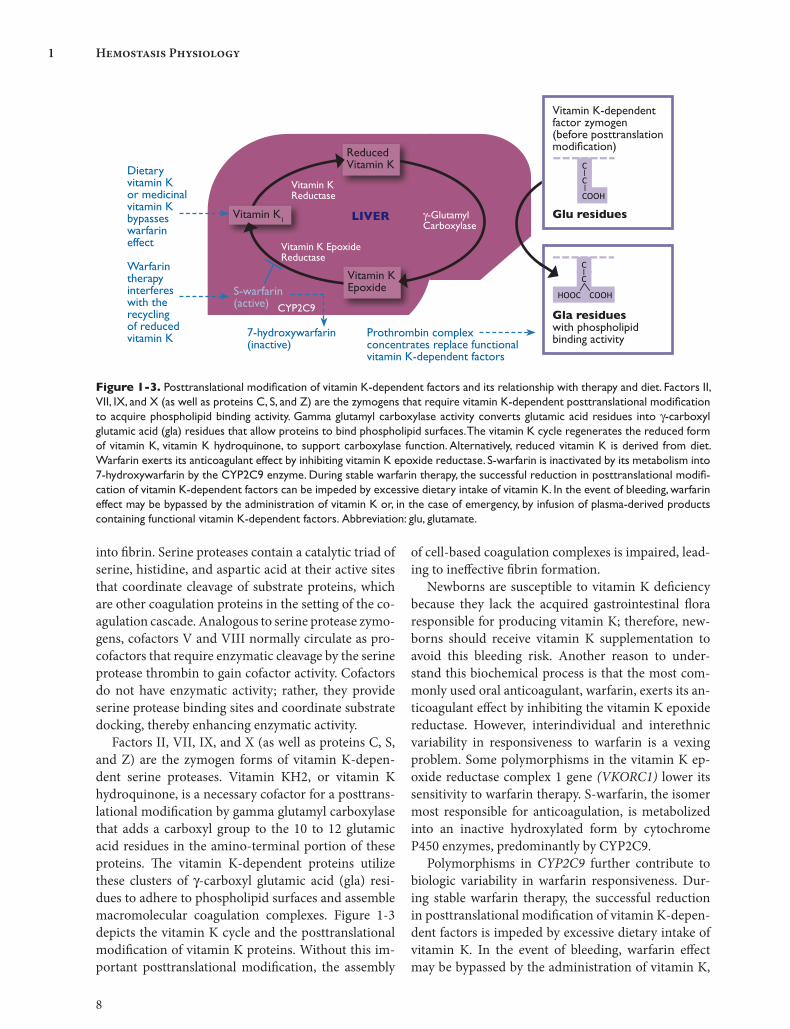
Factors II, VII, IX, and X (as well as proteins C, S, and Z) are the zymogen forms of vitamin K-depen-dent serine proteases. Vitamin KH2, or vitamin K hydroquinone, is a necessary cofactor for a posttrans-lational modification by gamma glutamyl carboxylase that adds a carboxyl group to the 10 to 12 glutamic acid residues in the amino-terminal portion of these proteins. The vitamin K-dependent proteins utilize these clusters of g-carboxyl glutamic acid (gla) resi-dues to adhere to phospholipid surfaces and assemble macromolecular coagulation complexes. Figure 1-3 depicts the vitamin K cycle and the posttranslational modification of vitamin K proteins. Without this im-portant posttranslational modification, the assembly
of cell-based coagulation complexes is impaired, lead-ing to ineffective fibrin formation.
Newborns are susceptible to vitamin K deficiency because they lack the acquired gastrointestinal flora responsible for producing vitamin K; therefore, new-borns should receive vitamin K supplementation to avoid this bleeding risk. Another reason to under-stand this biochemical process is that the most com-monly used oral anticoagulant, warfarin, exerts its an-ticoagulant effect by inhibiting the vitamin K epoxide reductase. However, interindividual and interethnic variability in responsiveness to warfarin is a vexing problem. Some polymorphisms in the vitamin K ep-oxide reductase complex 1 gene (VKORC1) lower its sensitivity to warfarin therapy. S-warfarin, the isomer most responsible for anticoagulation, is metabolized into an inactive hydroxylated form by cytochrome P450 enzymes, predominantly by CYP2C9.
Polymorphisms in CYP2C9 further contribute to biologic variability in warfarin responsiveness. Dur-ing stable warfarin therapy, the successful reduction in posttranslational modification of vitamin K-depen-dent factors is impeded by excessive dietary intake of vitamin K. In the event of bleeding, warfarin effect may be bypassed by the administration of vitamin K,
Reduced Vitamin K
LIVER
Vitamin K Epoxide
Vitamin K1 g-Glutamyl Carboxylase
Vitamin K Reductase
Vitamin K Epoxide Reductase
Dietary vitamin K or medicinal vitamin K bypasses warfarin effect
S-warfarin (active)
Warfarin therapy interferes with the recycling of reduced vitamin K Prothrombin complex
concentrates replace functional vitamin K-dependent factors
HOOC
CC
COOH
C
C
COOH
HOOC
CC
COOH
C
C
COOH
Gla residues with phospholipid binding activity
Vitamin K-dependent factor zymogen (before posttranslation modification)
Glu residues
7-hydroxywarfarin (inactive)
CYP2C9
Figure 1-3. Posttranslational modification of vitamin K-dependent factors and its relationship with therapy and diet. Factors II, VII, IX, and X (as well as proteins C, S, and Z) are the zymogens that require vitamin K-dependent posttranslational modification to acquire phospholipid binding activity. Gamma glutamyl carboxylase activity converts glutamic acid residues into g-carboxyl glutamic acid (gla) residues that allow proteins to bind phospholipid surfaces. The vitamin K cycle regenerates the reduced form of vitamin K, vitamin K hydroquinone, to support carboxylase function. Alternatively, reduced vitamin K is derived from diet. Warfarin exerts its anticoagulant effect by inhibiting vitamin K epoxide reductase. S-warfarin is inactivated by its metabolism into 7-hydroxywarfarin by the CYP2C9 enzyme. During stable warfarin therapy, the successful reduction in posttranslational modifi-cation of vitamin K-dependent factors can be impeded by excessive dietary intake of vitamin K. In the event of bleeding, warfarin effect may be bypassed by the administration of vitamin K or, in the case of emergency, by infusion of plasma-derived products containing functional vitamin K-dependent factors. Abbreviation: glu, glutamate.
9
Coagulation Pathway and Physiology 1
Prothrombin
Thrombin
Fibrinogen Fibrin Clot
Thrombokinase Calcium
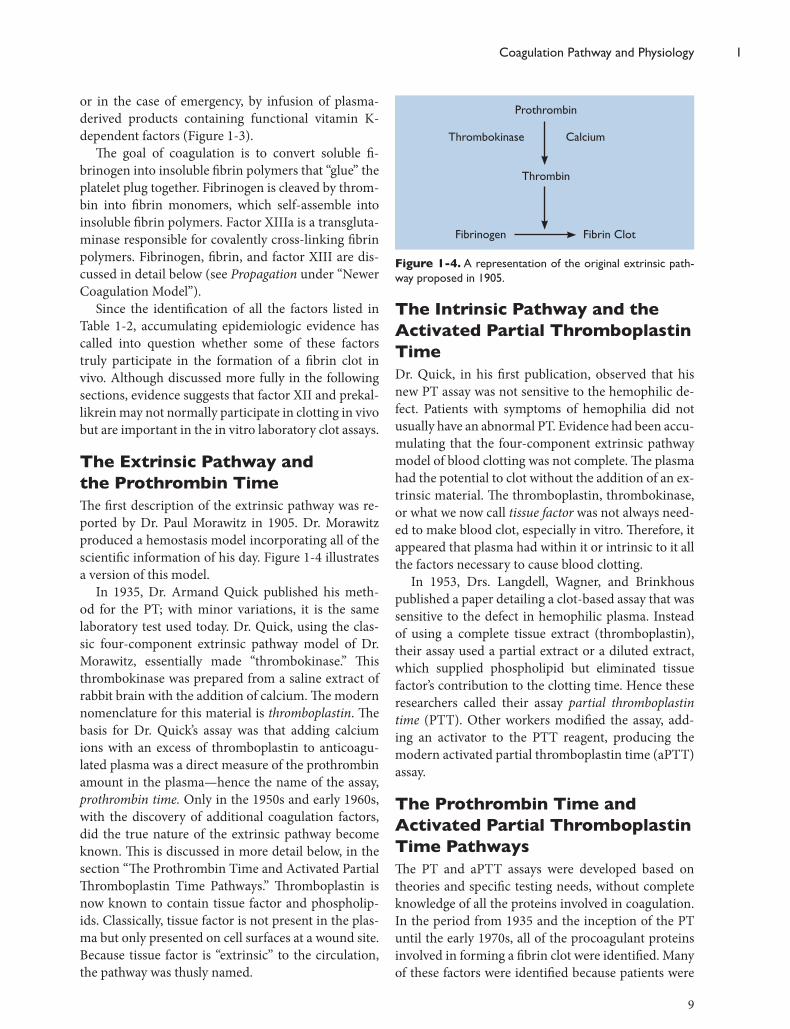
Figure 1-4. A representation of the original extrinsic path-way proposed in 1905.
or in the case of emergency, by infusion of plasma-derived products containing functional vitamin K-dependent factors (Figure 1-3).
The goal of coagulation is to convert soluble fi-brinogen into insoluble fibrin polymers that “glue” the platelet plug together. Fibrinogen is cleaved by throm-bin into fibrin monomers, which self-assemble into insoluble fibrin polymers. Factor XIIIa is a transgluta-minase responsible for covalently cross-linking fibrin polymers. Fibrinogen, fibrin, and factor XIII are dis-cussed in detail below (see Propagation under “Newer Coagulation Model”).
Since the identification of all the factors listed in Table 1-2, accumulating epidemiologic evidence has called into question whether some of these factors truly participate in the formation of a fibrin clot in vivo. Although discussed more fully in the following sections, evidence suggests that factor XII and prekal-likrein may not normally participate in clotting in vivo but are important in the in vitro laboratory clot assays.
The Extrinsic Pathway and the Prothrombin TimeThe first description of the extrinsic pathway was re-ported by Dr. Paul Morawitz in 1905. Dr. Morawitz produced a hemostasis model incorporating all of the scientific information of his day. Figure 1-4 illustrates a version of this model.
In 1935, Dr. Armand Quick published his meth-od for the PT; with minor variations, it is the same laboratory test used today. Dr. Quick, using the clas-sic four-component extrinsic pathway model of Dr. Morawitz, essentially made “thrombokinase.” This thrombokinase was prepared from a saline extract of rabbit brain with the addition of calcium. The modern nomenclature for this material is thromboplastin. The basis for Dr. Quick’s assay was that adding calcium ions with an excess of thromboplastin to anticoagu-lated plasma was a direct measure of the prothrombin amount in the plasma—hence the name of the assay, prothrombin time. Only in the 1950s and early 1960s, with the discovery of additional coagulation factors, did the true nature of the extrinsic pathway become known. This is discussed in more detail below, in the section “The Prothrombin Time and Activated Partial Thromboplastin Time Pathways.” Thromboplastin is now known to contain tissue factor and phospholip-ids. Classically, tissue factor is not present in the plas-ma but only presented on cell surfaces at a wound site. Because tissue factor is “extrinsic” to the circulation, the pathway was thusly named.
The Intrinsic Pathway and the Activated Partial Thromboplastin TimeDr. Quick, in his first publication, observed that his new PT assay was not sensitive to the hemophilic de-fect. Patients with symptoms of hemophilia did not usually have an abnormal PT. Evidence had been accu-mulating that the four-component extrinsic pathway model of blood clotting was not complete. The plasma had the potential to clot without the addition of an ex-trinsic material. The thromboplastin, thrombokinase, or what we now call tissue factor was not always need-ed to make blood clot, especially in vitro. Therefore, it appeared that plasma had within it or intrinsic to it all the factors necessary to cause blood clotting.
In 1953, Drs. Langdell, Wagner, and Brinkhous published a paper detailing a clot-based assay that was sensitive to the defect in hemophilic plasma. Instead of using a complete tissue extract (thromboplastin), their assay used a partial extract or a diluted extract, which supplied phospholipid but eliminated tissue factor’s contribution to the clotting time. Hence these researchers called their assay partial thromboplastin time (PTT). Other workers modified the assay, add-ing an activator to the PTT reagent, producing the modern activated partial thromboplastin time (aPTT) assay.
The Prothrombin Time and Activated Partial Thromboplastin Time Pathways The PT and aPTT assays were developed based on theories and specific testing needs, without complete knowledge of all the proteins involved in coagulation. In the period from 1935 and the inception of the PT until the early 1970s, all of the procoagulant proteins involved in forming a fibrin clot were identified. Many of these factors were identified because patients were

1 Hemostasis Physiology
10
found with deficiency states. Some of these patients had congenital bleeding disease, while others pre-sented with an abnormal prolongation in the PT and/or the aPTT without bleeding. It became clear that a fresh model of coagulation was needed.
In the early 1960s, a new synthesis of all hemosta-sis knowledge occurred, and the PT (or extrinsic) and aPTT (or intrinsic) coagulation pathways were pub-lished. Table 1-2 contains a list of coagulation factors and their functions. The coagulation pathways are illustrated in Figure 1-5. Although there have been some modifications since the original papers, these are the pathways with which most workers in hemostasis are familiar. Current evidence suggests the intrinsic and extrinsic hemostasis pathways that occur in test tubes might not be a correct representation of blood clotting in vivo. For example, patients deficient in fac-tor XII, prekallikrein, or high-molecular-weight ki-
ninogen have a significantly prolonged aPTT, but they do not have a bleeding phenotype. Logically, it would make sense that a deficiency of a factor at the start of a pathway would cause bleeding pathology; however, all evidence suggests this is not so. The intrinsic and ex-trinsic pathways as they have existed since their incep-tion are based on in vitro testing. The in vivo function appears to be different. It is important to be familiar with the older pathway model because the PT and the aPTT are still useful as diagnostic tests.
Newer Coagulation ModelFigure 1-6 illustrates a newer model of coagulation. In this figure, coagulation proteins are organized into macromolecular complexes on the phospholipid membranes of cells. Coagulation protein interac-tions are ineffective in the three-dimensional space of plasma; however, phospholipid-binding properties
Intrinsic Pathway Tested for by aPTT Extrinsic Pathway Tested for by PT
Negatively charged surface Tissue factor + Factor VIIa Factor VIIKallikrein
Prekallikrein
Factor XII Factor XIIa
Factor XI Factor XIa
Factor IX Factor IXa
Factor X Factor Xa
Factor Va
Factor II Thrombin
Factor VIIIa
Fibrinogen Fibrin monomer
Factor XIII
Fibrin polymer
Factor XIIIa
Cross-linked clotFinal Common Pathway
Figure 1-5. A model of the classic extrinsic and intrinsic coagulation pathways. For the sake of clarity, the calcium ions (Ca++) and phospholipids, which are two important cofactors for most coagulation reactions, have been omitted from the figure. Ca++
and phospholipids are necessary for most reactions, with the exception of the activation of factor XII and the activation of prekallikrein. The cofactor for the activation of prekallikrein, high-molecular-weight kininogen, has also been omitted from the figure for clarity. Abbreviations: aPTT, activated partial thromboplastin time; PT, prothrombin time.
11
Coagulation Pathway and Physiology 1
of vitamin K-dependent factors bring the interactions into a two-dimensional surface on cell membranes. Protein interactions are further coordinated by co-factors that optimize the ability of serine proteases to find their substrate on the two-dimensional surface. The contribution of phospholipid and cofactors has been compared to railroad tracks that allow the easy coupling of railroad cars to one another. Enzymatic activity of these complexes requires Ca++. Initiation, amplification, and propagation stages of coagula-tion are depicted in Figure 1-6. Of note, the figure lacks several proteins normally considered part of the classic intrinsic coagulation pathway: factor XII and prekallikrein. These contact factors are not es-sential for in vivo hemostasis induced by tissue factor, and contact factor deficiencies do not cause bleeding
disorders. Conversely, contact activation may occur under pathologic circumstances if contact activators are introduced into the circulation. As an example, long-chain polyphosphate (polyP) (1000-2000mers) from microbial organisms can activate the extrinsic pathway. Contact activation may or may not initiate the coagulation cascade, depending on the magnitude of the stimulus, but it is an important pathway linking coagulation and inflammation. Nevertheless, contact activation is not mentioned further in the description of the newer coagulation model below.
InitiationThis new coagulation model has extrinsic and intrinsic pathway limbs, but the in vivo process of hemostasis is initiated by cell-based tissue factor. Functional tis-
Adventitial Fibroblast
Tissue injury
Platelet
Tissue factor + Factor VII
Ca++ Tissue factor Factor VIIa
Extrinsic tenase
Ca++ Factor Va Factor Xa
Prothrombinase
ThrombinInitiation
Amplification
Propagation
Factor IX
Factor IXa
Factor X
Prothrombin
Tissue factor pathway inhibitor rapidly inhibits initiation
Diffusion to platelet
Thrombin
Factor Va PolyP
Thrombin
Thrombin
PolyP
Ca++ Factor VIIIa Factor IXa
Intrinsic tenase
Ca++ Factor Va Factor Xa
Prothrombinase
Factor XI
Factor VIII
Factor V
Factor XIa Factor IX
Factor X
Prothrombin
Fibrinogen
Factor XIII
Factor XIIIa Fibrin monomer
Fibrin polymerCross-linked clot
Thrombin
Ca++
?
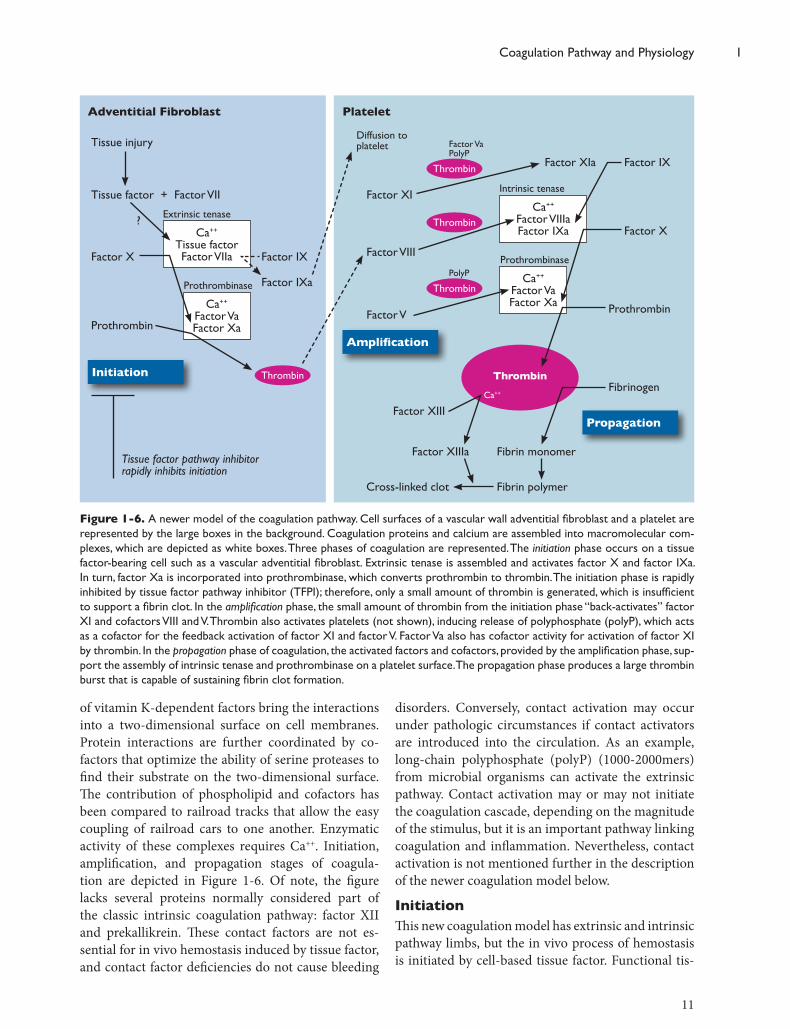
Figure 1-6. A newer model of the coagulation pathway. Cell surfaces of a vascular wall adventitial fibroblast and a platelet are represented by the large boxes in the background. Coagulation proteins and calcium are assembled into macromolecular com-plexes, which are depicted as white boxes. Three phases of coagulation are represented. The initiation phase occurs on a tissue factor-bearing cell such as a vascular adventitial fibroblast. Extrinsic tenase is assembled and activates factor X and factor IXa. In turn, factor Xa is incorporated into prothrombinase, which converts prothrombin to thrombin. The initiation phase is rapidly inhibited by tissue factor pathway inhibitor (TFPI); therefore, only a small amount of thrombin is generated, which is insufficient to support a fibrin clot. In the amplification phase, the small amount of thrombin from the initiation phase “back-activates” factor XI and cofactors VIII and V. Thrombin also activates platelets (not shown), inducing release of polyphosphate (polyP), which acts as a cofactor for the feedback activation of factor XI and factor V. Factor Va also has cofactor activity for activation of factor XI by thrombin. In the propagation phase of coagulation, the activated factors and cofactors, provided by the amplification phase, sup-port the assembly of intrinsic tenase and prothrombinase on a platelet surface. The propagation phase produces a large thrombin burst that is capable of sustaining fibrin clot formation.
1 Hemostasis Physiology
12
sue factor is constitutively expressed on fibroblasts (or adventitial fibroblasts) but may be induced on mono-cytes or endothelial cells. The latter cells contain inac-tive, cryptic tissue factor that may be decrypted after endothelial injury. To be more specific, monocyte-derived or endothelial-derived microparticles may de-liver tissue factor to the site of clot formation. For the sake of simplicity, the description in Figure 1-6 uses the vascular adventitial fibroblasts as the source of cell-based tissue factor. Vascular injury exposes blood to the constitutively expressed tissue factor on adven-titial fibroblasts. Tissue factor binds to factor VII in a 1:1 complex. Factor VII in the tissue factor-factor VII complex is rapidly activated by a yet-uncharacterized process that may involve factor Xa or other noncoagu-lation protease. The resultant macromolecular com-plex, extrinsic tenase, activates factor X or factor IX to activated serine proteases. In turn, the newly formed activated factor Xa complexes with its cofactor, factor Va, to form the prothrombinase complex. This com-plex converts prothrombin to thrombin by cleavage of an activation peptide, prothrombin F1.2; however, the amount of thrombin generated is insufficient to prop-agate a fibrin clot because TFPI potently inhibits the initiation phase of coagulation. See the “Endothelium” and “Protease Inhibitors” sections for more details on TFPI.
AmplificationIf conditions are permissive, the small amount of thrombin created during initiation can amplify key activated coagulation proteins necessary to support the intrinsic pathway. This is largely accomplished by the feedback activation of factor XI and two cofactors, factor V and factor VIII, by thrombin. Until recently, the activation of factor XI by thrombin was thought to be too slow to explain physiological hemostasis. Two recently described cofactors, platelet-derived polyP and factor Va, accelerate thrombin activation of factor XI. Platelet-derived polyP ranges from 60 to 100mers, much shorter than the microbial-derived polyP men-tioned above that activates the contact pathway. PolyP also accelerates autoactivation of factor XI by factor XIa. Platelets in the immediate vicinity of clot initia-tion may be necessary to provide polyP as well as a membrane surface for efficient amplification. Hence, the amplification phase of coagulation provides build-ing blocks for the enzyme complexes in the propaga-tion phase of coagulation.
PropagationThe propagation phase generates a large thrombin burst that sustains fibrin clot growth until regulatory mechanisms supervene. Activated platelets at the site of injury localize factor Va, factor VIIIa, and IXa to their surface and support the assembly of macromo-lecular complexes of the propagation phase of co-agulation. Factor XIa, provided by the amplification phase, converts factor IX to factor IXa. Factor IXa is incorporated into an intrinsic tenase complex to-gether with its cofactor, factor VIIIa. Once factor Xa is generated, it is incorporated into a prothrombinase composed of factor Xa and its cofactor, factor Va. The large burst of thrombin generated by the propagation phase supports growth of the fibrin clot.
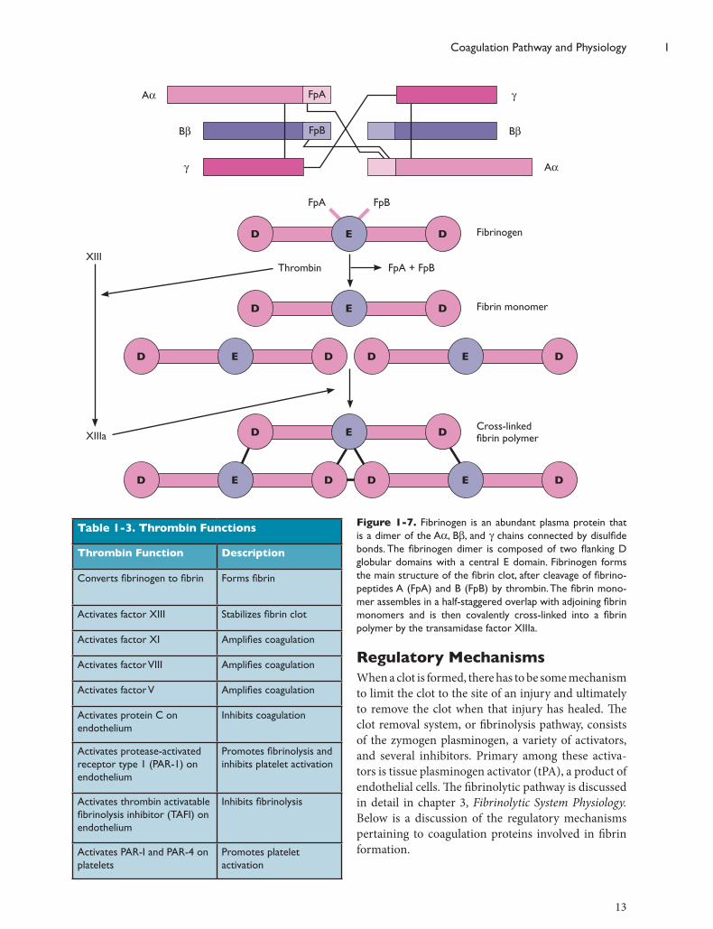
Fibrinogen is the ultimate substrate protein of the coagulation cascade and forms the principal structur-al protein of the fibrin clot. Fibrinogen, produced in the liver, is a dimer composed of three pairs of protein chains—Aa, Bb, and g—that are disulfide-linked at their amino-terminal ends. Fibrinogen, as viewed by molecular imaging techniques, is composed of three globular domains: a central E domain flanked by two identical D domains (Figure 1-7). Thrombin cleaves small peptides, termed fibrinopeptides A and B, from the Aa and Bb chains, respectively, to form a fibrin monomer. These monomers assemble into proto-fibrils in a half-staggered, side-to-side fashion that is stabilized by noncovalent interactions between fibrin molecules. The protofibrils laterally associate into thicker fibrin fibers and form the fibrin clot. This clot, however, is not stable and ultimately will dissociate if not covalently cross-linked. Thrombin activates factor XIII to factor XIIIa, a transglutaminase. Factor XIIIa, acting upon the glutamic acid and lysine side chains in the fibrin amino acid sequence, creates covalent bonds between fibrin monomer g chains, creating a stable clot. In addition, factor XIIIa can covalently cross-link a variety of other proteins into the forming fibrin clot, including plasminogen and antiplasmin. This prop-erty of factor XIIIa is important for the penultimate purpose of the clot: wound healing and tissue repair.
Thrombin has many properties other than the for-mation of the fibrin clot. Thrombin has direct effects on the other constituents of the coagulation triad: co-agulation proteins, platelets, and endothelial cells. The many functions of thrombin are listed in Table 1-3, including interactions with regulatory proteins. In the next section, some of these coagulation regulatory processes will be mentioned.
13
Coagulation Pathway and Physiology 1
Regulatory MechanismsWhen a clot is formed, there has to be some mechanism to limit the clot to the site of an injury and ultimately to remove the clot when that injury has healed. The clot removal system, or fibrinolysis pathway, consists of the zymogen plasminogen, a variety of activators, and several inhibitors. Primary among these activa-tors is tissue plasminogen activator (tPA), a product of endothelial cells. The fibrinolytic pathway is discussed in detail in chapter 3, Fibrinolytic System Physiology. Below is a discussion of the regulatory mechanisms pertaining to coagulation proteins involved in fibrin formation.
Table 1-3. Thrombin Functions
Thrombin Function Description
Converts fibrinogen to fibrin Forms fibrin
Activates factor XIII Stabilizes fibrin clot
Activates factor XI Amplifies coagulation
Activates factor VIII Amplifies coagulation
Activates factor V Amplifies coagulation
Activates protein C on endothelium
Inhibits coagulation
Activates protease-activated receptor type 1 (PAR-1) on endothelium
Promotes fibrinolysis and inhibits platelet activation
Activates thrombin activatable fibrinolysis inhibitor (TAFI) on endothelium
Inhibits fibrinolysis
Activates PAR-I and PAR-4 on platelets
Promotes platelet activation
Fibrinogen
Thrombin
g
Bb
g Aa
FpBBb
FpAAa
FpA FpB
FpA + FpB
D DE
Fibrin monomerD DE
XIII
XIIIa
D DE D DE
D DE
D DE D DE
Cross-linked fibrin polymer
Figure 1-7. Fibrinogen is an abundant plasma protein that is a dimer of the Aa, Bb, and g chains connected by disulfide bonds. The fibrinogen dimer is composed of two flanking D globular domains with a central E domain. Fibrinogen forms the main structure of the fibrin clot, after cleavage of fibrino-peptides A (FpA) and B (FpB) by thrombin. The fibrin mono-mer assembles in a half-staggered overlap with adjoining fibrin monomers and is then covalently cross-linked into a fibrin polymer by the transamidase factor XIIIa.
1 Hemostasis Physiology
14
protein and serves as a cofactor in protein C activa-tion. The functional fraction of protein S (40%) cir-culates free in the plasma, while the majority (60%) is reversibly bound to the complement inhibitor protein, C4b-binding protein. Physiologic and pathophysio-logic mechanisms such as pregnancy and acute phase response cause an increase in C4b-binding protein and decrease the fraction of free protein S. In addition to its role in APC generation, protein S is a cofactor for the inhibition of factor Xa by TFPI.
Protease InhibitorsTissue factor pathway inhibitor is a 33,000-dalton Kunitz-type serine protease inhibitor of the extrinsic pathway found in plasma, on endothelial cells, and on platelets after exposure to thrombin and collagen. TFPI has three Kunitz domains. The second Kunitz domain inhibits factor Xa reversibly by interaction with its active site. The first Kunitz domain of TFPI-Xa complex then inhibits the factor VIIa site of tissue factor-factor VIIa (TF-FVIIa) complex in a calcium-dependent interaction, resulting in an inactive quar-ternary complex. The third Kunitz domain does not have a characterized protease function but facilitates protein S interaction that enhances inhibition of factor Xa on phospholipid surfaces. Inhibition of TF-FVIIa is augmented when TFPI-Xa is associated with a cell sur-face. TFPI is predominantly found bound to the en-dothelial surface. Full-length TFPI, TFPIa, is not di-rectly linked to glycosyl phosphatidylinositol (GPI); it is tightly bound to an uncharacterized GPI-anchored protein on endothelial cells. To a lesser extent, TFPI is associated with glycosaminoglycans (GAGs) on the endothelium or is secreted into the plasma. A splice variant of TFPI, TFPIb, lacks the third Kunitz domain and is directly GPI-anchored on endothelial cells. Platelets contain only full-length TFPI. TFPI is nor-mally absent on the surface of platelets, but it can be
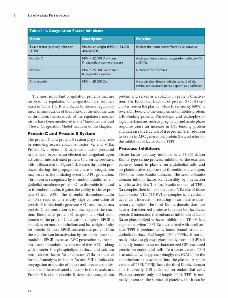
The most important coagulation proteins that are involved in regulation of coagulation are summa-rized in Table 1-4. It is difficult to discuss regulatory mechanisms outside of the context of the endothelium or thrombin; hence, much of the regulatory mecha-nisms have been mentioned in the “Endothelium” and “Newer Coagulation Model” sections of this chapter.
Protein C and Protein S SystemThe protein C and protein S system plays a vital role in removing excess cofactors, factor Va and VIIIa. Protein C, a vitamin K-dependent factor produced in the liver, becomes an efficient anticoagulant upon activation into activated protein C, a serine protease. This is illustrated in Figure 1-2. Excess thrombin pro-duced during the propagation phase of coagulation may serve as the initiating event in APC generation. Thrombin is recognized by thrombomodulin, an en-dothelial membrane protein. Once thrombin is bound to thrombomodulin, it gains the ability to cleave pro-tein C into APC. The thrombin-thrombomodulin complex requires a relatively high concentration of protein C to efficiently generate APC, and the plasma protein C concentration is too low support the reac-tion. Endothelial protein C receptor is a vital com-ponent of the protein C activation complex. EPCR is abundant on most endothelium and has a high affinity for protein C; thus, EPCR concentrates protein C on the endothelium for activation by thrombin-thrombo-modulin. EPCR increases APC generation by throm-bin-thrombomodulin by a factor of five. APC—along with protein S, a phospholipid surface, and calcium ions—cleaves factor Va and factor VIIIa to inactive forms. Proteolysis of factors Va and VIIIa limits clot propagation at the site of injury and prevents the cir-culation of these activated cofactors in the vasculature. Protein S is also a vitamin K-dependent coagulation
Table 1-4. Coagulation Factor Inhibitors
Name Description Function
Tissue factor pathway inhibitor (TFPI)
Molecular weight (MW) = 33,000 daltons (Da)
Inhibits the tissue factor/factor VIIa complex
Protein C MW = 62,000 Da; vitamin K-dependent serine protease
Activated form cleaves coagulation cofactors Va and VIIIa
Protein S MW = 75,000 Da; vitamin K-dependent protein
Cofactor for protein C
Antithrombin MW = 58,000 Da A serpin that directly inhibits several of the serine proteases; requires heparin as a cofactor
15
Coagulation Pathway and Physiology 1
induced by dual stimulation with collagen and throm-bin.
Antithrombin is a 58,000-dalton serine protease inhibitor (serpin) that is produced in the liver. Anti-thrombin has a five-stranded central b-sheet (the A-sheet), together with a heparin-binding D helix and a mobile reactive site loop containing an arginine393-serine394 bond that resembles the substrate of serine proteases such as thrombin. Antithrombin inhibition is not limited to thrombin, as the name might imply; rather, it has inhibitory activity also against factor Xa, factor IXa, factor XIa, and factor VIIa. Once a serine protease cleaves the bond, the protease is trapped by a covalent linkage to antithrombin, forming an inactive protease-antithrombin complex (see Figure 1-2). In its native state, antithrombin inactivates the proteases inefficiently because of conformational inaccessibility of the arginine-serine bond. Inhibition is accelerated approximately 1000-fold by the binding of heparin to arginine residues in the D helix of antithrombin, with a resultant conformational change and exposure of the reactive site loop. The in vivo source of heparin-like substance is heparan sulfate present on endothelium. Antithrombin requires a specific heparin pentasac-charide sequence to accelerate activity. The pentasac-charide sequence alone is sufficient to accelerate fac-tor Xa inhibition; however, longer heparin molecules (>26 saccharide units) are needed to accelerate throm-bin inhibition. These longer heparin molecules bind
both antithrombin and thrombin, facilitating their interaction. Antithrombin has an established role in the regulation of intravascular coagulation and is con-sidered the major serpin anticoagulant.
Heparin cofactor II is another serpin with activity against thrombin. Like antithrombin, heparin cofactor II activity is produced in the liver, inhibits thrombin, and has accelerated activity when bound to a GAG. The type of GAG required is less specific than for an-tithrombin; and heparin, heparan sulfate, and derma-tan sulfate all increase the rate of thrombin inhibition. Unlike antithrombin, heparin cofactor II inhibition is largely limited to thrombin. About 40% of heparin cofactor II localizes to extravascular tissues. Although the in vivo function of heparin cofactor II is not com-pletely characterized, it probably regulates thrombin generation in extravascular tissue, such as the intima of arterial walls, where it has been detected.
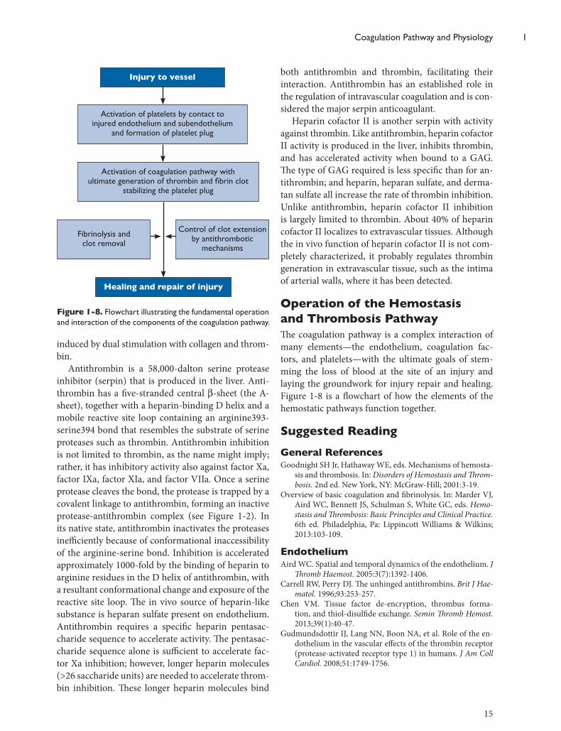
Operation of the Hemostasis and Thrombosis PathwayThe coagulation pathway is a complex interaction of many elements—the endothelium, coagulation fac-tors, and platelets—with the ultimate goals of stem-ming the loss of blood at the site of an injury and laying the groundwork for injury repair and healing. Figure 1-8 is a flowchart of how the elements of the hemostatic pathways function together.
Suggested ReadingGeneral References Goodnight SH Jr, Hathaway WE, eds. Mechanisms of hemosta-
sis and thrombosis. In: Disorders of Hemostasis and Throm-bosis. 2nd ed. New York, NY: McGraw-Hill; 2001:3-19.
Overview of basic coagulation and fibrinolysis. In: Marder VJ, Aird WC, Bennett JS, Schulman S, White GC, eds. Hemo-stasis and Thrombosis: Basic Principles and Clinical Practice. 6th ed. Philadelphia, Pa: Lippincott Williams & Wilkins; 2013:103-109.
EndotheliumAird WC. Spatial and temporal dynamics of the endothelium. J
Thromb Haemost. 2005:3(7):1392-1406. Carrell RW, Perry DJ. The unhinged antithrombins. Brit J Hae-
matol. 1996;93:253-257.Chen VM. Tissue factor de-encryption, thrombus forma-
tion, and thiol-disulfide exchange. Semin Thromb Hemost. 2013;39(1):40-47.
Gudmundsdottir IJ, Lang NN, Boon NA, et al. Role of the en-dothelium in the vascular effects of the thrombin receptor (protease-activated receptor type 1) in humans. J Am Coll Cardiol. 2008;51:1749-1756.
Injury to vessel
Activation of platelets by contact to injured endothelium and subendothelium
and formation of platelet plug
Activation of coagulation pathway with ultimate generation of thrombin and fibrin clot
stabilizing the platelet plug
Healing and repair of injury
Fibrinolysis and clot removal
Control of clot extension by antithrombotic
mechanisms
Figure 1-8. Flowchart illustrating the fundamental operation and interaction of the components of the coagulation pathway.