Embed Size (px)
Citation preview
Coagulation Disorders
Assistant ProfessorDr Talib Hussein Kamoona (CABM)Hematologist/Medical Oncologist
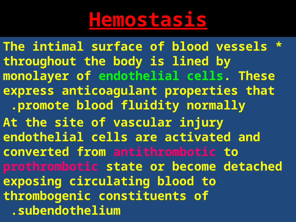
Hemostasis *The intimal surface of blood vessels throughout
the body is lined by monolayer of endothelial cells. These express anticoagulant properties that
promote blood fluidity normally .At the site of vascular injury endothelial cells are activated and converted from antithrombotic to prothrombotic state or become detached exposing circulating blood to thrombogenic constituents of
subendothelium .
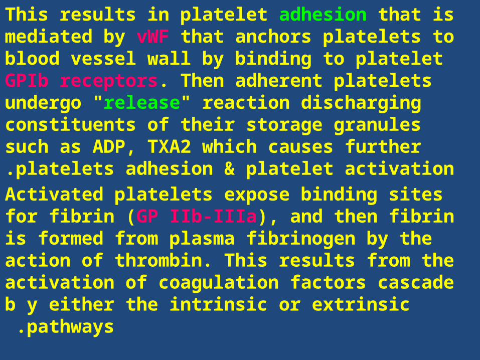
This results in platelet adhesion that is mediated by vWF that anchors platelets to blood vessel wall by binding to platelet GPIb receptors. Then adherent platelets undergo "release" reaction discharging constituents of their storage granules such as ADP, TXA2 which causes further platelets adhesion &
platelet activation .Activated platelets expose binding sites for fibrin (GP IIb-IIIa), and then fibrin is formed from plasma fibrinogen by the action of thrombin. This results from the activation of coagulation factors cascade
b y either the intrinsic or extrinsic pathways .

Coagulation Cascade

•A 10-year-old Caucasian male is to undergo an elective tonsillectomy. The pre-operative
history describes significant bleeding after a recent tooth extraction. The mother reports
that the bleeding occurred several hours after the tooth extraction, after they had returned
home from the dentist’s office.
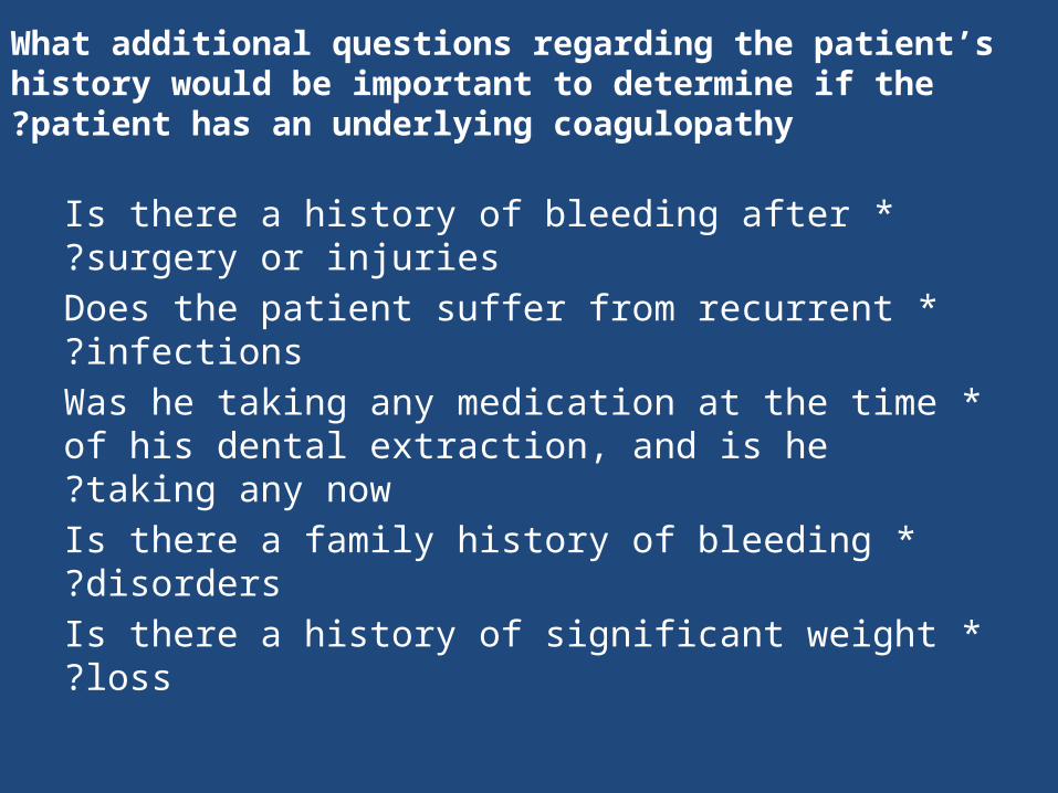
What additional questions regarding the patient’s history would be important to determine if the patient has an underlying coagulopathy?
*Is there a history of bleeding after surgery or injuries?
*Does the patient suffer from recurrent infections?
*Was he taking any medication at the time of his dental extraction, and is he taking any now?
*Is there a family history of bleeding disorders? *Is there a history of significant weight loss?

*Evaluation of prior bleeding has been shown to be predictive of a bleeding disorder. Evaluation of a patient’s history should focus on spontaneous bleeding, bleeding after trauma and surgical
procedures, and in women, the presence of menorrhagia . *Someone who does not have bleeding complications after trauma
or surgical challenges is less likely to have a significant coagulation disorder .
*Disorders of platelet function or number tend to produce immediate bleeding after surgical procedures or trauma, in contrast to disorders of the coagulation system, which may present as delayed bleeding after initial hemostasis was obtained.

*Family history, when positive, is also strongly associated with inherited bleeding disorders, although genetic coagulation disorders may also arise from spontaneous gene mutations.
*Since there are many medications that increase the risk of bleeding, one should take a history of all prescribed and over-the-counter medications.
*Family history, when positive, is also strongly associated with inherited bleeding disorders, although genetic coagulation disorders may also arise from spontaneous gene mutations.
Since there are many medications that increase the risk of bleeding, one should take a history of all prescribed and over-the-counter medications.

What aspects of the physical exam do you think would be most relevant in this case?
*Skin *Eyes
*Lymph nodes *Abdomen
*Joints
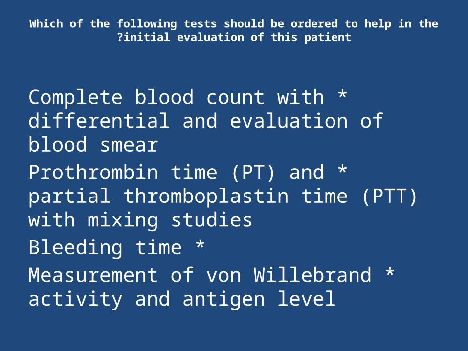
Which of the following tests should be ordered to help in the initial evaluation of this patient?
*Complete blood count with differential and evaluation of blood smear
*Prothrombin time (PT) and partial thromboplastin time (PTT) with mixing studies
*Bleeding time *Measurement of von Willebrand activity and
antigen level

Laboratory Test ResultsTest Patient Normal Range
White blood cell count 6,000/μL 4,500 – 15,000/μL
Hemoglobin 14 g/dL 11.7 – 15.5 g/dL
Platelet count 250,000/μL 150,000 – 400,000/μL
Peripheral smear evaluationNormal-appearing platelet morphology, no abnormal white blood cells, normal shape and distribution of red blood cells
Prothrombin Time (PT) 13.5 sec 12.3 – 14.5 sec
Partial Thromboplastin Time (PTT)
50 sec 24 – 34 sec
PTT 1:1 mixing study 30 sec 24 – 34 sec
Ristocetin co-factor activity 85% 40% – 200%
von Willebrand antigen 75% 50% – 180%
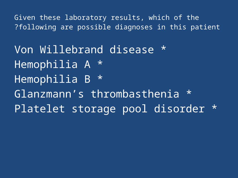
Given these laboratory results, which of the following are possible diagnoses in this patient?
*Von Willebrand disease *Hemophilia A *Hemophilia B
*Glanzmann’s thrombasthenia *Platelet storage pool disorder
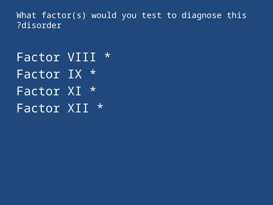
What factor(s) would you test to diagnose this disorder?
*Factor VIII *Factor IX *Factor XI *Factor XII
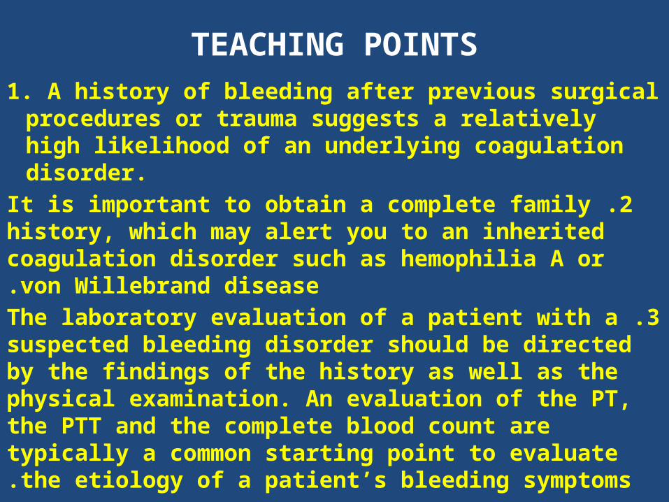
TEACHING POINTS1. A history of bleeding after previous surgical procedures
or trauma suggests a relatively high likelihood of an underlying coagulation disorder.
2 .It is important to obtain a complete family history, which may alert you to an inherited coagulation disorder such as hemophilia A or von Willebrand disease.
3 .The laboratory evaluation of a patient with a suspected bleeding disorder should be directed by the findings of the history as well as the physical examination. An evaluation of the PT, the PTT and the complete blood count are typically a common starting point to evaluate the etiology of a patient’s bleeding symptoms.

TEACHING POINTS
4 .Patients with a diagnosis of hemophilia A will typically have findings and symptoms that are directly related to the level of factor VIII activity. Patients with less than 5% of factor VIII activity are more likely to have spontaneous bleeds into joints (hemarthroses), while less severely affected patients with higher (but still abnormally low) factor VIII levels may have bleeding symptoms only after trauma or surgical procedures.
5 .There are other causes of prolonged PTT besides hemophilia A and B. These include hemophilia C (factor XI deficiency) and factor XII deficiency. Factors XII and XI are typically evaluated only if factors VIII and IX are normal.
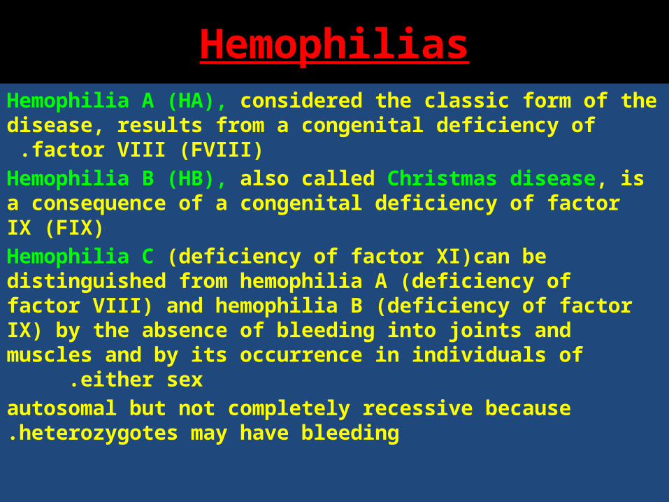
HemophiliasHemophilia A (HA), considered the classic form of the disease,
results from a congenital deficiency of factor VIII (FVIII) .Hemophilia B (HB), also called Christmas disease, is a consequence of a congenital deficiency of factor IX (FIX)Hemophilia C (deficiency of factor XI)can be distinguished from hemophilia A (deficiency of factor VIII) and hemophilia B (deficiency of factor IX) by the absence of bleeding into joints and muscles and by its occurrence in
individuals of either sex .autosomal but not completely recessive because heterozygotes may have bleeding.
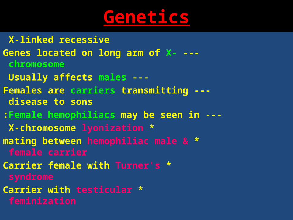
GeneticsX-linked recessive
--- Genes located on long arm of X-chromosome --- Usually affects males
--- Females are carriers transmitting disease to sons
--- Female hemophiliacs may be seen in : * X-chromosome lyonization
* mating between hemophiliac male & female carrier
* Carrier female with Turner's syndrome * Carrier with testicular feminization
S&S
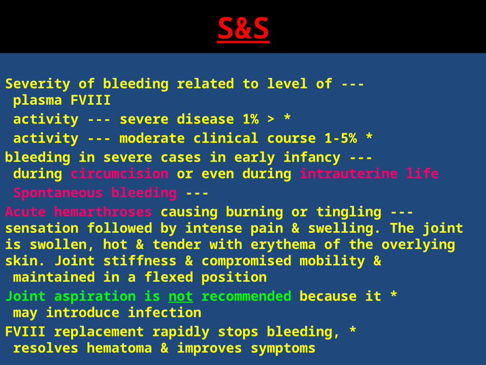
---Severity of bleeding related to level of plasma FVIII < * 1% activity --- severe disease
* 1-5% activity --- moderate clinical course --- bleeding in severe cases in early infancy during circumcision or
even during intrauterine life --- Spontaneous bleeding
--- Acute hemarthroses causing burning or tingling sensation followed by intense pain & swelling. The joint is swollen, hot & tender with erythema of the overlying skin. Joint stiffness & compromised mobility & maintained in a flexed position
* Joint aspiration is not recommended because it may introduce infection
* FVIII replacement rapidly stops bleeding, resolves hematoma & improves symptoms
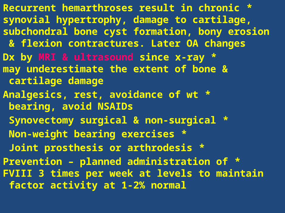
*Recurrent hemarthroses result in chronic synovial hypertrophy, damage to cartilage, subchondral bone cyst formation, bony erosion & flexion contractures. Later OA changes
* Dx by MRI & ultrasound since x-ray may underestimate the extent of bone & cartilage damage
* Analgesics, rest, avoidance of wt bearing, avoid NSAIDs
* Synovectomy surgical & non-surgical * Non-weight bearing exercises * Joint prosthesis or arthrodesis
* Prevention – planned administration of FVIII 3 times per week at levels to maintain factor activity at 1-2% normal
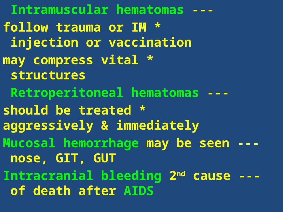
--- Intramuscular hematomas * follow trauma or IM injection or vaccination
* may compress vital structures --- Retroperitoneal hematomas
* should be treated aggressively & immediately --- Mucosal hemorrhage may be seen nose, GIT,
GUT --- Intracranial bleeding 2nd cause of death after
AIDS

Ask about the patient's family history and bleeding symptoms .
Male patients with severe hemophilia present at circumcision .
Easy bruising may occur at the start of ambulation or primary dentition .
The patient may have a history of hemarthroses and prolonged bleeding with surgical procedures, trauma, dental extraction, and he or she may have spontaneous
bleeding in soft tissues .A traumatic challenge relatively late in life may have to occur before mild or moderate hemophilia is diagnosed. Factors that elevate FVIII levels (e.g. stress, exercise) may mask mild hemophilia. Physiologically low levels of all vitamin K–dependent procoagulant factors may complicate the early diagnosis of hemophilia B.
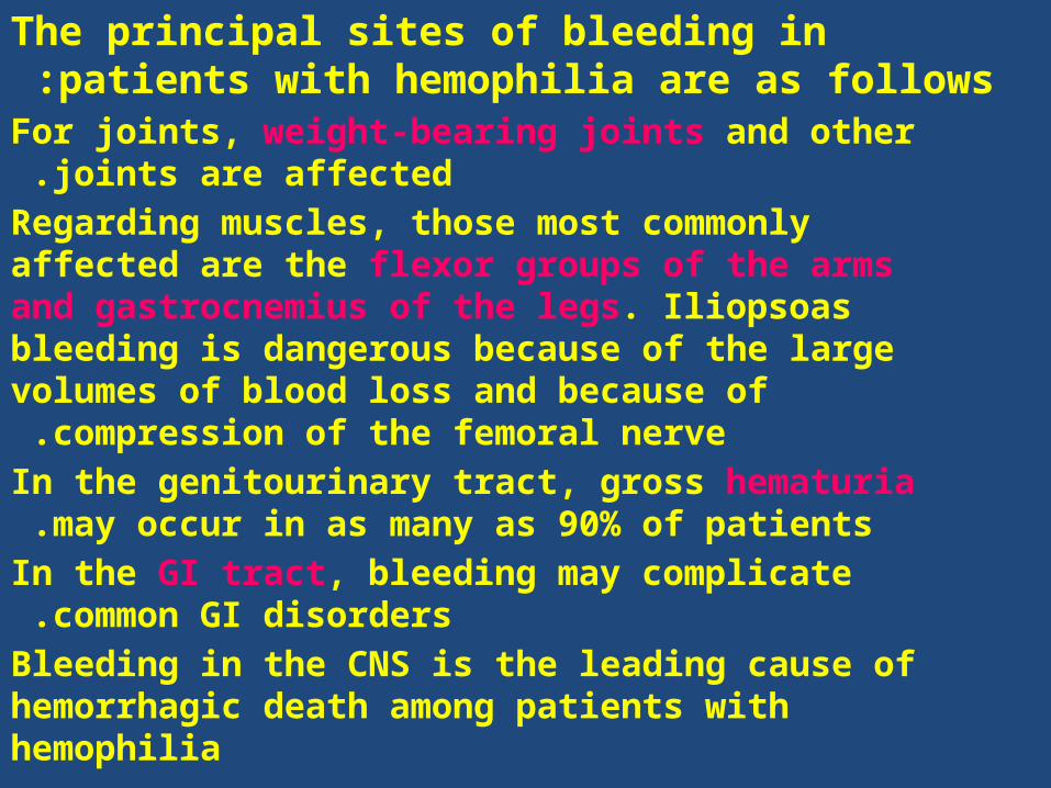
The principal sites of bleeding in patients with hemophilia are as follows :
For joints, weight-bearing joints and other joints are affected .
Regarding muscles, those most commonly affected are the flexor groups of the arms and gastrocnemius of the legs. Iliopsoas bleeding is dangerous because of the large volumes of blood loss and because of
compression of the femoral nerve .In the genitourinary tract, gross hematuria may occur in
as many as 90% of patients .In the GI tract, bleeding may complicate common GI
disorders .Bleeding in the CNS is the leading cause of hemorrhagic death among patients with hemophilia
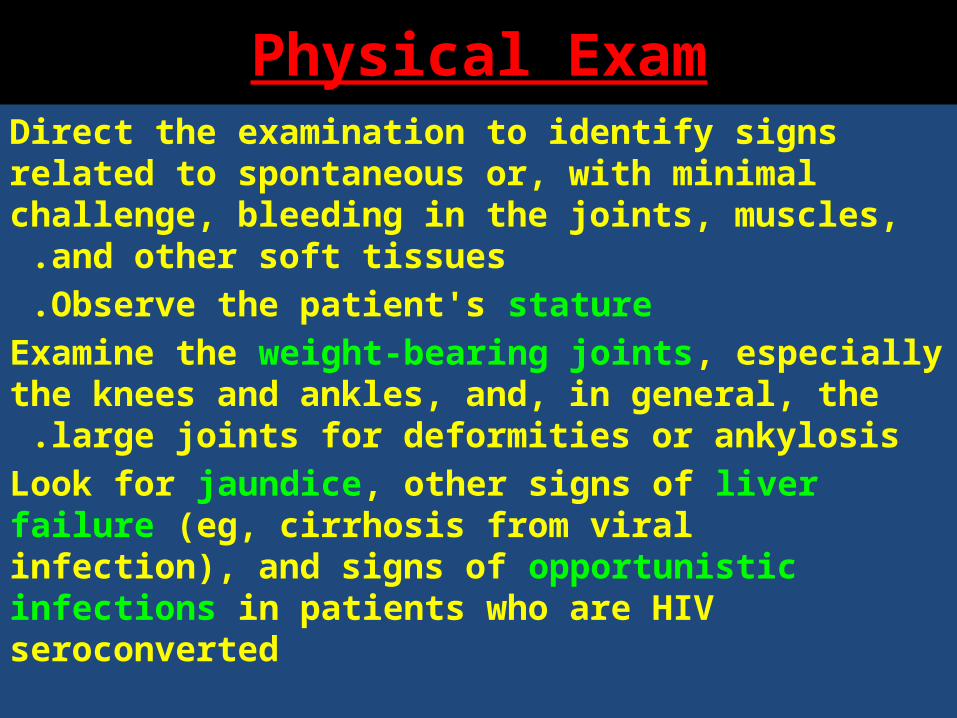
Physical ExamDirect the examination to identify signs related to spontaneous or, with minimal challenge, bleeding
in the joints, muscles, and other soft tissues .Observe the patient's stature .
Examine the weight-bearing joints, especially the knees and ankles, and, in general, the large joints
for deformities or ankylosis .Look for jaundice, other signs of liver failure (eg, cirrhosis from viral infection), and signs of opportunistic infections in patients who are HIV seroconverted
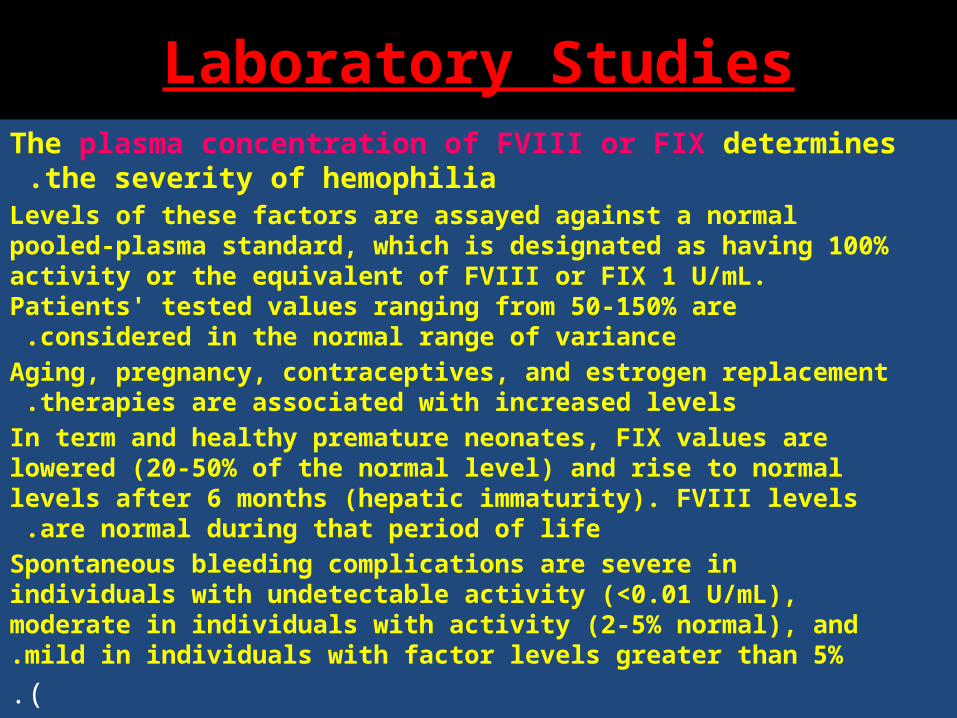
Laboratory StudiesThe plasma concentration of FVIII or FIX determines the severity of
hemophilia .Levels of these factors are assayed against a normal pooled-plasma standard, which is designated as having 100% activity or the equivalent of FVIII or FIX 1 U/mL. Patients' tested values ranging from
50-150% are considered in the normal range of variance .Aging, pregnancy, contraceptives, and estrogen replacement therapies
are associated with increased levels .In term and healthy premature neonates, FIX values are lowered (20-50% of the normal level) and rise to normal levels after 6 months (hepatic
immaturity). FVIII levels are normal during that period of life .Spontaneous bleeding complications are severe in individuals with undetectable activity (<0.01 U/mL), moderate in individuals with activity (2-5% normal), and mild in individuals with factor levels greater than 5%..)
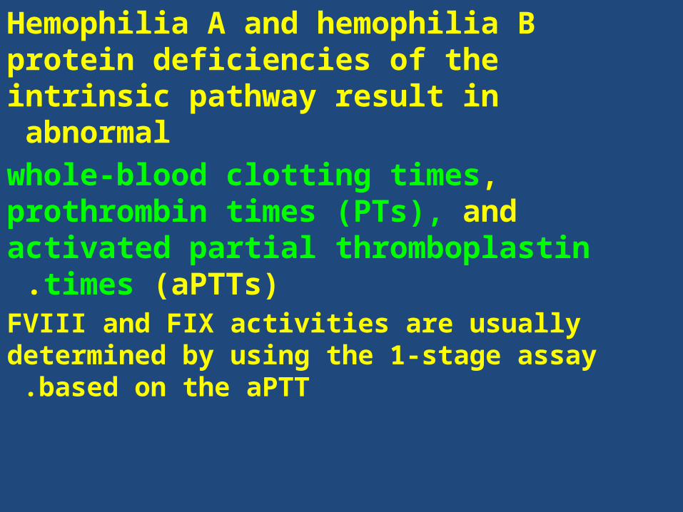
Hemophilia A and hemophilia B protein deficiencies of the intrinsic pathway result in abnormal
whole-blood clotting times, prothrombin times (PTs), and activated partial thromboplastin times
(aPTTs) .FVIII and FIX activities are usually determined by using
the 1-stage assay based on the aPTT .
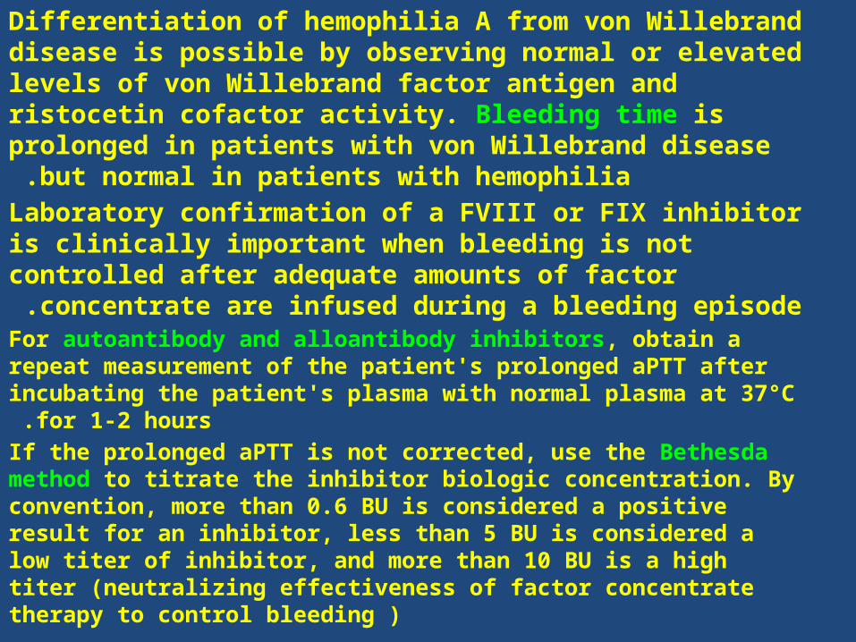
Differentiation of hemophilia A from von Willebrand disease is possible by observing normal or elevated levels of von Willebrand factor antigen and ristocetin cofactor activity. Bleeding time is prolonged in patients with von Willebrand
disease but normal in patients with hemophilia .Laboratory confirmation of a FVIII or FIX inhibitor is clinically important when bleeding is not controlled after adequate amounts of factor concentrate are infused during a bleeding
episode .For autoantibody and alloantibody inhibitors, obtain a repeat measurement of the patient's prolonged aPTT after incubating the
patient's plasma with normal plasma at 37°C for 1-2 hours .If the prolonged aPTT is not corrected, use the Bethesda method to titrate the inhibitor biologic concentration. By convention, more than 0.6 BU is considered a positive result for an inhibitor, less than 5 BU is considered a low titer of inhibitor, and more than 10 BU is a high titer (neutralizing effectiveness of factor concentrate therapy to control bleeding )
℞
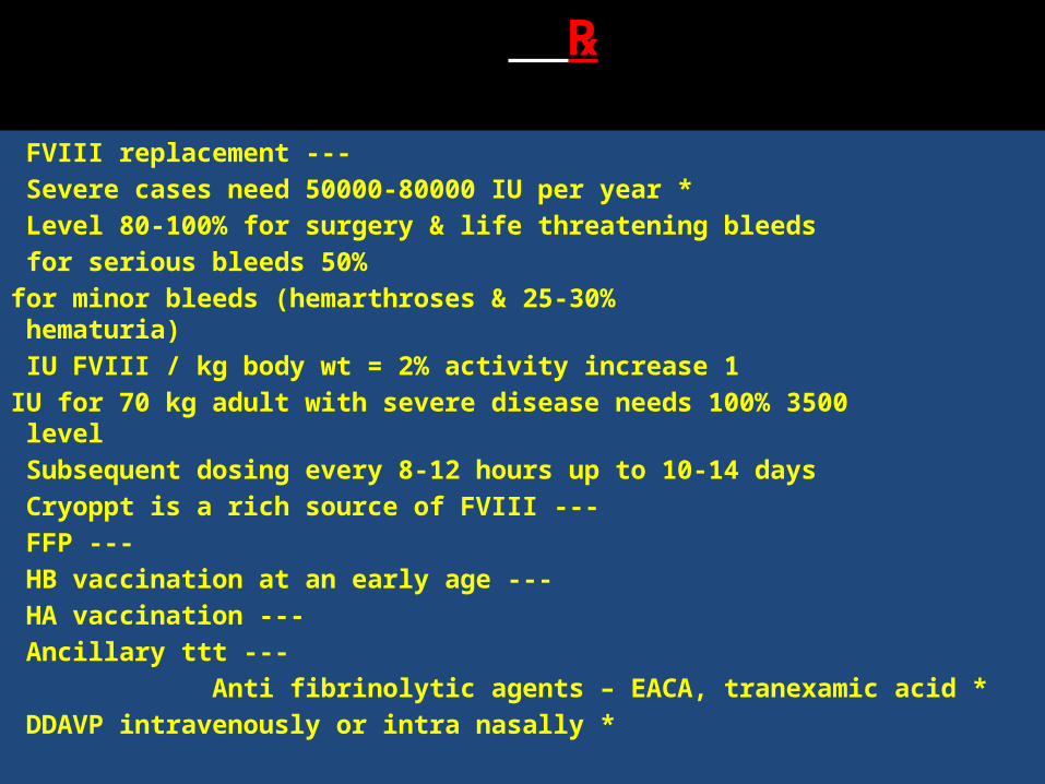
---FVIII replacement * Severe cases need 50000-80000 IU per year
Level 80-100% for surgery & life threatening bleeds 50% for serious bleeds
25-30% for minor bleeds (hemarthroses & hematuria) 1 IU FVIII / kg body wt = 2% activity increase
3500 IU for 70 kg adult with severe disease needs 100% level Subsequent dosing every 8-12 hours up to 10-14 days
--- Cryoppt is a rich source of FVIII --- FFP
---HB vaccination at an early age ---HA vaccination
---Ancillary ttt * Anti fibrinolytic agents – EACA, tranexamic acid
* DDAVP intravenously or intra nasally
PreventionProphylactic replacement of FVIII or FIX is used to maintain a measurable level at all times, with the goal of avoiding hemarthrosis and breaking the vicious cycle of repetitive bleeding and inflammation that results in destructive arthritis.This goal is achieved by administering factor 2-3 times a week .
The National Hemophilia Foundation has recommended the administration of primary prophylaxis, beginning at the age of 1-2 years.
Carrier testing may prevent births of individuals with major hemophilia. This testing can be offered to women interested in
childbearing who have a family history of hemophilia .Carrier testing is valuable for women who are related to obligate carrier
females or males with hemophilia .Prenatal diagnosis is important even if termination of the pregnancy is not desired because a cesarean delivery may be planned or
replacement therapy can be scheduled for the perinatal period .
Disseminated intravascular coagulation
(DIC)
(defibrination syndrome )
It is a consumptive coagulopathy that is caused by a wide variety of serious
disorders.
Etiology
*Infections --- G-ve bacterial sepsis --- Other bacteria, fungi, Rocky Mountain
spotted fever, viruses, malaria
Obstetric complications
---Amniotic fluid embolism ---Retained dead fetus ---Abruptio placentae
---Toxemia of pregnancy ---Septic abortion
Malignancies
----Pancreatic Ca ---Adenocarcinomas
---Acute promyelocytic leukemia (M3) ---Other neoplasms
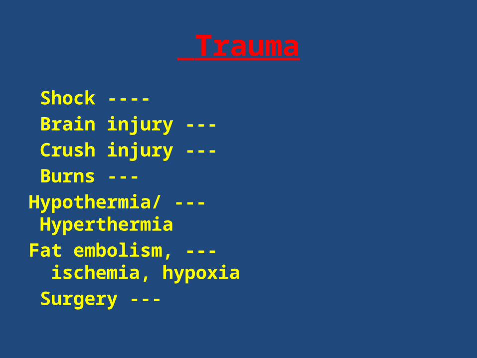
Trauma
---- Shock --- Brain injury --- Crush injury
--- Burns --- Hypothermia/ Hyperthermia
--- Fat embolism, ischemia, hypoxia --- Surgery
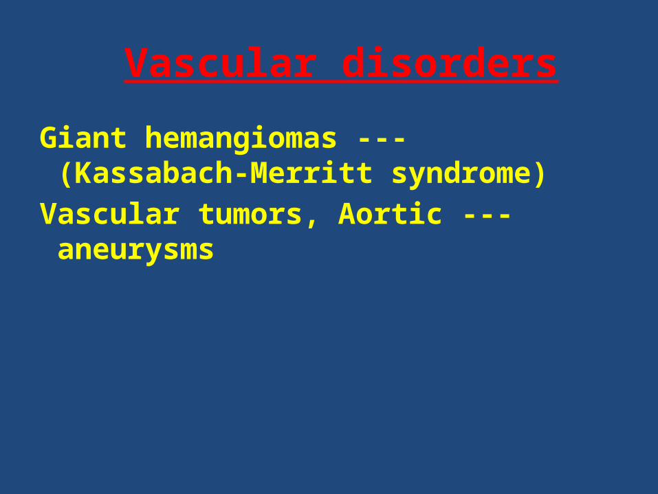
Vascular disorders
---Giant hemangiomas (Kassabach-Merritt syndrome)
---Vascular tumors, Aortic aneurysms
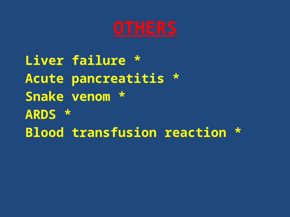
OTHERS
*Liver failure *Acute pancreatitis
*Snake venom *ARDS
*Blood transfusion reaction
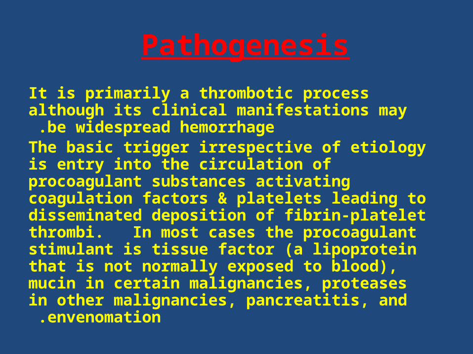
Pathogenesis It is primarily a thrombotic process although its clinical
manifestations may be widespread hemorrhage .The basic trigger irrespective of etiology is entry into the circulation of procoagulant substances activating coagulation factors & platelets leading to disseminated deposition of fibrin-platelet thrombi. In most cases the procoagulant stimulant is tissue factor (a lipoprotein that is not normally exposed to blood), mucin in certain malignancies, proteases
in other malignancies, pancreatitis, and envenomation.
S&S
Asymptomatic --- low grade DIC showing only Lab abnormalities
Thrombotic complications
---Trousseau's syndrome --- Gangrene of digits & extremities
--- Hemorrhagic necrosis of skin --- Purpura fulminans
Bleeding
---most common manifestation in acute cases ---Generalized & widespread
---Characteristically from the cannula site --- oozes from mucosal surfaces & orifices
Clinical manifestations of the causative disease
Dx
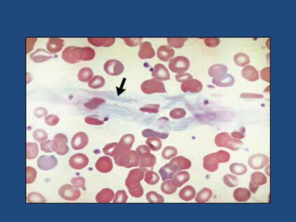
PT, aPTT & TT are prolonged Thrombocytopenia ,
Plasma Fibrinogen ↓FDPs ↑ (measured by latex agglutination or D-dimer assay)Blood film --- Schistocytes, fragmented RBCs
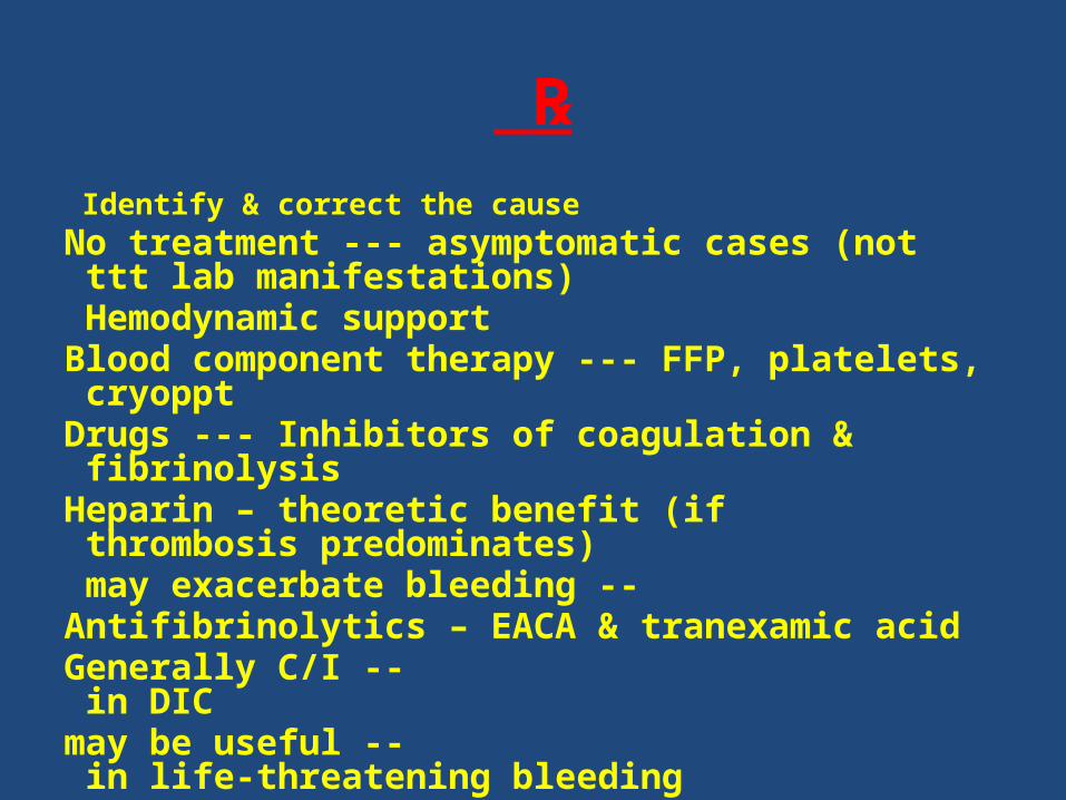
℞ Identify & correct the cause No treatment --- asymptomatic cases (not ttt lab manifestations) Hemodynamic support Blood component therapy --- FFP, platelets, cryoppt Drugs --- Inhibitors of coagulation & fibrinolysis
Heparin – theoretic benefit (if thrombosis predominates) -- may exacerbate bleeding
Antifibrinolytics – EACA & tranexamic acid -- Generally C/I in DIC
-- may be useful in life-threatening bleeding

Platelet Disorders
Thrombocytopenia
It is a condition in which platelet count is below normal level.
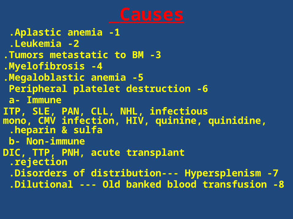
Causes 1 -Aplastic anemia .
2 -Leukemia .3 -Tumors metastatic to BM.
4 -Myelofibrosis.5 -Megaloblastic anemia.
6 -Peripheral platelet destruction a- Immune
ITP, SLE, PAN, CLL, NHL, infectious mono, CMV infection, HIV, quinine, quinidine, heparin & sulfa .
b- Non-immune DIC, TTP, PNH, acute transplant rejection .
7 -Disorders of distribution--- Hypersplenism .8 -Dilutional --- Old banked blood transfusion .
Idiopathic Thrombocytopenic Purpura: (ITP)
It is a bleeding disorder caused by autoimmune Abs destroying patient's own platelets by phagocytosis in the spleen by Mφ (to a lesser degree in the
liver).

Epidemiology *Children are affected by an acute type of illness
following URT infection (usually viral) . = *♀ ♂incidence .
*Adults are affected by a more gradual onset of disease with chronic course & without preceding
illness .*Age < 40 years
= : *♂ ♀3-4:1
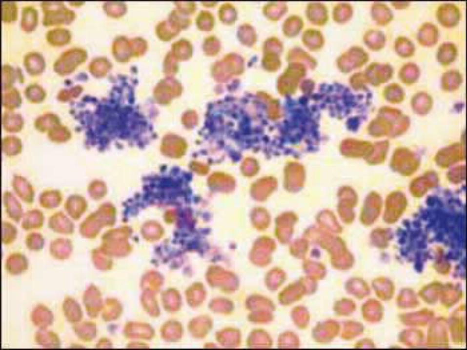
S & S Skin shows petechiae (pinpoint red hemorrhagic
spots) & ecchymoses. Mucosal bleeding --- bleeding gum, epistaxis, menorrhagia, metrorrhagia, GI bleeding & hematuria
Intracranial hemorrhage is seen in 1%. Risk of death 5% No splenomegaly.
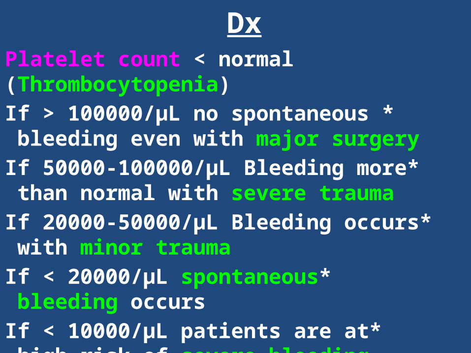
Dx Platelet count < normal (Thrombocytopenia)
* If > 100000/µL no spontaneous bleeding even with major surgery
* If 50000-100000/µL Bleeding more than normal with severe trauma
* If 20000-50000/µL Bleeding occurs with minor trauma
* If < 20000/µL spontaneous bleeding occurs * If < 10000/µL patients are at high risk of severe
bleeding
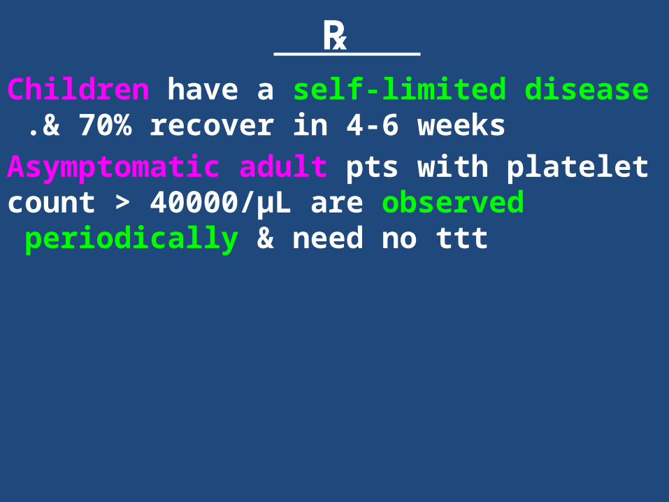
℞ Children have a self-limited disease & 70% recover in
4-6 weeks .Asymptomatic adult pts with platelet count > 40000/µL are observed periodically & need no ttt
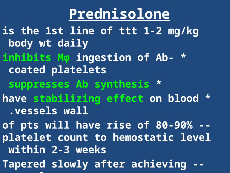
Prednisolone is the 1st line of ttt 1-2 mg/kg body wt daily
* inhibits Mφ ingestion of Ab-coated platelets * suppresses Ab synthesis
* have stabilizing effect on blood vessels wall . -- 80-90% of pts will have rise of platelet count to
hemostatic level within 2-3 weeks -- Tapered slowly after achieving normal count
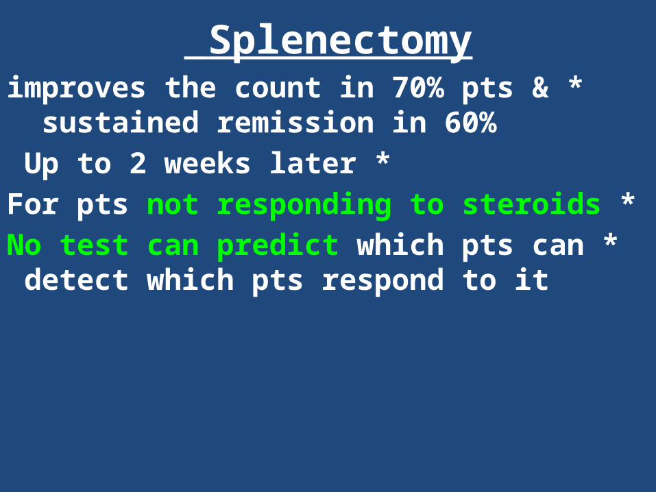
Splenectomy *improves the count in 70% pts & sustained
remission in 60% *Up to 2 weeks later
*For pts not responding to steroids *No test can predict which pts can detect which pts
respond to it
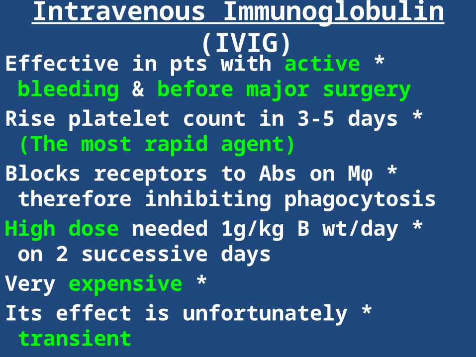
Intravenous Immunoglobulin (IVIG) *Effective in pts with active bleeding & before
major surgery * Rise platelet count in 3-5 days (The most rapid
agent) * Blocks receptors to Abs on Mφ therefore
inhibiting phagocytosis * High dose needed 1g/kg B wt/day on 2 successive
days * Very expensive
* Its effect is unfortunately transient
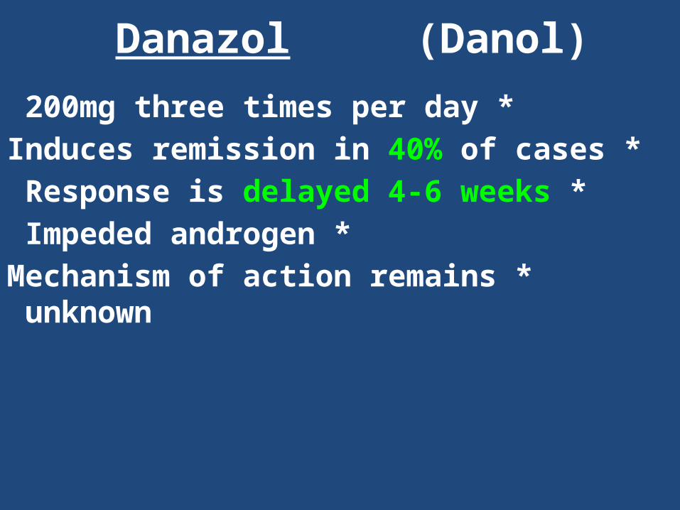
Danazol (Danol) *200mg three times per day
* Induces remission in 40% of cases * Response is delayed 4-6 weeks
* Impeded androgen * Mechanism of action remains unknown
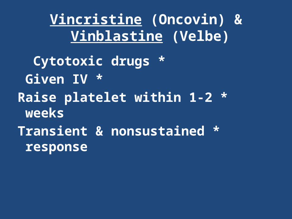
Vincristine (Oncovin) & Vinblastine (Velbe)
*Cytotoxic drugs *Given IV
*Raise platelet within 1-2 weeks *Transient & nonsustained response
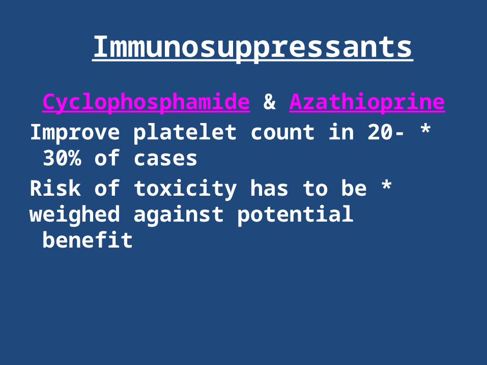
Immunosuppressants
Cyclophosphamide & Azathioprine * Improve platelet count in 20-30% of cases
* Risk of toxicity has to be weighed against potential benefit
ITP in Pregnancy
*Complicated by additional risk to the fetus (thrombocytopenia due to maternal Abs) , newborn intraventricular hemorrhage & GI bleeding
*C/S delivery is recommended to decrease risk of intracranial bleeding in the newborn.
Functional platelet disorders Hereditary
•Glanzmann's thrombasthenia •Bernard-Soulier syndrome
•Storage pool disease

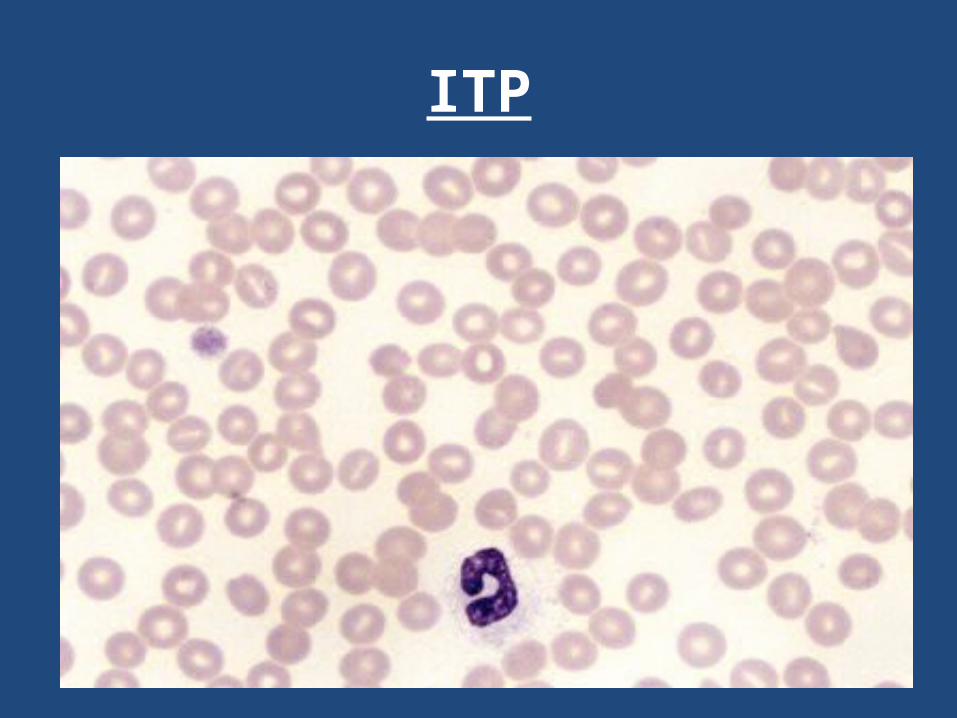
ITP
Thrombotic Dis
Hypercoagulable States are more likely to develop clots,venous and arterial thrombosis than healthy individuals. There is often a history of recurrent thromboembolism, thrombosis at a young age, pulmonary embolism and a family history of thrombosis. Venous thrombosis and are associated with significant morbidity and mortality.
Causes
•Hereditary – Antithrombin III def - Protein C def - Protein S def
- FV LeidenAcquired
Acquired
Advanced ageImmobilizationInflammationPregnancyOral contraception useObesityDiabetesHormonal replacement therapyCancer (especially adenocarcinoma)Lupus anticoagulantSickle cell anemia and other hemolytic anemias
Pathophysiology
The Virchow triad of underlying factors in venous thrombosis include hypercoagulability, venous stasis, and vascular damage. The risk factors mentioned above tip the balance of Virchow’s triad. Procoagulants are released in patients with cancer. Immobilization, obesity, and advanced age lead to reduced blood flow and venous stasis.
pregnancy
Thrombosis during pregnancy can be due to increased procoagulant factors, impaired fibrinolysis, venous stasis, and endothelial cell injury.[2] The risk of thrombosis is increased in
patients on hormonal replacement therapy .
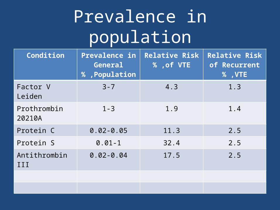
Prevalence in populationCondition Prevalence in
General Population% ,
Relative Risk of VTE% ,
Relative Risk of Recurrent VTE% ,
Factor V Leiden 3-7 4.3 1.3Prothrombin 20210A
1-3 1.9 1.4
Protein C 0.02-0.05 11.3 2.5Protein S 0.01-1 32.4 2.5Antithrombin III 0.02-0.04 17.5 2.5
Mortality/Morbidity
Morbidity and mortality in patients with hypercoagulable states and thrombophilia are primarily due to venous thrombosis and pulmonary embolism. Pulmonary embolism is associated with a 1-3% mortality rate. The incidence of factor V Leiden and prothrombin 20210A is significantly greater than that of protein C, protein S, and antithrombin III (ATIII) deficiencies. However, the risk of venous thrombosis in protein C, protein S, and antithrombin III (ATIII) deficiencies is greater than in factor V Leiden and
prothrombin 20210A, as shown in Table above .The risk for thrombosis can be markedly increased in patients with 2 or more risk factors for thrombosis. Any multiplicity of risk factors, albeit hereditary thrombophilias or acquired risks, increase the risk
for thrombosis .
Hereditary thrombophilia
These disorders should be suspected in patients has a history of recurrent venous thromboembolism, venous thrombosis in a person younger than 40 years, a familial history of venous thromboembolism, and thrombosis in unusual sites (eg, mesenteric vein, renal vein, hepatic and cerebral thrombosis).Purpura fulminans in infancy could suggest protein C deficiency.
Thrombophilic disorders are usually associated with venous thrombosis. However, protein S, protein C, ATIII deficiencies, and lupus anticoagulants have been associated with arterial
thrombosis .Patients with protein C and S deficiencies can develop warfarin-induced skin necrosis when placed on warfarin since protein C and
S are vitamin K–dependent factors and, hence are suppressed .
Lupus anticoagulants These antibodies occur in about 20% of patients with systemic lupus erythematosus (SLE), but they are also associated with other autoimmune diseases. Lupus anticoagulants may occur in patients taking phenothiazines, phenytoin, phenytoin, hydralazine, quinine, amoxicillin, and oral contraceptives.
Clinical criteria for indicating the presence of lupus anticoagulants (Sapporo criteria for the antiphospholipid syndrome) are as follows :
One or more arterial, venous, or small vessel thrombosis, affecting any organ or tissuePregnancy morbidity: The risk for maternal and fetal morbidity increases after the 10th week of pregnancy. Fetal mortality in pregnancy can
include spontaneous abortions, prematurity, and stillbirths .Three or more unexplained consecutive spontaneous abortions after the 10th week of gestation
Lab
PT ; aPTT; INRD-dimer assayAnti-cardiolipin Ab
treatment
HeparinLMWHWarfarinFondaparinux
Case2A 20-year-old college student returned to the U.S. following a summer break in which she traveled and
hiked extensively in New Zealand .Two days after her flight she awoke with swelling and pain in her left calf and thigh and noticed that the
skin of the leg appeared dusky blue in color .Because of these rather alarming symptoms, and the fact that her mother had a blood clot when she was young, she came immediately to the emergency room where noninvasive venous studies showed an extensive deep venous thrombosis involving the popliteal, femoral and iliac veins on the left.
Should this patient be evaluated for an underlying thrombophilia?
*Yes *No
Common Risk Factors for Thrombosis
ObesityInactivityPregnancyEstrogens/Birth Control PillsPost-surgeryMalignancy

Inherited Procoagulant StatesDefect Incidence in Population Percent of Patients with
Procoagulant States
1 .Factor V Leiden 5-10% 20-60%
2 .Prothrombin Gene Mutation 2-4% 6-8%
3 .Protein C Deficiency 1:200 <5%
4 .Protein S Deficiency -- <5%
5 .Antithrombin Deficiency 1:2-5,000 <1%
6 .Dysfibrinogenemia Not known ~1-2%
7 .Elevated Lipoprotein(a) Not known* --
Acquired Prothrombotic Medical States
Antiphospholipid antibody syndromeDisseminated intravascular coagulationHeparin-induced thrombocytopenia and thrombosis syndromeInflammatory bowel diseaseMyeloproliferative disordersNephrotic syndromeParoxysmal nocturnal hemoglobinuria (PNH)
What symptoms or history might be helpful in making the diagnosis ?
Do you have any illnesses?Have you ever had a thrombosis in the past?Has anyone else in your family had a thrombosis?
Have you ever experienced leg cramps while hiking?What medications are you on ?Was this your first trip overseas ?Have you ever been pregnant ?
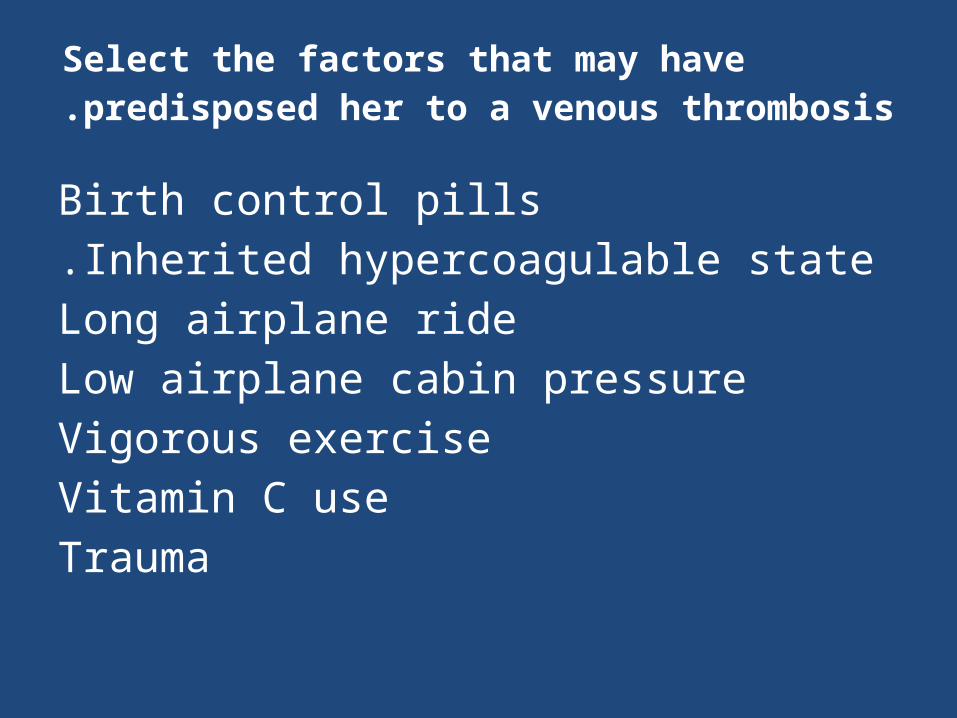
Select the factors that may have predisposed her to a venous thrombosis.
Birth control pillsInherited hypercoagulable state.
Long airplane rideLow airplane cabin pressureVigorous exerciseVitamin C useTrauma
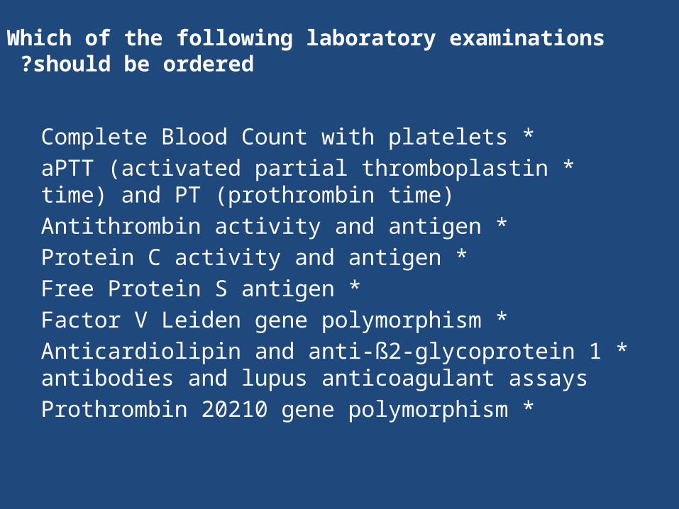
Which of the following laboratory examinations should be ordered?
*Complete Blood Count with platelets *aPTT (activated partial thromboplastin time) and PT
(prothrombin time) *Antithrombin activity and antigen
*Protein C activity and antigen *Free Protein S antigen
*Factor V Leiden gene polymorphism *Anticardiolipin and anti-ß2-glycoprotein 1 antibodies
and lupus anticoagulant assays *Prothrombin 20210 gene polymorphism
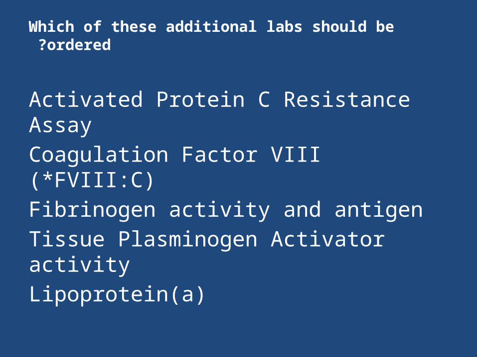
Which of these additional labs should be ordered ?
Activated Protein C Resistance AssayCoagulation Factor VIII (*FVIII:C)Fibrinogen activity and antigenTissue Plasminogen Activator activityLipoprotein(a)

Which one of the following laboratory tests reveals an abnormality?
Complete Blood CountBeta-HCGAntithrombin III activityProtein C activityProtein S activityProthrombin Gene mutationHomocysteineAnticardiolipin AntibodyLupus Inhibitor Evaluation
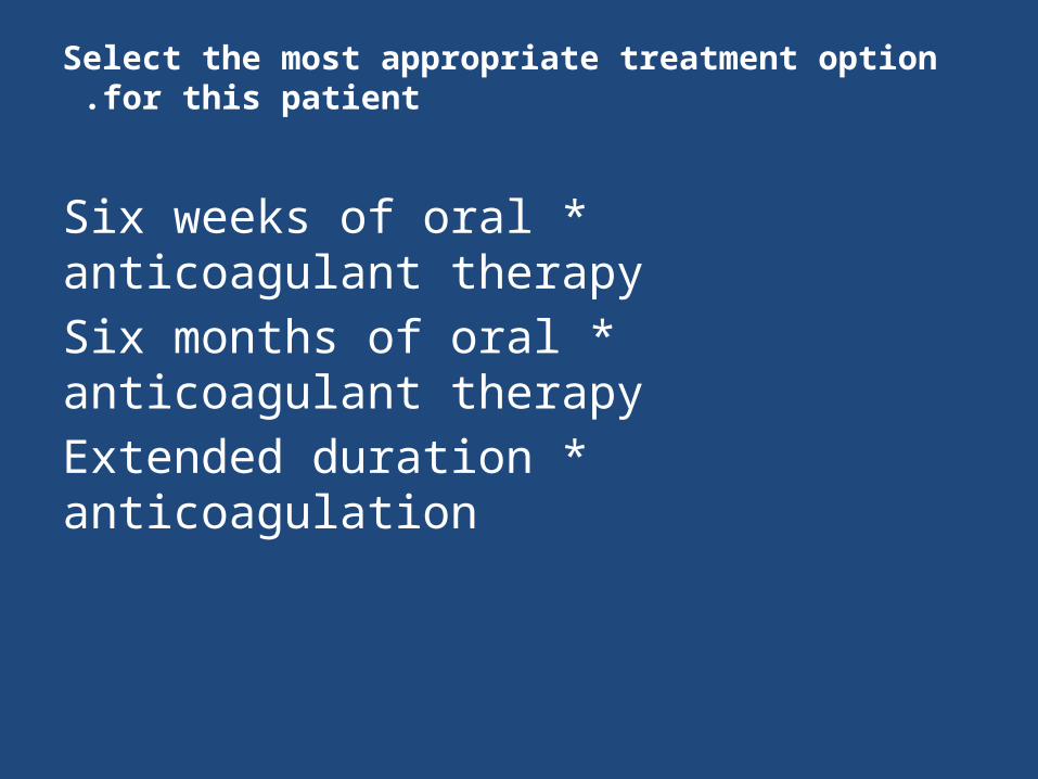
Select the most appropriate treatment option for this patient .
*Six weeks of oral anticoagulant therapy *Six months of oral anticoagulant therapy
*Extended duration anticoagulation
Should her family be screened for protein C deficiency ?
*No *Yes
The patient's sister was found to be heterozygous for protein C deficiency. She has asked your advice about
the risks of pregnancy. What do you tell her ?
*The risks are minimal. *She is at increased risk for thrombosis during
pregnancy and otherwise.
The patient's sister (who has asymptomatic protein C deficiency) has also asked your advice about taking oral contraceptives. Select the most appropriate advice.
*Oral contraceptives are an acceptable choice given the low risk of thrombosis.
*She should consider alternate forms of contraception.
Four clues to a hypercoagulable state
*Thrombosis at an early age *Recurrent thromboses
*Thrombosis at an unusual site *Family history of thrombosis
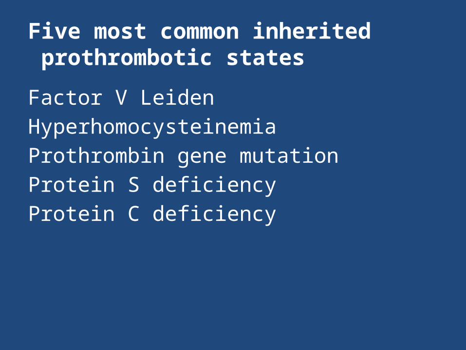
Five most common inherited prothrombotic states
Factor V LeidenHyperhomocysteinemiaProthrombin gene mutationProtein S deficiencyProtein C deficiency
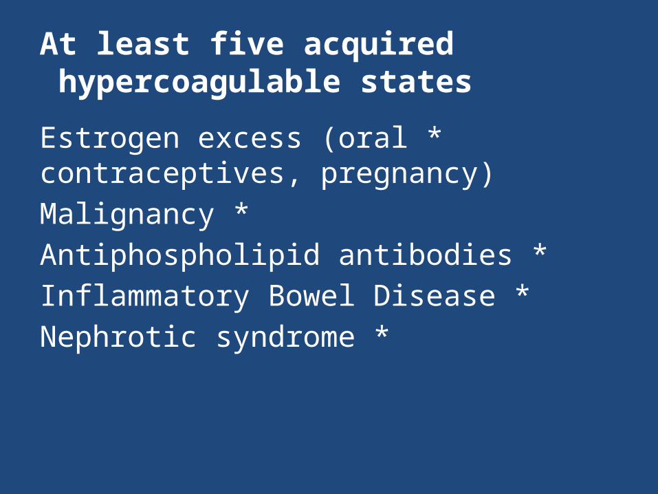
At least five acquired hypercoagulable states
*Estrogen excess (oral contraceptives, pregnancy)
*Malignancy *Antiphospholipid antibodies *Inflammatory Bowel Disease
*Nephrotic syndrome


![The Hematologist - Final PDF of September October 2010[1]](https://img.pdfslide.us/doc/110x75/577d35911a28ab3a6b90cc96/the-hematologist-final-pdf-of-september-october-20101.jpg)