Embed Size (px)
Citation preview

Surface Science 603 (2009) 3099–3103
Contents lists available at ScienceDirect
Surface Science
journal homepage: www.elsevier .com/ locate/susc
CO adsorption and dissociation on iron oxide supported Pt particles
Y.-N. Sun, Z.-H. Qin, M. Lewandowski, S. Shaikhutdinov *, H.-J. FreundAbteilung Chemische Physik, Fritz-Haber-Institut der Max-Planck-Gesellschaft, Faradayweg 4-6, Berlin 14195, Germany
a r t i c l e i n f o
Article history:Received 8 June 2009Accepted for publication 24 August 2009Available online 28 August 2009
Keywords:PlatinumIron oxidesCO adsorptionCO dissociationStrong metal support interaction
0039-6028/$ - see front matter � 2009 Elsevier B.V. Adoi:10.1016/j.susc.2009.08.022
* Corresponding author. Tel.: +49 030 8413 4114; fE-mail address: [email protected] (S. Shaik
a b s t r a c t
We studied CO adsorption on Pt particles deposited on well-ordered Fe3O4(1 1 1) thin films grown onPt(1 1 1) by temperature programmed desorption (TPD). A highly stepped Pt(1 1 1) surface producedby ion sputtering and annealing at 600 K was studied for comparison. Structural characterization wasperformed by scanning tunneling microscopy and Auger electron spectroscopy. The TPD spectra revealedthat in addition to the desorption peaks at �400 and 480 K, assigned to CO adsorbed on Pt(1 1 1) facetsand low-coordination sites respectively, the Pt nanoparticles annealed at 600 K exhibit a desorption stateat �270 K. This state is assigned to initial stages of strong metal support interaction resulting in partialFe–Pt intermixing. On both Pt/Fe3O4(1 1 1) and stepped Pt(1 1 1) surfaces CO is found to dissociate at500 K. The results suggest that CO dissociation and carbon accumulation occur on the low-coordinatedPt sites.
� 2009 Elsevier B.V. All rights reserved.
1. Introduction
Adsorption of carbon monoxide on platinum surfaces is one ofthe most explored reactions in surface science (e.g., see [1]).Numerous studies performed on clean Pt single crystal surfacesshowed that CO preferentially occupies atop sites on Pt surfacesand also bridge sites at increasing coverage. The binding energyof CO depends on the Pt surface structure. From surfaces withthe low Miller indices CO desorbs in a relatively broad signal, be-tween 300 and 550 K, in temperature programmed desorption(TPD) spectra [2–6]. Stepped and kinked Pt surfaces typically exhi-bit two distinct desorption states, i.e. at �400 and 500 K [6–8],which have been assigned to the (1 1 1)-like terraces and low-coor-dination sites, respectively. A similar assignment was applied forsupported Pt particles [9–12]. For example, Pt particles, depositedat low coverage both on amorphous alumina and a-Al2O3(0 0 0 1)supports at 300 K, showed a single desorption peak at �510 K,while the larger aggregates revealed an additional desorption stateat �400 K [9,10].
It was generally believed that Pt does not appreciably dissociateCO, although controversial results were reported [5–8,13–16].Somorjai and co-workers first showed that CO dissociation on Ptis, in fact, a surface structure sensitive reaction [13]. Using X-rayphotoelectron spectroscopy, they observed carbon deposition uponCO adsorption on the Pt(s)-6(1 1 1) � (7 1 0) surface. The Pt atomsat steps and kinks were assigned to the sites for CO dissociation.Using field emission microscopy for a Pt single crystal rod, Li and
ll rights reserved.
ax: +49 030 8413 4105.hutdinov).
Vanselow [15] also showed that the kinked areas facilitate CO dis-sociation. The latter was clearly observed at elevated CO pressures.Using sum frequency generation spectroscopy at 40 Torr of CO andAuger electron spectroscopy (AES) McCrea et al. showed that COdissociation occurs on Pt(1 1 1), Pt(5 5 7) and Pt(1 0 0) at 673,548 and 500 K, respectively [17]. The authors suggested that disso-ciation proceeds via CO-induced surface roughening at high pres-sures and temperatures, but this mechanism was revisited in themore recent work by Rupprechter et al. [16].
When supported on reducible transition metal oxides such asCeO2 and TiO2, Pt particles exhibit a so-called strong metal supportinteraction (SMSI) [11,18,19] resulting in a dramatic decrease of COuptake due to particles’ encapsulation by the reduced oxide sup-port at elevated temperatures (typically, above 700 K). We have re-cently demonstrated that Pt particles supported on well-orderediron oxide Fe3O4(1 1 1) films also undergo the SMSI effect viaencapsulation [20,21]. Scanning tunneling microscopy (STM) stud-ies showed that the top facets of the Pt particles annealed at tem-peratures above 800 K exhibit the structure of a FeO(1 1 1) filmgrown on Pt(1 1 1).
In this work, we studied CO adsorption on Pt particles sup-ported on Fe3O4(1 1 1) by TPD, AES, and STM. Perfect and highlystepped Pt(1 1 1) surfaces were used as reference materials. The re-sults indicate that vacuum annealing at 600 K, i.e. before the FeOovergrowth has been observed, causes Fe migration on/into thePt particles as the initial stage of the SMSI. On both Pt/Fe3O4(1 1 1) and stepped Pt(1 1 1) surfaces CO is found to dissoci-ate at 500 K resulting in carbon deposition. The TPD results suggestthat CO dissociation and carbon accumulation occur on the low-coordinated Pt sites.

3100 Y.-N. Sun et al. / Surface Science 603 (2009) 3099–3103
2. Experimental
The experiments were performed in two UHV chambers (TPDand STM, base pressure below 3 � 10�10 mbar) equipped with alow energy electron diffraction/Auger electron spectroscopy setup(LEED/AES, from Specs), and a quadrupole mass-spectrometer(QMS, Hiden HAL 301). In the TPD chamber the Pt (1 1 1) crystal(�10 mm in diameter, 1.5 mm in thickness, from Mateck) wasspot-welded to two parallel Ta wires used for resistive heatingand also for cooling by filling a manipulator rod with liquid nitro-gen. The temperature was measured by a chromel–alumel thermo-couple spot-welded to the backside of the crystal.
In the STM chamber the Pt(1 1 1) crystal, mounted to a Pt sam-ple holder, was heated by electron bombardment from the back-side. The temperature was controlled using a chromel–alumelthermocouple spot-welded to the edge of the crystal. In bothchambers the crystal temperature was controlled using a feedbackcontrol system (Schlichting Phys. Instrum.)
The preparation of thin Fe3O4(1 1 1) films on Pt(1 1 1) is de-scribed elsewhere [22,23]. Briefly, one monolayer (ML) of Fe isdeposited onto clean Pt(1 1 1) at 300 K and subsequently annealedin 10�6 mbar O2 at 1000 K for 2 min to form a FeO(1 1 1) mono-layer film. Repeated cycles of 5 ML Fe deposition and oxidation re-sults in Fe3O4(1 1 1) films as judged by LEED and STM. The averagethickness of the films used in this work is about 10 nm.
Iron and Pt (both 99.95%, Goodfellow) were deposited usingelectron beam assisted evaporators (Focus EFM3). During deposi-tion, the sample was biased with a retarding potential to preventmetal ions from being accelerated towards the sample. Calibrationof Pt deposition rate in the TPD chamber was performed with aquartz microbalance.
3. Results and discussion
Fig. 1a and b show typical large-scale STM images of Pt/Fe3O4(1 1 1) surfaces that were annealed in UHV at 600 K for5 min. The vacuum annealing was performed in order to eliminatestructural changes during CO TPD experiments. At sub-monolayercoverage Pt forms two-dimensional islands, while large, well-fac-eted Pt particles are formed at higher coverage. Although atomicresolution was not achieved on Pt deposits (but on bare support,see [20,21]), it is conceivable that Pt particles grow on aFe3O4(1 1 1) film via the same epitaxial relationships as betweenthe film and the Pt(1 1 1) substrate underneath. The height of theparticles seen in Fig. 1a is about 0.5 nm, on average, that roughlycorresponds to 2 layers of Pt(1 1 1). At high coverages the particles
Fig. 1. STM images of the Pt/Fe3O4(1 1 1) surface annealed to 600 K in UHV for 5 min acontrast, shows a Pt(1 1 1) single crystal surface sputtered by 1 keV Ar+ ions at 300 K ancurrent are VS = 1.4 V, I = 1 nA (a) 1.4 V and 0.7 nA (b) 0.7 V and 0.4 nA (c).
grow both in lateral size and height (up to �1 nm) while exposingatomically flat Pt(1 1 1) top facets as shown in Fig. 1b.
Fig. 2a shows CO TPD spectra of a Pt/Fe3O4(1 1 1) surface an-nealed to 600 K as a function of Pt coverage. The low-temperaturesignals, i.e. below 200 K, were observed on the pristine films priorto Pt deposition (not shown here). The highest desorption state,found for the clean films, i.e. at �230 K (see also [24]), has beenassociated with defect sites, which are now decorated by Ptand are, therefore, not visible in the spectra presented. Thus,three desorption peaks, centered at 270, 400 and 480 K, withthe coverage-dependent intensity ratios are related to Pt particles.The latter peak is more pronounced at the low Pt coverage,whereas the signals at 400 and 270 K gain intensity with increas-ing coverage.
On the basis of the literature results [9–12], the desorptionstates at 400 and 480 K can be explained in terms of CO adsorbedon Pt(1 1 1) facets and low-coordination sites, respectively. To val-idate this assignment, Fig. 2b depicts a TPD spectrum of CO ob-tained on the Pt(1 1 1) surface produced by 1 keV Ar+ ionsputtering at 300 K and subsequent annealing at 600 K for 5 min.This treatment yields a rough surface with a small width ofPt(1 1 1) terraces and a high density of low-coordination sites (stepedges and kinks), as illustrated by the STM image in Fig. 1c (seealso [25]). Besides the main TPD signal from the Pt(1 1 1) surface(centered at 400 K), an additional peak at �510 K is clearly seen,that is assigned to CO adsorption on the low-coordinated Pt atoms.The absence of the 270 K state on the roughened Pt(1 1 1) surfaceindicates that this state is intrinsic to the Pt/Fe3O4(1 1 1) surface.
One could, in principle, associate this feature with other than(1 1 1) facets, constituting a particle surface, such as (1 0 0) and,to a lesser extent, (1 1 0) [26]. However, to the best of our knowl-edge, these two surfaces do not exhibit such a low-temperaturedesorption peak [27–31]. In fact, the Pt(1 0 0) surface exhibits adesorption peak at higher temperature than that of Pt(1 1 1). Finitesize effects are unlikely, too: The particles contain hundreds of Ptatoms, on average, and showed almost bulk behaviour in X-rayphotoelectron spectra (not shown). If the 270 K state were the me-tal/oxide interfacial sites, one would expect to have more of thesesites at low Pt coverage where the particles are smaller and thedensity is higher (see Fig. 1). In fact, Fig. 2a shows that the intensityof the 270 K signal scales with Pt coverage. Bearing in mind that Ptis prone to the SMSI effect with transition metal oxides via encap-sulation at elevated temperatures, in particular occurring on theFe3O4(1 1 1) films at �800 K [20,21], we have tentatively linkedthe 270 K signal to the initial stages of SMSI effects. This interac-tion could, in principle, result in the support material (Fe and O)migration onto the Pt particles at 600 K.
t 0.8 ML Pt (a) and 2.6 ML (b) Pt coverages. Image (c), presented in differentiatedd then annealed to 600 K for 5 min. Image size is 100 � 100 nm; tunneling bias and

Fig. 2. (a) TPD spectra of CO on Pt/Fe3O4(1 1 1) annealed at 600 K for 5 min as afunction of Pt coverage as indicated. (b) TPD spectra of CO on the stepped Pt(1 1 1)surface prepared by 1 keV Ar+ sputtering at 300 K and annealing at 600 K for 5 min.Subsequently, 0.1 ML of Fe were deposited onto Pt(1 1 1)S surface and annealed for5 min at 600 K prior CO adsorption. 7.5 L of CO were dosed at 100 K in each case.The heating rate is 3 K/s.
Fig. 3. Repeated TPD spectra of 7.5 L CO adsorbed at 100 K on 1.7 ML Pt/Fe3O4(1 1 1). The 28 (CO) and 44 (CO2) amu signals are shown. The heating rateis 3 K/s.
Y.-N. Sun et al. / Surface Science 603 (2009) 3099–3103 3101
An O spillover onto Pt seems to be hardly possible since theaffinity of Pt for oxygen is obviously lower than that of Fe. There-fore, we first address Fe migration onto Pt. To examine this hypoth-esis, we studied CO adsorption on 0.1 ML Fe deposited onto thestepped Pt(1 1 1) surface at 300 K and subsequently annealed inUHV at 600 K for 5 min, i.e. as in the case of Pt/Fe3O4. For thislow Fe coverage one would expect Fe decorating the step edges.However, according to infrared studies [32] and Monte-Carlo sim-ulations [33], the Fe atoms may also migrate into the sub-surfaceregion of Pt at temperatures above �450 K. Nonetheless, Fig. 2bshows that the intensity of the high temperature peak (�510 K)on the Fe/Pt(1 1 1) surface is reduced by a factor of 2, while theCO capacity of the (1 1 1) terraces is almost unchanged. Interest-ingly, the new CO desorption state emerges below 300 K, i.e. verysimilar to that observed on the Pt/Fe3O4 surface (see Fig. 2a).Weakening of the CO bond on Pt–Fe surfaces has previously beenreported for the Pt-terminated Pt80Fe20(1 1 1)-(2�2) surface,where a main desorption peak is observed at �340 K [34]. The sim-ilar downshift of CO desorption by surface alloying with other met-als has also been reported, e.g. on Pt–Sn [35] and Pt–Ce [36]surfaces. Therefore, the results indicate that the 270 K peakobserved on the Pt particles originates from Fe migration ontothe Pt particles or partial Pt–Fe intermixing upon heating toelevated temperatures.
Fig. 3 shows TPD results for CO adsorbed on 1.7 ML Pt/Fe3O4(1 1 1) at 100 K, where also the CO2 signal (44 amu) wasmonitored. Two CO2 desorption peaks are observed at �150 and�500 K, which are definitely not due to CO cracking in the mass-spectrometer. The signal at 150 K has been detected on pristineFe3O4(1 1 1) films and thus assigned to CO2 adsorption on theoxide surface from the vacuum background upon cooling the sam-ple to 100 K. The experiments with CO on O-precovered Pt(1 1 1)revealed CO2 formation in a broad peak between 300 and 400 K(not shown), which is missing in these spectra. Although there issome CO2 intensity at around 200 and 300 K, which could, in prin-ciple, be assigned to CO + O reaction on Pt(1 0 0) facets [31], thesignal does not change upon repeating the spectra and most likelyoriginates from the heating wires, etc. and thus can be neglected.Therefore, the results show no evidence for O spillover onto Ptparticles.
The most prominent signal at �500 K must be attributed to thereaction limited desorption of CO2 that forms on the Pt/Fe3O4(1 1 1) interface. The isotopic experiments with C18O re-vealed that the O atoms for this reaction come from the iron oxidefilm as solely the formation of C16O18O was observed on the Pt/Fe3
16O4(1 1 1) surface. Since CO on the (1 1 1) facets desorbs atmuch lower temperatures, CO2 can only be formed from CO morestrongly bound to the sites that are interfacial in nature. This find-ing further supports the conclusion that the 270 K state cannot beassigned to the metal/oxide interface. In addition, the results showthat CO on the interface sites desorbs at the same temperature (i.e.480 K) as for other low-coordination sites such as edges andcorners.
Fig. 3 shows that the CO2 production at 500 K gradually de-creases in repeated CO TPD runs, indicating that oxygen reactedwith CO cannot be replenished under these conditions. Interest-
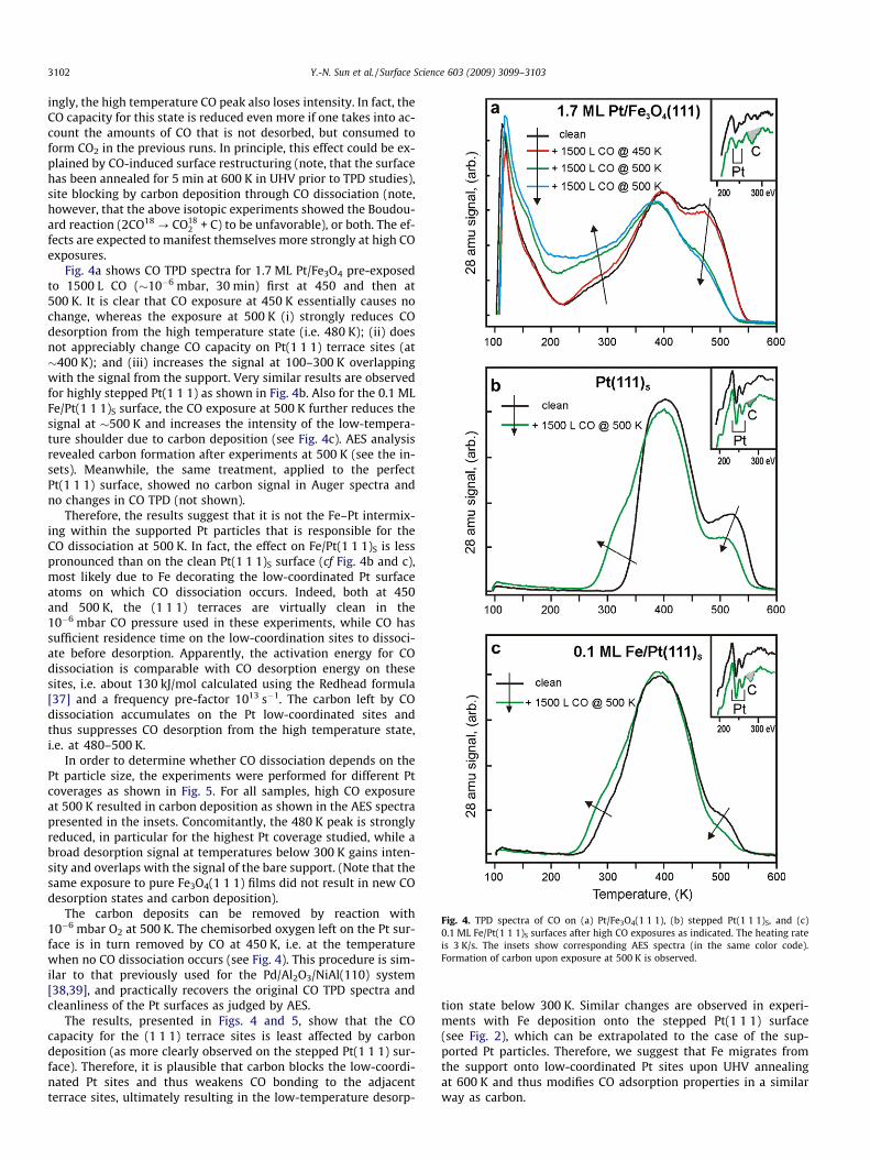
Fig. 4. TPD spectra of CO on (a) Pt/Fe3O4(1 1 1), (b) stepped Pt(1 1 1)S, and (c)0.1 ML Fe/Pt(1 1 1)S surfaces after high CO exposures as indicated. The heating rateis 3 K/s. The insets show corresponding AES spectra (in the same color code).Formation of carbon upon exposure at 500 K is observed.
3102 Y.-N. Sun et al. / Surface Science 603 (2009) 3099–3103
ingly, the high temperature CO peak also loses intensity. In fact, theCO capacity for this state is reduced even more if one takes into ac-count the amounts of CO that is not desorbed, but consumed toform CO2 in the previous runs. In principle, this effect could be ex-plained by CO-induced surface restructuring (note, that the surfacehas been annealed for 5 min at 600 K in UHV prior to TPD studies),site blocking by carbon deposition through CO dissociation (note,however, that the above isotopic experiments showed the Boudou-ard reaction (2CO18 ? CO18
2 + C) to be unfavorable), or both. The ef-fects are expected to manifest themselves more strongly at high COexposures.
Fig. 4a shows CO TPD spectra for 1.7 ML Pt/Fe3O4 pre-exposedto 1500 L CO (�10�6 mbar, 30 min) first at 450 and then at500 K. It is clear that CO exposure at 450 K essentially causes nochange, whereas the exposure at 500 K (i) strongly reduces COdesorption from the high temperature state (i.e. 480 K); (ii) doesnot appreciably change CO capacity on Pt(1 1 1) terrace sites (at�400 K); and (iii) increases the signal at 100–300 K overlappingwith the signal from the support. Very similar results are observedfor highly stepped Pt(1 1 1) as shown in Fig. 4b. Also for the 0.1 MLFe/Pt(1 1 1)S surface, the CO exposure at 500 K further reduces thesignal at �500 K and increases the intensity of the low-tempera-ture shoulder due to carbon deposition (see Fig. 4c). AES analysisrevealed carbon formation after experiments at 500 K (see the in-sets). Meanwhile, the same treatment, applied to the perfectPt(1 1 1) surface, showed no carbon signal in Auger spectra andno changes in CO TPD (not shown).
Therefore, the results suggest that it is not the Fe–Pt intermix-ing within the supported Pt particles that is responsible for theCO dissociation at 500 K. In fact, the effect on Fe/Pt(1 1 1)S is lesspronounced than on the clean Pt(1 1 1)S surface (cf Fig. 4b and c),most likely due to Fe decorating the low-coordinated Pt surfaceatoms on which CO dissociation occurs. Indeed, both at 450and 500 K, the (1 1 1) terraces are virtually clean in the10�6 mbar CO pressure used in these experiments, while CO hassufficient residence time on the low-coordination sites to dissoci-ate before desorption. Apparently, the activation energy for COdissociation is comparable with CO desorption energy on thesesites, i.e. about 130 kJ/mol calculated using the Redhead formula[37] and a frequency pre-factor 1013 s�1. The carbon left by COdissociation accumulates on the Pt low-coordinated sites andthus suppresses CO desorption from the high temperature state,i.e. at 480–500 K.
In order to determine whether CO dissociation depends on thePt particle size, the experiments were performed for different Ptcoverages as shown in Fig. 5. For all samples, high CO exposureat 500 K resulted in carbon deposition as shown in the AES spectrapresented in the insets. Concomitantly, the 480 K peak is stronglyreduced, in particular for the highest Pt coverage studied, while abroad desorption signal at temperatures below 300 K gains inten-sity and overlaps with the signal of the bare support. (Note that thesame exposure to pure Fe3O4(1 1 1) films did not result in new COdesorption states and carbon deposition).
The carbon deposits can be removed by reaction with10�6 mbar O2 at 500 K. The chemisorbed oxygen left on the Pt sur-face is in turn removed by CO at 450 K, i.e. at the temperaturewhen no CO dissociation occurs (see Fig. 4). This procedure is sim-ilar to that previously used for the Pd/Al2O3/NiAl(110) system[38,39], and practically recovers the original CO TPD spectra andcleanliness of the Pt surfaces as judged by AES.
The results, presented in Figs. 4 and 5, show that the COcapacity for the (1 1 1) terrace sites is least affected by carbondeposition (as more clearly observed on the stepped Pt(1 1 1) sur-face). Therefore, it is plausible that carbon blocks the low-coordi-nated Pt sites and thus weakens CO bonding to the adjacentterrace sites, ultimately resulting in the low-temperature desorp-
tion state below 300 K. Similar changes are observed in experi-ments with Fe deposition onto the stepped Pt(1 1 1) surface(see Fig. 2), which can be extrapolated to the case of the sup-ported Pt particles. Therefore, we suggest that Fe migrates fromthe support onto low-coordinated Pt sites upon UHV annealingat 600 K and thus modifies CO adsorption properties in a similarway as carbon.

Fig. 5. TPD spectra of CO on Pt/Fe3O4(1 1 1) annealed at 600 K for 5 min as afunction of Pt coverage as indicated. Then each sample was treated with 1500 L ofCO at 500 K. Subsequently, the sample was exposed to 540 L O2 at 500 K and 1500 LCO at 450 K. 7.5 L of CO were dosed at 100 K in each case. Heating rate is 3 K/s. Thecorresponding AES spectra are shown in the insets in the same color code.
Y.-N. Sun et al. / Surface Science 603 (2009) 3099–3103 3103
4. Conclusions
Comparative STM, AES and CO TPD study of Pt/Fe3O4(1 1 1), per-fect Pt(1 1 1) and highly stepped Pt(1 1 1) surfaces showed that the
Pt nanoparticles annealed at 600 K exhibit a new desorption stateat �270 K. This state is assigned to initial stages of strong metalsupport interaction resulting in Fe migration onto the Pt particles,although Fe diffusion into the particles at these temperatures can-not be excluded.
Both on Pt/Fe3O4(1 1 1) and stepped Pt(1 1 1) surfaces CO isfound to dissociate at 500 K resulting in deposition of carbon, sup-porting the previous literature results on the key role of low-coor-dinated sites in CO dissociation on Pt. The results also suggest thatcarbon accumulation occurs on the low-coordinated Pt sites. Car-bon deposits can be removed by mild oxidation at 500 K.
Acknowledgements
We thank the Fonds der Chemischen Industrie for financialsupport.
References
[1] R. Imbihl, G. Ertl, Chem. Rev. 95 (1995) 697.[2] R.W. McCabe, L.D. Schmidt, Surf. Sci. 66 (1977) 101.[3] G. Ertl, M. Neumann, K.M. Streit, Surf. Sci. 64 (1977) 393.[4] H. Steininger, S. Lehwald, H. Ibach, Surf. Sci. 123 (1982) 264.[5] D.M. Collins, W.E. Spicer, Surf. Sci. 69 (1977) 85.[6] H. Hopster, H. Ibach, Surf. Sci. 77 (1978) 109.[7] B.E. Hayden, K. Kretzschmar, A.M. Bradshaw, R.G. Greenler, Surf. Sci. 149
(1985) 394.[8] M.R. McClellan, J.L. Gland, F.R. McFeeley, Surf. Sci. 112 (1981) 63.[9] E.I. Altman, R.J. Gorte, Surf. Sci. 172 (1986) 71.
[10] E.I. Altman, R.J. Gorte, Surf. Sci. 195 (1988) 392.[11] D.R. Mullins, K.Z. Zhang, Surf. Sci. 513 (2002) 163.[12] D.L. Doering, H. Poppa, J.T. Dickinson, J. Vac. Sci. Technol. 20 (1982) 827.[13] Y. Iwasawa, R. Mason, M. Textor, G.A. Somorjai, Chem. Phys. Lett. 44 (1976)
468.[14] B. Lang, R.W. Joyner, G.A. Somorjai, Surf. Sci. 30 (1972) 454.[15] X.Q.D. Li, R. Vanselow, Catal. Lett. 2 (1989) 113.[16] G. Rupprechter, T. Dellwig, H. Unterhalt, H.J. Freund, J. Phys. Chem. B 105
(2001) 3797.[17] K. McCrea, J.S. Parker, P. Chen, G. Somorjai, Surf. Sci. 494 (2001) 238.[18] M.A. Vannice, C.C. Twu, J. Catal. 82 (1983) 213.[19] S.J. Tauster, S.C. Fung, R.L. Garten, J. Am. Chem. Soc. 100 (1978) 170.[20] Z.H. Qin, M. Lewandowski, Y.N. Sun, S. Shaikhutdinov, H.J. Freund, J. Phys.
Chem. C 112 (2008) 10209.[21] Z.H. Qin, M. Lewandowski, Y.N. Sun, S. Shaikhutdinov, H.J. Freund, J. Phys.:
Condens. Matter 21 (2009) 134019.[22] W. Weiss, W. Ranke, Prog. Surf. Sci. 70 (2002) 1.[23] W. Weiss, M. Ritter, Phys. Rev. B 59 (1999) 5201.[24] C. Lemire, R. Meyer, V.E. Henrich, S. Shaikhutdinov, H.J. Freund, Surf. Sci. 572
(2004) 103.[25] T. Michely, G. Comsa, Surf. Sci. 256 (1991) 217.[26] K.H. Hansen, T. Worren, S. Stempel, E. L�gsgaard, M. Bäumer, H.J. Freund, F.
Besenbacher, I. Stensgaard, Phys. Rev. Lett. 83 (1999) 4120.[27] H.P. Bonzel, R. Ku, J. Chem. Phys. 58 (1973) 4617.[28] G. Kneringer, F.P. Netzer, Surf. Sci. 49 (1975) 125.[29] R.W. McCabe, L.D. Schmidt, Surf. Sci. 60 (1976) 85.[30] R.K. Sharma, W.A. Brown, D.A. King, Surf. Sci. 414 (1998) 68.[31] R.B. Shumbera, H.H. Kan, J.F. Weaver, J. Phys. Chem. C 112 (2008) 4232.[32] T. Wadayama, H. Osano, T. Maeyama, H. Yoshida, K. Murakami, N. Todoroki, S.
Oda, J. Phys. Chem. C 112 (2008) 8944.[33] C. Pint, G. Bozzolo, J.E. Garcés, Surf. Sci. 602 (2008) 559.[34] A. Ali, M. Abon, P. Beccat, J.C. Bertolini, B. Tardy, Surf. Sci. 302 (1994) 121.[35] C. Xu, B.E. Koel, Surf. Sci. Lett. 304 (1994) L505.[36] B. Vermag, M. Juel, S. Raaen, Phys. Rev. B 73 (2006) 033407.[37] P.A. Redhead, Vacuum 12 (1962) 203.[38] S. Shaikhutdinov, M. Heemeier, J. Hoffmann, I. Meusel, B. Richter, M. Bäumer,
H. Kuhlenbeck, J. Libuda, H.J. Freund, R. Oldman, S.D. Jackson, C. Konvicka, M.Schmid, P. Varga, Surf. Sci. 501 (2002) 270.
[39] S. Schauermann, J. Hoffmann, V. Johanek, J. Hartmann, J. Libuda, Phys. Chem.Chem. Phys. 4 (2002) 3909.