Embed Size (px)
Citation preview

Clostridium difficileClostridium difficile: an overview of the : an overview of the changes in our understanding the changes in our understanding the
organism over the last 30 yearsorganism over the last 30 yearsMaja Rupnik
Institute of Public Health Maribor and Faculty of Medicine UM, Maribor, Slovenia
Institute of Public Health Maribor
Faculty of MedicineUniversity of Maribor
Recognition of the pathogenic role of Recognition of the pathogenic role of Clostridium difficileClostridium difficile
• 1977 First description that a Clostridium sp. caused AAD in hamsters (subsequently identified as C. difficile). Bartlett et al
• 1978 Identification of C. difficile as the cause of PMC in man Bartlett et al
• 1979 Development of a selective medium for C.difficile. George et al
• 1981 Demonstration that C. difficile produces two toxins. Banno et al and Taylor et al

• Diseasedisease is toxin mediated CDI is only a disturbance and easy to treatimportance of normal gut floraassociation with antibiotics
• Toxins and pathogenesistwo toxins are main virulence factorssynergistic action
• EpidemiologyC. difficile is human nosocomial pathogen(typing systems, diagnostics)
C. difficile toxins
• Toxin A and Toxin B• purification• cytotoxicity, effects on animals• antibodies• molecular biology

C. difficile C. difficile pathogenicity locus (PaLoc)pathogenicity locus (PaLoc)
tcdBtcdR tcdE tcdCtcdA
Hc H Hc Hc H Hc P P Hc H
cdu1
cdu1
cdu2cdu2' cdd1
cdd1
cdd2cdd3cdd4
PP
115 bp
PaLoc19 kb
C. difficile tox-
C. difficile VPI 10463
1 956
1128
1155
1275
1849 2710 aa546
C C C C
N C
C C C C
N C
Hydrofobicregion
Region with rare amino acids
EA1 AS1 A1 KV1 IA1 IA2 itd.
E F G H A D I A B
Oligopeptide repeats(CROPs)
Oligonucleotide repeats (SRONs)
2366
TcdB
TcdA
C. difficile toxins A and B share structural properties

C. difficile toxins act on intracellular targets
• Bacterial protein toxins act onMembraneReceptors Intracellular targets
• Binding• Internalization• (Specific) modification of (specific) targets
C-terminal receptor binding domain
• Repetitive structures involved in carbohydrate binding
• Receptorspoorly knowncarbohydrate structures containing Galβ1-4GlcNAc (TcdA)carbohydrate linkage to protein or lipid is unknown
TcdA – apical side (of T-84 cells)TcdB – basolateral side (of T-84 cells)

Structure of TcdA repetitive regions
•crystal structure of a 127 aa fragment within repetitive region(Ho et al., PNAS, 2005 )
•solenoid structure of 3 to 5 SRs connected with 1 LR
•carbohydrate recognition(Greco et al., Nature Struct Mol Biol, 2006)
•optimal for multivalent binding to the cell
Catalytic domain and molecular mechanism of action
• cytotoxic effect of LCTs• disruption of actin filaments
control type D type S
toxin treatedcontrol
Clostridial toxins acting on actin cytoskeleton:• actin ADP-ribosylating toxins
clostridial binary toxinsC3 like toxins
• glycosyltransferase activitylarge clostridial toxins
new and unique mechanism of actionfor bacterial protein toxins(Just et al., Nature, 1995)

Clostridial toxins - glycosylation of small GTPases
Substrate (small GTPases)Rho subfamily (Rho, Rac, Cdc42)
Ras subfamily (Ras, Rap, Ral)
Co-substrate (activated sugars)UDP-Glc (C. difficile, C. sordellii)
UDP-GlcNAc (C. novyi)
GTPase
GTPase
Rho/Ras
Rho/Ras
Thr
Thr
+
+
glc
glc
UDP
UDP
toxin
Jank, T. et al. Glycobiology 2007
Crystal structure of the catalytic domain of toxin B
• Enzymatic activity (DXD and Trp)
• Substrate specificitysugar molecule – Ile383, Gln 385(Jank et al., JBC, 2005)GTPase – not known
• Crystal structure of 543 fragment(Reinert et al., JMB, 2005)
Catalytic core (234 residues)type A family of glycosyltransferases
Additional subdomainsunknown function4 N terminal helices
interaction with membrane?C. sordelii TcsL 18 aabinding to phosphatidylserine

Pathways of internalization of secreted bacterial toxins
late endosomes retrograde transport to ER
binding to the receptor
endocytosis
membrane translocation membrane translocation
diphteria toxinantrax toxinC. difficile toxins
cholera toxinshiga toxin
LCT entry into host cell – pore formation• endosomes, pH↓ → conformational change• insertion of hydrophobic regions into endosomal membrane• pore formation on cells and artificial membranes
TcdA – cell type specific; strictly cholesterol dependent(Giesemann, JBC, 2006)
TcdB – not cholesterol dependent(Barth et al., JBC, 2001)

Falnes and Sandvig, 2000
Entry of catalytic domain into the cell
• Toxins with two or more subunits
subunit B – bindingsubunit A – enzymatic
• Single chain proteins
cleavage of catalytic domain?
C C C C
N C
catalytic domain binding domain
polyclonal anti-C. difficile serummonoclonal antibody 2CV
cleaved labeled toxin TcdB
detected on SDS PAGE gelWestern blots with two different antibodies
Toxin TcdB is cleaved in vitro in the presence of cell lysate
200 kDA
60 kDA
Rupnik et al., Microbiology, 2005

Cleavage site defines catalytic domain
holotoxin
Binding/translocationdomain
Catalytic domain
Characteristics of proteolytic activity
pH dependentlow at pH 5.0, optimal at pH 8.0
not inhibited byEDTA, EGTA,various protease inhibitorsheating, protease K (later studies)
inhibited byCa2+, pepstatin A
In vitro cleavage reaction:
Cy3 labeled toxincell lysate
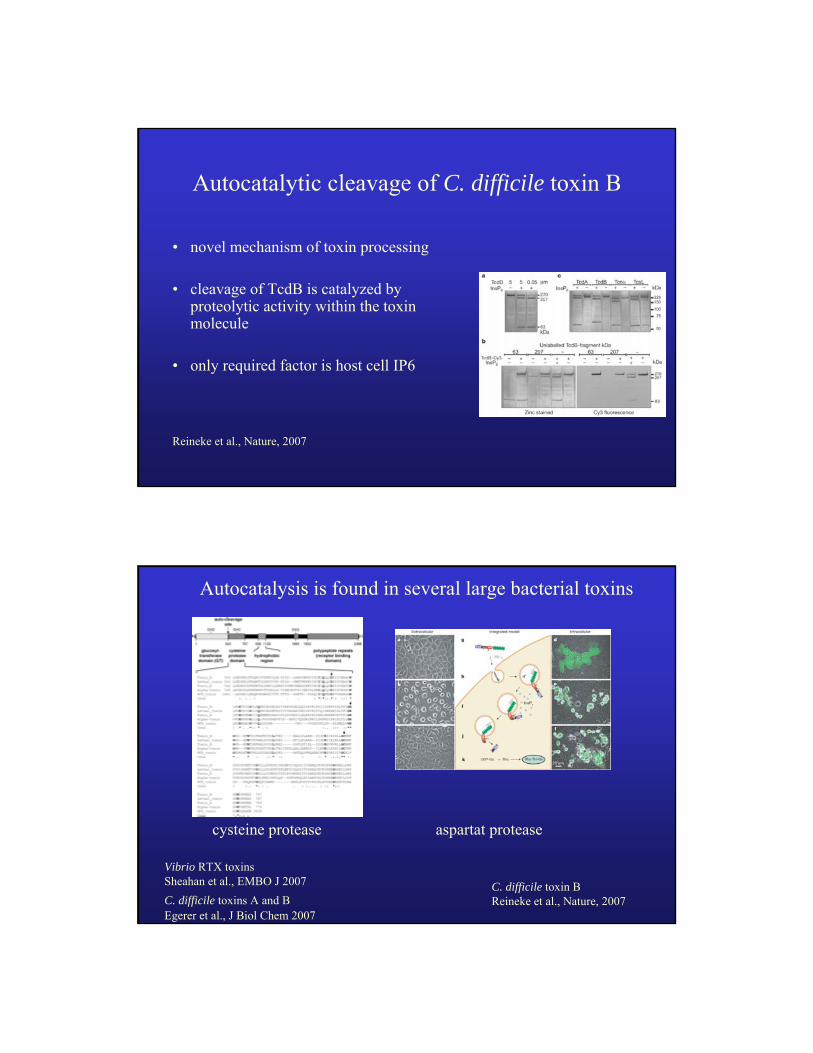
Autocatalytic cleavage of C. difficile toxin B
• novel mechanism of toxin processing
• cleavage of TcdB is catalyzed by proteolytic activity within the toxin molecule
• only required factor is host cell IP6
Reineke et al., Nature, 2007
Autocatalysis is found in several large bacterial toxins
cysteine protease aspartat protease
Vibrio RTX toxins Sheahan et al., EMBO J 2007 C. difficile toxin B
Reineke et al., Nature, 2007C. difficile toxins A and BEgerer et al., J Biol Chem 2007

Structure and function of LCTs
• toxins – main virulence factors• basis for new therapeutic targets• non-antibiotic treatment
inhibition of binding, inhibition of pore formation, inhibition of catalytic centre, inhibition of proteolysis
Large clostridial toxins (LCT)Large clostridial toxins (LCT)
C. difficile C. difficile (TcdA, TcdB) (TcdA, TcdB) C. sordellii C. sordellii (TcsH, TcsL) (TcsH, TcsL) C. novyi C. novyi (Tcn(Tcnαα))C. C. perfringensperfringens ((TcpLTcpL))
•• size (250size (250--300 kDa) 300 kDa)
•• cytotoxicitycytotoxicity
•• gglycosyltransferaseslycosyltransferases
•• autoproteolyticautoproteolytic activityactivity
GTPaza
GTPaza
Rho/Ras
Rho/Ras
Thr
Thr
+
+
glc
glc
UDP
UDP
toksin

Role of the toxins in the pathogenesisC. difficile always produces both toxins (A+B+ strains)
Early hamster experiments (Lyerly et al., 1986)purified A – symptoms (local)purified B – no symptomspurified B with low concentr. A – symptoms (systemic)
K/O mutants tcdB and tcdA - hamster ↓tcdB only - hamster ↓tcdA only - hamster ↑
C. difficile strains can produce up to 3 toxinsA-B+ strains – high virulence for humans
Variability in C. difficile toxin genes
D type
S type
B-0 (VPI 10463; M30307)
B-0 (VPI 10463; X53138)
B-VI
B-IV
B-III
B- IX
B-X (8864; AJ011301)
B-VIII (1470; Z23277)
B-VIII (CF2; AF217292)
C.sordellii TcsL (X82638)
A-X (8864; AJ011301)
A-IV
A-VI
A-IX
A-0 (VPI 10463; M30307)
A-0 (VPI 10463; X92982)
C.novyi TcnA (Z48636)
0.1
D type
S type
Rupnik, FEMS Rev. 2008

C. difficile binary toxin CDT
• Additional virulence factor
• Prevalence of binary toxin producing strains is increasing(Spigagalia and Mastrantonio, JMM, 2004)
time interval before 1990 1991-1999 2000-2001% of CDT+ str. 0 24 45
• Binary toxin positive strains more likely associated with severe disease(Barbut t al., JMM, 2005; Terhes et al., JCM, 2004)
cdtA cdtB
1 1383 1 2631
C. difficile CD196
CDT locus (4.3 kb)
N C
catalytic domain translocation and binding domain
50 kDa C N 100 kDa
C. difficile types
Entire genomePCR ribotyping
PFGE
REA
MLVA
MLST
PaLoc regiontoxinotyping
slpA
Toxin productiontoxin A, TcdA
toxin B, TcdB
binary toxin CDT
Phenotypicserotyping

C. difficile – mostly used typing methodsRibotyping REA PFGE(Europe) (USA) (North America)
PCR of 16S-23S rDNA intergenic spacer region160 ribotypes
Stubbs, JCM 1999;Bidet, JCM 2000
HindIII restriction of whole DNA
>100 REA groups(Rea Types)
Gerding D., Chicago, USA
SmaI restriction of whole DNA
no large international collection
Changes in human hostChanges in human host
• Hospital-associated v.s. community-associated infections
• Increase in population previously at low risk
• Spread of a specific typestoxinotype VIII (serogroup F; 017, A-B+CDT+)toxinotype III (type BI/NAP1/027; 027, A+B+CDT+)toxinotype V (type 078, A+B+CDT+)
• Emergence of hypervirulent types

C. difficile types in humans and animals
• diversity of C. difficile animal strains is lower than in human isolates
• same ribotypes are found in animals and humans
• different types are currently predominately associated with animals or with humans
C. difficile types in humans and animals
• cats and dogs, humans (Australia) (O’Neill et al., Epidemiol. Infect. 1993)no overlap between animal and human strainsgood correlation between animal and veterinary clinic environment
strains
• calves (Canada) (Rodruigez-Palacios et al., Emerg.Infect.Dis., 2006)8 ribotypes 7 of them also in human isolates (same time/geogr. area)078 (V), 017 (VIII), 027 (III), 033 (XI), 077 (0), 014 (0)
• calves and pigs (USA) (Keel et al., 2007)4 ribotypesall of them known in human isolates078 (V), 017 (VIII), 033 (XI), 002 (0), 126 (ND)

C. difficile toxinotype V/078 and animals
• 6 toxinotypes (III, IV, V, VIII, XI, XII) out of 27 known described in animal hosts
• toxinotype V/078 present in high proportion in different animal hosts worldwide and in food
horsespigletscattle
• current ‘epidemic type’ in animals? • particularly adapted to animals?
Summary
• C. difficile is an important human and animal pathogen
• infection and transmition: hospital environment (direct or indirect contact), animals, food;
• changes in virulence and antibiotic susceptibility• additional virulence factors to toxins TcdA and
TcdB (CDT, adhesins, sporulation…)