Embed Size (px)
Citation preview
ORIGINAL PAPER
Cloning and characterization of Plasmodium vivaxthioredoxin peroxidase-1
Hassan Hakimi & Masahito Asada &
Jose Ma. M. Angeles & Noboru Inoue &
Shin-ichiro Kawazu
Received: 1 December 2011 /Accepted: 10 February 2012 /Published online: 6 March 2012# Springer-Verlag 2012
Abstract Reactive oxygen species produced from hemo-globin digestion and the host immune system could haveadverse effects on malaria parasites. To protect them-selves, malaria parasites are highly dependent on theantioxidant enzymes, including superoxide dismutasesand thioredoxin-dependent peroxidases. To date, severalthioredoxin peroxidases (TPx) have been characterized inPlasmodium falciparum, but the TPx in Plasmodiumvivax has not yet been characterized. The complete se-quence of gene coding for thioredoxin peroxidase-1 of P.vivax (PvTPx-1) was amplified by PCR and cloned.Using the recombinant PvTPx-1 (rPvTPx-1), polyclonalantibody was produced in mice for immunolocalizationof the enzyme in the parasite. The antioxidant activity ofrPvTPx-1 was evaluated by mixed-function oxidationassay. PvTPx-1 has two conserved cysteine residues inthe amino acid sequence at the positions 50 and 170which formed a dimer under a non-reducing condition.Using a thiol mixed-function oxidation assay, the antiox-idant activity of rPvTPx-1 was revealed. Indirect immu-nofluorescence microscopy with the specific antibodyindicated that PvTPx-1 was expressed in the cytoplasmof the erythrocytic stage of the parasite in a dots-likepattern. The results suggest that P. vivax uses TPx-1 toreduce and detoxify hydrogen peroxides in order tomaintain their redox homeostasis and proliferation inthe host body.
Introduction
Although huge efforts have been made to control malaria, itstill remains as one of the most important global healthproblems. Plasmodium vivax, the second most prevalentmalaria species after Plasmodium falciparum, has a widergeographical distribution and mostly co-exists with P.falciparum in endemic countries (Arevalo-Herrera et al.2010). P. vivax causes 25–40% of malaria cases worldwide(Dharia et al. 2010) resulting to about 80 to 300 millionclinical cases every year (Arevalo-Herrera et al. 2010).In spite of the public health importance of this humanmalaria parasite, less attention has been given on researchesabout P. vivax (Mendis et al. 2001).
Since malaria parasites proliferate in the human bodyand the mosquito vector that are oxygen-rich environ-ments, the parasites are exposed to the toxic effects ofreactive oxygen species (ROS) which might cause dam-ages to their macromolecules (Kawazu et al. 2008; Kehret al. 2010). Digestion of hemoglobin by malaria para-sites produces heme, the prominent source of ROS inthe parasite (Muller 2004). Moreover, the immune sys-tem of the host also induces the production of ROS toact against the parasites (Baert et al. 1999; Kawazu etal. 2008). In order to proliferate in the host body,malaria parasites protect themselves from the damagescaused by ROS (Kehr et al. 2010). Since Plasmodiumlacks catalase and genuine glutathione peroxidase, thiore-doxin peroxidases play a vital role in the reduction of ROS(Kawazu et al. 2008; Kehr et al. 2010; Muller 2004).
Thioredoxin peroxidase-1 (TPx-1), which is primarilylocated in the cytosol (Kawazu et al. 2008), belongs to thefamily of thiol-specific antioxidant proteins (Wood et al.2003). TPx-1 reduces and detoxifies hydrogen peroxidesthrough the action of the redox-active cysteine (Wood et
H. Hakimi :M. Asada : J. M. M. Angeles :N. Inoue :S.-i. Kawazu (*)National Research Center for Protozoan Diseases,Obihiro University of Agriculture and Veterinary Medicine,Inada-cho,Obihiro, Hokkaido 080-8555, Japane-mail: [email protected]
Parasitol Res (2012) 111:525–529DOI 10.1007/s00436-012-2864-3
al. 2003). Based on the number and position of cysteine(Cys) residue, TPxs are divided into three types, namely:1-Cys, typical 2-Cys, and atypical 2-Cys type (Vaca-Paniaguaet al. 2009; Wood et al. 2003).
The susceptibility of the malaria parasite to the distur-bance of their antioxidant system makes it a great potentialtarget for the development of new drugs (Muller 2004).Artimisinin compounds, established as antimalarial in the1990s, kill the parasites by generating free radicals andincreasing oxidative stress (Warrell et al. 2002). In recentyears, several researches have been made on the TPx of P.falciparum with six TPxs already characterized (Kawazu etal. 2000; Rahlfs and Becker 2001; Kawazu et al. 2008;Richard et al. 2011). However, the TPx in P. vivax hasnot been characterized yet. In this study, we cloned andcharacterized the TPx-1 from P. vivax.
Materials and methods
Cloning of the gene coding for the TPx-1 of P. vivax
The complete sequence of the gene coding for the TPx-1of P. vivax (PvTPx-1) was amplified using the primersets: 5′-CAT ATG CCG ACA TAC GTA GGA AAG G-3′and 5′-GGA TTC TTA TAA TGT GGA CAA ATA CTGCGC-3′ (start and stop codons were underlined). Theprimers were designed using PlasmoDB (2011) database(accession number: PVX_118545) with the restrictionenzyme sites NdeI and BamHI (italic). The PvTPx-1 genewas amplified from genomic DNA of the P. vivax sal 1strain. The gene was shown as a single copy and had nointrons in the genome. The PCR was done in 50 μl ofreaction mixture containing 1.5 mM MgCl2, 200 μM ofeach dNTP and 1 μM of each primer as the final con-centration, and 0.5 U of Taq DNA polymerase (Invitro-gen, Tokyo, Japan). Each PCR cycle consists of
denaturation at 94°C for 15 s, annealing at 50°C for20 s, and extension at 68°C for 2 min and was repeated35 times. It was followed by 68°C for 10 min as finalextension. The expected length of the PCR product was588 bp, and the gene sequence was analyzed using ABIPrism 3100 Genetic Analyzer (Applied Biosystems,Carlsbad, CA).
Expression and purification of recombinant PvTPx-1
The PCR product was digested with restriction enzymesNdeI and BamHI and then inserted into pET-28a expres-sion vector (EMD Biosciences, San Diego, CA) by over-night incubation with ligation mixture (Takara, Otsu,Japan) at 15°C. The recombinant plasmid containing thecoding sequence of PvTPx-1 was transfected into Escher-ichia coli (strain BL21). The transformed colony wasincubated in SOB medium (BD, Sparks, MD) containingkanamycin (50 μg/ml) at 37°C until the optical density at600 nm reached 0.6. Expression of the recombinantPvTPx-1 (rPvTPx-1) was induced by adding 1 mM iso-propyl thio-β-D-galactoside (IPTG) and an additional 4 hof incubation. The bacterial culture was harvested bycentrifugation, resuspended in the lysis buffer (100 mMsodium phosphate, pH 8.0; 10 mM Tris–Cl, pH 8.0), andsonicated for 10 min. Histidine-tagged recombinant pro-tein in the supernatant was incubated with Ni-NTAagarose beads (Qiagen Inc., Valencia, CA). After wash-ing two times, the captured protein was eluted by in-creasing the imidazol concentration in the elution bufferup to 200 mM. The purity of the recombinant proteinwas evaluated by 12% sodium dodecyl sulfate poly-acrylamide gel electrophoresis (SDS/PAGE) under re-ducing conditions and subsequent Coomassie BrilliantBlue staining. The concentration of each expressed pro-tein was measured using BCA assay (Thermo Scientific,Rockford, IL).
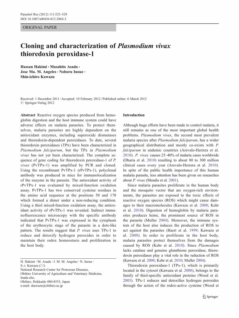
1 MASYVGREAPYFKAEAVFADNTFGEVNLHDFIGKKYVLLYFYPLDFTFVCPSEIIALDKA 601 MPTYVGKEAPFFKAEAVFGDNSFGEVNLTQFIGKKYVLLYFYPLDFTFVCPSEIIALDKA 60
61 LDAFKERNVELIGCSVDSKYTHLAWKKTPLTKGGIGNIQHTLISDITKSISRSYNVLFGD 12061 LDAFHERNVELLGCSVDSKYTHLAWKKTPLAKGGIGNIKHTLLSDITKSISKDYNVLFDD 120
21 SVSLRAFVLIDKQGVVQHLLVNNLAIGRSVEEVLRIIDAVQHHEQHGDVCPANWKKGKVA 180 21 SVSLRAFVLIDMNGIVQHLLVNNLAIGRSVDEILRIIDAIQHHEKYGDVCPANWQKGKVS 180
81 MKPSEEGVSEYL L 195 81 MKPSEEGVAQYL 195
PfTPx-1
PfTPx-1
PfTPx-1
PfTPx-1
PvTPx-1
PvTPx-1
PvTPx-1
PvTPx-1
*
*
SS
LTK
Fig. 1 Amino acid sequence alignment of PfTPx-1 and PvTPx-1.Asterisks indicate the conserved cysteine residues and peroxisometargeting signal 1 (PTS1), and the PTS1-like motifs at the C-terminus
of PfTPx-1 and PvTPx-1 are underlined, respectively. The identicalresidues between the two sequences are boxed
526 Parasitol Res (2012) 111:525–529
Antioxidant activity assay
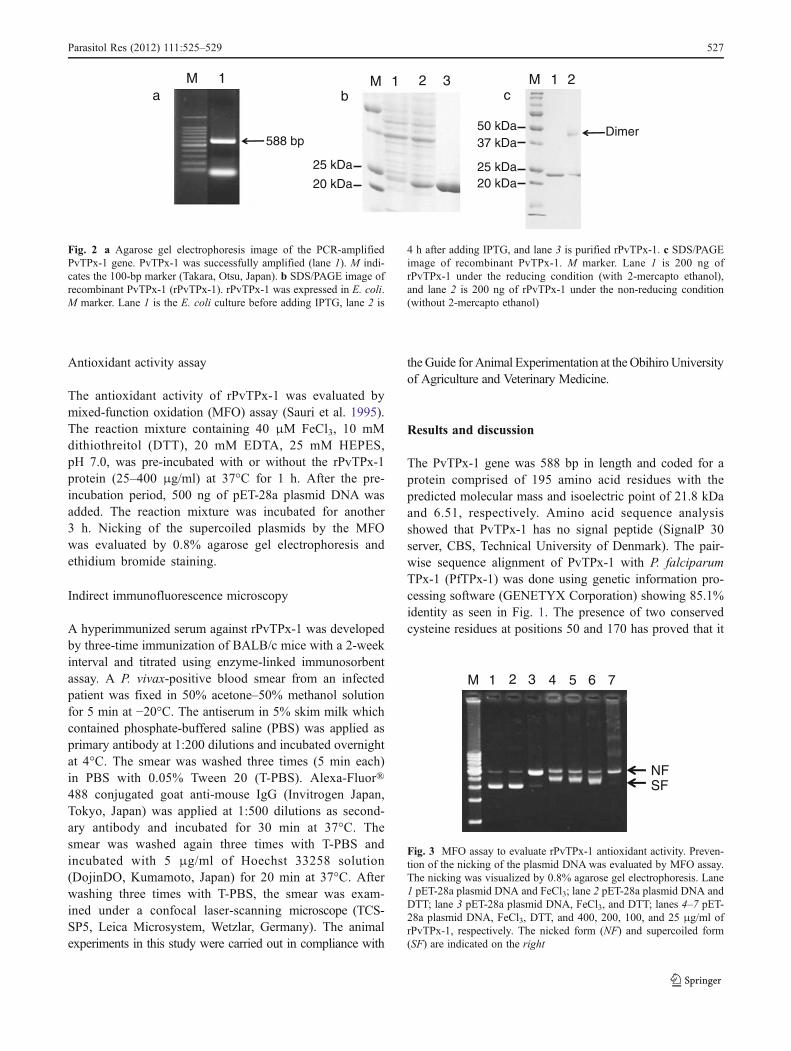
The antioxidant activity of rPvTPx-1 was evaluated bymixed-function oxidation (MFO) assay (Sauri et al. 1995).The reaction mixture containing 40 μM FeCl3, 10 mMdithiothreitol (DTT), 20 mM EDTA, 25 mM HEPES,pH 7.0, was pre-incubated with or without the rPvTPx-1protein (25–400 μg/ml) at 37°C for 1 h. After the pre-incubation period, 500 ng of pET-28a plasmid DNA wasadded. The reaction mixture was incubated for another3 h. Nicking of the supercoiled plasmids by the MFOwas evaluated by 0.8% agarose gel electrophoresis andethidium bromide staining.
Indirect immunofluorescence microscopy
A hyperimmunized serum against rPvTPx-1 was developedby three-time immunization of BALB/c mice with a 2-weekinterval and titrated using enzyme-linked immunosorbentassay. A P. vivax-positive blood smear from an infectedpatient was fixed in 50% acetone–50% methanol solutionfor 5 min at −20°C. The antiserum in 5% skim milk whichcontained phosphate-buffered saline (PBS) was applied asprimary antibody at 1:200 dilutions and incubated overnightat 4°C. The smear was washed three times (5 min each)in PBS with 0.05% Tween 20 (T-PBS). Alexa-Fluor®488 conjugated goat anti-mouse IgG (Invitrogen Japan,Tokyo, Japan) was applied at 1:500 dilutions as second-ary antibody and incubated for 30 min at 37°C. Thesmear was washed again three times with T-PBS andincubated with 5 μg/ml of Hoechst 33258 solution(DojinDO, Kumamoto, Japan) for 20 min at 37°C. Afterwashing three times with T-PBS, the smear was exam-ined under a confocal laser-scanning microscope (TCS-SP5, Leica Microsystem, Wetzlar, Germany). The animalexperiments in this study were carried out in compliance with
theGuide for Animal Experimentation at the Obihiro Universityof Agriculture and Veterinary Medicine.
Results and discussion
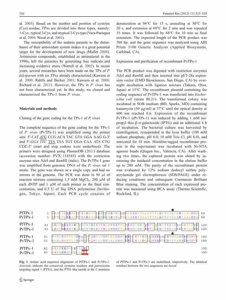
The PvTPx-1 gene was 588 bp in length and coded for aprotein comprised of 195 amino acid residues with thepredicted molecular mass and isoelectric point of 21.8 kDaand 6.51, respectively. Amino acid sequence analysisshowed that PvTPx-1 has no signal peptide (SignalP 30server, CBS, Technical University of Denmark). The pair-wise sequence alignment of PvTPx-1 with P. falciparumTPx-1 (PfTPx-1) was done using genetic information pro-cessing software (GENETYX Corporation) showing 85.1%identity as seen in Fig. 1. The presence of two conservedcysteine residues at positions 50 and 170 has proved that it
aM
588 bp
25 kDa
20 kDa
bM 1 2 31
cM 1 2
25 kDa20 kDa
50 kDa37 kDa
Dimer
Fig. 2 a Agarose gel electrophoresis image of the PCR-amplifiedPvTPx-1 gene. PvTPx-1 was successfully amplified (lane 1). M indi-cates the 100-bp marker (Takara, Otsu, Japan). b SDS/PAGE image ofrecombinant PvTPx-1 (rPvTPx-1). rPvTPx-1 was expressed in E. coli.M marker. Lane 1 is the E. coli culture before adding IPTG, lane 2 is
4 h after adding IPTG, and lane 3 is purified rPvTPx-1. c SDS/PAGEimage of recombinant PvTPx-1. M marker. Lane 1 is 200 ng ofrPvTPx-1 under the reducing condition (with 2-mercapto ethanol),and lane 2 is 200 ng of rPvTPx-1 under the non-reducing condition(without 2-mercapto ethanol)
M 1 2 3 4 5 6 7
NFSF
Fig. 3 MFO assay to evaluate rPvTPx-1 antioxidant activity. Preven-tion of the nicking of the plasmid DNAwas evaluated by MFO assay.The nicking was visualized by 0.8% agarose gel electrophoresis. Lane1 pET-28a plasmid DNA and FeCl3; lane 2 pET-28a plasmid DNA andDTT; lane 3 pET-28a plasmid DNA, FeCl3, and DTT; lanes 4–7 pET-28a plasmid DNA, FeCl3, DTT, and 400, 200, 100, and 25 μg/ml ofrPvTPx-1, respectively. The nicked form (NF) and supercoiled form(SF) are indicated on the right
Parasitol Res (2012) 111:525–529 527
was a typical 2-Cys type TPx. SDS/PAGE under a non-reducing condition showed that the protein forms a dimerresulting from an intersubunit disulfide bond (Fig. 2c).TPx-1 has two conserved active Cys namely peroxidaticCys and resolving Cys (Wood et al. 2003). The perox-idatic Cys of one subunit is attacked by resolving Cys inthe C-terminus of the other subunit and making a disul-fide bond between two molecules in the action for re-ducing peroxide (Wood et al. 2002, 2003). The 2-Cysdisulfide in the oxidized enzyme is reduced by anotherbiothiol such as thioredoxin wherein the dimer dissoci-ates into two regenerated monomers as reduced enzymes(Wood et al. 2003).
In order to evaluate the enzyme activity and develop anantibody against PvTPx-1, the recombinant protein wasproduced (Fig. 2b). The antioxidant activity of rPvTPx-1was evaluated by MFO assay (Fig. 3). The production ofhydroxyl radicals in the reaction mixture of the assaydamages the DNA (Sauri et al. 1995). In the absence ofrPvTPx-1, FeCl3 and DTT produced hydroxyl radicalsgiving nicks in the supercoiled plasmid DNA and thuschanging the running behavior of the DNA in the aga-rose gel (Fig. 3, lane 3). However, in the reaction mix-tures containing 400, 200, and 100 μg/ml of rPvTPx-1,the nicking of the plasmid DNA was not observed(Fig. 3, lanes 4–6). This suggested that rPvTPx-1 hasantioxidant activity.
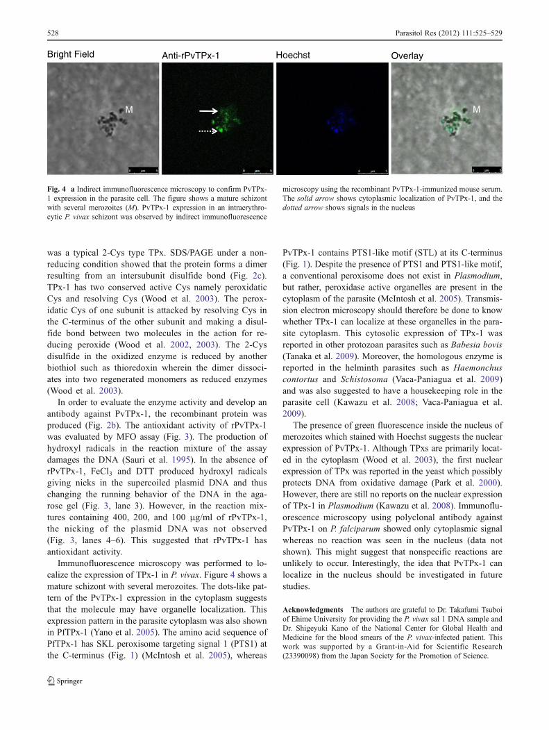
Immunofluorescence microscopy was performed to lo-calize the expression of TPx-1 in P. vivax. Figure 4 shows amature schizont with several merozoites. The dots-like pat-tern of the PvTPx-1 expression in the cytoplasm suggeststhat the molecule may have organelle localization. Thisexpression pattern in the parasite cytoplasm was also shownin PfTPx-1 (Yano et al. 2005). The amino acid sequence ofPfTPx-1 has SKL peroxisome targeting signal 1 (PTS1) atthe C-terminus (Fig. 1) (McIntosh et al. 2005), whereas
PvTPx-1 contains PTS1-like motif (STL) at its C-terminus(Fig. 1). Despite the presence of PTS1 and PTS1-like motif,a conventional peroxisome does not exist in Plasmodium,but rather, peroxidase active organelles are present in thecytoplasm of the parasite (McIntosh et al. 2005). Transmis-sion electron microscopy should therefore be done to knowwhether TPx-1 can localize at these organelles in the para-site cytoplasm. This cytosolic expression of TPx-1 wasreported in other protozoan parasites such as Babesia bovis(Tanaka et al. 2009). Moreover, the homologous enzyme isreported in the helminth parasites such as Haemonchuscontortus and Schistosoma (Vaca-Paniagua et al. 2009)and was also suggested to have a housekeeping role in theparasite cell (Kawazu et al. 2008; Vaca-Paniagua et al.2009).
The presence of green fluorescence inside the nucleus ofmerozoites which stained with Hoechst suggests the nuclearexpression of PvTPx-1. Although TPxs are primarily locat-ed in the cytoplasm (Wood et al. 2003), the first nuclearexpression of TPx was reported in the yeast which possiblyprotects DNA from oxidative damage (Park et al. 2000).However, there are still no reports on the nuclear expressionof TPx-1 in Plasmodium (Kawazu et al. 2008). Immunoflu-orescence microscopy using polyclonal antibody againstPvTPx-1 on P. falciparum showed only cytoplasmic signalwhereas no reaction was seen in the nucleus (data notshown). This might suggest that nonspecific reactions areunlikely to occur. Interestingly, the idea that PvTPx-1 canlocalize in the nucleus should be investigated in futurestudies.
Acknowledgments The authors are grateful to Dr. Takafumi Tsuboiof Ehime University for providing the P. vivax sal 1 DNA sample andDr. Shigeyuki Kano of the National Center for Global Health andMedicine for the blood smears of the P. vivax-infected patient. Thiswork was supported by a Grant-in-Aid for Scientific Research(23390098) from the Japan Society for the Promotion of Science.
Bright Field Anti-rPvTPx-1 Hoechst Overlay
M M
Fig. 4 a Indirect immunofluorescence microscopy to confirm PvTPx-1 expression in the parasite cell. The figure shows a mature schizontwith several merozoites (M). PvTPx-1 expression in an intraerythro-cytic P. vivax schizont was observed by indirect immunofluorescence
microscopy using the recombinant PvTPx-1-immunized mouse serum.The solid arrow shows cytoplasmic localization of PvTPx-1, and thedotted arrow shows signals in the nucleus
528 Parasitol Res (2012) 111:525–529
References
Arevalo-Herrera M, Chitnis C, Herrera S (2010) Current status ofPlasmodium vivax vaccine. Hum Vaccines 6:124–132
Baert CB, Deloron P, Viscogliosi E, Delgado-Viscogliosi P, Camus D,Dive D (1999) Cloning and characterization of iron-containingsuperoxide dismutase from the human malaria species Plasmodi-um ovale, P. malaria and P. vivax. Parasitol Res 85:1018–1024
Dharia NV, Bright AT, Westenberger SJ, Barnes SW, Batalov S,Kuhen K, Borboa R, Federe GC, McClean CM, Vinetz JM,Neyra V, Llanos-Cuentas A, Barnwell JW, Walker JR, WinzelerEA (2010) Whole-genome sequencing and microarray analysisof ex vivo Plasmodium vivax reveal selective pressure on puta-tive drug resistance genes. Proc Natl Acad Sci USA 107:20045–20050
Kawazu S, Komaki-Yasuda K, Oku H, Kano S (2008) Peroxiredoxinsin malaria parasites: parasitologic aspects. Parasitol Int 57:1–7
Kawazu S, Tsuji N, Hatabu T, Kawai S, Matsumoto Y, Kano S (2000)Molecular cloning and characterization of a peroxiredoxin fromthe human malaria parasite Plasmodium falciparum. Mol Bio-chem Parasitol 109:165–169
Kehr S, Sturm N, Rahlfs S, Przyborski JM, Becker K (2010) Compart-mentation of redox metabolism in malaria parasites. PLoS Pathog6:e1001242
McIntosh MT, Elliot DA, Joiner KA (2005) Plasmodium falciparum:discovery of peroxidase active organelles. Exp Parasitol 111:133–136
Mendis K, Sina BJ, Marchesini P, Carter R (2001) The neglectedburden of Plasmodium vivax malaria. AmJTrop Med Hyg64:97–106
Muller S (2004) Redox and antioxidant systems of the malaria parasitePlasmodium falciparum. Mol Microbiol 53:1291–1305
Park SG, Cha M, Jeong W, Kim I (2000) Distinct physiologicalfunctions of thiol peroxidase isoenzymes in Saccharomyces cer-evisiae. J Biol Chem 275:5723–5732
PlasmoDB (2011) Plasmodium Genomic Resource, PlasmoDB. At:http://www.plasmodb.org/plasmo. Accessed 24 June 2011
Rahlfs S, Becker K (2001) Thioredoxin peroxidases of the malariaparasite Plasmodium falciparum. Eur J Biochem 268:1404–1409
Richard D, Bartfai R, Volz J, Ralph SA, Muller S, Stunnenberg HG,Cowman AF (2011) A genome-wide chromatin-associatednuclear peroxiredoxin from the malaria parasite Plasmodiumfalciparum. J Biol Chem 286:11746–11755
Sauri H, Butterfield L, Kim A, Shau H (1995) Antioxidant functionof recombinant human natural killer enhancing factor. BiochemBiophys Res Commun 208:964–969
Tanaka M, Sakurai T, Yokoyama N, Inoue N, Kawazu S (2009)Cloning and characterization of peroxiredoxin in Babesia bovis.Parasitol Res 105:1473–1477
Vaca-Paniagua F, Parra-Unda R, Landa A (2009) Characterizationof one typical 2-Cys peroxiredoxin gene of Taenia solium andTaenia crassiceps. Parasitol Res 105:781–787
Warrell DA, Watkins WM, Winstanley PA (2002) Treatment andprevention of malaria. In: Warrell DA, Gilles HM (eds) Essentialmalariology, 4th edn. Arnold, London, pp 270–271
Wood ZA, Poole LB, Hantgan RR, Karplus PA (2002) Dimers todoughnuts: redox-sensitive oligomerization of 2-cysteine peroxir-edoxins. Biochemistry 41:5493–5504
Wood ZA, Schroder E, Robin Harris J, Poole LB (2003) Structure,mechanism and regulation of peroxiredoxins. Trends Biochem Sci28:32–40
Yano K, Komaki-Yasuda K, Kobayashi T, Takemae H, Kita K, Kano S,Kawazu S (2005) Expression of mRNAs and proteins for perox-iredoxins in Plasmodium falciparum erythrocytic stage. ParasitolInt 54:35–41
Parasitol Res (2012) 111:525–529 529