Embed Size (px)
Citation preview

PB 161PBInternational Journal of Scientific Study | April 2017 | Vol 5 | Issue 1 161 International Journal of Scientific Study | April 2017 | Vol 5 | Issue 1
Clinico-etiological Study of Tinea Corporis: Emergence of Trichophyton mentagrophytesMuhilan Jegadeesan1, Sheela Kuruvila2, Shashikala Nair3
1Assistant Professor, Department of Dermatology, Venereology and Leprosy, Saveetha Medical College, Chennai, Tamil Nadu, India, 2Professor and Head, Department of Dermatology, Venereology and Leprosy, Pondicherry Institute of Medical Sciences, Kalapet, Puducherry, India, 3Professor, Department of Microbiology, Pondicherry Institute of Medical Sciences, Kalapet, Puducherry, India
being, belonging to any race or geographical location, during the course of his or her lifetime will be infected by dermatophytes at some point.2,3 Dermatophytes are a group of closely related fungi belonging to three different genera (Trichophyton, Microsporum, and Epidermophyton) that produce a skin infection, in humans and other animals, termed dermatophytosis, commonly referred to as “Ringworm” or “Tinea.”4,5 These species are further classified as geophilic, zoophilic, or anthropophilic based on whether they predominantly reside in the soil, on animals, or on humans, respectively.6 There is significant variability in the incidence and distribution of these fungal infections worldwide as the prevalence of the different species varies with geographic regions, climatic conditions, local cultural
INTRODUCTION
Superficial fungal skin infections are quite common and affect millions of people the world over. Dermatophytes are the most common causative agents of these superficial fungal infections with an estimated 10-20% lifetime risk of acquiring one.1 It is indeed possible that almost every human
Original Article
AbstractBackground: Tinea corporis is a superficial fungal infection of the glabrous skin of the trunk and extremities caused by closely related organisms of the three genera – Trichophyton, Microsporum, and Epidermophyton, collectively known as dermatophytes. The prevalence of the different species varies according to geographic and climatic regions. Trichophyton rubrum has been reported from various worldwide (up to 80%) and Indian studies (up to 88%) as the most common causative organism of tinea corporis.
Aim: The aim of the study was to study the clinico-etiological profile of tinea corporis in a tertiary care center.
Materials and Methods: A total of 74 patients clinically diagnosed with tinea corporis fulfilling the inclusion criteria were recruited into the study. A detailed clinical history was obtained, and clinical examination was done and documented. Scrapings from the active margins of the skin lesions were taken for potassium hydroxide (KOH) mount and fungal culture on Saboraud’s dextrose agar.
Results: KOH microscopy was positive in 91.9% of patients. The sensitivity of KOH microscopy in the study was 94.2% and specificity was 40% with a positive predictive value of 95.6% and a negative predictive value of 33.3%. Fungal growth was observed in 74.3% of the samples cultured. Trichophyton mentagrophytes was the most common dermatophyte isolated (64%) followed by Trichophyton tonsurans (20%) and T. rubrum (12%).
Conclusion: T. rubrum only accounted for 12% of cases in this study. T. mentagrophytes was the most common pathogen isolated which has not been previously reported from our geographical location, and only very few reports from other parts of the world. The emergence of this organism warrants a renewed look into the antifungal susceptibility patterns for combating this common superficial fungal infection efficiently.
Key words: Dermatophytes, Fungal culture, Fungal skin infection, Potassium hydroxide mount, Tinea corporis, Trichophyton mentagrophytes, Trichophyton rubrum
Access this article online
www.ijss-sn.com
Month of Submission : 02-2017 Month of Peer Review : 03-2017 Month of Acceptance : 03-2017 Month of Publishing : 04-2017
Corresponding Author: Dr. Muhilan Jegadeesan, Department of Dermatology, Venereology and Leprosy, Saveetha Medical College and Hospital, Thandalam - 602 105, Kanchipuram, Tamil Nadu, India. Phone: +91-9442112111. E-mail: [email protected]
Print ISSN: 2321-6379Online ISSN: 2321-595X
DOI: 10.17354/ijss/2017/180

Jegadeesan, et al.: Clinico-etiological Study of Tinea Corporis
162 163162International Journal of Scientific Study | April 2017 | Vol 5 | Issue 1 163 International Journal of Scientific Study | April 2017 | Vol 5 | Issue 1
practices, and socioeconomic conditions.7,8 Areas with high humidity, overcrowding, and poor hygienic conditions are the predisposing factors for dermatophytosis making it one of the major public health problems in many countries.9
The dermatophyte infection of the glabrous skin of the trunk and extremities is termed tinea corporis and can be caused by any of the dermatophytes though most frequently attributed to the prevailing fungi of that particular region.10 It is the most common dermatophyte infection in India and abroad.6,8 Clinically presents with single or multiple, confluent, annular and polycyclic plaques with varied inflammatory responses. Milder lesions show peripheral scaling and minimal erythema while highly inflammatory lesions show pustular margins and marked erythema.11 Trichophyton rubrum is responsible for up to 80% of cases of tinea corporis worldwide while in India it is accountable for up to 88% of cases followed by Trichophyton mentagrophytes (up to 35% of cases).6,8 Microsporum canis is associated with 14% of tinea corporis infections worldwide and in India, making it the third most common causative organism, followed by Epidermophyton floccosum (up to 8%).
Recent studies have shown that there has been a significant change in the worldwide distribution of these dermatophytes over the century, due to constant competition for their specific environment, leading to emergence of the predominant species and displacement of the others.8 These changes are ascertained by laboratory cultures of infected cutaneous tissues collected continuously, and the data used to compare the past and present trends, to predict increasing antifungal resistance and the need for newer drugs.12
The diagnosis of tinea corporis is mostly clinical though it is prudent and occasionally essential to involve laboratory testing which includes direct microscopy of the specimen in 10% potassium hydroxide (KOH) solution and fungal culture in Sabouraud’s dextrose agar (SDA) medium.13 In experienced hands, KOH microscopy has been widely advocated to be more sensitive and reliable than fungal cultures for demonstrating dermatophytes as culture techniques have a limited role because of the expense and time involved.13,14 Given the nature of species variability region-wise, and the recent rise in antifungal resistance observed clinically, this study was undertaken to identify the clinico-etiological profile of tinea corporis.
MATERIALS AND METHODS
This study was conducted in the Department of Dermatology, Pondicherry Institute of Medical Sciences, Pondicherry, India. A total of 74 patients clinically diagnosed with tinea corporis and fulfilling the inclusion
criteria were taken into the study. Patients under 16 years of age and those who had undertaken any topical or systemic antifungal therapy in the past 2 months at the time of presentation were excluded from the study. A detailed clinical history was taken and physical examination done in all the cases, and the data were entered in a proforma. The infected lesions were scraped to obtain specimens to confirm the presence of fungal infection by microscopic examination and culture. The lesions were thoroughly cleaned free of any debris by using 70% alcohol to reduce bacterial contamination. The area was then allowed to dry thoroughly. Scrapings of the skin were taken with a no. 15 sterile surgical blade held vertically to the skin from the edge of the lesions. Scrapings were collected directly onto the slide for KOH-microscopy and onto a sterile folded paper (which kept the specimen dry) placed inside a sterile glass container, for transport to the microbiology lab for culture.
For KOH microscopy, a drop of 10% KOH was added to the sample collected on the slide. A cover slip was applied with gentle pressure to drain away excess KOH. The slides were kept at room temperature for 20 min for clearing of the keratin. Slides were then examined microscopically at 400× magnification. The test was considered positive when long, branching, septate, hyaline hyphae were present.
For fungal culture, the scales obtained in the sterile container were transferred into a set of 4 screw-capped tubes, two containing SDA and two with Emmon’s modified SDA, and sealed with parafilm. One of each was incubated at 25°C and 37°C. Observation for growth was done every week for a total of 6 weeks. If growth was observed, a lacto-phenol cotton blue (LPCB) preparation was done from the culture for species identification. The species identification was done by growth characteristics on the culture media, the reverse of the colonies, conidial morphology, arrangement, and biochemical tests such as growth on urease medium and cornmeal agar. If the species could not be identified, then a microslide culture was done from that culture specimen using a square block of agar on a glass slide and incubated at 25°C. After growth on slide culture, the agar was removed, and the coverslip was examined microscopically after staining with LPCB, and the species of dermatophyte was identified. Cultures that did not show any growth at the end of 6 weeks were considered negative.
OBSERVATIONS AND RESULTS
Out of the 74 patients included in the study, 42 were male (56.76%) and 32 were female (43.24%). The age-group and gender-wise distribution of the patients are shown in Figure 1. In this study, of the 74 patients diagnosed with tinea corporis, 32 patients were found to have associated

Jegadeesan, et al.: Clinico-etiological Study of Tinea Corporis
162 163162International Journal of Scientific Study | April 2017 | Vol 5 | Issue 1 163 International Journal of Scientific Study | April 2017 | Vol 5 | Issue 1
tinea cruris (43.24%), 3 patients with tinea faciei (4.05%) and 3 others with tinea unguium (4.05%).
KOH microscopy was positive in 68 (91.90%) of the patients while only 55 (74.30%) patients showed growth on fungal culture. Of the 68 patients with positive KOH findings, 17 patients (22.97%) had a negative culture and of the 6 patients who had a negative KOH, 4 patients (5.40%) had a positive culture. KOH microscopy and culture were both positive in 51 patients (68.92%) and both negative in 2 patients (2.70%). The sensitivity of KOH in the study was calculated to be 94.20% and specificity as 40.00% with a positive predictive value of 95.59% and a negative predictive value of 33.33%.
On fungal culture, as depicted in Figure 2, T. mentagrophytes was the most common organism isolated (32 samples, 43.24%) followed by Trichophyton tonsurans in 10 samples (13.51%). T. rubrum was grown in 6 (8.11%) samples and E. floccosum in 2 (2.70%). Four (5.48%) cultures grew Candida spp., 1 (1.35%) grew Cladosporium spp. and 19 samples (25.68%) did not show any growth. Of the dermatophytes cultured, Trichophyton spp. constituted 96% (48 out of 50 samples) and T. mentagrophytes,
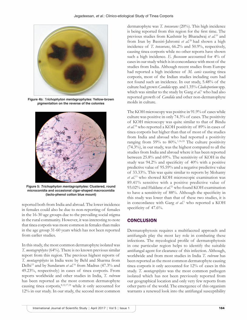
32 out of 50 samples, was the most common dermatophyte isolated (64%) followed by T. tonsurans (20%). T. rubrum accounted for 12% and E. floccosum for 4%. Microsporum spp. was not isolated from any of the patients who were included in this study. The mycological pattern of dermatophytes isolated in the study is depicted in Figure 3. The culture characteristics and fungal identification of Trichophyton mentagrophytes are depicted in Figures 4a, b and 5 respectively.
DISCUSSION
This study showed that the majority of the patients with tinea corporis were in the age group 21-40 (48.65%) which is similar to findings reported in other studies. This could be due to the fact that this is the group of the population indulging in greater physical activity, such as agriculture, outdoor activities, and sports that leads to increased sweating and therefore predispose to the disease. The study showed an overall predominance of tinea corporis in males (56.76%). This is in concordance with studies
Figure 1: Age-group and gender-wise distribution of the study population
Figure 2: Fungal culture
Figure 3: Mycological pattern of dermatophytes
Figure 4a: Trichophyton mentagrophytes: Cream colored colonies with raised central tufts and suede-like surface grown
on Sabouraud’s dextrose agar medium
Jegadeesan, et al.: Clinico-etiological Study of Tinea Corporis
164 165164International Journal of Scientific Study | April 2017 | Vol 5 | Issue 1 165 International Journal of Scientific Study | April 2017 | Vol 5 | Issue 1
reported both from India and abroad. The lower incidence in females could also be due to non-reporting of females in the 16-30 age groups due to the prevailing social stigma in the rural community. However, it was interesting to note that tinea corporis was more common in females than males in the age group 31-60 years which has not been reported from earlier studies.
In this study, the most common dermatophyte isolated was T. mentagrophytes (64%). There is no known previous similar report from this region. The previous highest reports of T. mentagrophytes in India were by Behl and Sharma from Delhi15 and by Sundaram et al.16 from Madras (47.3% and 49.23%, respectively) in cases of tinea corporis. From reports worldwide and other studies in India, T. rubrum has been reported as the most common dermatophyte causing tinea corporis,8,12,17,18 while it only accounted for 12% in our study. In our study, the second most common
dermatophyte was T. tonsurans (20%). This high incidence is being reported from this region for the first time. The previous studies from Kashmir by Bharadwaj et al.19 and from Iran by Bassiri-Jahromi et al.20 had shown a high incidence of T. tonsurans, 66.2% and 50.9%, respectively, causing tinea corporis while no other reports have shown such a high incidence. E. floccosum accounted for 4% of cases in our study which is in concordance with most of the studies from India. Although recent studies from Europe had reported a high incidence of M. canis causing tinea corporis, most of the Indian studies including ours had not found such an incidence. In our study, 5.48% of the culture had grown Candida spp. and 1.35% Cladosporium spp. which was similar to the study by Garg et al.1 who had also reported growth of Candida and other non-dermatophyte molds in culture.
The KOH microscopy was positive in 91.9% of cases while culture was positive in only 74.3% of cases. The positivity of KOH microscopy was quite similar to that of Bindu et al.18 who reported a KOH positivity of 89% in cases of tinea corporis but higher than that of most of the studies from India and abroad who had reported a positivity ranging from 59% to 80%.1,16,18 The culture positivity (74.3%), in our study, was the highest compared to all the studies from India and abroad where it has been reported between 25.8% and 69%. The sensitivity of KOH in the study was 94.2% and specificity of 40% with a positive predictive value of 95.59% and a negative predictive value of 33.33%. This was quite similar to reports by Mohanty et al.13 who showed KOH microscopic examination was 89.41% sensitive with a positive predictive value of 93.02% and Haldane et al.21 who found KOH examination to have a sensitivity of 88%. Although the specificity in this study was lower than that of these two studies, it is in concordance with Garg et al.1 who reported a KOH specificity of 47.6%.
CONCLUSION
Dermatophytosis requires a multifaceted approach and antifungals play the most key role in combating these infections. The mycological profile of dermatophytosis in one particular region helps to identify the suitable antifungal agent for clearance of this infection. Although, worldwide and from most studies in India T. rubrum has been reported as the most common dermatophyte causing tinea corporis it only accounted for 12% of cases in this study. T. mentagrophytes was the most common pathogen isolated which has not been previously reported from our geographical location and only very few reports from other parts of the world. The emergence of this organism warrants a renewed look into the antifungal susceptibility
Figure 4b: Trichophyton mentagrophytes: Yellow-brown pigmentation on the reverse of the colonies
Figure 5: Trichophyton mentagrophytes: Clustered, round microconidia and occasional cigar-shaped macroconidia
(lacto-phenol cotton blue mount)

Jegadeesan, et al.: Clinico-etiological Study of Tinea Corporis
164 165164International Journal of Scientific Study | April 2017 | Vol 5 | Issue 1 165 International Journal of Scientific Study | April 2017 | Vol 5 | Issue 1
How to cite this article: Jegadeesan M, Kuruvila S, Nair S. Clinico-etiological Study of Tinea Corporis: Emergence of Trichophyton mentagrophytes. Int J Sci Stud 2017;5(1):161-165.
Source of Support: Nil, Conflict of Interest: None declared.
patterns for combating this common superficial fungal infection efficiently.
REFERENCES
1. Garg J, Tilak R, Garg A, Prakash P, Gulati AK, Nath G. Rapid detection of dermatophytes from skin and hair. BMC Res Notes 2009;2:60.
2. Gräser Y, Scott J, Summerbell R. The new species concept in dermatophytes-a polyphasic approach. Mycopathologia 2008;166:239-56.
3. Rippon JW. The pathogenic fungi and the pathogenic actinomycetes. In: Medical Mycology. 3rd ed. Philadelphia, PA: W. B. Saunders; 1988. p. 169-275.
4. Hay RJ, Ashbee HR. Mycology. In: Burns T, Breathnach S, Cox N, Griffiths C, editors. Rook’s Textbook of Dermatology. 8th ed. Oxford: Wiley-Blackwell Science Ltd.; 2010. p. 36, 94.
5. Ghannoum MA, Isham NC. Dermatophytes and dermatophytoses. In: Anaissie EJ, editor. Clinical Mycology. 2nd ed. Philadelphia, PA: Churchill Livingstone; 2009. p. 375-84.
6. Kanwar AJ, De D. Superficial fungal infections. In: Valia AR, Valia G, editors. IADVL Textbook of Dermatology. 3rd ed. Mumbai: Bhilani Publishers; 2008. p. 252-97.
7. Havlickova B, Czaika VA, Friedrich M. Epidemiological trends in skin mycoses worldwide. Mycoses 2008;51 Suppl 4:2-15.
8. Seebacher C, Bouchara JP, Mignon B. Updates on the epidemiology of dermatophyte infections. Mycopathologia 2008;166:335-52.
9. Ranganathan S, Menon T, Selvi SG, Kamalam A. Effect of socio-economic status on the prevalence of dermatophytosis in Madras. Indian J Dermatol Venereol Leprol 1995;61:16-8.
10. Degreef H. Clinical forms of dermatophytosis (ringworm infection). Mycopathologia 2008;166:257-65.
11. Weitzman I, Summerbell RC. The dermatophytes. Clin Microbiol Rev 1995;8:240-59.
12. Foster KW, Ghannoum MA, Elewski BE. Epidemiologic surveillance of cutaneous fungal infection in the United States from 1999 to 2002. J Am Acad Dermatol 2004;50:748-52.
13. Mohanty JC, Mohanty SK, Sahoo RC, Sahoo A, Prahara CN. Diagnosis of superficial mycoses by direct microscopy - A statistical evaluation. Indian J Dermatol Venereol Leprol 1999;65:72-4.
14. Andrews MD, Burns M. Common tinea infections in children. Am Fam Physician 2008;77:1415-20.
15. Behl PN, Sharma MD. Incidence of mycotic infections in Delhi. Indian J Dermatol 1957;3:5-7.
16. Sundaram BM, Badrinath S, Subramanian S. Clinico-mycological study of dermatomycoses in Madras (India). Mykosen 1986;29:230-4.
17. Venkatesan G, Singh AJ, Murugesan AG, Janaki C, Shankar SG. Trichophyton rubrum - The predominant etiological agent in human dermatophytoses in Chennai, India. Afr J Microbiol Res 2007;1:9-12.
18. Bindu V, Pavithran K. Clinico-mycological study of dermatophytosis in Calicut. Indian J Dermatol Venereol Leprol 2002;68:259-61.
19. Bhardwaj G, Hajini GH, Khan IA, Masood Q, Khosa RK. Dermatophytoses in Kashmir, India. Mykosen 1987;30:135-8.
20. Bassiri-Jahromi S, Khaksari AA. Epidemiological survey of dermatophytosis in Tehran, Iran, from 2000 to 2005. Indian J Dermatol Venereol Leprol 2009;75:142-7.
21. Haldane DJ, Robart E. A comparison of calcofluor white, potassium hydroxide, and culture for the laboratory diagnosis of superficial fungal infection. Diagn Microbiol Infect Dis 1990;13:337-9.
![SCIENCE CHINA Life Sciences - Springer · tions, such as tinea capitis, tinea corporis, tinea inguinalis, tinea manus, tinea unguium and tinea pedis [1–3]. Unlike](https://img.pdfslide.us/doc/110x75/5d1b54ac88c993283c8ce38a/science-china-life-sciences-springer-tions-such-as-tinea-capitis-tinea-corporis.jpg)