Embed Size (px)
Citation preview
CLINICAL VALIDATION TEST OF ARTIFICIAL
INTELLIGENCE ASSISTED IN SKIN CANCER
SCREENING SYSTEM ON SMARTPHONE
APPLICATION
BY
MISS THORFUN TREEWATANAKUL
A THESIS SUBMITTED IN PARTIAL FULFILLMENT OF
THE REQUIREMENTS FOR THE DEGREE OF
MASTER OF SCIENCE (DERMATOLOGY)
CHULABHORN INTERNATIONAL COLLEGE OF MEDICINE
THAMMASAT UNIVERSITY
ACADEMIC YEAR 2017
COPYRIGHT OF THAMMASAT UNIVERSITY
Ref. code: 25605929040508OMC


(1)
Thesis Title CLINICAL VALIDATION TEST OF
ARTIFICIAL INTELLIGENCE ASSISTED
IN SKIN CANCER SCREENING SYSTEM
ON SMARTPHONE APPLICATION
Author Miss Thorfun Treewatatnakul
Degree Master of science
Major Field/Faculty/University Dermatology
Chulabhorn International College of Medicine
Thammasat university
Thesis Advisor Asst. Prof. Panlop Chakkavittumrong, M.D.
Thesis Co-Advisor Saroj Suvanasuthi M.D., Ph.D., ABHRS.
Assoc. Prof. Charturong Tantibundhit, Ph.D.
Academic Years 2017
ABSTRACT
Background: Deep learning has been reported to outperform other
handcrafted machine learning ( ML) algorithms in classification of pigmented skin
lesions. To develop reliable, portable, automated diagnosis system with high diagnostic
performances is essential for skin cancer screening and diagnosis regardless of
specialized dermatologists.
Objectives: To validate the diagnostic performances of artificial
intelligence (AI) assisted in skin cancer screening system compared to Board-certified
dermatologists versus experienced dermoscopic specialized dermatologists using
dermoscopic images in clinical practice.
Methods: Retrospective, descriptive study using 200 randomly selected
dermoscopic images of pigmented skin lesions ( PSLs) ; 31 melanomas, 65 nevi, 52
seborrheic keratoses, 6 squamous cell carcinomas, 39 basal cell carcinomas, and 7 other
lesions from the medical records in Samitivej Sukhumvit Hospital, Bangkok, Thailand.
We examined our AI system’ s performance against three board- certified
dermatologists versus three experienced dermoscopic specialized dermatologists.
Ref. code: 25605929040508OMC

(2)
Results: AI system showed higher sensitivity ( 67. 7% ) in melanoma
diagnosis compared to Board-certified dermatologists ( 22. 6% ) and almost the same
level as dermoscopic specialized dermatologists (69.9%).
Conclusion: Our artificial intelligence system using deep learning method
achieves performance in diagnosis of melanoma with a same level as Board-certified
dermatologists. However, AI system still need further trainings to improve its outcomes
before applying in clinical settings especially in both squamous cell carcinomas and
basal cell carcinomas categories.
Keywords: Artificial intelligence, deep learning, dermoscopy, melanoma, skin cancer,
screening system, mobile application
Ref. code: 25605929040508OMC

(3)
ACKNOWLEDGEMENTS
Foremost, this work would not have been possible without the kindly
support from my beloved advisor, Asst. Prof. Panlop Chakkavittumrong, M.D. His
guidance helped me in every step in this research. My enormous gratefulness towards
him is indescribably unforgettable.
Moreover, I really appreciate for the learning opportunities given by my
committee chair, Saroj Suvanasuthi M.D., Ph.D., ABHRS. His motivation led me to
start this amazing project. It has been a period of intense learning for me, not only in
the knowledge, but also on a personal mindset. He has been giving me many fruitful
advices to complete this thesis successfully.
I sincerely thank to the rest of my committee for their support: Assoc. Prof.
Charturong Tantibundhit, Ph. D. , my co-advisor, and his artificial intelligence for
dermatology project team. They dedicated themselves to this research. And they have
introduced me to see the new world, the world of AI.
Most importantly, I would like to thank to my parents who always standby
my side no matter what.
Miss Thorfun Treewatanakul
Ref. code: 25605929040508OMC

(4)
TABLE OF CONTENTS
Page
ABSTRACT (1)
ACKNOWLEDGEMENTS (3)
LIST OF TABLES (7)
LIST OF FIGURES (8)
LIST OF ABBREVIATIONS (11)
CHAPTER 1 INTRODUCTION 1
1.1 Background and Rationale 1
1.2 Research question 2
1.3 Specific objective 2
1.4 Hypothesis 3
1.5 Keywords 3
1.6 Operation definition 3
1.7 Ethical consideration 3
1.8 Limitation 3
1.9 Expected benefits and application 3
1.10 Obstacles and strategies to solve the problems 4
CHAPTER 2 REVIEW OF LITERATURE 6
2.1 Pigmented skin lesions 6
2.1.1 Benign 6
2.1.2 Skin cancers 12
2.2 Skin cancer diagnosis 15
Ref. code: 25605929040508OMC

(5)
2.2.1 Visual examination (Naked eyes) 15
2.2.2 Non-invasive imaging tools 16
2.2.2.1 Dermoscopy 19
(1) The Heuristic approach 20
(2) The Analytical approach 25
2.2.3 Histopathological examination 26
2.3 Computer aided diagnosis in skin cancers 27
2.3.1 Artificial intelligence 30
2.3.2 Deep learning 31
CHAPTER 3 RESEARCH METHODOLOGY 38
3.1 Materials 38
3.1.1 Dermoscopic images of pigmented skin lesions 38
3.1.1.1 Test dataset 38
(1) Sample size 38
(2) Inclusion criteria 38
(3) Exclusion criteria 39
3.1.1.2 Trained dataset 39
3.2 Research design 42
3.2.1 Research location 42
3.2.2 Research procedures 43
3.2.3 Outcome measurements 45
3.3 Data analysis 45
CHAPTER 4 RESULTS AND DISCUSSION 48
4.1 Results 48
4.1.1 Diagnostic performances of each type of pigmented skin 48
lesions
4.1.1.1 Board-certified dermatologists 48
4.1.1.2 Experienced dermoscopic specialized dermatologists 51
Ref. code: 25605929040508OMC

(6)
4.1.1.2 Artificial intelligence system (Deep learning) 54
4.1.2 Comparison of skin cancer diagnostic performances among 56
three groups
4.1.3 Comparison of pigmented skin lesions 61
diagnostic performances among three groups
4.2 Discussion 66
CHAPTER 5 CONCLUSIONS AND RECOMMENDATIONS 71
5.1 Conclusions 71
5.2 Recommendations 71
REFERENCES 72
APPENDICES 80
APPENDIX A CASE RECORD FORM 81
APPENDIX B TEST DATASET FOR DERMATOLOGISTS 82
APPENDIX C ANSWER SHEET 83
APPENDIX D SMARTPHONE APPLICATION OF AI ASSISTED 84
IN SKIN CANCER SCREENING SYSTEM
BIOGRAPHY 85
Ref. code: 25605929040508OMC

(7)
LIST OF TABLES
Tables Page
1.1 Administration and time schedule 6
2.2.2 In vivo imaging techniques for the diagnosis of skin cancer 17
2.2.2.1.1a ABCD rule of dermoscopy (Modified 1994) 23
2.2.2.1.1b The 7-point checklist 24
2.2.2.1.1c Menzies’ method for the diagnosis of melanoma 25
2.3.2 Area under ROC curve of different methods on diagnosis of 35
melanoma, nevus, and seborrheic keratosis
3.1.1.2 Sources of trained dataset 40
4.1.1.1 Mean diagnostic performances of Board-certified dermatologists 48
in diagnosis of different pigmented skin lesions
4.1.1.2 Mean diagnostic performances of experienced dermoscopic 51
specialized dermatologists in diagnosis of different pigmented skin
lesions
4.1.1a Interclass correlation coefficient (ICC) 52
4.1.1.3 Diagnostic performances of artificial intelligence system in 54
diagnosis of different pigmented skin lesions
4.2 Sources of trained dataset 68
Ref. code: 25605929040508OMC

(8)
LIST OF FIGURES
Figures Page
2.1.1.1 Acquired melanocytic nevi 7
2.1.1.2 Seborrheic keratosis 7
2.1.1.3 Spitz nevus 8
2.1.1.4 Reed nevus 8
2.1.1.5 Blue nevus 9
2.1.1.6 Nevus spilus 9
2.1.1.7 Congenital melanocytic nevi 10
2.1.1.8 Atypical melanocytic nevus 11
2.1.1.9 Solar lentigines 11
2.1.1.11 Dermatofibroma 12
2.1.2.1 The four most common types of melanoma 13
2.1.2.2 Basal cell carcinoma 14
2.1.2.3 Squamous cell carcinoma 15
2.2.2.1.1a The Pattern Analysis (Two steps algorithm) 20
2.2.2.1.1b,c Stepwise evaluation of dermoscopic features of Pattern Analysis 21
(Two steps algorithm)
2.2.2.1.1d Criteria for melanocytic lesions 22
2.2.2.1.1e Benign and malignant patterns of volar aspects 22
2.2.2.1.2 The analytic approach (Chaos & Clues method) 26
2.3 CAD steps to help in diagnosis of pigmented skin lesions 27
2.3.2a Flowchart of algorithms used to classify melanoma, 28
seborrheic kerratosis, and nevus.
2.3.2b Dermoscopic images of malignant melanoma from ISIC-ISBI 28
Challenge 2017
2.3.2c Example network architectures for CNNs. 29
Right: a residual network with 34 parameter layers
2.3.2d A deep DenseNet with three dense blocks. 29
2.3.2e Diagram of 3-Binary DenseNet121 classifier and 35
3-Binary ResNet50 Classifier
Ref. code: 25605929040508OMC

(9)
2.3.2f Average ROC ± SD of four algorithms and the average AUC ± SD 36
for melanoma(MM) classification
2.3.2g Flowchart of scientific development design in this study 37
3.1.1a, b Examples of dermoscopic images of melanoma 41
3.1.1c FotoFinder Hub® system 42
(FotoFinder Systems GmbH, Deutschland)
3.2.2 Flow chart of study procedures 43
3.2.2.3a,b Examples of validation test for dermatologists 44
3.3.1 Diagnostic parameters 46
4.1.2.1a Diagnostic performances in diagnosis of different types of skin 50
cancers; three Board-certified dermatologists and mean values
4.1.2.2a Diagnostic performances in diagnosis of different types of skin 53
cancers; three experienced dermoscopic specialized dermatologists
and mean values
4.1.1.3 Confusion matrix of AI system performance 55
4.1.2.1 Sensitivities in diagnosis of different types of skin cancers 56
compared among three groups
4.1.2.2 Specificities in diagnosis of different types of skin cancers 57
compared among three groups
4.1.2.3 Accuracies in diagnosis of different types of skin cancers 58
compared among three groups
4.1.2.4 Positive predictive values(PPV) in diagnosis of different types of 59
skin cancers compared among three groups
4.1.2.5 Negative predictive values(NPV) in diagnosis of different types of 60
skin cancers compared among three groups
4.1.3.1 Sensitivities in diagnosis of different types of benign pigmented 61
skin lesions compared among three groups
4.1.3.2 Specificities in diagnosis of different types of benign pigmented 62
skin lesions compared among three groups
4.1.3.3 Accuracies in diagnosis of different types of benign pigmented 63
skin lesions compared among three groups
Ref. code: 25605929040508OMC

(10)
4.1.3.4 Positive predictive values(PPV) in diagnosis of different types of 64
benign pigmented skin lesions compared among three groups
4.1.3.5 Negative predictive values(NPV) in diagnosis of different types of 65
benign pigmented skin lesions compared among three groups
4.2.1a,b,c Clinical dermoscopic images in test dataset 66
4.2.2a Confusion matrix of AI system performance: 67
melanoma classification
4.2.2b Confusion matrix of AI system performance: 69
BCC classification
4.2.2c Confusion matrix of AI system performance: 70
SCC classification
Ref. code: 25605929040508OMC

(11)
LIST OF ABBREVIATIONS
Symbols/Abbreviations Terms
α Alpha
AI Artificial intelligence
β Beta
BCC Basal cell carcinoma
BMC-IRB Institutional Review Board of Bangkok Hospital
Medical Center
CAD Computer aided detection
CADx Computer aided diagnosis
cm Centimeter
CNNs Convolutional neural networks
CSLM Confocal scanning laser microscopy
CT scan Computerized Tomography scan
DARPA
DenseNets
DL
DNA
e.g.
The Defense Advanced Research Projects Agency
Densely Convolutional Networks
Deep learning
Deoxyribonucleic acid
For example
ELM Epiluminescence microscopy
Fig Figure
FN False negative
FP False positive
GANs Generative Adversarial Networks
GPU Graphics processing unit
HFU
ICC
High frequency ultrasound
Interclass Correlation Coefficient
ISIC International Skin Image Collaboration
Ref. code: 25605929040508OMC

(12)
ISIC- ISBI International Symposium on Biomedical Imaging
Hosted by the International Skin Image
Collaboration
KNN
ML
mm
K-Nearest Neighbors
Machine learning
millimeter
MM Malignant melanoma
MRI Magnetic resonance imaging
n
NPV
Sample size
Negative predictive values
NV Nevus
OCT
Others
p
p0
Optical coherence tomography
Other diagnosis
Proportion
Reference value
PET Positron emission tomography
PPV
PSLs
ResNets
RMC-IRB
ROC curve
Positive predictive values
Pigmented skin lesions
Deep Residual Neural Networks
Institutional Review Board of Bangkok Hospital
Medical Center
Receiver Operating Characteristic curve
SD Standard deviation
SebK
SK
SPSS
Seborrheic keratosis
Seborrheic keratosis
Statistical Package for Social Sciences
3D
TBSE
TDS
Three dimensions
Total body skin examination
Total dermoscopy score
TLM Transillumination
TN True negative
Ref. code: 25605929040508OMC

(13)
TP
U.S.
WGAN-GP
True positive
United States of America
Wasserstein-Generative Adversarial Network with
gradient penalty
XLM Cross-polarization epiluminescence
Ref. code: 25605929040508OMC

1
CHAPTER 1
INTRODUCTION
1.1 Background and Rationale
Pigmented skin lesions ( PSLs) are included both benign and malignancy.
Melanoma, a malignant form of PSLs, is the most aggressive and life- threatening skin
cancer. The statistic of global cancers shows that the incidence rates and mortality rates
of malignant melanoma are increased every year. Although early stages are highly
survivable, melanoma can rapidly spread and become fatal. 5-year survival rate of
melanoma drops from 97% in the earliest stage to 10% in the latest stage. Therefore,
early detection of melanoma is critical to reduce morbidity and mortality rates of
patients. However, overdiagnosis may lead to unnecessary biopsies, which possibly
results in adverse effects.
Diagnosing melanoma begins with visual examination. Only 60% of
clinical accuracy in diagnosis with naked eyes has been reported for dermatologists in
the specialized centers. Dermoscopy, which is a microscopic imaging tool for
pigmented skin lesions diagnosis, significantly improved the accuracy compared with
inspection by naked eyes but only in specialized well- trained physicians. Moreover,
diagnosis of early stage of melanoma is still challenging even for experienced
dermatologists.
To overcome these limitations, computer aided diagnosis system has been
introduced. Deep learning, a subtype of machine learning, has been used in several
fields due to outperformance over other handcrafted machine learning algorithms
particularly in visual task such as face recognition, object classification, playing
strategic board game like Go, and medical screening which has been shown to exceed
human performances.
Thai researchers team has trained AI with four high potential algorithms in
classification of melanomas, nevi, and seborrheic keratoses by using dermoscopic
images from International Skin Imaging Collaboration (ISIC) 2017 dataset. The result
showed that Densely Convolutional Network ( DenseNet- 121) , one of four deep
Ref. code: 25605929040508OMC

2
learning algorithm network, performed the best in sensitivity, specificity, and accuracy
up to 80-90% . Moreover, with the help of artificial training images generated from
WGAN- GP can solve the problem of scarcity of training data and improve
classification outcome of melanoma.
1.2 Research question
Although the computer aided diagnosis of melanoma had high sensitivity,
specificity, and accuracy under experimental conditions, the use of this method in real
clinical settings is still unknown.
In this research, we aimed to validate the diagnostic performances of the
first artificial intelligence assisted skin cancer screening system in Thailand and
compare the diagnostic ability to Board- certified dermatologists and experienced
dermoscopic specialized dermatologists using clinical dermoscopic images.
1.3 Specific objective
The primary objective is to validate the diagnostic performances of the
artificial intelligence ( deep learning) in skin cancers diagnosis including sensitivity,
specificity, accuracy, positive predictive values, and negative predictive values using
clinical dermoscopic images.
The secondary objective is to compare the diagnostic performances of the
artificial intelligence with dermoscopic specialized dermatologists vs Board-certified
dermatologists in skin cancers diagnosis using dermoscopic images.
1.4 Hypothesis
Artificial intelligence (Deep learning) might achieve performances in
diagnosis of skin cancers with a same level as Board-certified dermatologist or
dermoscopic specialized dermatologist.
Ref. code: 25605929040508OMC

3
1.5 Keywords
Artificial intelligence
Deep learning
Dermoscopy
Melanoma
Skin cancer screening system
Mobile application
1.6 Operation definition
Clinical dermoscopic images of pigmented skin lesions
1.7 Ethical consideration
The study protocol was granted by Institutional Review Board of Bangkok
Hospital Medical Center (BMC-IRB).
1.8 Limitation
Lack of clinical dermoscopic images of skin cancers especially squamous
cell carcinomas and basal cell carcinomas used to train artificial intelligence. In
addition, colors of images from multi-sources images were different which could lead
AI in low diagnostic performances.
1.9 Expected benefits and application
Early detection of melanoma is critical. Advances in computer aided
classification of pigmented skin lesions could potentially assist dermatologists or
medical practitioners in improving diagnostic accuracy especially in early stage
melanoma. Moreover, getting to a dermatologist is rarely easy. To make the algorithm
system compatible with mobile application can greatly extend the accessibility of
Ref. code: 25605929040508OMC

4
dermatologists outside of the hospitals and even in the remote areas which lack of
specialists. This artificial intelligence technology will allow patients to self-follow up
in suspicious pigmented skin lesions and early detect skin cancers from anywhere.
Therefore, it provides low-cost access and high reliability to vital diagnostic care.
However, rigorous prospective validation of this artificial intelligence assisted skin
cancer screening system is necessary before it can be used in clinical practice. So that,
this research is designed to test this system as well as compare its performance with
standard process.
1.10 Obstacles and strategies to solve the problems
The lack of dermoscopic images of skin cancers especially squamous cell
carcinomas and basal cell carcinomas caused low diagnostic performances for AI. The
problem was solved by combining clinical dermoscopic images from medical records
into the trained dataset for AI. Also, dermoscopic images from reliable textbook were
included in the trained dataset.
For adjusting the color of images, the researchers applied color constancy
using the Shades of Grey method to improve the outcome.
Ref. code: 25605929040508OMC

5
Table 1.1 Administration and time schedule
2017 2018
DE
C
JAN
FE
B
MA
R
AP
R
MA
Y
JUN
JUL
Y
Research proposal
Research ethics
Experiment
Data analysis & conclusion
Proceeding
Publishing
Ref. code: 25605929040508OMC

6
CHAPTER 2
REVIEW OF LITERATURE
2.1 Pigmented skin lesions
Pigmented skin lesions (PSLs) which refer to lesions that are brown, black,
blue, grey or red in color, are often melanocytic. (1) They can be classed as benign or
malignant. Most pigmented skin lesions are reported as benign nevi, however a small
number will be malignancy. (2) They are very similar in morphologies, colors, and
textures. To distinguish between malignant and benign moles is challenging task for
dermatologists. (3, 4) The following section summarizes the common pigmented skin
lesions.
2.1.1 Benign
Benign pigmented skin lesions (PSLs) are harmless, although they
are closely related to malignant melanomas. The common benign PSLs are such as
acquired melanocytic nevi, seborrheic keratoses, blue nevi, atypical or dysplastic nevi,
congenital nevi, pigmented Spitz nevi etc.
2.1.1.1 Acquired melanocytic nevi
Acquired melanocytic nevi, commonly called benign moles,
are usually small, pigmented macules, papules, or nodules with sharply demarcated
border. They are classified into three groups as listed below. (5)
• Junctional nevus: usually small, brown to black macule
(Figure 2.1.1.1a)
• Intradermal nevus: a dome-shaped skin-colored or light to
dark brown papule or nodule (Figure 2.1.1.1b)
• Compound nevus: can be light to dark brown papule or
nodule (Figure2.1.1.1c)
Ref. code: 25605929040508OMC

7
a) b) c)
Figure 2.1.1.1 Acquired melanocytic nevi. (5)
a) junctional nevus b) intradermal nevus c) compound nevus
2.1.1.2 Seborrheic keratosis
Seborrheic keratosis is a benign epithelial skin neoplasm. It can
appear on any site of body especially on face and trunk, but not palms and soles. It
usually begins as flat, well-circumscribed, black or brown patches. Then, it may
become polypoidal with verrucous and dull surface. "Stuck-on" appearance is the key
feature. Its color varies from yellowish to brownish-black. (5, 6)
Figure 2.1.1.2 Seborrheic keratosis showing stuck-on appearance. (7)
Ref. code: 25605929040508OMC

8
2.1.1.3 Spitz nevi
It is characterized by a small (<1 cm), dome-shaped, tan or pink
nodule with often a history of recent rapid growth. It is very difficult to distinguish
from melanoma.
Figure 2.1.1.3 Spitz nevus (8)
Available from https://www.dermnetnz.org/topics/spitz-naevus/
2.1.1.4 Reed nevi
It is characterized as dark brown to black papule or plaque,
usually smaller than Spitz nevus. It is often seen in young women around thirty. Lower
extremities are common sites.
Figure 2.1.1.4 Reed nevus. (9)
Ref. code: 25605929040508OMC

9
2.1.1.5 Blue nevi
The clinical presentation of blue nevi is acquired, firm blue to
gray to black, sharply demarcated papule or nodule. About 50% are seen on the dorsal
aspect of the hands and feet. Although it is benign, some types of blue nevi may have
an elevated risk for development of melanoma.
Figure 2.1.1.5 Blue nevus. A well-circumscribed, blue, dome-shaped papule. (5)
2.1.1.6 Nevus spilus
It consists of a light brown macule which vary in size and
multiple dark brown small macules (2-3 mm) or papules scattered throughout the
pigmented background.
Figure 2.1.1.6 Nevus spilus. Multiple brown macules and papules superimposed upon
a tan patch. (5)
Ref. code: 25605929040508OMC
10
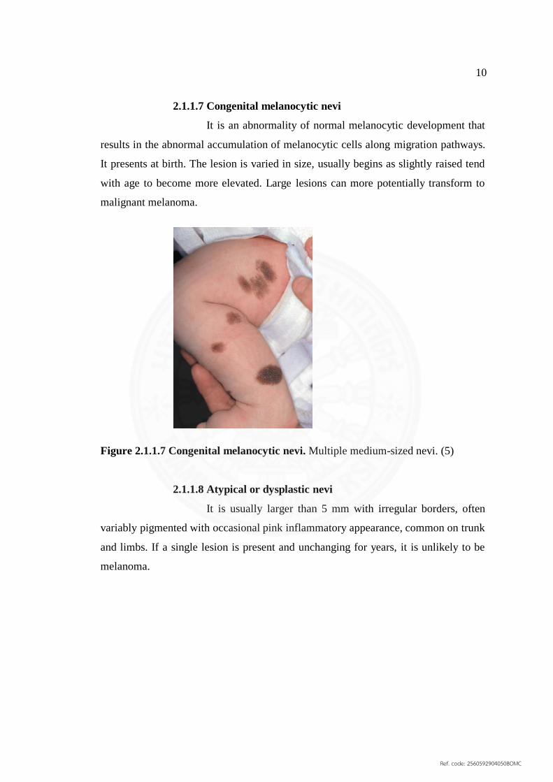
2.1.1.7 Congenital melanocytic nevi
It is an abnormality of normal melanocytic development that
results in the abnormal accumulation of melanocytic cells along migration pathways.
It presents at birth. The lesion is varied in size, usually begins as slightly raised tend
with age to become more elevated. Large lesions can more potentially transform to
malignant melanoma.
Figure 2.1.1.7 Congenital melanocytic nevi. Multiple medium-sized nevi. (5)
2.1.1.8 Atypical or dysplastic nevi
It is usually larger than 5 mm with irregular borders, often
variably pigmented with occasional pink inflammatory appearance, common on trunk
and limbs. If a single lesion is present and unchanging for years, it is unlikely to be
melanoma.
Ref. code: 25605929040508OMC

11
Figure 2.1.1.8 Atypical melanocytic nevus. There is asymmetry as well as several
shades of brown, simulating the clinical features seen in cutaneous melanoma. (5)
2.1.1.9 Solar lentigines
They usually present with numerous small (<0.5 mm) brown
macules. Sun-exposed areas such as face, arms, and hands are common sites.
Figure 2.1.1.9 Solar lentigines. (5)
2.1.1.10 Café-au-lait macules
They may be described as homogenous light to dark brown
macules with well-defined margins, usually 2-5 cm in diameter, but may vary in size.
They can be located anywhere on the body except mucous membranes. These skin
lesions are found in both normal population and McCune-Albright syndrome patients.
Ref. code: 25605929040508OMC

12
2.1.1.11 Dermatofibromas
Dermatofibroma is characterized as a button-like dermal
nodule commonly seen on the extremities. “Dimple” sign is the key clinical finding. It
can be pigmented or non-pigmented.
Figure 2.1.1.11 Dermatofibroma. Hyperpigmented firm papule on the lower
extremity. (5)
2.1.2 Malignancy
Skin cancers are abnormal growth of skin cells which most often
develops on chronically sun exposed skin. There are three main types of skin cancers
including malignant melanoma, basal cell carcinoma, and squamous cell carcinoma.
Melanomas are often pigmented unlike others.
2.1.2.1 Melanoma
Melanoma is the most aggressive skin cancer. It can develop
anywhere on the body and occur either on normal-appearing skin or existing mole. (10)
Typical features are asymmetry of the lesion, irregular borders, vary in color, diameter
greater than 5 mm, growth of nodules and regression of lesions. Although melanomas
are usually pigmented, they can also be amelanotic. There are four major subtypes of
melanoma which can be classified according to clinical presentation and histological
features. (11)
Ref. code: 25605929040508OMC

13
Superficial spreading melanoma is primarily macule that can
slowly develop into a nodule or plaque, often with multiple colors and areas of
regression.
Nodular melanoma is often presented as brown to black nodules
with eroded or bleeding ulcer.
Lentigo maligna melanoma usually arises slowly from
melanoma in situ on the sun-damaged skin.
Acral lentiginous melanoma is typically located on periphery. It
is primarily an irregular, poorly defined border pigmentation, later becomes nodule in
an invasive growth phase.
Figure 2.1.2.1 The four most common types of melanoma: clinical and
dermoscopic images (5)
a) Small superficial melanoma
b) Nodular type melanoma
c) Small facial melanoma in situ (lentigo maligna)
d) Acral melanoma
Ref. code: 25605929040508OMC

14
2.1.2.2 Pigmented basal cell carcinoma
Basal cell carcinoma (BCC) is a tumor that arises within sun-
damaged skin. The major risk factor is UV radiation. There are four main
clinicopathologic types including nodular, superficial, morpheaform, and
fibroepithelial BCC.(5) Although most BCCs are amelanotic, pigmented BCCs can be
observed more commonly in those with dark skin types. Classic presentations are
pearly rolled border and central hemorrhagic crust or telangiectasia.
Figure 2.1.2.2 Basal cell carcinoma, nodular subtype (5)
2.1.2.3 Pigmented squamous cell carcinoma
Squamous cell carcinoma (SCC) is a common type of skin
cancers. It can appear on any part of the body including lips and genitals. The color
usually varies from erythematous to skin-colored, rarely pigmented variants. SCCs are
often papulonodular, but can be plaque-like, papillomatous or exophytic. It can be
classified into three groups depending on histopathology of lesions.
Actinic keratosis is considered as precancerous or premalignant
tumor because atypical keratinocytes are confined within epidermis.
Squamous cell carcinoma in situ which is commonly known as
Bowen’s disease, is often not aggressive. The most common presentation are
erythematous scaly patches or plaques.
Invasive squamous cell carcinoma is the aggressive form of
SCC.
Ref. code: 25605929040508OMC

15
Figure 2.1.2.3 Squamous cell carcinoma on patient’s face with sun damaged skin. (7)
Available from https://www.aad.org/public/diseases/skin-cancer/squamous-cell-
carcinoma
2.2 Skin cancer diagnosis
Malignant melanoma is a lethal form of skin cancers resulting from DNA
mutation of melanocytes. (12) The global cancer statistics show that the number of
new patients and mortality rates of melanoma are steadily increased every year. (13)
This current year, the American Cancer Society estimates that 87,000 new cases and
9,000 deaths will occur in U.S. due to the disease. (14) In the advanced stages of
melanoma are incurable and the treatments are mainly palliative, including surgery,
immunotherapy, chemotherapy, targeted therapy, and/or radiation therapy. (11, 15-17)
Therefore, screening system in early melanoma is thought to improve the prognosis and
reduce morbidity and mortality rates of patients.
2.2.1 Visual examination (Naked eyes)
Most malignant melanomas arise on the skin surface and primarily
diagnosed by visual examination. The key principle for skin cancer screening
techniques is total body skin examination (TBSE). (3)
The clinical diagnosis of dermatologists is based on three analysis
steps of pigmented skin lesion. First step is excluding non-melanocytic lesions and
searching for suspicious melanocytic lesions. There are many methods used for
Ref. code: 25605929040508OMC

16
identifying suspicious melanocytic lesions such as ABCDE criteria (18) and the
Glasgow 7-point checklist (19). The ABCDE approach has been widely used in clinical
practice. The rule which “A” stands for asymmetry, “B” stands for border irregularity,
“C” stands for color ununiform, “D” stands for diameter greater than six mm, and “E”
stands for elevation and/or enlargement of a lesion. (20) Moreover, EFG is being added
to the ABCD rule for nodular lesions, including “F” which stands for firm, and “G”
stands for growing for one month. (2) Second step is comparative analysis, which is
looking for the “ugly duckling sign” or the moles that are not alike the others in the
same patient. Last step is to search for rapid growth or recent change of lesions like in
“E” and “G” in ABCD rule with additional EGF.
However, unaided visual inspection of pigmented skin lesions is
suboptimal. (21, 22) Only 60% of clinical accuracy in diagnosis with naked eyes has
been reported even for expert dermatologists in the specialized centers. (23) Another
study showed that sensitivity in diagnosis of clinical melanoma of experienced
dermatologists is approximately 70%. (24)
2.2.2 Non-invasive imaging tools
Although the best way and the most reliable method to
differentiate between benign and malignant lesions are histopathological examination
from skin biopsy, there is limitation in scar formation. Therefore, it is greatly important
to develop tools for diagnosis skin cancers which have more accuracy than using only
naked eyes and also avoid unnecessary excision of benign moles.
Numerous imaging modalities in vivo diagnosis of melanoma
have been developed including total cutaneous photography, dermoscopy, confocal
scanning laser microscopy ( CSLM) , high frequency ultrasound(HFU), magnetic
resonance imaging ( MRI) , optical coherence tomography ( OCT) , positron emission
tomography (PET) and multispectral imaging. (3, 25, 26) These non-invasive in vivo
imaging tools are important in screening process and tend to improve early detection.
Each technique has different pros and cons shown in Table 2.2.2
Ref. code: 25605929040508OMC

17
Table 2.2.2 In vivo imaging techniques for the diagnosis of skin cancer (25)
Methods Advantages Limitations
Photography
- Affordable and easy data
management.
- Monitoring patients with many
dysplastic nevi.
- Useful in the follow-up
management and easy comparison
for detecting changes that may be
suggestive of malignancy.
- Limited morphologic
information.
Dermoscopy
ELM
(oil/slide mode
and polarizing
mode)
- Facilitating 20–70% magnification
of the skin.
- Dermoscopic features of skin
lesions are correlated to
histopathologic characteristics.
- Identifying foci of melanoma to
help pathologist in decision of where
to section specimen.
- Liquid immersion provides
increased illumination and resolution
and sharper and less distorted colors.
- Polarizing mode can avoid
nosocomial infections.
- Qualitative and
potentially subjective.
- Low magnification in
routinely used
instruments.
Multispectral
imaging
-Melafind
-Solar scan
- Spectral imaging is quantitative and
more objective.
- Less interphysician variability.
- Processes in tumor
invasion depth cannot be
evaluated accurately.
Ref. code: 25605929040508OMC

18
-Spectrophoto
metric
intracutaneous
analysis
- SIA scope can detect very small
skin lesions.
- Skin chromophores can be analyzed
- Price of instrument is
expensive to use in
routine clinical
application.
- Formal training and
experience is required.
Laser- based
enhanced
diagnosis
-Confocal
scanning laser
microscopy
-Reflectance
confocal
microscopy
-Spectrally
encoded
confocal
microscopy
- Can provide information of skin
lesions at variable depths and
examination at a quasi-histological
resolution without biopsy.
- High resolution allows imaging of
deeper layers of tissue structures.
- No tissue damage because of low-
power laser.
-Processes in tumor
invasion depth cannot be
evaluated accurately.
-Training and experience
is required.
Optical
coherence
tomography
- Depth of invasion can be better
measured with OCT.
- Noninvasive assessment and
monitor of inflammatory skin
diseases.
-Limited resolution does
not allow a distinguish
between benign versus
malignant lesions.
-Limited to thin tumors
because of the strong
scattering of epidermal
tissue.
Ref. code: 25605929040508OMC

19
Ultrasound
imaging
- Can provide dynamic information
such as perfusion phase of lymph
nodes and blood vessels that can be
facilitated in staging of the skin
cancers.
-Accuracy of results
depend heavily on
the skill of examiner and
anatomic site of lesion.
Magnetic
resonance
imaging
- Obtaining information on thickness
and volume of melanoma, also the
depth of tumor and underlying tissue
involvement.
- Expensive to use in
routine clinical
application
2.2.2.1 Dermoscopy
Dermoscopy has become an essential tool for dermatologists to
distinguish between benign and malignant pigmented lesions. It links clinical and
pathologic characteristics by improving the visualization of morphological details
which cannot be seen with naked eyes examination. (25) So far, this method is the
fastest way to detect skin cancers and most widely used tool in dermatologic clinics.
There are different techniques such as solar scan, epiluminescence microscopy (ELM),
cross-polarization epiluminescence ( XLM) , and side transillumination ( TLM) which
can potentially provide better morphological details for better visualization.
Several publications have been proven the benefit outcomes
using dermoscopy in screening system for skin cancers. This microscopic examination
significantly improves the clinical diagnosis of pigmented skin lesions (27-29) and
enables better diagnosis as compared to unaided eyes. (30, 31) A meta-analysis of
several studies showed that dermoscopic experienced practitioners had high
performances in melanoma diagnosis of sensitivity 89% and specificity 79%. (32)
Moreover, A multicenter study showed that the use of dermoscopy increased sensitivity
in melanoma diagnosis and decreased the number of unnecessary biopsied benign
Ref. code: 25605929040508OMC

20
lesions. ( 4 , 2 6 ) In European consensus-based interdisciplinary guideline 2016
recommended to use digital dermoscopy in screening and following up high risk
patients. (11) Nowadays, there are two major approaches for dermoscopic images; the
Heuristic approach or Pattern analysis and the Analytical approach or Chaos and Clues.
(33)
(1) The Heuristic approach is also called "The Pattern Analysis."
It provides a two steps algorithm to diagnose pigmented skin lesions shown in Figure
2.2.2.1.1a
Figure 2.2.2.1.1a The Pattern Analysis (Two steps algorithm)
First of all, you need to classify pigmented skin lesions into
melanocytic and non-melanocytic categories by using stepwise evaluation of
dermoscopic features shown in Figure 2.2.2.1.1b, c
Ref. code: 25605929040508OMC

21
Figure 2.2.2.1.1b, c Stepwise evaluation of dermoscopic features of the Pattern
Analysis (Two steps algorithm) (34)
The melanocytic lesion is considered following criteria
including pigment network, branched streaks, streaks, negative network, aggregated
globules, homogenous blue pigmentation, pseudonetwork (face), or parallel pattern
(palms, soles, and mucosa). (see Figure 2.2.2.1.1d and 2.2.2.1.1e)
Pigmented skin lesions
Melanocytic Non-melanocytic
Melanocytic lesions
Dermoscopic features
Pigment network
Pseudonetwork Aggregated globules
Pigment network
Pseudo network
Aggregated globules
Branched streaks
Parallel pattern
Homogenous blue pigmentation
Multiple milia-like cysts
Comedo-like openings
Light brown fingerprint-like structures
Cerebriform patterns
Arborizing vessels
Leaf-like structures
Large blue-grey ovoid nests
Spoke-wheel areas
Ulceration
Red blue lacunas
Red-bluish to reddish black
Homogenous areas
Melanocytic lesion
Blue nevus
Seborrheic keratosis
Basal cell carcinoma
Melanocytic lesion
Vascular lesion
b
c
Ref. code: 25605929040508OMC

22
Figure 2.2.2.1.1d Criteria for melanocytic lesions (33)
Figure 2.2.2.1.1e Benign and malignant dermoscopic patterns of volar areas (33)
Second, you need to consider whether the melanocytic lesion
is benign, suspected or malignant. In this step, many different algorithms can be
proposed such as the 7-point checklist (35), Three-point checklist (36), Pattern analysis
(37), ABCD rule(38), Menzies’ method etc. (33) These followings are the summaries
of common approaches.
Nevus
Acral lentiginous
malignant melanoma
Ref. code: 25605929040508OMC

23
ABCD rule of dermoscopy being introduced by Stolz and
coworkers has been proven to be a reliable method of melanoma diagnosis. In 1994,
Nachbar et al. (38) studied on the accuracy of the ABCD rule resulting that specificity
was about 90% and sensitivity was around 92%.
The ABCD rule represents the second step of a two- step
algorithm. ( 6 ) First, pigmented skin lesion will be classified as melanocytic or non-
melanocytic. When melanocytic lesion is diagnosed, this calculated rule will be applied.
For the ABCD rule calculation will be scored and interpreted
according to Table 2.2.2.1.1a.
Table 2.2.2.1.1a ABCD rule of dermoscopy (Modified 1994) (38)
Criteria
Description
Score
Weight
factor
Asymmetry Assess both colors and structures of
horizontal and vertical axes
0-2 1.3
Borders Abrupt ending of pigment pattern at the
periphery in 0-8 segments (all axes)
0-8 0.1
Colors Presence of up to six colors white, red,
light-brown, dark brown, blue-gray, and
black)
1-6 0.5
Differential
structures
Presence of network, structureless or
homogeneous areas, streaks, dots, and
globules
1-5 0.5
Total Dermoscopy
Score(TDS)
Interpretation
<4.75
Benign melanocytic lesion
Ref. code: 25605929040508OMC

24
4.8-5.45
Suspicious lesion; close follow-up or excision recommended
>5.45
Highly suspicious lesion for melanoma
Formula for calculating TDS:
[ (A score x 1.3) + (B score x 0.1) + (C score x 0.5) + (D score x 0.5) ]
The 7-point checklist was studied to evaluate the seven features
which were frequently associated with histopathologic examination of melanoma.
To diagnose melanoma using this approach, the criteria either
1 major plus 1 minor or 3 minor criteria is required. (see Table 2.2.2.1.1b).
Table 2.2.2.1.1b The 7-point checklist. A minimum total score of 3 is required for the
diagnosis of melanoma (6)
Criteria
Score
Major criteria:
1. Atypical pigment network
2
2. Blue-whitish veil 2
3. Atypical vascular pattern 2
Minor criteria:
4. Irregular streaks
1
5. Irregular pigmentation 1
6. Irregular dots/globules 1
7. Regression structures 1
The Menzies’ method is an approach based on the recognition
of two negative dermoscopic features and nine positive features seen in Table
Ref. code: 25605929040508OMC

25
2.2.2.1.1d. For melanoma diagnosis, a lesion must neither have negative feature and
must have at least one out of nine positive features.
Table 2.2.2.1.1c Menzies’ method for the diagnosis of melanoma. (6)
Criteria
Negative features Symmetry of pattern
Presence of a single color
Positive features
1.Blue-white veil
2.Multiple brown dots
3.Pseudopods
4. Radial streaming
5. Scar-like depigmentation
6. Peripheral black dots/globules
7. Multiple (5-6) colors
8. Multiple blue/gray dots
9. Broadened network
(2) The analytical approach is based on the Chaos & Clues
method. First, pigmented skin lesion must be thoroughly decided whether chaotic or
not based on its color and pattern in both horizontal and vertical axes. If the lesion is
not chaotic, there is no further intervention. In the other hand, if the lesion is chaotic,
you must look for a clue in the diagnosis of melanoma which is shown in Figure
2.2.2.1.2. If the lesion has at least one of the clues, biopsy might be considered. (33)
Ref. code: 25605929040508OMC

26
Figure 2.2.2.1.2 The analytical approach (Chaos & Clues method) (33)
From two main approaches of dermoscopic images, we can
conclude that the main principles are based on colors and structures. However,
dermoscopic features vary between lesions from different sites of body, with particular
locations such as face, nails, palms and soles, and mucous membranes have unique
pigmentation patterns. (4)
In literature reviews, the sensitivity of melanoma diagnosis
increased by 20% and the specificity increased by 10% when using dermoscopy
compared to the naked eyes examination. There was no significantly different between
their overall performance of different algorithms. (39) However, due to the complexity
of features and patterns, the accuracy in diagnosis using dermoscopic examination has
limitations especially for inexperienced dermatologists. (40, 41) The diagnostic
accuracy of dermoscopy is even worse in general practitioners. (42) The sensitivity
using dermoscopy for melanoma diagnosis is approximatedly 80%-90%, based on the
experience of the dermatologists. The specificity of this method were up to 90% for the
experts, while general practitioners drop into 62%-63%. (25, 26, 31, 41)
2.2.3 Histopathological examination
Histopathological examination is the gold standard for
pigmented skin lesions diagnosis and staging in many guidelines (11, 16, 43, 44),
although the rate of discordant readings between pathologists can be high. Up to 50%
discordance rate among pathologists has been reported. (45, 46) Thus, the diagnostic
accuracy of melanoma remains problematic independent of the method used for
diagnosis.
Ref. code: 25605929040508OMC

27
2.3 Computer aided diagnosis in skin cancers
Computer aided detection (CAD) and computer aided diagnosis ( CADx)
software are important tools in different fields of medical imaging for diagnosis and
evaluation. These technologies may assist physicians to gain “second opinion” to their
diagnoses. In clinical situation, automated system has been widely applied in detection
of lesions such as lung tumor on chest x-ray or CT scans (47), polyp or tumor detection
in CT colonography(48), and breast lesion detection in mammography. (49, 50)
Computer aided diagnosis has also been used to analyze skin lesions and other
diagnostic images. (51, 52) In dermatologic field, the practical value of the integration
of this advanced computer into pigmented skin lesions diagnosis for dermatologists still
needs further investigations and validations. (41, 42, 53-55)
Due to low diagnostic accuracy of malignant melanomas in non-specialized
physicians, the scarcity of well-trained dermatologists, limitation in diagnosis of early
melanoma, and acknowledge that a dermatologist’s clinical approaches and diagnosis
are based on morphologic factors such as color, shape etc. beyond dermoscopic
inspection of a lesion, have led many institutes worldwide to develop the automated
diagnostic tool for melanoma screening.
The development of computational methods helps general physicians as
well as dermatologists to give faster and more accurate diagnoses. After several
successful studies on computer aided diagnosis for melanoma (56-60), the better
algorithms have been developed every year. Recent developments in artificial
intelligence called deep learning have raised expectations for the researchers all over
the world that fully automated diagnostic software will become available to detect skin
cancers especially malignant melanoma without human expertise. (25, 61, 62)
Most of automated systems for screening of melanoma are programed to
imitate the decision making by the dermatologist when approaching pigmented skin
lesion images. They were primarily developed to gain better performances especially
in specificity and sensitivity in melanoma diagnosis when compared to Board-certified
dermatologists. Although the software is being processed for various imaging
modalities, two main approaches are clinical photography and dermoscopic images. (3,
25, 42, 58, 59, 63-65)
Ref. code: 25605929040508OMC

28
General principle of CAD and CADx system are based on four steps. First,
image preprocessing techniques are used to locate the lesions and allows reducing
various artifacts like hairs, air bubbles, ruler markings etc. presented in the images.
Then, it focuses on the lesion by using image segmentation method. When the lesion is
located, different shape, texture, color and other morphological features will be
extracted and used to process in classification as the last step.
These following lists are the CAD steps to help in diagnosis of pigmented
skin lesions. (Figure 2.3) (25)
1. Image preprocessing
2. Image segmentation
3. Feature extraction
4. Classification
Ref. code: 25605929040508OMC

29
Figure 2.3 CAD steps to help in diagnosis of pigmented skin lesions
Recently, there have been over hundreds of research studied on
dermoscopy and automated computational system in diagnosis of pigmented skin
lesions. Many approaches to these topics have been proposed to reach higher diagnosis
performances. (66) For examples
• Mathematical features for the border evaluation of pigmented skin
lesion images.
• New different approaches in melanoma segmentation including color
clustering, wavelet analysis, Markov tree features etc.
• Several developed classifiers
Numerous studies have developed more effective CADx systems that can
distinguish benign versus malignant pigmented skin lesions by utilizing digital
dermoscopic images with high diagnostic performances almost the same level to
dermatologists. (67-71) Comparing performances among different systems is difficult
Shape feature
extraction Texture feature
extraction
Color feature
extraction
Feature selection
Classification
Ref. code: 25605929040508OMC

30
because the outcomes were depending to the specific data set used for each experiment.
Also, other reasons such as different features and image sets, different classifier
parameters and different learning procedures make it difficult to compare among
different algorithms. A major problem which occurred in most systems and researches
is the lack of publicly available databases of dermoscopic images to train algorithm.
In conclusion, the clinical value of automated dermoscopic image
classifying systems is currently needed further investigations. (67)
2.3.1 Artificial intelligence
According to the Oxford Living Dictionary, the term artificial
intelligence (AI) means “the theory and development of computer systems able to
perform tasks that normally require human intelligence, such as visual perception,
speech recognition, decision-making and translation between languages.”
Early AI research in the 1940s explored issues about programmable
digital computer for mathematical problems. (72) The field of AI research was founded
in 1956 in Dartmouth College. Later, the US Department of Defense applied this type
of work and started training computers to mimic basic human reasoning. Investment
and interest in AI were significantly increased in the first decades of 21st century, when
it was successfully integrated to many problems in both educational and industrial
fields. (73)
AI system is the software which be able to gather input data with
quick processing approaches, then allowing the system to learn automatically from
patterns or features shown in the dataset and finally solve the problems. AI is a field of
study that integrates many basic knowledge principles, methods, experiments, and
technologies, as well as the following major subfields listed below.
• Machine learning is an automatedly analyzing model. It applies
methods from neuronal model to search automatically for hidden
data without being programmed from humans.
• A neural network is one of machine learning that contains of
millions of dots connected together like neuronal model in
human’s brain. It processes data by correlating data between each
Ref. code: 25605929040508OMC

31
dot. The method needs several passes and layers at the data to line
up the connections and deliver the answer.
• Deep learning uses large neural networks with multiple layers of
processing units. It can deal with large amount of data and solves
many difficult tasks. Common applications with evidence of high
performances include image and speech recognition.
2.3.3 Deep learning
Deep learning (DL) was developed in 1980s from the traditional
neural network paradigm of artificial intelligence which mimicked model of neurons in
the brain. (74) Today, the most useful neural network models are composed of
thousands of multi-layered artificial neurons that are parameterized by exponentially
more biases and weights that require massive datasets to estimate. However, once these
networks are trained on sufficiently large high quality labeled datasets, they generally
outperform other machine learning methods. The keyword of deep learning is that
multiple layers in processing method are not programed by human beings, they are
learned from data. (75) Furthermore, the exponential growth in computational power
and the recent emergence of GPU computation, together with the abundance of large
data sets to train on makes deep learning application more practical now than ever
before.
Deep learning has received high attention during recent years for their
capability to convert large amount of information into highly thinking procedures
which mimic human’s brain using machine learning methods. Recently, this neural
network has been used in several fields, e. g. , speech recognition, face recognition,
object classification, and medical screening, due to outperformance over other machine
learning algorithms. (76) Such attention has been growing in the field of medical image
detection and diagnosis, particularly in pigmented skin lesions all over the world. (25,
61, 62, 65, 77-79)
In 2015, Google developed AI called AlphaGo to beat World
champion human Go player using deep learning. Also, deep convolutional neural
networks (CNNs) which are technique in deep learning, showed high potential for
processing in many difficult tasks especially fine-grained object categories task which
Ref. code: 25605929040508OMC

32
can benefit in classification of skin lesion appearances like in the work by Esteva et al.
(80) which demonstrated that AI was able to classify skin cancers with the same level
as experienced dermatologists.
Recently, a group of Thai researchers from Chulalongkorn university
and Thammasat university, has trained AI with four high potential algorithms in
classification of melanomas, nevi, and seborrheic keratoses (see Figure 2.3.2a) by using
dermoscopic images from International Skin Imaging Collaboration ( ISIC) Challenge
2017 dataset. (see Figure 2.3.2b)
Figure 2.3.2a Flowchart of algorithms used to classify melanoma, seborrheic keratosis,
and nevus.
Figure 2.3.2b Dermoscopic images of malignant melanoma from ISIC-ISBI Challenge
2017. Available from http://isic-archive.com/
Ref. code: 25605929040508OMC

33
Four algorithms in classification process included Densely
Convolutional Network (DenseNets- 121), Binary-DenseNets-121, Deep residual
neural networks (ResNets-50), and Binary-ResNets-50.
(1) Convolutional Neural Networks (CNNs)
Convolutional Neural Networks (CNNs) have been used mainly
for visual recognition propose. (81) These networks are technique in deep learning (82)
that can extract features with deeper networks automatically during training.
(2) Deep Residual Neural Networks (ResNets)
ResNet is a type of CNN that inserts shortcut connections as
extra layers, which turn the network into its counterpart residual version. It bypasses
signal from one layer to the other layer via identity connections. This network can be
used when the input and output are in the same dimension. (83) (see Figure 2.3.2c)
(3) Densely Convolutional Network (DenseNet)
Densely Convolutional Network (DenseNet) is a network
that directly connects each layer to the other layers in the network in a feed forward
manner. (see Figure 2.3.2d) DenseNets can solve the vanishing gradient problem, reuse
feature, and reduce the number of parameters. (81) Moreover, DenseNets connection
helps to reduce overfitting of model with limited training dataset.
(4) Binary classifiers
Binary classifiers are techniques to filter each classification into
two classes for examples MM vs. rest, SebK vs. rest, NV vs. rest. (see Figure 2.3.2e)
Ref. code: 25605929040508OMC

34
Figure 2.3.2c Example network architectures for CNNs. Right : a residual network with
34 parameter layers. (83)
Figure 2.3.2d A deep DenseNet with three dense blocks (81)
Ref. code: 25605929040508OMC

35
Figure 2.3.2e Diagram of 3-Binary DenseNet121 classifier and 3-Binary ResNet50
classifier. The results are based on weighted vote accuracy from each sub classifier.
(84)
The result showed that DenseNet- 121, one of four deep learning
algorithm network which directly connects each layer to the other layers in the network,
performed the best in sensitivity, specificity, and accuracy up to 80-90% . (84) (see
Figure 2.3.2f and Table 2.3.2) Moreover, with the help of artificial training images
generated from integration of Generative Adversarial Networks ( GANs) , a powerful
form of generative model which can approximately sample from high dimensional
distributions like natural images, can solve the problem of scarcity of training data and
improve classification outcome of melanoma.
Table 2.3.2 Area under ROC curve of different methods on diagnosis of melanoma,
nevus, and seborrheic keratosis
Algorithm models
% Average AUC ± SD
Melanoma Nevus Seborrheic
keratosis
DenseNets-121 82.96 ± 1.23 86.91 ± 0.98 93.62 ± 0.68
Binary-DenseNets-121 82.82 ±5.49 85.90 ±1.48 92.94 ±1.07
ResNets-50 80.24 ± 2.49 84.91 ± 0.87 91.17 ± 0.95
Binary-ResNets-50 80.07 ±6.11 85.56 ±1.77 91.87 ±1.23
Ref. code: 25605929040508OMC

36
From the successful improvement of automated melanoma
recognition using DenseNet algorithm, they have explored further on other skin cancers
to be proved on diagnostic performance.
Although AI system had impressive results under the experimental
conditions, dermoscopic images used to train and assess effectiveness from previous
study were based on the ISIC-ISBI challegnge 2017. The researchers doubt whether AI
can classify the real clinical dermoscopic images in Asian patients or not.
Figure 2.3.2f Average ROC ± SD of four algorithms and the average AUC ± SD for
melanoma (MM) classification
Ref. code: 25605929040508OMC
37
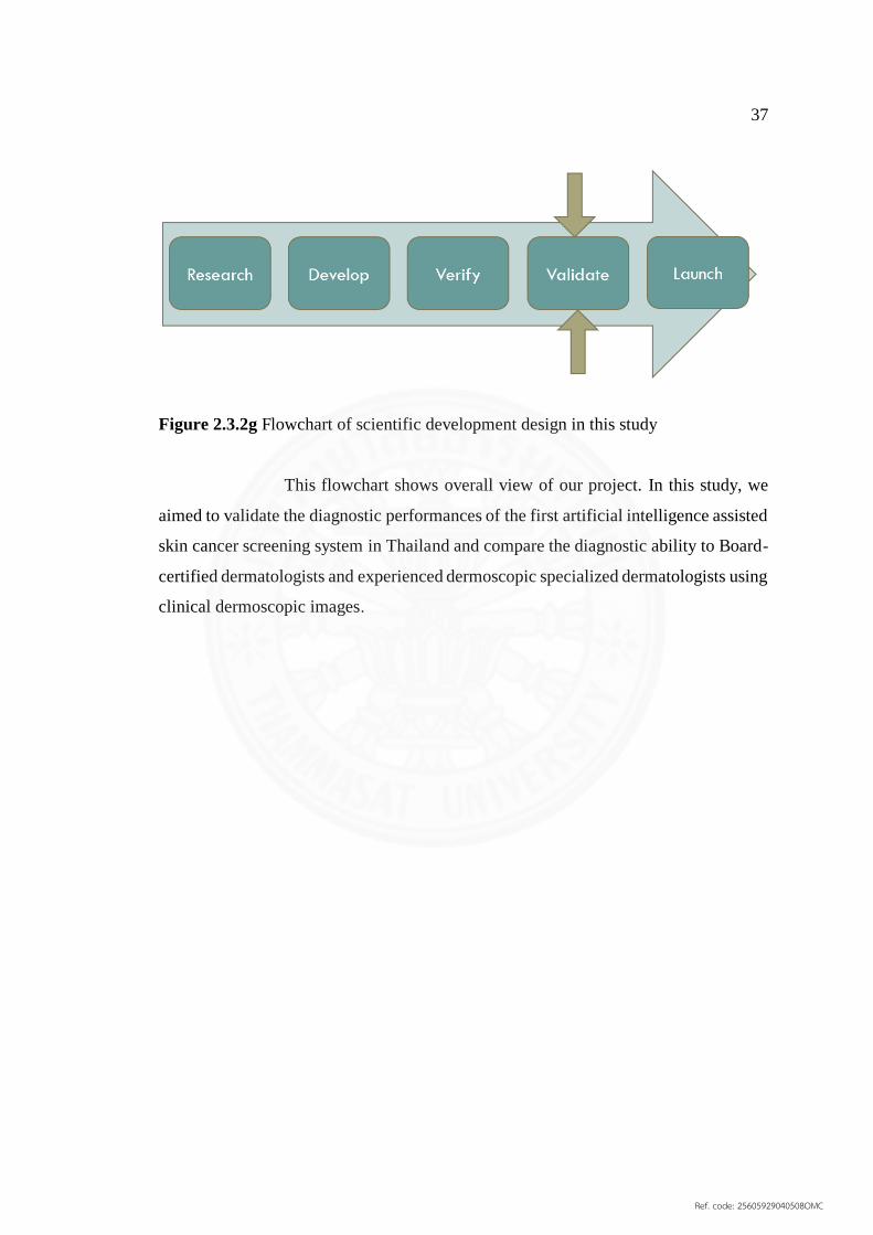
Figure 2.3.2g Flowchart of scientific development design in this study
This flowchart shows overall view of our project. In this study, we
aimed to validate the diagnostic performances of the first artificial intelligence assisted
skin cancer screening system in Thailand and compare the diagnostic ability to Board-
certified dermatologists and experienced dermoscopic specialized dermatologists using
clinical dermoscopic images.
Ref. code: 25605929040508OMC

38
CHAPTER 3
RESEARCH METHODOLOGY
3.1 Materials
3.1.1 Dermoscopic images of pigmented skin lesions
3.1.1.1 Test dataset
(1) Sample size
Clinical dermoscopic images of pigmented skin lesions
including Melanoma, Squamous cell carcinoma, Basal cell carcinoma, Seborrheic
keratosis, Nevus, and other skin lesions from the medical records in Samitivej
Sukhumvit Hospital, Bangkok, Thailand from January 2014 to December 2017. All
lesions were biopsied for histopathological examination to confirm diagnosis. All
images were taken with FotoFinder Hub® system ( FotoFinder Systems GmbH,
Deutschland) and were saved in JPG format.
Sample Size determination
The sample size was calculated from the formula of Testing
for one population proportion formula
Reference value (p0) = 1
Proportion (p) = 0.95
α = 0.05
β = 0.1
Sample size(n) = 200
(2) Inclusion criteria
2.1) Dermoscopic images of pigmented skin lesions including
Melanoma, Squamous cell carcinoma, Basal cell carcinoma, Seborrheic keratosis,
Nevus, and other skin lesions from the medical records in Samitivej Sukhumvit
Hospital, Bangkok, Thailand from January 2014 to December 2017
Ref. code: 25605929040508OMC

39
2.2) All images must be confirmed diagnosis by
histopathological examination
(3) Exclusion criteria
3.1) Inadequate image qualities: poor focus, too much artifacts
3.2) Images which are included multiple lesions
3.3) Images which lesions encompassed the entire field of
view
3.4) Images with non-histopathological examined lesions
3.5) Images which exists in trained dataset for AI
3.1.1.2 Trained dataset
(1) Study population
Dermoscopic images of pigmented skin lesions including
Malignant melanoma (MM), Squamous cell carcinoma (SCC), Basal cell carcinoma
(BCC), Seborrheic keratosis (SK), and Nevus (NV) used to train and access
effectiveness of artificial intelligence system in this study were based on four sources
as listed below. (see Table 3.1.1.2)
1) ISIC-ISBI Challenge 2017: 2000 images; melanoma 374
images, nevus 1372 images, seborrheic keratosis 254
images
2) Medical records in Samitivej Sukhumvit Hospital,
Bangkok, Thailand from January 2014 to December 2017:
82 images; melanoma 25 images, seborrheic keratosis 52
images, SCC 3 images, and BCC 2 images
3) Medical textbooks: 269 images; melanoma 217 images,
seborrheic keratosis 40 images, SCC 8 images, and BCC 4
images
• Dermatoscopy in clinical practice second edition
• Dermoscopy: an illustrated self-assessment guide
• Compendium of surface microscopic and dermoscopic
features
• Handbook of dermoscopy
Ref. code: 25605929040508OMC

40
4) Journal articles: 120 images; SCC 18 images and BCC 102
images
Table 3.1.1.2 Sources of trained dataset
TRAINED
DATASET
MM SK NV BCC SCC
ISIC 2017 374 254 1372 - -
Smitivej
hospital
25 - - 22 3
Textbooks 217 40 - 47 8
Journal
articles
- - - 102 18
Total 616 346 1372 171 29
MM: Melanoma, SK: Seborrheic keratosis, NV: Nevus, BCC: Basal cell carcinoma, SCC: Squamous cell carcinoma
Ref. code: 25605929040508OMC

41
a)
b)
Figure 3.1.1a and 3.1.1b Examples of dermoscopic images of melanoma
a) From ISIC-ISBI Challenge 2017
b) From medical records in Samitivej Sukhumvit Hospital; images were taken with
FotoFinder Hub® system
Ref. code: 25605929040508OMC

42
Figure 3.1.1c FotoFinder Hub® system (FotoFinder Systems GmbH, Deutschland)
3.2 Research design
Retrospective, descriptive study
3.2.1 Study location
Skin and laser clinic at Samitivej Sukhumvit Hospital, Bangkok,
Thailand
Ref. code: 25605929040508OMC

43
3.2.2 Study procedures
Figure 3.2.2 Flow chart of study procedures
3.2.2.1 This study was approved by Institutional Review Board of
Bangkok Hospital Medical Center (BMC-IRB) before starting the experiment.
3.2.2.2 Gathered clinical dermoscopic images of pigmented skin
lesions including Melanomas, Basal cell carcinomas, Squamous cell carcinomas,
Seborrheic keratoses, and Nevi from the medical records in Samitivej Sukhumvit
Hospital from January 2014 to December 2017. All lesions were biopsied for
histopathological examination to confirm diagnosis.
Data collection
(1) Genders
(2) Ages
(3) Dermoscopic images of lesions
(4) Locations of lesions
(5) Diagnosis of pigmented skin lesions
(6) Histopathological examination from skin biopsies
3.2.2.3 Created the validation test using randomly computerized
selected 200 clinical dermoscopic images including 31 melanomas, 39 basal cell
carcinomas, 6 squamous cell carcinomas, 52 seborrheic keratoses, 65 nevi, and 7 other
lesions including 2 cherry hemangiomas, 2 telangiectasias, tattoo, dermatofibroma, and
clear cell acanthoma.
Gathered dermoscopic images of pigmented skin lesions
Created a clinical validation test
The validation test was read by 3 dermoscopic specialized dermatologists vs 3 Board-certified dermatologists vs AI system
Analyzed diagnostic parameters
Ref. code: 25605929040508OMC

44
Each dermoscopic image was provided one correct answer out
of six choices; melanoma, basal cell carcinoma, squamous cell carcinoma, seborrheic
keratosis, nevus, and other diagnosis.
a)
b)
Figure 3.2.2.3a,b Examples of validation test for dermatologists
LE SI ON 1 : WH AT I S D I A GN OSI S?
A. Melanoma B. Seborrheic keratosis C. Nevus D. Basal cell carcinoma E. Squamous cell carcinoma O. Other diagnosis
Ref. code: 25605929040508OMC

45
3.2.2.4 This validation test was read by three Board-certified
dermatologists versus three dermoscopic specialized dermatologists versus artificial
intelligence system (Deep learning).
All readers were blinded to the diagnosis and clinical images.
No additional clinical information was given to the dermatologists. No time
restrictions. All readers could complete the test over multiple sittings.
3.2.2.5 Analyzed diagnostic parameters of each type of pigmented
skin lesions among three groups.
3.2.3 Outcome measurements
3.2.3.1 Sensitivity for diagnosis each type of pigmented skin lesions
3.2.3.2 Specificity for diagnosis each type of pigmented skin lesions
3.2.3.3 Accuracy for diagnosis each type of pigmented skin lesions
3.2.3.4 Positive predictive value (PPV) for diagnosis each type of
pigmented skin lesions
3.2.3.5 Negative predictive value (NPV) for diagnosis each type of
pigmented skin lesions
3.2.3.6 Compare diagnostic performances among three groups:
Board-certified dermatologists versus dermoscopic
specialized dermatologists versus artificial intelligence
system (Deep learning).
3.3 Data analysis
3.3.1 Diagnostic performance analysis
The primary outcomes were diagnostic performances of three
groups on each type of pigmented skin lesions including sensitivity, specificity,
accuracy, positive predictive values, and negative predictive values. The values were
calculated in percentage (%) based on the following standard formulae.
3.3.1.1 Sensitivity for diagnosis each type of pigmented skin lesions
Sensitivity = TP
TP + FN
Ref. code: 25605929040508OMC

46
3.3.1.2 Specificity for diagnosis each type of pigmented skin lesions
Specificity = TN
TN + FP
3.3.1.3 Accuracy for diagnosis each type of pigmented skin lesions
Accuracy = (TP + TN)
(TP + TN + FP + FN)
3.3.1.4 Positive predictive values for diagnosis each type of
pigmented skin lesions
Positive predictive values = TP
TP + FP
3.3.1.5 Negative predictive values for diagnosis each type of
pigmented skin lesions
Negative predictive values = TN
TN + FN
Figure 3.3.1 Diagnostic parameters (85)
When evaluate the group values from several readers in the same
group, the mean values of each diagnostic parameters were used in our analysis.
The secondary outcomes were comparison among three groups on
diagnostic performances of each type of pigmented skin lesions.
Ref. code: 25605929040508OMC

47
3.3.2 Statistical analysis
Artificial intelligence system (Deep learning) submitted
predictions of each dermoscopic image with one out of six choices including 0, 1, 2, 3,
4, 5. Also, all dermatologists submitted predictions of each image with one out of six
choices including A, B, C, D, E, O.
Each score was checked 6 times with correct ( 1. 0) and incorrect
(0.0) answer in each choice.
Kappa analysis and Interclass correlation coefficient ( ICC) were
used to evaluate readers’ performance correlation among each dermatologist group.
In all graphs, the baseline value is 0.00. Statistical analyses used
SPSS (version 10.0).
Ref. code: 25605929040508OMC

48
CHAPTER 4
RESULTS AND DISCUSSION
4.1 Results
4.1.1 Diagnostic performances of each type of pigmented skin lesions
4.1.1.1 Board-certified dermatologists
MM BCC SCC SK NV Others
Sensitivity (%) 22.6 42.7 16.7 63.5 31.8 95.2
Specificity (%) 93.7 97.1 94.0 82.5 92.3 75.6
Accuracy (%) 82.7 86.5 91.7 77.5 72.7 76.3
PPV (%) 42.0 79.2 9.9 58.6 64.3 12.8
NPV (%) 86.9 87.5 97.3 86.1 74.2 99.8
From Table 4.1.1.1, mean sensitivities of Board- certified
dermatologists in melanoma, BCC, and SCC diagnosis were 22.6%, 42.7%, and 16.7%
respectively. In contrast, mean specificities in diagnosis of melanoma, BCC, and SCC
were high as 93.7%, 97.1%, and 94.0% respectively. Mean accuracies were 82.7, 86.5,
and 91.7%. Moreover, mean positive predictive values in melanoma, BCC, and SCC
diagnosis were 42.0%, 79.2%, and 9.9%. Mean negative predictive values were 86.9%,
87.5%, and 97.3% respectively.
Benign pigmented skin lesions which are seborrheic keratosis,
nevus, and other diagnosis, mean sensitivities in diagnosis of Board-certified
dermatologists were 63.5%, 31.8%, and 95.2%. Mean specificities in diagnosis in
seborrheic keratosis, nevus, and other diagnosis were 82.5%, 92.3%, and 75.6%. Mean
accuracies were 77.5, 72.7, and 76.3% respectively. In addition, mean positive
predictive values in diagnosis of seborrheic keratosis, nevus, and other pigmented skin
MM: Melanoma, BCC: Basal cell carcinoma, SCC: Squamous cell carcinoma,
SK: Seborrheic keratosis, NV: Nevus, Others: Other diagnosis
Table 4.1.1.1 Mean diagnostic performances of Board-certified dermatologists in
diagnosis of different pigmented skin lesions
Ref. code: 25605929040508OMC

49
lesions were 58.6%, 64.3%, and 12.8%. Mean negative predictive values were 86.1%,
74.2%, and 99.8% respectively.
Intermediate to excellent agreement beyond each lesion was
observed among the readers in this group. ( see Table 4.1.1a) Also, figure 4.1.2.1a
shows the good correlation among three dermatologists in diagnosis of different types
of skin cancers.
Ref. code: 25605929040508OMC

50
Figure 4.1.2.1a Diagnostic performances in diagnosis of different types of skin
cancers; three Board-certified dermatologists and mean values.
Ref. code: 25605929040508OMC

51
4.1.1.2 Experienced dermoscopic specialized dermatologists
Table 4.1.1.2 Mean diagnostic performances of experienced dermoscopic specialized
dermatologists in diagnosis of different pigmented skin lesions
MM BCC SCC SK NV Others
Sensitivity (%) 69.9 66.7 72.2 83.3 63.6 90.5
Specificity (%) 82.2 98.2 95.7 97.1 93.6 99.1
Accuracy (%) 80.3 92.0 95.0 93.5 83.8 98.8
PPV (%) 43.5 89.4 38.5 90.9 84.1 80.3
NPV (%) 93.8 92.4 99.1 94.3 84.4 99.7
Dermoscopic specialized dermatologists’ diagnostic
performances were high in almost all parameters.
For skin cancer classification, mean sensitivities in melanoma,
BCC, and SCC diagnosis were 69. 9% , 66. 7% , and 72. 2% respectively. Mean
specificities in melanoma, BCC, and SCC diagnosis were 82. 2% , 98. 2% , and 95. 7%
respectively. Mean accuracies were 80.3, 92.0, and 95.0%. In addition, mean positive
predictive values in diagnosis of melanoma, BCC, and SCC were 43.5%, 89.4%, and
38.5%. Mean negative predictive values were 93.8.9%, 92.4%, and 99.1% respectively.
(see Table 4.1.1.2)
Diagnostic performances in benign pigmented skin lesions
showed that mean sensitivities in seborrheic keratosis, nevus, and other lesions were
83.3%, 63.6%, and 90.5%. Mean specificities were 97.1%, 93.6%, and 99.1%
respectively. Mean accuracies were 93.5, 83.8, and 98.8%. Moreover, mean positive
predictive values in diagnosis of seborrheic keratosis, nevus, and other pigmented skin
lesions were 90.9%, 84.1%, and 80.3%. Mean negative predictive values were 94.3%,
84.4%, and 99.7% respectively.
MM: Melanoma, BCC: Basal cell carcinoma, SCC: Squamous cell carcinoma,
SK: Seborrheic keratosis, NV: Nevus, Others: Other diagnosis
Ref. code: 25605929040508OMC

52
All lesions were classified with excellent agreement among all
readers in the group. (see Table 4.1.1a) Figure 4.1.2.2a also shows the good correlation
in skin cancers diagnosis among three dermoscopic specialized dermatologists.
Table 4.1.1a Interclass correlation coefficient (ICC)
ICC
MM BCC SCC SK NV Others
Dermoscopic
specialized
dermatologists
0.896
0.900
0.868
0.954
0.870
0.936
Dermatologist 0.579 0.846 0.599 0.858 0.718 0.982
MM: Melanoma, BCC: Basal cell carcinoma, SCC: Squamous cell carcinoma,
SK: Seborrheic keratosis, NV: Nevus, Others: Other diagnosis
Ref. code: 25605929040508OMC

53
Figure 4.1.2.2a Diagnostic performances in diagnosis of different types of skin
cancers; three experienced dermoscopic specialized dermatologists and mean values.
Ref. code: 25605929040508OMC
54
4.1.1.3 Artificial intelligence system (Deep learning)
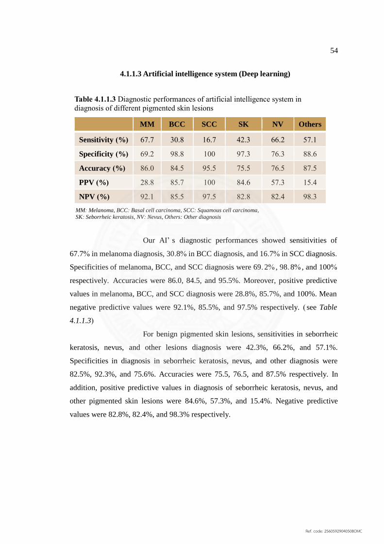
MM BCC SCC SK NV Others
Sensitivity (%) 67.7 30.8 16.7 42.3 66.2 57.1
Specificity (%) 69.2 98.8 100 97.3 76.3 88.6
Accuracy (%) 86.0 84.5 95.5 75.5 76.5 87.5
PPV (%) 28.8 85.7 100 84.6 57.3 15.4
NPV (%) 92.1 85.5 97.5 82.8 82.4 98.3
Our AI’ s diagnostic performances showed sensitivities of
67.7% in melanoma diagnosis, 30.8% in BCC diagnosis, and 16.7% in SCC diagnosis.
Specificities of melanoma, BCC, and SCC diagnosis were 69. 2% , 98. 8% , and 100%
respectively. Accuracies were 86.0, 84.5, and 95.5%. Moreover, positive predictive
values in melanoma, BCC, and SCC diagnosis were 28.8%, 85.7%, and 100%. Mean
negative predictive values were 92.1%, 85.5%, and 97.5% respectively. ( see Table
4.1.1.3)
For benign pigmented skin lesions, sensitivities in seborrheic
keratosis, nevus, and other lesions diagnosis were 42.3%, 66.2%, and 57.1%.
Specificities in diagnosis in seborrheic keratosis, nevus, and other diagnosis were
82.5%, 92.3%, and 75.6%. Accuracies were 75.5, 76.5, and 87.5% respectively. In
addition, positive predictive values in diagnosis of seborrheic keratosis, nevus, and
other pigmented skin lesions were 84.6%, 57.3%, and 15.4%. Negative predictive
values were 82.8%, 82.4%, and 98.3% respectively.
Table 4.1.1.3 Diagnostic performances of artificial intelligence system in
diagnosis of different pigmented skin lesions
MM: Melanoma, BCC: Basal cell carcinoma, SCC: Squamous cell carcinoma,
SK: Seborrheic keratosis, NV: Nevus, Others: Other diagnosis
Ref. code: 25605929040508OMC

55
BCC
SCC
NV
SK
Others
BCC SCC NV SK Others
21
1
0
9
0
0
22
12
0
3
2
0
2
1
1
1
0
1
19
0
0
43
1
2
7
0
0
19
22
4
0
0
0
3
0
4
Predicted level
Figure 4.1.1.3 Confusion matrix of AI system performance
Valu
e l
evel
MM
MM
MM: Melanoma, BCC: Basal cell carcinoma, SCC: Squamous cell carcinoma,
SK: Seborrheic keratosis, NV: Nevus, Others: Other diagnosis
Ref. code: 25605929040508OMC

56
4.1.2 Comparison of skin cancer diagnostic performances among three
groups
4.1.2.1 Sensitivities
In melanoma diagnosis, AI system showed higher sensitivity
compared to Board-certified dermatologists ( 67. 7% vs 22. 6% ) and almost the same
level as dermoscopic specialized dermatologists (69.9%).
For BCC diagnosis, AI system had lower sensitivity (30.8%)
compared to other groups (Board-certified dermatologist 42.7% vs Dermoscopists
66.7%).
For SCC diagnosis, AI system had lower sensitivity compared
to Dermoscopic s specialized dermatologists (16.7% vs 72.2%), but the same level with
Board-certified dermatologists (16.7%). (see Figure 4.1.2.1)
Figure 4.1.2.1 Sensitivities in diagnosis of different types of skin cancers compared
among three groups
Ref. code: 25605929040508OMC

57
4.1.2.2 Specificities
Specificities in diagnosis of melanoma, BCC, and SCC among
three groups were not significantly different. (see Figure 4.1.2.2)
In melanoma diagnosis, specificities of AI system, Board-
certified dermatologists, and dermoscopic specialized dermatologists were 69.2%,
93.7%, and 82.2% respectively.
In BCC diagnosis, specificities of AI system, Board-certified
dermatologists, and dermoscopic specialized dermatologists were 69.2%, 93.7%, and
82.2% respectively.
In SCC diagnosis, specificities of AI system, Board-certified
dermatologists, and dermoscopic specialized dermatologists were 98.8%, 97.1%, and
98.2% respectively. (see Figure 4.1.2.2)
Figure 4.1.2.2 Sensitivities in diagnosis of different types of skin cancers compared
among three groups
Ref. code: 25605929040508OMC

58
4.1.2.3 Accuracies
AI system had higher accuracies in diagnosis of melanoma and
SCC compared to other groups. (see Figure 4.1.2.3)
In melanoma diagnosis, accuracies of AI system, Board-
certified dermatologists, and dermoscopic specialized dermatologists were 86.0%,
92.7%, and 80.3% respectively.
For BCC diagnosis, AI system had lower accuracy as 84.5%
compared to Board-certified dermatologists as 86.5%, and dermoscopic specialized
dermatologists as 92%.
For SCC diagnosis, accuracies of AI system, Board-certified
dermatologists, and dermoscopic specialized dermatologists were 95.5%, 91.7%, and
95% respectively.
Figure 4.1.2.3 Accuracies in diagnosis of different types of skin cancers compared
among three groups
Ref. code: 25605929040508OMC

59
4.1.2.4 Positive predictive values (PPV)
AI system showed higher PPV than other groups in melanoma
diagnosis, but lower in BCC diagnosis. (see Figure 4.1.2.4)
In melanoma diagnosis, PPV of AI system, Board-certified
dermatologists, and dermoscopic specialized dermatologists were 57.9%, 42.0%, and
43.5% respectively.
In BCC diagnosis, PPV of AI system, Board-certified
dermatologists, and dermoscopic specialized dermatologists were 58.3%, 79.2%, and
89.4% respectively.
In SCC diagnosis, PPV of AI system, Board-certified
dermatologists, and dermoscopic specialized dermatologists were 20.0%, 9.9%, and
38.5% respectively.
Figure 4.1.2.4 Positive predictive values (PPV) in diagnosis of different types of skin
cancers compared among three groups
Ref. code: 25605929040508OMC

60
4.1.2.5 Negative predictive values (NPV)
AI system had higher NPV than other groups in BCC diagnosis.
For melanoma and SCC diagnosis, AI system had lower NPV than dermoscopic
specialized dermatologists, but higher than Board-certified dermatologists. (see Figure
4.1.2.5)
In melanoma diagnosis, NPV of AI system, Board-certified
dermatologists, and dermoscopic specialized dermatologists were 89%, 86.9%, and
93.8% respectively.
In BCC diagnosis, NPV of AI system, Board-certified
dermatologists, and dermoscopic specialized dermatologists were 92.8%, 87.5%, and
92.4% respectively.
In SCC diagnosis, NPV of AI system, Board-certified
dermatologists, and dermoscopic specialized dermatologists were 97.4%, 97.3%, and
99.1% respectively.
Figure 4.1.2.5 Negative predictive values (NPV) in diagnosis of different types
of skin cancers compared among three groups
Ref. code: 25605929040508OMC

61
4.1.3 Comparison of benign pigmented skin lesions diagnostic
performances among three groups
4.1.3.1 Sensitivities
AI system had lower sensitivities in seborrheic keratosis and
other lesions compared to other groups, in contrast, higher sensitivity in nevus
diagnosis. (see Figure 4.1.3.1)
In seborrheic keratosis diagnosis, sensitivities of AI system,
Board- certified dermatologists, and dermoscopic specialized dermatologists were
26.9%, 63.6%, and 83.3% respectively.
For nevus diagnosis, sensitivities of AI system, Board-certified
dermatologists, and dermoscopic specialized dermatologists were 72.3%, 31.8%, and
63.6% respectively.
For other lesions diagnosis, sensitivities of AI system, Board-
certified dermatologists, and dermoscopic specialized dermatologists were 57.1%,
95.2%, and 90.5% respectively.
Figure 4.1.3.1 Sensitivities in diagnosis of different types of benign pigmented skin
lesions compared among three groups
Ref. code: 25605929040508OMC

62
4.1.3.2 Specificities
Specificities in benign pigmented lesions diagnosis among
three groups were similar. (see Figure 4.1.3.2)
In seborrheic keratosis diagnosis, specificities of AI system,
Board- certified dermatologists, and dermoscopic specialized dermatologists were
92.6%, 82.5%, and 97.1% respectively.
For nevus diagnosis, specificities of AI system, Board-certified
dermatologists, and dermoscopic specialized dermatologists were 78.5%, 92.3%, and
93.6% respectively.
For other lesions diagnosis, specificities of AI system, Board-
certified dermatologists, and dermoscopic specialized dermatologists were 88.6%,
75.6%, and 99.1% respectively.
Figure 4.1.3.2 Specificities in diagnosis of different types of benign pigmented skin
lesions compared among three groups
Ref. code: 25605929040508OMC

63
4.1.3.3 Accuracies
Accuracies in benign pigmented lesions diagnosis among three
groups were similar. (see Figure 4.1.3.3)
In seborrheic keratosis diagnosis, accuracies of AI system,
Board- certified dermatologists, and dermoscopic specialized dermatologists were
75.5%, 77.5%, and 93.5% respectively.
For nevus diagnosis, accuracies of AI system, Board-certified
dermatologists, and dermoscopic specialized dermatologists were 76.5%, 72.7%, and
83.8% respectively.
For other lesions diagnosis, accuracies of AI system, Board-
certified dermatologists, and dermoscopic specialized dermatologists were 87.5%,
76.3%, and 98.8% respectively.
Figure 4.1.3.3 Accuracies in diagnosis of different types of benign pigmented skin
lesions compared among three groups
Ref. code: 25605929040508OMC
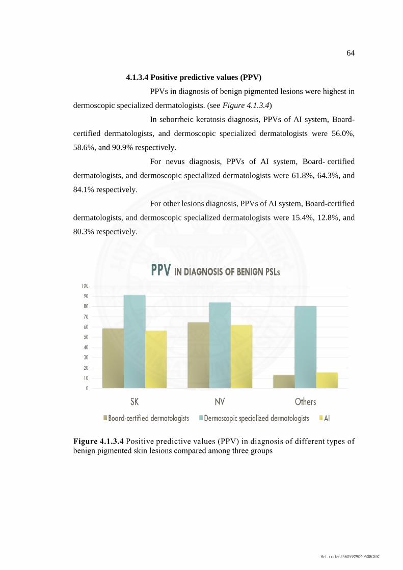
64
4.1.3.4 Positive predictive values (PPV)
PPVs in diagnosis of benign pigmented lesions were highest in
dermoscopic specialized dermatologists. (see Figure 4.1.3.4)
In seborrheic keratosis diagnosis, PPVs of AI system, Board-
certified dermatologists, and dermoscopic specialized dermatologists were 56.0%,
58.6%, and 90.9% respectively.
For nevus diagnosis, PPVs of AI system, Board- certified
dermatologists, and dermoscopic specialized dermatologists were 61.8%, 64.3%, and
84.1% respectively.
For other lesions diagnosis, PPVs of AI system, Board-certified
dermatologists, and dermoscopic specialized dermatologists were 15.4%, 12.8%, and
80.3% respectively.
Figure 4.1.3.4 Positive predictive values (PPV) in diagnosis of different types of
benign pigmented skin lesions compared among three groups
Ref. code: 25605929040508OMC

65
4.1.3.5 Negative predictive values (NPV)
NPVs in benign pigmented lesions diagnosis among three
groups were not different. (see Figure 4.1.3.5)
In seborrheic keratosis diagnosis, NPVs of AI system, Board-
certified dermatologists, and dermoscopic specialized dermatologists were 78.3%,
86.1%, and 94.3% respectively.
For nevus diagnosis, NPVs of AI system, Board- certified
dermatologists, and dermoscopic specialized dermatologists were 85.5%, 74.2%, and
84.4% respectively.
For other lesions diagnosis, NPVs of AI system, Board-
certified dermatologists, and dermoscopic specialized dermatologists were 98.3%,
99.8%, and 99.7% respectively.
Figure 4.1.3.5 Negative predictive values (NPV) in diagnosis of different types of
benign pigmented skin lesions compared among three groups
Ref. code: 25605929040508OMC

66
4.2 Discussion
From previous study, artificial intelligence (AI) system showed
outperformances in melanoma, nevus, and seborrheic keratosis diagnosis up to 80-90%
of area under ROC curve. All dermoscopic images used to train and assess effectiveness
of all classifiers in that work were based on the ISIC Challenge 2017 data.
This is the first study to demonstrate clinical validation test of artificial
intelligence (AI) assisted in skin cancer screening system in Thailand using clinical
dermoscopic images and compared with Board-certified dermatologists vs experienced
dermoscopic specialized dermatologists.
Our validation test was very challenging that even Board- certified
dermatologists performed with low sensitivity (22.6%) in melanoma diagnosis. In test
dataset, 31 correctly dermoscopic images of melanoma were included 18 malignant
melanomas and 13 melanomas in situ. Normally, to distinguish among three lesions
which are melanoma, melanoma in situ, and nevus, are very difficult task for even the
expertise due to similarity in morphologies, colors, and textures as shown in Figure
4.2a and 4.2b
a) b) c)
Figure 4.2.1a, b, c Clinical dermoscopic images in test dataset of
a) melanoma
b) melanoma in situ
c) nevus
From confusion matrix of AI system performance, AI mostly misdiagnosed
of melanoma into nevus from 9 out of 31 images. And 4 out of 9 false negatives which
Ref. code: 25605929040508OMC

67
account for almost 50%, are melanoma in situ condition. This might also interpret that
false negatives in melanoma are majorly due to similarity among melanoma, melanoma
in situ, and nevus. (see Figure 4.2.2)
Figure 4.2.2a Confusion matrix of AI system performance: melanoma classification
However, AI system had higher sensitivity (67.7%) in melanoma diagnosis
compared to Board-certified dermatologists ( 22. 6% ) and almost the same level as
dermoscopic specialized dermatologists (69.9%).
For other types of skin cancers diagnosis which either basal cell carcinoma
or squamous cell carcinoma, AI system’s diagnostic performances were still
disappointing. AI had lower sensitivities compared to Board-certified dermatologists
and experienced dermoscopic specialized dermatologists (30.8% vs 42.7% vs 66.7% in
Ref. code: 25605929040508OMC

68
BCC diagnosis, and 16.7% vs 16.7% vs 72.2% in SCC diagnosis). This might be the
reflection from scarcity of dermoscopic images of BCC and SCC used to train AI
system. (see Table 4.2)
Table 4.2 Sources of trained dataset
TRAINED
DATASET
MM SK NV BCC SCC
ISIC 2017 374 254 1372 - -
Smitivej
hospital
25 - - 22 3
Textbooks 217 40 - 47 8
Journal
articles
- - - 102 18
Total 616 346 1372 171 29
Interestingly, most of false negatives for BCC and SCC classification were
melanomas which were still malignant. This showed that AI performances in
differentiating malignancy and benign were acceptable.
For BCC classification, if we re-classify into cancer and non-cancer
categories, sensitivity in diagnosis increases significantly up to 81.18%. (see Figure
4.2.2b)
Ref. code: 25605929040508OMC

69
Figure 4.2.2b Confusion matrix of AI system performance: BCC classification
For SCC classification, most of false negatives were melanoma. If we re-
classify into cancer and non-cancer categories, sensitivity in diagnosis increases up to
66.67%. (see Figure 4.2.2c)
Ref. code: 25605929040508OMC

70
Figure 4.2.2c Confusion matrix of AI system performance: SCC classification
In benign pigmented skin lesions diagnosis, AI system had highest
sensitivity in nevus category, but lowest in seborrheic keratosis and other lesions
categories. From the results, we might conclude that the more images used to train AI,
the better diagnostic performances.
Although diagnostic performance of our AI system in diagnosis of skin
cancers was not achieved with high diagnostic performances as the results in our
previous experiment using ISIC 2017 images as validation test, this was the first start
to develop AI system to be applied in real clinical setting.
Scarcity of dermoscopic images used to train AI algorithm was the major
limitation for improving diagnostic accuracy.
Ref. code: 25605929040508OMC

71
CHAPTER 5
CONCLUSIONS AND RECOMMENDATIONS
5.1 Conclusions
Our artificial intelligence system using deep learning computer vision
algorithm achieved performance in diagnosis of melanomas with similar level as Board-
certified dermatologists. However, in other skin cancers classification which were basal
cell carcinomas and squamous cell carcinomas, AI still needed to be trained more to
improve diagnostic accuracy.
5.2 Recommendations
5.2.1 AI system still need further trainings to improve its outcomes before
applying in clinical settings especially in melanoma in situ, basal cell carcinoma and
squamous cell carcinoma categories.
5.2.2 The more images you train AI, the better outcome you can get in the
future. Other sources of images may gather from other medical centers or open public
to physicians.
5.2.3 Add on algorithm such as ABCD rule to AI system to be trained to
reach better performances.
5.2.4 In practice, classification of cancer versus non-cancer is the most
essential point. Too many categories in classification may not helpful in case that AI
system still has learning limitation.
5. 2. 5 Larger sample size and prospective study may be necessary for the
further studies.
Ref. code: 25605929040508OMC

72
REFERENCES
1. Oakley A. Pigmented skin lesions New Zealand2015 [Available from:
https://www.dermnetnz.org/topics/pigmented-skin-lesions.
2. S. B. The assessment, history taking and differential diagnosis of pigmented
skin lesions. Dermatological nursing. 2015;14(4):18-22.
3. Garcia R, Korotkov K, Korotkov K, Garcia R. Computerized analysis of
pigmented skin lesions: A review.
4. Thomas L, Puig S. Dermoscopy, Digital Dermoscopy and Other Diagnostic
Tools in the Early Detection of Melanoma and Follow-up of High-risk Skin Cancer
Patients. Acta Dermato-Venereologica. 2017;97:14-21.
5. Bolognia J, Jorizzo J, Schaffer J. Dermatology, third edition. London: Elsevier;
2012.
6. Soyer P, Argenziano G, Ruocco V, Chimenti S. Dermoscopy of Pigmented Skin
Lesions2001. 483-98 p.
7. Goldsmith L, Katz S, Gilchrest B, Paller A, Leffell D. Fitzpatrick's Dermatology
in general medicine, 8th edition. United States of America: The McGraw-Hill
Companies, Inc. ; 2012.
8. Ngan V. Spitz naevus 2003 [Available from: https://www.dermnetnz.org/topic-
s/spitz-naevus/.
9. Yoradjian A, Enokihara MM, Paschoal FM. Spitz nevus and Reed nevus. Anais
brasileiros de dermatologia. 2012;87(3):349-57; quiz 58-9.
10. Gibson L. Melanoma: Mayo clinic; 2017 [Available from: https://www.mayo-
clinic.org/diseases-conditions/melanoma/symptoms-causes/syc-20374884.
11. Garbe C, Peris K, Hauschild A, Saiag P, Middleton M, Bastholt L, et al.
Diagnosis and treatment of melanoma. European consensus-based interdisciplinary
guideline - Update 2016. Eur J Cancer. 2016;63:201-17.
12. Society AC. Cancer facts & figures 2016 Atlanta2016 [Available from:
https://www.cancer.org/research/cancer-facts-statistics/all-cancer-facts-
figures/cancer-facts-figures-2016.html.
Ref. code: 25605929040508OMC

73
13. Valachovic E, Zurbenko I. Multivariate analysis of spatial–temporal scales in
melanoma prevalence. Cancer Causes & Control. 2017;28(7):733-43.
14. Siegel RL, Miller KD, Jemal A. Cancer Statistics, 2017. CA Cancer J Clin.
2017;67(1):7-30.
15. Bhattacharya A, Young A, Wong A, Stalling S, Wei M, Hadley D. Precision
Diagnosis Of Melanoma And Other Skin Lesions From Digital Images. AMIA Joint
Summits on Translational Science proceedings AMIA Joint Summits on Translational
Science. 2017;2017:220-6.
16. Davey RJ, van der Westhuizen A, Bowden NA. Metastatic melanoma
treatment: Combining old and new therapies. Crit Rev Oncol Hematol. 2016;98:242-
53.
17. Bichakjian CK, Halpern AC, Johnson TM, Foote Hood A, Grichnik JM, Swetter
SM, et al. Guidelines of care for the management of primary cutaneous melanoma.
American Academy of Dermatology. Journal of the American Academy of
Dermatology. 2011;65(5):1032-47
18. Harries M, Malvehy J, Lebbe C, Heron L, Amelio J, Szabo Z, et al. Review:
Treatment patterns of advanced malignant melanoma (stage III–IV) – A review of
current standards in Europe. European Journal of Cancer. 2016;60:179-89.
19. Abbasi NR, Shaw HM, Rigel DS, Polsky D, Friedman RJ, Kopf AW, et al.
Early diagnosis of cutaneous melanoma: Revisiting the ABCD criteria. JAMA, The
Journal of the American Medical Association. 2004(22):2771.
20. Mackie RM, Doherty VR. Seven-point checklist for melanoma. Clinical &
Experimental Dermatology. 1991;16(2):151.
21. Gordon R. Skin Cancer: An Overview of Epidemiology and Risk Factors.
Seminars in Oncology Nursing. 2013;29(3):160-9.
22. Morton, Mackie. Clinical accuracy of the diagnosis of cutaneous malignant
melanoma. British Journal of Dermatology. 1998;138(2):283.
23. Lindelöf B, Hedblad MA. Accuracy in the Clinical Diagnosis and Pattern of
Malignant Melanoma at a Dermatological Clinic. The Journal of Dermatology.
1994;21(7):461.
24. Grin CM, Kopf AW, Welkovich B, Bart RS, Levenstein MJ. Accuracy in the
clinical diagnosis of malignant melanoma. Arch Dermatol. 1990;126(6):763-6.
Ref. code: 25605929040508OMC

74
25. Gachon J, Beaulieu P, Sei JF, Gouvernet J, Claudel JP, Lemaitre M, et al. First
prospective study of the recognition process of melanoma in dermatological practice.
Arch Dermatol. 2005;141(4):434-8.
26. Masood A, Al-Jumaily AA. Computer aided diagnostic support system for skin
cancer: a review of techniques and algorithms. Int J Biomed Imaging.
2013;2013:323268.
27. Modern non-invasive diagnostic techniques in the detection of early cutaneous
melanoma. Journal of Dermatological Case Reports; 2014. p. 1-8.
28. Argenziano G, Fabbrocini G, Carli P, De Giorgi V, Sammarco E, Delfino M.
Epiluminescence Microscopy for the Diagnosis of Doubtful Melanocytic Skin Lesions:
Comparison of the ABCD Rule of Dermatoscopy and a New 7-Point Checklist Based
on Pattern Analysis. Archives of Dermatology. 1998;134(12):1563.
29. Binder M. Epiluminescence microscopy of small pigmented skin lesions: Short-
term formal training improves the diagnostic performance of dermatologists. 1997:197.
30. Pehamberger H, Binder M, Steiner A, Wolff K. In Vivo Epiluminescence
Microscopy: Improvement of Early Diagnosis of Melanoma. Journal of Investigative
Dermatology. 1993;100(3):356S-62S.
31. Carli P, De Giorgi V, Crocetti E, Mannone F, Massi D, Chiarugi A, et al.
Improvement of malignant/benign ratio in excised melanocytic lesions in the
'dermoscopy era': a retrospective study 1997-2001. The British journal of dermatology.
2004;150(4):687-92.
32. Carli P, de Giorgi V, Chiarugi A, Nardini P, Weinstock MA, Crocetti E, et al.
Report: Addition of dermoscopy to conventional naked-eye examination in melanoma
screening: a randomized study. Journal of the American Academy of Dermatology.
2004;50:683-9.
33. Kittler H, Pehamberger H, Wolff K, Binder M. Diagnostic accuracy of
dermoscopy. Lancet Oncology. 2002(3):159.
34. Hossam D, Sadek A, Saied N. Dermoscopy: A Literature Review. Egyptian
Dermatology Online Journal. 2015;11(1):1-32.
35. Scope A, Benvenuto-Andrade C, Agero ALC, Marghoob AA. Nonmelanocytic
Lesions Defying the Two-Step Dermoscopy Algorithm. Dermatologic Surgery.
2006;32(11):1398.
Ref. code: 25605929040508OMC
75
36. Dal Pozzo V, Benelli C, Roscetti E. The seven features for melanoma: a new
dermoscopic algorithm for the diagnosis of malignant melanoma. European journal of
dermatology : EJD. 1999;9(4):303-8.
37. Soyer HP, Argenziano G, Zalaudek I, Corona R, Sera F, Talamini R, et al.
Three-point checklist of dermoscopy. A new screening method for early detection of
melanoma. Dermatology (Basel, Switzerland). 2004;208(1):27-31.
38. Pehamberger H, Steiner A, Wolff K. In vivo epiluminescence microscopy of
pigmented skin lesions. I. Pattern analysis of pigmented skin lesions. Journal of the
American Academy of Dermatology. 1987;17:571-83.
39. Nachbar F, Stolz W, Merkle T, Cognetta AB, Vogt T, Landthaler M, et al. The
ABCD rule of dermatoscopy. High prospective value in the diagnosis of doubtful
melanocytic skin lesions. Journal of the American Academy of Dermatology.
1994;30(4):551-9.
40. Rajpara SM, Botello AP, Townend J, Ormerod AD. Systematic review of
dermoscopy and digital dermoscopy/ artificial intelligence for the diagnosis of
melanoma. The British journal of dermatology. 2009;161(3):591-604.
41. Kittler H, Pehamberger H, Wolff K, Binder M. Review: Diagnostic accuracy of
dermoscopy. Lancet Oncology. 2002;3:159-65.
42. Piccolo D, Ferrari A, Peris K, Daidone R, Ruggeri B, Chimenti S. Dermoscopic
diagnosis by a trained clinician vs. a clinician with minimal dermoscopy training vs.
computer-aided diagnosis of 341 pigmented skin lesions: a comparative study. British
Journal of Dermatology. 2002;147(3):481-6.
43. Chang W-Y, Huang A, Yang C-Y, Lee C-H, Chen Y-C, Wu T-Y, et al.
Computer-Aided Diagnosis of Skin Lesions Using Conventional Digital Photography:
A Reliability and Feasibility Study. PLoS ONE. 2013(11).
44. Smoller BR. Histologic criteria for diagnosing primary cutaneous malignant
melanoma. Modern pathology : an official journal of the United States and Canadian
Academy of Pathology, Inc. 2006;19 Suppl 2:S34-40.
45. Balch CM, Gershenwald JE, Soong SJ, Thompson JF, Atkins MB, Byrd DR, et
al. Final version of 2009 AJCC melanoma staging and classification. Journal of clinical
oncology : official journal of the American Society of Clinical Oncology.
2009;27(36):6199-206.
Ref. code: 25605929040508OMC

76
46. Ackerman AB. Discordance among expert pathologists in diagnosis of
melanocytic neoplasms. Human pathology. 1996;27(11):1115-6.
47. Farmer ER, Gonin R, Hanna MP. Discordance in the histopathologic diagnosis
of melanoma and melanocytic nevi between expert pathologists. Human pathology.
1996;27(6):528-31.
48. Prakashini K, Babu S, Rajgopal KV, Kokila KR. Role of Computer Aided
Diagnosis (CAD) in the detection of pulmonary nodules on 64 row multi detector
computed tomography. Lung India. 2016;33(4):391-7.
49. Jiang L, Uitert RV, Jianhua Y, Petrick N, Franaszek M, Huang A, et al. Wavelet
method for CT colonography computer-aided polyp detection. Medical Physics.
2008;35(8):3527.
50. Houssami N, Given-Wilson R, Ciatto S. Early detection of breast cancer:
Overview of the evidence on computer-aided detection in mammography screening.
Journal of Medical Imaging & Radiation Oncology. 2009;53(2):171-6.
51. Fenton JJ, Taplin SH, Carney PA, Abraham L, Sickles EA, D'Orsi C, et al.
Influence of computer-aided detection on performance of screening mammography.
The New England Journal of Medicine. 2007(14):1399.
52. Fujita H, Katafuchi T, Uehara T, Nishimura T. Application of Artificial Neural
Network to Computer-Aided Diagnosis of Coronary Artery Disease in Myocardial
SPECT Bull's-eye Images. Journal of Nuclear Medicine. 1992;33(2):272.
53. Lindahl, Palmer, Edenbrandt, Lindahl D. Myocardial SPET: artificial neural
networks describe extent and severity of perfusion defects. Clinical Physiology.
1999;19(6):497-503.
54. Fernandez-Penas P. Diagnostic and neural analysis of skin cancer (DANAOS).
A multicentre study for collection and computer-aided analysis of data from pigmented
skin lesions using digital dermoscopy. British Journal of Dermatology. 2003;149:801-
9.
55. Schmid-Saugeona P, Guillodb J, Thirana J-P. Towards a computer-aided
diagnosis system for pigmented skin lesions. Computerized Medical Imaging and
Graphics. 2003;27:65-78.
Ref. code: 25605929040508OMC

77
56. Zortea M, Schopf TR, Thon K, Geilhufe M, Hindberg K, Kirchesch H, et al.
Performance of a dermoscopy-based computer vision system for the diagnosis of
pigmented skin lesions compared with visual evaluation by experienced dermatologists.
Artificial Intelligence in Medicine. 2014;60(1):13-26.
57. Hall PN, Claridge E, Smith JDM. Computer screening for early detection of
melanoma -- is there a future? British Journal of Dermatology. 1995;132(3):325.
58. Cascinelli N, Ferrario M, Tonelli T, Leo E. A possible new tool for clinical
diagnosis of melanoma: The computer. Journal of the American Academy of
Dermatology. 1987;16(Part 1):361-7.
59. Cristofolini M, Bauer P, Boi S, Cristofolini P, Micciolo R, Sicher MC.
Diagnosis of cutaneous melanoma: accuracy of a computerized image analysis system
(Skin View). Skin Research & Technology. 1997;3(1):23.
60. Ferri M. Computer-aided diagnosis of melanocytic lesions.
61. Rubegni P. Automated diagnosis of pigmented skin lesions.
62. Premaladha J, Ravichandran KS. Novel Approaches for Diagnosing Melanoma
Skin Lesions Through Supervised and Deep Learning Algorithms. Journal of Medical
Systems. 2016;40(4):96.
63. Liu Z, Sun J, Smith L, Smith M, Warr R, Liu Z, et al. Distribution quantification
on dermoscopy images for computer-assisted diagnosis of cutaneous melanomas.
Medical & Biological Engineering & Computing. 2012;50(5):503-13.
64. Mokhtari M, Rezaeian M, Gharibzadeh S, Malekian V. Computer aided
measurement of melanoma depth of invasion in microscopic images. Micron.
2014;61:40-8.
65. Jaworek-Korjakowska J, Kłeczek P. Automatic Classification of Specific
Melanocytic Lesions Using Artificial Intelligence. BioMed Research International.
2016;2016:1-17.
66. Li L, Clark A, Wang JZ, editors. A Computer-Aided Diagnostic Tool for
Melanoma. 2014 International Conference on Computational Science and
Computational Intelligence; 2014 10-13 March 2014.
67. Kruk M, Świderski B, Osowski S, Kurek J, Słowińska M, Walecka I. Melanoma
recognition using extended set of descriptors and classifiers. EURASIP Journal on
Image & Video Processing. 2015;2015(1):1.
Ref. code: 25605929040508OMC

78
68. Patient acceptance and diagnostic utility of automated digital image analysis of
pigmented skin lesions. Journal of the European Academy of Dermatology and
Venereology. 2012(3):368.
69. Ercal F, Chawla A, Stoecker WV, Lee HC, Moss RH. Neural network diagnosis
of malignant melanoma from color images. IEEE transactions on bio-medical
engineering. 1994;41(9):837-45.
70. Elbaum M, Kopf AW, Rabinovitz HS, Langley RGB, Kamino H, Mihm JMC,
et al. Reports: Automatic differentiation of melanoma from melanocytic nevi with
multispectral digital dermoscopy: A feasibility study. Journal of the American
Academy of Dermatology. 2001;44(Part 1):207-18.
71. Seidenari S, Pellacani G, Pepe P. Digital videomicroscopy improves diagnostic
accuracy for melanoma. Journal of the American Academy of Dermatology. 1998;39(2
Pt 1):175-81.
72. Rubegni P. Digital dermoscopy analysis and artificial neural network for the
differentiation of clinically atypical pigmented skin lesions: A retrospective study.
73. Monga K. Mining Medical Records: A Case for Artificial Intelligence in Health
Systems. Journal of AHIMA. 2017;88(10):54-6.
74. Moor J. The Dartmouth College Artificial Intelligence Conference: The Next
Fifty Years. 2006:87.
75. LeCun Y, Bengio Y, Hinton G. Deep learning. Nature. 2015;521:436.
76. Andrew J, Anant M. Deep learning for digital pathology image analysis: A
comprehensive tutorial with selected use cases. Journal of Pathology Informatics, Vol
7, Iss 1, Pp 29-29 (2016). 2016(1):29.
77. Nasr-Esfahani E, Samavi S, Karimi N, Soroushmehr SM, Jafari MH, Ward K,
et al. Melanoma detection by analysis of clinical images using convolutional neural
network. Conference proceedings : Annual International Conference of the IEEE
Engineering in Medicine and Biology Society IEEE Engineering in Medicine and
Biology Society Annual Conference. 2016;2016:1373-6.
78. Marchetti MA, Codella NCF, Dusza SW, Gutman DA, Helba B, Kalloo A, et
al. Results of the 2016 International Skin Imaging Collaboration International
Symposium on Biomedical Imaging challenge: Comparison of the accuracy of
Ref. code: 25605929040508OMC

79
computer algorithms to dermatologists for the diagnosis of melanoma from
dermoscopic images. Journal of the American Academy of Dermatology. 2017.
79. Kirkwood J. The Human Element in ARTIFICIAL INTELLIGENCE: As the
promise of machine learning grows, so does the need for clinical expertise and
collaboration. Clinical Laboratory News. 2017;43(11):12-6.
80. Esteva A, Kuprel B, Novoa RA, Ko J, Swetter SM, Blau HM, et al.
Dermatologist-level classification of skin cancer with deep neural networks. Nature.
2017;546(7639).
81. Huang G, Liu Z, Maarten L, Weinberger K. Densely Connected Convolutional
Networks. 2017:2261.
82. An Ensemble of Fine-Tuned Convolutional Neural Networks for Medical
Image Classification. IEEE Journal of Biomedical and Health Informatics, Biomedical
and Health Informatics, IEEE Journal of, IEEE J Biomed Health Inform. 2017(1):31.
83. Deep Residual Learning for Image Recognition. IEEE; 2016. p. 770.
84. Sawankachirdwilai P, Pusowan A, Kethirun P, Sombune P, Tantibundhit C.
Automated melanoma recognition in dermoscopic images: comparison of ResNets,
DenseNets, Their 3-Binary counterparts, and GANs Integration to improve
classification results. IEEE transactions on bio-medical engineering. 2018.
85. Liberman M. Biomedical nerdview 2014 [Available from: http://languagelog.l-
dc.upenn.edu/nll/?p=14835.
Ref. code: 25605929040508OMC

APPENDICES
Ref. code: 25605929040508OMC

81
APPENDIX A
CASE RECORD FORM
Collecting dermoscopic images of pigmented skin lesions from medical
records in Samitivej Sukhumvit hospital.
Ref. code: 25605929040508OMC

82
APPENDIX B
TEST DATASET FOR DERMATOLOGISTS
Test dataset contains 200 dermoscopic images of pigmented skin lesions. Each
image provides multiple choices including; A. Melanoma, B. Seborrheic keratosis, C.
Nevus, D. Basal cell carcinoma, E. Squamous cell carcinoma.
Ref. code: 25605929040508OMC

83
APPENDIX C
ANSWER SHEET
Answer sheet of test dataset for dermatologists. This test is allowed to
choose only one correct answer of each image.
Ref. code: 25605929040508OMC
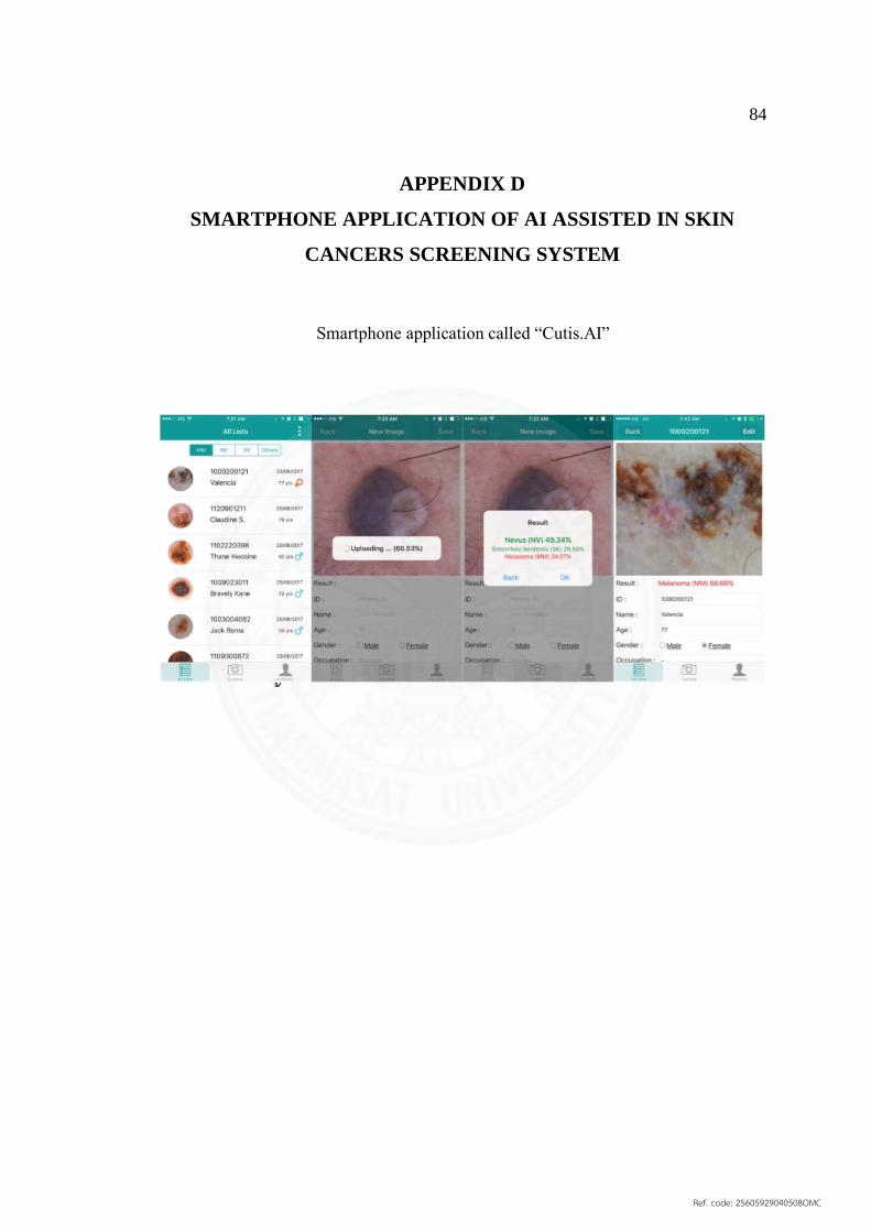
84
APPENDIX D
SMARTPHONE APPLICATION OF AI ASSISTED IN SKIN
CANCERS SCREENING SYSTEM
Smartphone application called “Cutis.AI”
Ref. code: 25605929040508OMC

85
BIOGRAPHY
Name Miss Thorfun Treewatanakul
Date of Birth January 15, 1990
Educational Attainment
2009-2014 Doctor of Medicine,
Chiangmai university
Work Position Master degree student
Department of Dermatology,
Chulabhorn International College of Medicine
Thammasat university
Scholarship N/A
Publications N/A
Work Experiences 2015-2016 Internship at Nakornping Hospital
Chiangmai, Thailand
Ref. code: 25605929040508OMC