Embed Size (px)
Citation preview

Clinical Translational Evaluation of Al18F-NOTA-FAPI for Fibroblast Activation Protein TargetedTumour ImagingShuailiang Wang
Institute of Medical Technology, Peking University Health Science CenterXin Zhou
Peking University Cancer Hospital & InstituteXiaoxia Xu
Peking University Cancer Hospital & InstituteJin Ding
Peking University Cancer Hospital & InstituteSong Liu
Peking University Cancer Hospital & InstituteXingguo Hou
Peking University Cancer Hospital & InstituteNan Li
Peking University Cancer Hospital & InstituteHua Zhu
Peking University Cancer Hospital & InstituteZhi Yang ( [email protected] )
Peking University Cancer Hospital & Institute https://orcid.org/0000-0003-2084-5193
Research Article
Keywords: Fibroblast activation protein, A118F, PET/CT, Tumour imaging, Clinical translational evaluation
Posted Date: February 24th, 2021
DOI: https://doi.org/10.21203/rs.3.rs-231262/v1
License: This work is licensed under a Creative Commons Attribution 4.0 International License. Read Full License
Version of Record: A version of this preprint was published at European Journal of Nuclear Medicine andMolecular Imaging on June 24th, 2021. See the published version at https://doi.org/10.1007/s00259-
Clinical Translational Evaluation of Al18F-NOTA-FAPI for Fibroblast Activation
Protein Targeted Tumour Imaging
Shuailiang Wang1,2, Xin Zhou2, Xiaoxia Xu2, Jin Ding2, Song Liu2, Xingguo Hou2, Nan
Li2, Hua Zhu2*, Zhi Yang1,2*
1Institute of Medical Technology, Peking University Health Science Center, Beijing
100191, China; 2Key Laboratory of Carcinogenesis and Translational Research
(Ministry of Education/Beijing), Department of Nuclear Medicine, Peking University
Cancer Hospital & Institute, Beijing 100142, China.
Corresponding author:
Hua Zhu, Peking University Cancer Hospital & Institute, No. 52 Fu-Cheng Rd., Beijing,
100142, China. E-mail: [email protected]
Zhi Yang, Peking University Cancer Hospital & Institute, No. 52 Fu-Cheng Rd., Beijing,
100142, China. E-mail: [email protected]
Abstract
Purpose In this study, a novel Al18F-NOTA-FAPI probe was developed for fibroblast
activation protein (FAP) targeted tumour imaging, which was available to achieve curie
level radioactivity by automatic synthesizer. The tumour detection efficacy of Al18F-
NOTA-FAPI was further validated both in preclinical and clinical translational studies.
Methods The radiolabeling procedure of Al18F-NOTA-FAPI was optimized. Cell
uptake and competitive binding assay were completed with U87MG and A549 cell lines,
to evaluate the affinity and specificity of Al18F-NOTA-FAPI probe. The biodistribution,
pharmacokinetics, radiation dosimetry and tumour imaging efficacy of Al18F-NOTA-
FAPI probe were researched with healthy Kunming (KM) and/or U87MG model mice.
After the approval of ethical committee, Al18F-NOTA-FAPI probe was translated into
clinical for the PET/CT imaging of first 10 cancer patients.
Results The radiolabeling yield of Al18F-NOTA-FAPI was 33.8 ± 3.2% through
manually operation (n = 10), with the radiochemical purity over than 99% and the
specific activity of 9.3-55.5 MBq/nmol. Whole body effective dose of Al18F-NOTA-
FAPI was estimated to be 1.24E-02 mSv/MBq, lower than several other FAPI probes
(68Ga-FAPI-04, 68Ga-FAPI-46 and 68Ga-FAPI-74). In U87MG tumour bearing mice,
Al18F-NOTA-FAPI showed good tumor detection efficacy from the results of micro
PET/CT imaging and biodistribution studies. In organ biodistribution study of human
patients, Al18F-NOTA-FAPI showed lower SUVmean than 2-[18F]FDG in most organs,
especially in liver (1.1 ± 0.2 vs. 2.0 ± 0.9), brain (0.1 ± 0.0 vs. 5.9 ± 1.3), and bone
marrow (0.9 ± 0.1 vs. 1.7 ± 0.4). Meanwhile, Al18F-NOTA-FAPI do not show extensive
bone uptakes, and was able to find out more tumour lesions than 2-[18F]FDG in the
PET/CT imaging of several patients.
Conclusion Al18F-NOTA-FAPI probe was successfully fabricated and applied in
fibroblast activation protein targeted tumour PET/CT imaging, which showed excellent
imaging quality and tumour detection efficacy in U87MG tumour bearing mice as well
as in human cancer patients.
Trial registration: Chinese Clinical Trial Registry ChiCTR2000038080. Registered 09
September 2020. http://www.chictr.org.cn/showproj.aspx?proj=61192
Keyword Fibroblast activation protein; Al18F; PET/CT; Tumour imaging; Clinical
translational evaluation
Declarations
Funding This work was supported by National Natural Science Foundation of China
projects No. 81871386 and 81871387, Yangfan project No. ZYLX201816, Dengfeng
project No. DFL20191102, and Science Foundation of Peking University Cancer
Hospital-2020-18.
Conflicts of interest The authors declare that they have no conflict of interest.
Availability of data and material Not applicable.
Code availability Not applicable.
Authors' contributions ZY and HZ conceived and designed this research. SW was
responsible to all the experiments, data collection and analysis, and also wrote the
manuscript. XZ and XX were responsible to the recruitment of patients and image
analysis. JD, SL and XH were involvode in the preparation of radiopharmaceuticals and
also took part in most of the animal experiments. All of the authors joined in the
embellishment of the article.
Ethics approval All procedures involving human participants were carried out in
accordance with the Ethics Committee of Peking University Cancer Hospital (2019
KT95), and registered in Chinese Clinical Trial Registry (ChiCTR2000038080). All
animal studies were performed according to a protocol approved by the Peking
University Cancer Hospital Animal Care and Use Committee.
Consent to participate Written informed consents were obtained from all participants
included in the study.
Consent for publication Not applicable.
Clinical Trial Registration This study was approved by the Ethics Committee of
Peking University Cancer Hospital (2019 KT95), and registered in Chinese Clinical
Trial Registry (ChiCTR2000038080).
Acknowledgments
We gratefully appreciate all the chemists, nurses, and technicians from the Department
of Nuclear Medicine, Peking University Cancer Hospital for their contributions to
tracer administration and PET/CT imaging.
Introduction
Fibroblast activation protein (FAP) is a type II transmembrane glycoprotein consisting
of 760 amino acids, which belongs to the serine protease family and is selectively
expressed in the stroma fibroblasts associated with epithelial cancers [1]. Reactive
tumour stroma or fibrosis generally presents with increased number of activated
fibroblasts that usually express FAP, whereas normal stroma in most adult organs only
contains a small number of quiescent or resting fibroblasts with low or undetectable
FAP expression, making FAP a novel metabolic target in cancer theranostics [2, 3]. Till
now, plenty of therapies targeting FAP have been explored, including FAP inhibitors
[4], peptide drug complexes [5, 6], antibodies [7], CAR-T cell therapy [8], vaccines [9]
and tumour immunotherapy [10].
Especially in recent several years, (4-Quinolinoyl)glycyl-2-cyanopyrrolidine based
organic small molecules that exhibited excellent affinity with FAP [11], known as FAP
inhibitor (FAPI), have been radiolabeled with different radionuclides including 68Ga,
90Y, 99mTc, 64Cu and 225Ac, and have been translated into clinical for the nuclear imaging
and radionuclide therapy of various types of cancer [12-15]. Earlier study indicated that
several highly prevalent cancers showed remarkably high uptake and image contrast on
68Ga-FAPI PET/CT imaging [13]. More importantly, in contrast to 2-[18F]FDG, no diet
or fasting is required in preparation for the 68Ga-FAPI examination, and the image
acquisition in 68Ga-FAPI imaging can be potentially started much earlier than 2-
[18F]FDG imaging [16].
However, 68Ga-FAPI PET/CT imaging suffered the disadvantage of radionuclide supply,
since 68Ga was usually eluted from 68Ge-68Ga generator, one single synthesis would
only achieve a small amount of radiopharmaceutics avaliable to 2-4 patients.
Meanwhile, the supply of 68Ga-FAPI to distant centres that requiring this probe would
be restricted because of the short half-life of 68Ga (t1/2 = 68 min). Remolding of 68Ga-
FAPI into 18F radiolabeled probes would fundamentally resolve its intrinsic
disadvantages mentioned above, in consideration of the longer half-life of 18F (t1/2 =
109.8 min) and the availability of 18F from cyclotron that owned by many medical
centres. Additionally, the lower positron energy of 18F (Emean = 0.25 MeV vs. 0.83
MeV of 68Ga) rendered it a shorter positron range than 68Ga (0.6 mm vs. 3.5 mm), which
would result in a higher spatial resolution on PET/CT imaging [17]. Most recently, two
18F radiolabeled FAPI probes has been reported, namely 18F-FAPI-74 and [18F]FGlc-
FAPI, respectively [18, 19]. Both of this two probes showed excellent tumour imaging
efficacy, however, the structure of this novel Al18F-NOTA-FAPI probe was different
from them.
Herein, we report the rapid and efficient radiolabeling strategy of a novel Al18F-NOTA-
FAPI probe and carried out its preclinical investigations. Furthermore, Al18F-NOTA-
FAPI is translated into clinical application for the PET/CT imaging of patients with
different types of cancer, and its imaging efficacy is compared with 2-[18F]FDG.
Materials and methods
Chemicals and reagents
Metal basis chemicals used in this study include the following: potassium hydrogen
phthalate (KHP) was purchased from Acros Organics (USA), anhydrous aluminum
chloride (AlCl3) and sodium acetate were purchased from Alfa Aesar (China)
Chemicals Co. Ltd. Reagents including ethanol and acetonitrile were purchased from
Honeywell International Inc (USA), trifluoroacetic acid (TFA) was purchased from
Shanghai Aladdin Biochemical Technology Co., Ltd (China). All chemicals and
reagents were applied directly without further purification. NOTA-FAPI precursor was
purchased from HUAYI Co. Ltd (China). Reagents including phosphate buffer saline
(PBS), cell culture medium, fetal bovine serum (FBS), penicillin-streptomycin solution
(PS), Glutamax, non-essential amino acid (NEAA) and trypsin for cell culturing and
following experiments were obtained from Gibco (Thermo Fisher Scientific, China).
Cell culture and tumour models
U87MG and A549 cancer cell lines were obtained from National Collection of
Authenticated Cell Cultures. U87MG cells were cultivated in Dulbecco modified Eagle
medium (DMEM high glucose) containing 10% FBS, 1% PS, 1% Glutamax and 1%
NEAA at 37 oC in 5% carbon dioxide. A549 cells were cultivated in Roswell Park
Memorial Institute (RPMI 1640) medium containing 10% FBS, 1% PS at 37 oC in 5%
carbon dioxide.
U87MG and A549 tumour bearing BALB/c Nude mice (6-8 weeks, female, 16-20 g)
were purchased from Beijing Vital River Laboratory Animal Technology Co. Ltd
(China). Mice of specific pathogen free (SPF) grade were breeding in individual
ventilated cages (IVC) with free available of food and water. Tumour on the right flank
of mice were allowed to reach an volume of approximately 0.5-0.8 cm in diameter, and
animals were further allpied for following imaging and in vivo biodistribution
experiments. All animal experiments were completed in accordance with relavant
guides and regulations of Beijing Cancer Hospital.
Radiopharmaceutical preparation
As shown in Fig. 1, radiolabeling of Al18F-NOTA-FAPI was completed based on a
previous reported method with some modifications [20]. Briefly, 18F was produced
from HM-20 medical cyclotron (Sumitomo Corporation, Japan), loaded on QMA
cartridge (Waters Corporation, USA) and further eluted with saline (0.3-0.5 mL). For
the radiolabeling, 18F in saline (0.1 mL, 555-3330 MBq) was mixed with AlCl3 (6 μL,
2 mM) and KHP (6 μL, 0.5 M) for 5 min under room temperature to form the [Al18F]2+
conjugate, NOTA-FAPI precursor (5 μL, 4 mM) was further added to the reaction
system and heated at 110 oC for 15 min. After that, the reaction system was diluted with
5 mL of ultrapure water and purified with Sep-Pak Light C18 cartridge (Waters
Corporation, USA). Final product was formulated through the elution of C18 cartridge
with 0.5 mL of 80% ethanol and 6 mL of saline consecutively, and further passed
through a 0.2 μm syringe filter (Pall Corpotation, USA). The radiochemical purity of
Al18F-NOTA-FAPI was analyzed by a radio high performance liquid chromatograph
(1200 Series, Agilent, USA) with gradient elution: 0-3 min, 95% water (0.1% TFA) +
5% acetonitrile (0.1% TFA); 18 min, 5% water (0.1% TFA) + 95% acetonitrile (0.1%
TFA); 20 min, 95% water (0.1% TFA) + 5% acetonitrile (0.1% TFA).
Stability and partition coefficient
The in vitro stability of Al18F-NOTA-FAPI was assessed in saline and 5% human serum
albumin (HSA) for 1 h, 2 h and 4 h with radio-HPLC. For in vivo stability assessment,
KM mice (female, 18-20 g, n = 3) was administered with approximately 37 MBq of
Al18F-NOTA-FAPI intravenously, urine and blood was collected at 10 min, 0.5 h and 1
h post-injection, radiochemical purity of Al18F-NOTA-FAPI in the urine and blood of
mice was analyzed with radio-HPLC.
The octanol-water partition coefficient of Al18F-NOTA-FAPI was detected by mixting
10 μL of purified Al18F-NOTA-FAPI with 590 μL of PBS (0.1 M, pH 7.4) and 600 μL
of octanol in a 1.5 mL tube (n = 5). The mixture was vortexed for 3 min and further
centrifugated at 3000 × rpm for 5 min, a small portion (10 μL) was removed from each
phase and further counted with a γ counter (Wizard II, Perkin Elmer Inc., Germany).
The partition coefficient was calculated as the counts in octanol divided by the counts
in phosphate buffered saline, the value was expressed as logD7.4 (mean ± SD).
Cell uptake and competitive binding assay
For cell uptake experiment, cells were seeded in 24-well plates overnight (2 × 10⁵ per
well). Cells were washed with PBS (pH 7.4, 0.01 M) twice and replaced with fresh
medium without FBS. Cells were incubated with 74 kBq of Al18F-NOTA-FAPI in 1 mL
of medium for 10, 30 and 60 min at 37 oC. In the blocking group, an extra amount (1
μg/well) of non-radiolabeled precursor was added. After that, cells were washed twice
with cold PBS (pH 7.4, 0.01 M) and lysed with 200 μL of NaOH (0.1 M). The lysate
was collected and counted with a γ counter. The results were normalized to 1 × 10⁶ cells
and expressed as percentage of added dose (%AD/10^6 cell). The experiment was
repeated three times for each time point.
For competitive binding assay, different concentrations of unlabeled precursor (0.1 nM
to 10 μM) was added to cells with fixed activity of radiolabeled Al18F-NOTA-FAPI (74
kBq/well). The cells were incubated at 37 oC for 30 min, washed with cold PBS (pH
7.4, 0.01 M) for two times and further lysed with 200 μL of NaOH (0.1 M). The lysate
was collected and counted with a γ counter. The experiment was repeated three times
for each concentration. The IC50 of NOTA-FAPI was acquired using the non-linear
curve fitting of GraphPad Prism 8.3.0 software (GraphPad Software, San Diego, CA,
USA).
Small animal biodistribution and pharmacokinetics study
For animal biodistribution study, KM mice (female, 4 weeks, 18-20 g) were devided
into 5 groups (n = 3) and injected with Al18F-NOTA-FAPI (200 μL, 1.85 MBq/per
mouse) intravenously, 1% injected dose of Al18F-NOTA-FAPI was prepared and set as
contrast. At 5 min, 30 min, 1 h, 2 h, 4 h post-injection, KM mice were sacrificed after

anesthetization, organs of interest were collected, weighted, and counted with a γ
counter. As a standard, 10 samples of 1% injected dose were taken from the injection
and counted. The biodistribution of Al18F-NOTA-FAPI in different organs of KM mice
was expressed as percentage injected dose per gram (%ID/g, mean ± SD).
For biodistribution of Al18F-NOTA-FAPI in tumour bearing mice, U87MG and A549
(negative control) tumour bearing mice were grouped and injected intravenously with
200 μL of radiotracer (~ 1.11 MBq). At 1 h post injtection, mice were anesthetized and
sacrificed, blood samples and organs of interest were collected, weighted and counted
with a γ counter. For the block group, mice were co-injtected with 20 nmol (15.4 μg)
of non-radiolabeled FAPI precursor.
Pharmacokinetics study was accoplished using female KM mice (n = 5). Briefly, each
mouse was intravenously injected with 3.7 MBq of Al18F-NOTA-FAPI. A small portion
of blood was acqiured through the orbital venous of mice at 1 min, 3 min, 5 min, 10
min, 15 min, 30 min, 45 min, 60 min, 90 min, 120 min, 180 min and 240 min post-
injection. The blood samples were weighted before counted using a γ counter. The
results were calculated as percentage injected dose per gram (%ID/g, mean ± SD).
Radiation dosimetry estimation
The internal radiation dosimetry of Al18F-NOTA-FAPI probe in adult female patients
was estimated according to the biodistribution results in KM mice. Area under the time-
activity curves (AUCs) of different organs were calculated using GraphPad Prism 8.3.0,
and the results were further analyzed using OLINDA/EXM software (version 1.0;
Hermes Medical Solutions AB) to estimate the absorbed dose of each organ, as well as
the effective dose of whole body.
Micro PET/CT imaging in mice bearing U87MG and A549 xenograft
Tumour bearing BALB/c Nude mice were injected with 200 μL of Al18F-NOTA-FAPI
(~7.4 MBq), at 1 h post injection, mice were anesthetized and placed on the imaging
bed to perform the micro PET-CT imaging (SuperNova, PINGSENG Healthcare,
China). PET images were acquired for 10 minutes and reconstruted with attenuation
correction based on the CT data (CT-AC reconstruction). Regions of interest (ROI)

were drawn on the CT images and further mapped on PET, SUVmax of tumour and
muscle were calculated and referred to as tumour to muscle ratio. In the block group,
mouse were co-injected with an extra amount (20 nmol) of non-radiolabeled precursor.
Patient enrollment
The clinical translational study of Al18F-NOTA-FAPI was approved by the Ethics
Committee (approval No. 2019KT95) of Beijing Cancer Hospital, and registered in
Chinese Clinical Trial Registry (ChiCTR2000038080), all patients enrolled in this
study have signed a written informed consent. The include criteria were as follows, 18
to 75 years old cancer patients or suspected cancer patients, ability to provide a written
informed consent, normal liver and kidney function. The exclusion criteria included the
following: liver and renal dysfunction, pregnancy or current lactation and patients that
suffered psychiatry disease or claustrophobia.
PET/CT imaging in cancer patients
For tracer administration, all patients received intravenously injection through the
elbow or opisthenar vein with the radioactivity ranged from 173.5 MBq to 256.8 MBq.
At 60-90 min post-injection, patients were required for urination and received static
PET/CT scan (Siemens, Erlangen, Germany) from the top of the skull to the middle of
the femur. A Siemens workstation (syngo.via Client 4.1) was used for image processing.
Two experienced nuclear medicine physicians independently reviewed all images, and
any discordant results were resolved by consensus. The SUVmax and SUVmean values
of tumour lesions as well as normal organs were quantified by delineation of ROI
directly on the PET/CT iamges.
RESULTS
Radiopharmaceutical preparation
The synthetic procedure of Al18F-NOTA-FAPI was presented in Fig. 1, and the
molecular structures of this probe as well as earlier reported [18F]FGlc-FAPI were
compared directly. This study evaluated several parameters which might influence the
radiolabeling yield, including the amount of AlCl3 (2 mM), KHP (0.5 M) and precursor
(4 mM), as well as the temperature and raction volume. As shown in Fig. S1, all
parameters exhibited significant impact on radiolabeling yield. Meanwhile, the optimal
radiolabeling parameters are as follows, both the volumes of AlCl3 and KHP were 6 μL,
the amount of precursor was 5 μL, the volume of 18F ions in saline was 100 μL. The
reaction mixture was heated at 110 oC for 15 min, and further purified with Sep-Pak
Light C18 cartridge. The highest radiolabeling yield under the optimal reaction
conditions, appoximately 36.5% can be achieved under manual operation (33.8 ± 3.2%
on average, n = 10). The specific activity of Al18F-NOTA-FAPI was around 9.3-55.5
MBq/nmol, according to the activity of 18F ions added. Radiochemical purity of the
final product was higher than 99%. Furthermore, Al18F-NOTA-FAPI with much higher
activity (> 100 mCi) can be achieved under automatic synthesis with this optimal
reaction condition.
Stability and partition coefficient
Both the in vitro and in vivo stability of Al18F-NOTA-FAPI were determined with
radio-HPLC. The radiopharmaceutical showed excellent stability in vitro, including in
saline and 5% HSA, kept almost intact within 4 h (radiochemical purity > 95%, Fig.
S2). Furthermore, the stability of Al18F-NOTA-FAPI was also perfect in vivo, although
slightly compromised compared with its in vitro stability. Within 1 h post injection,
radiochemical purity of the radiopharmaceutical were higher than 90%, both in blood
and urine. Meanwhile, the partition coefficient of Al18F-NOTA-FAPI was calculated as
logD7.4 = -1.88 ± 0.01, with the majority of radioactivity concentrared in the water phase
(>98%), indicating the superior hydrophilicity of Al18F-NOTA-FAPI.
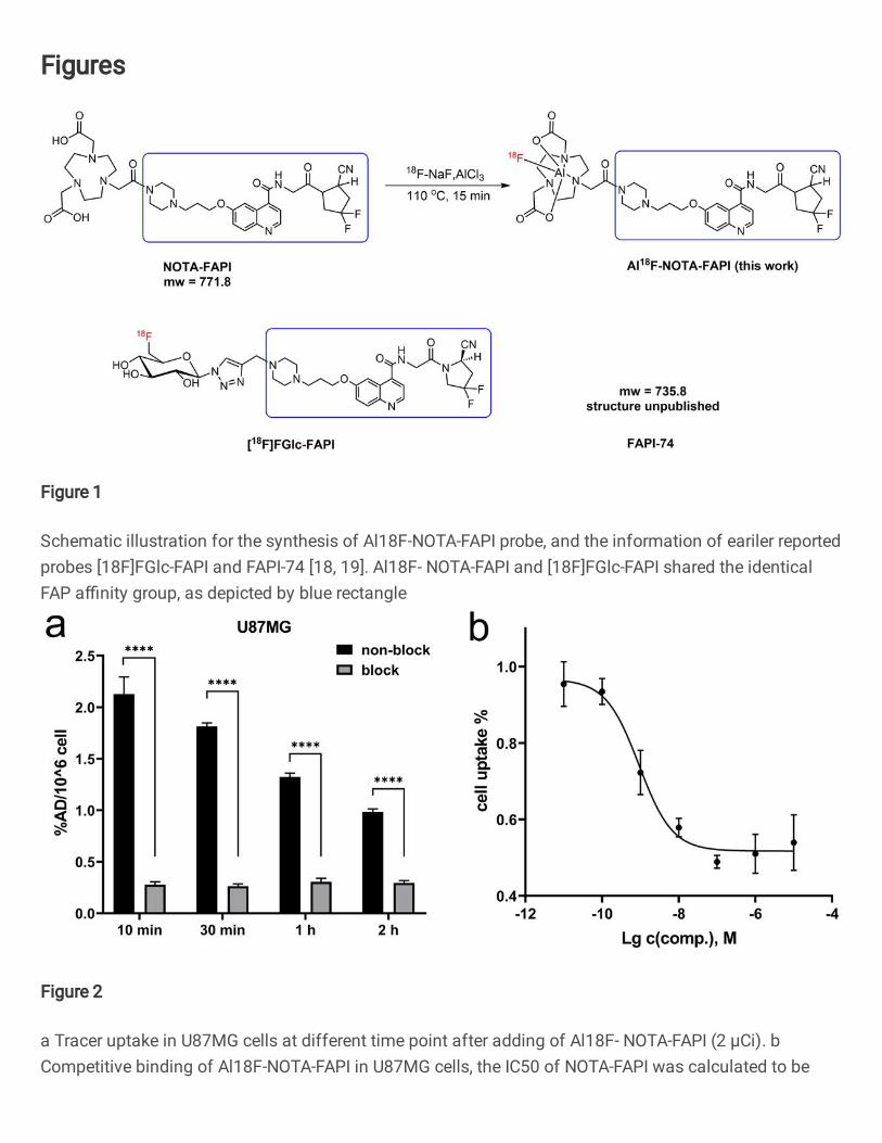
Cell uptake and IC50 of NOTA-FAPI
As shown in Fig. 2a, in FAP positive U87MG cells, highest uptake of Al18F-NOTA-
FAPI was observed at 10 min (2.13 ± 0.17 AD%), which can be blocked by the adding
of excess non-radiolabeled precursor (0.28 ± 0.03 AD%), indicating the specific
binding between Al18F-NOTA-FAPI and fibroblast activation protein. The uptake of
Al18F-NOTA-FAPI in U87MG cells decreased from 10 min to 2 h. While in FAP
negative A549 cells, much lower and stable uptake of the radiopharmaceutical was
observed at all time points (Fig. S3). Moreover, the IC50 of NOTA-FAPI was calculated
to be 1.73 ± 0.93 nM (Fig. 2b), which was lower than several other FAPI probes (Table
S1), indicating the high affinity between NOTA-FAPI and FAP.
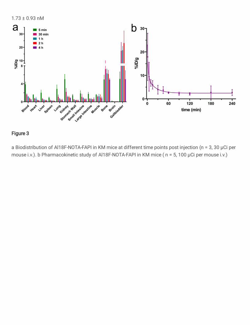
Small animal biodistribution and pharmacokinetics study
In KM mice, the tracer showed rapid clearence through the kidney, with extremely low
uptake in most normal organs including liver, lung, spleen and brain (Fig. 3a). At 1 h
post injection, the highest uptake was observed in the gallbladder (17.83 ± 2.92%ID/g),
followed by the bone (6.34 ± 0.98%ID/g) and muscle (1.25 ± 0.47%ID/g). The tracer
uptake in most organs decrased rapidly along with time, except in the bone and
gallbladder. A comparison of the biodistribution at 1 h post injection in KM mice
between 68Ga-FAPI and Al18F-NOTA-FAPI was presented in Tabel 1. In all tested
organs, the uptake of Al18F-NOTA-FAPI was lower than 68Ga-DOTA-FAPI-04,
especially in the spleen, stomach wall and muscle (Fig. S4).
Furthermore, pharmacokinetics study of Al18F-NOTA-FAPI in mice provided
conclusive evidence for its rapid clearance from body. As shown in Fig. 3b and equation
(1), pharmacokinetics of Al18F-NOTA-FAPI followed two phase decay in mice, with
an extremely short biodistribution half life of only 1.583 min, and half life of the
elimination phase was only 14.75 min. Ct = 2.46 + 13.82 × e−0.44t + 6.84 × e−0.05t...............................(1)
In U87MG tumour bearing xenografts, Al18F-NOTA-FAPI showed extremely high
accumulation in the tumour region (35.29 ± 1.00 %ID/g ) and low uptake in most organs
at 1 h post-injection, as shown in Fig. 4a. The uptake of Al18F-NOTA-FAPI in the
tumour was slightly higher than 68Ga-DOTA-FAPI-04 both at 1 h and 4 h post-injection
(Fig. 4b), and the uptake can be almost completely blocked (0.50 ± 0.11 %ID/g, p <
0.0001) by the adding of non-radiolabeled NOTA-FAPI. In A549 xenograft mice, the
uptake of Al18F-NOTA-FAPI in the tumour tissue was much lower, only reached 3.28
± 0.66 %ID/g (Fig. S5). Al18F-NOTA-FAPI showed relatively high uptake in the bone
structure, both in U87MG (5.25 ± 0.68 %ID/g) and A549 (6.77 ± 0.93 %ID/g) tumour

bearing mice. However, the uptake in bone can be lowered (3.27 ± 0.28 %ID/g) by co-
injection with non-radiolabeled NOTA-FAPI in U87MG tumour bearing mice (p < 0.01,
Figure 4a). Alike 68Ga-DOTA-FAPI-04 (Fig. S6), the retention of Al18F-NOTA-FAPI
in the tumour was not satisfied, with a decreasement of 33.72% from 1 h to 4 h post-
injection.
Radiation dosimetry estimation
The internal radiation dosimetry in adult female patients was estimated based on the
biodistribution results in KM mice. As shown in Tabel 2, most organs exhibited low
absorbed dose as well as effective dose. The osteogenic cell had the highest absorbed
dose (2.47E-02 mGy/MBq), followed by the red marrow (1.80E-02 mGy/MBq) and
small intestine wall (1.54E-02 mGy/MBq). The whole body effective dose of Al18F-
NOTA-FAPI was calculated to be 1.24E-02 mSv/MBq.
Small animal PET/CT imaging
In A549 tumour bearing mice, no significant uptake of Al18F-NOTA-FAPI was
observed at the tumour region (SUVmax = 0.87), the tracer showed rapid clearence
from body, with only high uptake in the gallbladder was noticed. However, in U87MG
tumour xenografts, the tumour showed extremely high uptake of Al18F-NOTA-FAPI
(SUVmax = 3.53), meanwhile, high uptake in the gallbladder was also noticed.
Interestingly, high uptake of Al18F-NOTA-FAPI in the tumour region of U87MG
xenografts can be almost totally blocked by co-injection of non-radiolabeled FAPI
precursor, while most of the injected radiopharmaceutics were excreted through the
biliary and intestine tract, indicating the specific uptake of Al18F-NOTA-FAPI in the
tumour area. (Fig. 5a-c). Futhermore, immunohistochemical staining results indicating
high expression of FAP in U87MG xenograft but not in A549 tumour xenograft (Fig.
5d and 5e).
PET/CT imaging in cancer patients
Al18F-NOTA-FAPI was successfully translated into clinical imaging of cancer patients
with various tumour types, including lung cancer, pancreas cancer, colorectal cancer,
prostate cancer and lymphoma patients (Table 3). In most normal organs of patients,

the SUVmean values in Al18F-NOTA-FAPI PET/CT imaging were lower than that in
2-[18F]FDG PET/CT imaging, especially in the liver (1.1 ± 0.2 vs. 2.0 ± 0.9), brain (0.1
± 0.0 vs. 5.9 ± 1.3), spleen (0.7 ± 0.1 vs. 1.8 ± 0.4) and bone marrow (0.9 ± 0.1 vs. 1.7
± 0.4) (Fig. 6a). However, the SUVmean of pancreas (3.0 ± 2.0 vs. 1.4 ± 0.4), muscle
(1.6 ± 0.4 vs. 0.7 ± 0.1), submaxillary gland (3.5 ± 1.5 vs. 1.6 ± 0.5) and parotid gland
(1.8 ± 0.7 vs. 0.9 ± 0.2) were higher in Al18F-NOTA-FAPI PET/CT imaging than 2-
[18F]FDG PET/CT. The SUVmean of gallbladder was high in Al18F-NOTA-FAPI
PET/CT imaging (7.6 ± 10.5) of several but not all patients, which was consistently low
in 2-[18F]FDG PET/CT imaging (0.8 ± 0.4). In Al18F-NOTA-FAPI PET/CT imaging,
patients showed higher tumor/spleen and tumor/brain SUVmean ratios than 2-
[18F]FDG PET/CT imaging, as shown in Fig. 6b. Meanwhile, the SUVmax and
SUVmean values were compared between Al18F-NOTA-FAPI and 2-[18F]FDG PET/CT
imaging, as shown in Fig. S7, where no siginificant differences were noticed. The
SUVmax and SUVmean values in the primary tumour lesions of individual patient were
presented in Table S3.
Fig. 7 shows the representative images of a female pancreas cancer patient (64 years
old) underwent Al18F-NOTA-FAPI PET/CT imaging. The tumour lesion in the pancreas
showed low uptake in 2-[18F]FDG PET/CT imaging (SUVmax = 2.7, Fig. 7a-c), while
in the Al18F-NOTA-FAPI PET/CT imaging, the lesion showed extremely high uptake
of radiotracer (SUVmax = 36.3, Fig. 7d-f). Furthermore, plenty of metastatic lymph
nodes in the abdomen of this patient were clearly visualized in Al18F-NOTA-FAPI
PET/CT imaging, while not obvious in 2-[18F]FDG PET/CT imaging. Fig. 8 shows
another representative images of a lung cancer patient. The 59 years old female patient
was diagnosed with a nodule in the superior lobe of left lung, which showed
inappreciable uptake of 2-[18F]FDG (SUVmax = 1.3, Fig. 8a-c). However, in Al18F-
NOTA-FAPI PET/CT imaging, the nodule was further validated as a malignancy lesion
with relatively high uptake of Al18F-NOTA-FAPI (SUVmax = 6.6, Fig. 8d-f).
Interestingly, unlike the high uptake of Al18F-NOTA-FAPI in the gallbladder in animal
micro-PET/CT imaging, such phenomenon was not validated in some human patients,
and the tracer uptake in the bone of human patients was negligible.
Discussion
Fibroblast activation protein targeted tumour imaging strategy developed rapidly in
recent several years, especially utilizing FAP specific small molecule inhibitors.
Although plenty of work needs to be further finished, FAPI has been deemed the most
competitive radiotracer with 18F-FDG in clinical tumour PET/CT imaging, because of
its fast clearance from body, universality in the diagnosis of various types of cancer,
and high tumour to background ratio. As we have successfully fabricated the Al18F
labeled PSMA ligand for tumour detection [20], it is also convenient to translate the
68Ga-DOTA-FAPI into Al18F-NOTA-FAPI probe. In this study, we investigated both
the preclinical and clinical utilization of Al18F-NOTA-FAPI probe, which showed great
significance in the PET/CT imaging of cancer patients.
Compared with 68Ga-FAPI, Al18F-NOTA-FAPI showed faster clearance from body in
mice biodistribution, rendered lower uptake in most normal organs. Meanwhile, the
lower effective dose of Al18F-NOTA-FAPI (1.24E-02 mSv/MBq) calculated in this
study might also ascribe to its faster clearance. The effective dose of Al18F-NOTA-FAPI
was almost the same with 18F-FAPI-74 (0.014 mSV/MBq) reported earlier, which
contains a NOTA chelator and can also be radiolabeled with 18F-AlF[18]. Although the
structure of FAPI-74 was not public yet, its molecular weight was reported to be 735.8
g/mol, different from the probe (mw. 771.82) utilized in this study.
The high uptake in the gallbladder of mice was a characteristic of Al18F-NOTA-FAPI,
since such phenomenon was not noticed in 68Ga-FAPI biodistribution. This can be
ascribed to the higher lipophilicity of Al18F-NOTA-FAPI, since the LogD7.4 of Al18F-
NOTA-FAPI was calculated to be -1.88 ± 0.01, bigger than the LogD7.4 of 68Ga-DOTA-
FAPI-04 (-2.87 ± 0.02). High accumulation of radiopharmaceutics in the bone of mice
was appreciable, which was validated by small animal PET/CT imaging, especially in
the shoulder joints, knee joints, skull and the spine of mice, which might be the
explanation of the high absorbed dose and effective dose in osteogenic cells and red
marrow, respectively. Such phenomenon was also noticed in previous study with the
[18F]FGlc-FAPI probe, which might because of the relatively high expression of FAP
in the osteoblast and bone marrow stem cells (BMSC) of mice, hence the high
accumulation of FAPI probe in the skeleton of mice was ascribed to physiological
uptake [19].
FAP was expressed primarily on the cancer associated fibroblasts but not on tumour
cells, only several cancer cell lines showed high expression of FAP, including several
glioblastoma cell line [21]. Indeed, much higher tumour cell uptake of Al18F-NOTA-
FAPI in U87MG cell line was noticed, compared with FAP low expressed A549 cell
line in vitro. Since the uptake of Al18F-NOTA-FAPI in U87MG cells can be blocked by
extra adding of non-radiolabeled precursor, the specific binding between the
radiopharmaceutical and FAP can be thus confirmed. Interestingly, the highest uptake
of Al18F-NOTA-FAPI in U87MG cells was observed at 10 min, and decreased in the
following 2 hours, which was not reported by previous studies. We ascribe this to the
rapid binding of Al18F-NOTA-FAPI with FAP and internalization in U87MG cells,
since the IC50 of NOTA-FAPI was lower than any previously reported FAPI probes
(Table S2) [12, 22].
Also, from the biodistribution analysis results of tumour bearing mice, tracer uptake in
U87MG xenografts was approximate 10 folds higher than that in A549 xenografts,
while no significant differences were noticed in most normal organs. Meanwhile, in
small animal PET/CT imaging, extremely high uptake of Al18F-NOTA-FAPI in the
tumour region of U87MG xenograft can be observed, the tumour-to-muscle SUVmax
ratio was 23.5, much higher than A549 mice xenograft (tumour/muscle SUVmax ratio
= 6.2). While in the mice co-injected with non-radiolabeled NOTA-FAPI, most of the
radioactivity was accumulated in the gallbladder and intestine, further indicated the
specific binding of Al18F-NOTA-FAPI to FAP. Moreover, in this study, the
immunohistological chemistry staining of both A549 and U87MG tumours rescted
from mice also demonstrated the high expression of FAP in U87MG xenografts.
In the PET/CT imaging of human cancer patients, there existed several differences
compared with micro PET/CT imaging with mice. Firstly, the high uptake in the
gallbladder of mice was not appreciable in several human patients, which might because
the fasting of mice will promote the secretion of bile, while eating in some human
patients might accelerate the removal of gallbladder contents, hence the difference of
SUVmean in human gallbladder was not significant between Al18F-NOTA-FAPI and 2-
[18F]FDG PET/CT imaging. Secondly, in the PET/CT imaging of most human cancer
patients, similar to 2-[18F]FDG, uptake of Al18F-NOTA-FAPI in the bone was not
significant (SUVmean = 0.7 ± 0.2), we ascribe such phenomenon to the lower
expression level of FAP in human bone structure. Although in small animal
biodistribution experiment, the accumulation of FAPI probes were relatively high.
Furthermore, the low SUVmean in the bone cortex and bone marrow of patients would
indicating the superior biostability of Al18F-NOTA-FAPI in human body. In our
previous clinical translational study utilizing the Al18F-PSMA-BCH probe, the
SUVmean were also low in the PET/CT imaging of prostate cancer patients [20]. Taken
together, Al18F-NOTA conjugate radiolabeled probes hold great feasibility and potential
for clinical translational studies.
In the PET/CT imaging of several patients enrolled in this study, Al18F-NOTA-FAPI
was able to detect more tumour lesions compared with 2-[18F]FDG, and was able to
differentiate benign from malignant, thus can provide more effective information for
the clinical treatment decisions of patients. As indicated in Fig. S7 and Table S3,
primary tumour lesions of all patients showed high SUVmax in Al18F-NOTA-FAPI
PET/CT imaging, while in several patients (No. 4, 5 and 8), the SUVmax was much
lower in 2-[18F]FDG PET/CT imaging, which might result in pseudo-negative diagnosis
in these patients. However, in the Al18F-NOTA-FAPI PET/CT imaging of several
patients (No. 1, 2, 3 and 9), primary tumour lesions showed lower SUVmax and
SUVmean than that in 2-[18F]FDG PET/CT imaging, which might remind us not to
overestimate the tumour diagnosing efficacy of FAPI probes versus 2-[18F]FDG. In this
study, the differences of SUVmax and SUVmean in primary tumours were not
significant between Al18F-NOTA-FAPI and 2-[18F]FDG PET/CT imaging, this was not
surprising since the differences of the uptake in primary tumours between 68Ga-DOTA-
FAPI-04 and 2-[18F]FDG PET/CT imaging of several cancer types were also not
significant, as demonstrated by Chen et al [23]. However, when comparing the
SUVmean values in primary tumours and normal organs, Al18F-NOTA-FAPI showed
higher tumor-to-background ratios than 2-[18F]FDG thanks to its low uptake in normal
organs, especially in the spleen and brain. Therefore, Al18F-NOTA-FAPI was a
promising candidate and alternative to 68Ga-DOTA-FAPI-04 for the popularization and
application of FAPI PET/CT in clinical tumour imaging territory, and might act as
supplementary to 2-[18F]FDG PET/CT imaging, to provide more helpful informations
for the precision diagnostic of individual patient.
Conclusion
In the present study, we developed the Al18F-NOTA-FAPI probe for fibroblast
activation protein targted imaging. Al18F-NOTA-FAPI can be achieved under
convenient manual operation with high radiolabeling yield and specific activity. NOTA-
FAPI in this study showed higher affinity with FAP compared with several other FAPI
probes. Al18F-NOTA-FAPI showed excellent imaging efficacy in U87MG tumour
bearing mice and was successfully translated into the clinical PET/CT imaging of
cancer patients. Furthermore, Al18F-NOTA-FAPI can be achieved by batch of
production with high radioactivity using automatic synthesis, render it a promising
candidate and alternative to 68Ga-DOTA-FAPI-04.
Compliance with ethical standards
Conflicts of interest: The authors declare that they have no conflict of interest.
Ethics approval: All procedures involving human participants were carried out in
accordance with the Ethics Committee of Peking University Cancer Hospital (2019
KT95), and registered in Chinese Clinical Trial Registry (ChiCTR2000038080). All
animal studies were performed according to a protocol approved by the Peking
University Cancer Hospital Animal Care and Use Committee.
Informed consent: Written informed consents were obtained from all participants
included in the study.

Funding: This work was supported by National Natural Science Foundation of China
projects No. 81871386 and 81871387, Yangfan project No. ZYLX201816, Dengfeng
project No. DFL20191102, and Science Foundation of Peking University Cancer
Hospital-2020-18.
References
1. Liu R, Li H, Liu L, Yu J, Ren X. Fibroblast activation protein: a potential
therapeutic target in cancer. Cancer Biol Ther. 2012;13:1239.
2. Kalluri R. The biology and function of fibroblasts in cancer. Nat Rev Cancer. 2016;
16:58298.
3. Sánchez-Garrido MA, Habegger KM, Clemmensen C, Holleman C, Müller TD,
Perez-Tilve D, et al. Fibroblast activation protein (FAP) as a novel metabolic target.
Mol Metab. 2016;5:101524.
4. Teichgräber V, Monasterio C, Chaitanya K, Boger R, Gordon K, Dieterle T, et al.
Specific inhibition of fibroblast activation protein (FAP)-alpha prevents tumor
progression in vitro. Adv Med Sci. 2015; 60:26472.
5. Chen M, Lei X, Shi C, Huang M, Li X, Wu B, et al. Pericyte-targeting prodrug
overcomes tumor resistance to vascular disrupting agents. J Clin Invest.
2017;127:2689701.
6. Wang J, Li Q, Li X, Yuan W, Huang S, Cai S, et al. A novel FAPα-based Z-Gly-
Pro epirubicin prodrug for improving tumor-targeting chemotherapy. Eur J
Pharmacol. 2017;815:16672.
7. Scott AM, Wiseman G, Welt S, Adjei A, Lee FT, Hopkins W, et al. A phase I dose-
escalation study of Sibrotuzumab in patients with advanced or metastatic fibroblast
activation protein-positive cancer. Clin Cancer Res. 2003;9:163947.
8. Wang LS, Lo A, Scholler J, Sun J, Majumdar RS, Kapoor V, et al. Targeting
fibroblast activation protein in tumor stroma with chimeric antigen receptor T Cells
can inhibit tumor growth and augment host immunity without severe toxicity.
Cancer Immunol Res. 2014;2:15466.
9. Loeffler M, Krüger JA, Niethammer AG, Reisfeld RA. Targeting tumor-associated
fibroblasts improves cancer chemotherapy by increasing intratumoral drug uptake.
J Clin Invest. 2006;116:195562.
10. Lee J, Fassnacht M, Nair S, Boczkowski D, Gilboa E. Tumor immunotherapy
targeting fibroblast activation protein, a product expressed in tumor-associated
fibroblasts. Cancer Res. 2005;65:1115663.
11. Jansen K, Heirbaut L, Verkerk R, Cheng JD, Joossens J, Cos P, et al. Extended
structure-activity relationship and pharmacokinetic investigation of (4-
quinolinoyl)glycyl-2-cyanopyrrolidine inhibitors of fibroblast activation protein
(FAP). J Med Chem. 2014;57:305374.
12. Lindner T, Loktev A, Altmann A, Giesel F, Kratochwil C, Debus J, et al.
Development of quinoline-based theranostic ligands for the targeting of fibroblast
activation protein. J Nucl Med. 2018;59:141522.
13. Kratochwil C, Flechsig P, Lindner T, Abderrahim L, Altmann A, Mier W, et al.
68Ga-FAPI PET/CT: tracer uptake in 28 fifferent kinds of cancer. J Nucl Med.
2019;60:8015.
14. Lindner T, Altmann A, Krämer S, Kleist C, Loktev A, Kratochwil C, Giesel F, Mier
W, Marme F, Debus J, Haberkorn U. Design and development of 99mTc-labeled
FAPI tracers for SPECT imaging and 188Re therapy. J Nucl Med. 2020;61:150713.
15. Watabe T, Liu Y, Kaneda-Nakashima K, Shirakami Y, Lindner T, Ooe K, et al.
Theranostics targeting fibroblast activation protein in the tumor stroma: 64Cu- and
225Ac-labeled FAPI-04 in pancreatic cancer xenograft mouse models. J Nucl Med.
2020;61:5639.
16. Giesel FL, Kratochwil C, Lindner T, Marschalek MM, Loktev A, Lehnert W, et al.
68Ga-FAPI PET/CT: biodistribution and preliminary dosimetry estimate of 2
DOTA-containing FAP-targeting agents in patients with various cancers. J Nucl
Med. 2019;60:38692.
17. Sabri S, Patrick C, Felix MM. The battle on time, money and precision: Da[18F] id
vs. [68Ga]liath. Eur J Nucl Med Mol Imaing. 2020;47:29446.

18. Giesel F, Adeberg S, Syed M, Lindner T, Jimenez LD, Mavriopoulou E, et al. FAPI-
74 PET/CT using either 18F-AlF or cold-kit 68Ga-labeling: biodistribution,
radiation dosimetry and tumor delineation in lung cancer patients. J Nucl Med.
2020; https://doi.org/10.2967/jnumed.120.245084.
19. Toms J, Kogler J, Maschauer S, Daniel C, Schmidkonz C, Kuwert T, et al.
Targeting fibroblast activation protein: radiosynthesis and preclinical evaluation of
an 18F-labeled FAP inhibitor. J Nucl Med. 2020;61:180613.
20. Liu T, Liu C, Xu X, Liu F, Guo X, Li N, et al. Preclinical evaluation and pilot
clinical study of Al18F-PSMA-BCH for prostate cancer PET imaging. J Nucl Med.
2019;60:128492.
21. Mentlein R, Hattermann K, Hemion C, Jungbluth AA, Held-Feindt J. Expression
and role of the cell surface protease seprase/fibroblast activation protein-α (FAP-α)
in astroglial tumors. Biol Chem. 2011;392:199207.
22. Loktev A, Lindner T, Burger EM, Altmann A, Giesel F, Kratochwil C, et al.
Development of fibroblast activation protein-targeted radiotracers with improved
tumor retention. J Nucl Med. 2019;60:14219.
23. Chen H, Pang Y, Wu J, Zhao L, Hao B, Wu J, et al. Comparison of [68Ga]Ga-
DOTA-FAPI-04 and [18F] FDG PET/CT for the diagnosis of primary and metastatic
lesions in patients with various types of cancer. Eur J Nucl Med Mol Imaging.
2020;47:182032.
Figure Captions
Fig. 1 Schematic illustration for the synthesis of Al18F-NOTA-FAPI probe, and the
information of eariler reported probes [18F]FGlc-FAPI and FAPI-74 [18, 19]. Al18F-
NOTA-FAPI and [18F]FGlc-FAPI shared the identical FAP affinity group, as depicted
by blue rectangle
Fig. 2 a Tracer uptake in U87MG cells at different time point after adding of Al18F-
NOTA-FAPI (2 μCi). b Competitive binding of Al18F-NOTA-FAPI in U87MG cells,
the IC50 of NOTA-FAPI was calculated to be 1.73 ± 0.93 nM

Fig. 3 a Biodistribution of Al18F-NOTA-FAPI in KM mice at different time points post
injection (n = 3, 30 μCi per mouse i.v.). b Pharmacokinetic study of Al18F-NOTA-FAPI
in KM mice ( n = 5, 100 μCi per mouse i.v.)
Fig. 4 a Biodistribution of Al18F-NOTA-FAPI in U87MG tumour bearing BALB/c
Nude mice (n = 3). The uptake of Al18F-NOTA-FAPI in tumour decreased rapidly from
1 h to 4 h post-injection, and can be blocked by the co-injection of non-radiolabeled
NOTA-FAPI (20 nmol). b Comparison of tracer uptake in the tumour of U87MG
tumour bearing mice between 68Ga-DOTA-FAPI-04 and Al18F-NOTA-FAPI. * p<0.05,
** p<0.01, **** p<0.0001
Fig. 5 Micro PET imaging of Al18F-NOTA-FAPI in a A549, b U87MG and c blocked
U87MG tumour bearing mice. The white arrow indicates the tumour xenograft, and the
red arrow indicates the gallbladder. d The immunohistochemical staining of A549
xenograft indicated low expression of FAP (20× amplification). e The
immunohistochemical staining of U87MG xenograft indicated high expression of FAP
in tumour (20× amplification)
Fig. 6 a Comparison of SUVmean values in normal organs between Al18F-NOTA-FAPI
and 2-[18F]FDG PET/CT imaging. b Comparison of tumor/spleen and tumor/brain
SUVmean ratios between Al18F-NOTA-FAPI and 2-[18F]FDG PET/CT imaging. * p <
0.05, ** p < 0.01, *** p < 0.001, **** p < 0.0001. n = 9 (patient No. 10 was excluded
because the lacking of 2-[18F]FDG PET/CT imaging)
Fig. 7 Comparison between 2-[18F]FDG and Al18F-NOTA-FAPI PET/CT imaging of
the 64 years old female pancreas cancer patient No. 5. a Maximum intensity projection,
b CT images and c PET/CT images of 2-[18F]FDG imaging, the white arrow indicated
the pancreas, which showed relatively low uptake of 2-[18F]FDG (SUVmax = 2.7). d
Maximum intensity projection, e CT images and f PET/CT images of Al18F-NOTA-
FAPI imaging, the white arrow indicated extremely high uptake of Al18F-NOTA-FAPI
in the pancreas (SUVmax = 36.3)
Fig. 8 Comparison between 2-[18F]FDG and Al18F-NOTA-FAPI PET/CT imaging of
the 59 years old female lung cancer patient No. 8. a Maximum intensity projection, b
CT images and c PET/CT images of 2-[18F]FDG imaging, the white arrow indicated the
lesion in the left superior lobe, which showed low uptake of 2-[18F]FDG (SUVmax =
1.3). d Maximum intensity projection, e CT images and f PET/CT images of Al18F-
NOTA-FAPI imaging, the white arrow indicated relatively high uptake of Al18F-NOTA-
FAPI in the lesion (SUVmax = 6.6), which was further validated as malignancy
Figures
Figure 1
Schematic illustration for the synthesis of Al18F-NOTA-FAPI probe, and the information of eariler reportedprobes [18F]FGlc-FAPI and FAPI-74 [18, 19]. Al18F- NOTA-FAPI and [18F]FGlc-FAPI shared the identicalFAP a�nity group, as depicted by blue rectangle
Figure 2
a Tracer uptake in U87MG cells at different time point after adding of Al18F- NOTA-FAPI (2 μCi). bCompetitive binding of Al18F-NOTA-FAPI in U87MG cells, the IC50 of NOTA-FAPI was calculated to be
1.73 ± 0.93 nM
Figure 3
a Biodistribution of Al18F-NOTA-FAPI in KM mice at different time points post injection (n = 3, 30 μCi permouse i.v.). b Pharmacokinetic study of Al18F-NOTA-FAPI in KM mice ( n = 5, 100 μCi per mouse i.v.)
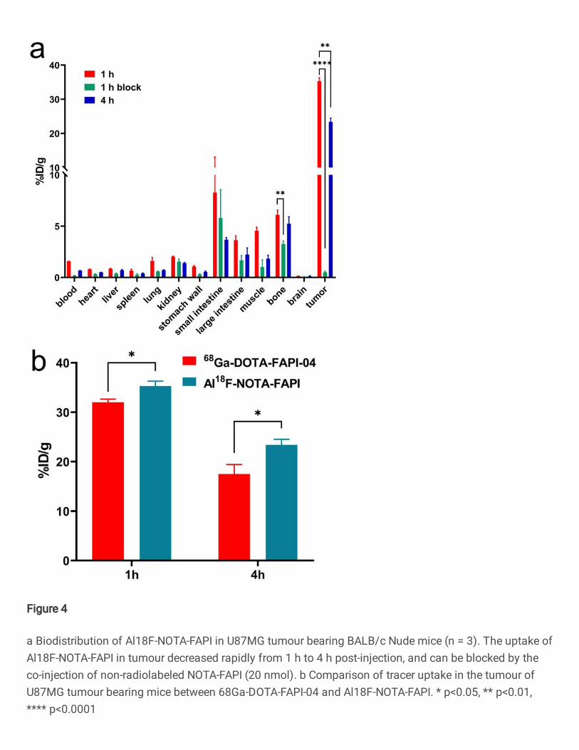
Figure 4
a Biodistribution of Al18F-NOTA-FAPI in U87MG tumour bearing BALB/c Nude mice (n = 3). The uptake ofAl18F-NOTA-FAPI in tumour decreased rapidly from 1 h to 4 h post-injection, and can be blocked by theco-injection of non-radiolabeled NOTA-FAPI (20 nmol). b Comparison of tracer uptake in the tumour ofU87MG tumour bearing mice between 68Ga-DOTA-FAPI-04 and Al18F-NOTA-FAPI. * p<0.05, ** p<0.01,**** p<0.0001

Figure 5
Micro PET imaging of Al18F-NOTA-FAPI in a A549, b U87MG and c blocked U87MG tumour bearing mice.The white arrow indicates the tumour xenograft, and the red arrow indicates the gallbladder. d Theimmunohistochemical staining of A549 xenograft indicated low expression of FAP (20× ampli�cation). eThe immunohistochemical staining of U87MG xenograft indicated high expression of FAP in tumour (20×ampli�cation)
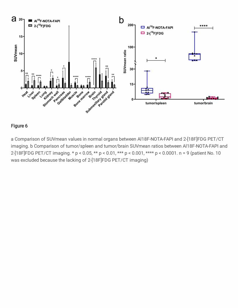
Figure 6
a Comparison of SUVmean values in normal organs between Al18F-NOTA-FAPI and 2-[18F]FDG PET/CTimaging. b Comparison of tumor/spleen and tumor/brain SUVmean ratios between Al18F-NOTA-FAPI and2-[18F]FDG PET/CT imaging. * p < 0.05, ** p < 0.01, *** p < 0.001, **** p < 0.0001. n = 9 (patient No. 10was excluded because the lacking of 2-[18F]FDG PET/CT imaging)
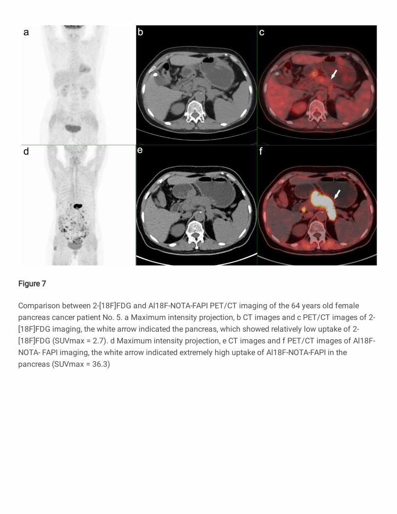
Figure 7
Comparison between 2-[18F]FDG and Al18F-NOTA-FAPI PET/CT imaging of the 64 years old femalepancreas cancer patient No. 5. a Maximum intensity projection, b CT images and c PET/CT images of 2-[18F]FDG imaging, the white arrow indicated the pancreas, which showed relatively low uptake of 2-[18F]FDG (SUVmax = 2.7). d Maximum intensity projection, e CT images and f PET/CT images of Al18F-NOTA- FAPI imaging, the white arrow indicated extremely high uptake of Al18F-NOTA-FAPI in thepancreas (SUVmax = 36.3)
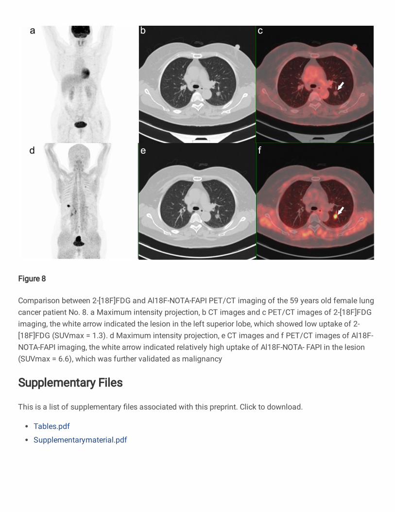
Figure 8
Comparison between 2-[18F]FDG and Al18F-NOTA-FAPI PET/CT imaging of the 59 years old female lungcancer patient No. 8. a Maximum intensity projection, b CT images and c PET/CT images of 2-[18F]FDGimaging, the white arrow indicated the lesion in the left superior lobe, which showed low uptake of 2-[18F]FDG (SUVmax = 1.3). d Maximum intensity projection, e CT images and f PET/CT images of Al18F-NOTA-FAPI imaging, the white arrow indicated relatively high uptake of Al18F-NOTA- FAPI in the lesion(SUVmax = 6.6), which was further validated as malignancy
Supplementary Files
This is a list of supplementary �les associated with this preprint. Click to download.
Tables.pdf
Supplementarymaterial.pdf