Embed Size (px)
Citation preview

Clinical StudyThe Impact of Thrombocytopenia on Outcome in Patients withAcute Coronary Syndromes: A Single Center Retrospective Study
Andreja SinkoviI1 and Maja Majal2
1Department of Medical Intensive Care, University Clinical Center Maribor, Ljubljanska 5, SI-2000 Maribor, Slovenia2Department of Hematology, University Clinical Center Maribor, Ljubljanska 5, SI-2000 Maribor, Slovenia
Correspondence should be addressed to Andreja Sinkovic; [email protected]
Received 1 May 2015; Accepted 14 July 2015
Academic Editor: Zsolt Molnar
Copyright © 2015 A. Sinkovic and M. Majal. This is an open access article distributed under the Creative Commons AttributionLicense, which permits unrestricted use, distribution, and reproduction in any medium, provided the original work is properlycited.
Background. In acute coronary syndromes (ACS), treated by combined antithrombotic therapy and percutaneous coronaryinterventions (PCI), thrombocytopenia may occur. Our aim was to evaluate predictors and the impact of thrombocytopenia onmortality in high-risk ACS patients.Methods. We retrospectively evaluated high-risk ACS patients.Thrombocytopenia was definedas platelet count <140.000/mL or a drop in platelet count of >50% during in-hospital stay. We compared demographic, laboratory,clinical, and mortality data between nonthrombocytopenic and thrombocytopenic ACS patients and evaluated independentpredictors of thrombocytopenia. Results. In 371 ACS patients, thrombocytopenia was observed in 21.3%. Thrombocytopenicpatients were significantly older and, less likely treated by PCIs (72.1% versus 89.7%, 𝑝 < 0.001) and combined antithrombotictherapy, with increased incidence of in-hospital complications and the use of additional treatments, but with increased mortality at30 days (27.8% versus 10.2%, 𝑝 < 0.001) and 6 months (35.4% versus 13.6%, 𝑝 < 0.001) when compared to nonthrombocytopenicpatients. The use of antibiotics, transfusions, insertion of intra-aortic balloon pump (IABP), and prior stroke independentlypredicted thrombocytopenia. Conclusions. Thrombocytopenia, observed in about 20% of high-risk ACS patients, was associatedsignificantly with in-hospital complications andmortality. Predictors of thrombocytopenia were the use of antibiotics, transfusions,insertion of IABP, and prior stroke.
1. Introduction
Acute coronary syndromes (ACS) with (STEMI) or without(NSTEMI) ST-segment elevation aremainly the consequenceof acute coronary atherothrombosis [1]. The most effectivetherapy is percutaneous coronary intervention (PCI), eitherprimary PCI in STEMI or early PCI in high-risk NSTEMIpatients, resulting in coronary recanalization andmyocardialreperfusion [2, 3].
PCI in STEMI and high-risk NSTEMI patients shouldbe accompanied by combination of antiplatelet and antico-agulant drugs to prevent thrombus formation at the site ofcoronary intervention [2, 3]. Antiaggregatory effect of anti-platelet drugs together with pronounced anticoagulant effectof heparins, either unfractionated or of lowmolecular weight,can also lead to increased risk of bleeding and increasingmortality in high-riskACSpatients [1, 4]. Platelet dysfunction
can be particularly expressed within the first hours after PCIs,related with the loading doses of antiplatelet agents. If thisleads to clinical thrombocytopenia, mortality in ACS patientsmay exceed 20% [5].
Thrombocytopenia is defined mostly as a decrease ofplatelet count below referenced lower limit of normal ora drop in platelet count of more than 50% during in-hos-pital stay [6]. A low platelet count in high-risk ACS patientscan be the consequence of different causes. It may be eitherimmunomediated due to heparins, glycoprotein receptor IIb/IIIa (GPIIb/IIIa) inhibitors, or thienopyridines or consump-tional, due to PCI or insertion of intra-aortic balloon pump(IABP) or due to acute heart failure [7]. In the GRACE reg-istry, 0.3% of thrombocythemia was heparin-induced (HIT),0.6% was glycoprotein-associated (GAT), and 0.7% was ofother origins [8]. Profound thrombocytopenia (<100.000)was observed in 2.5% of myocardial infarcts after abciximab
Hindawi Publishing CorporationBioMed Research InternationalVolume 2015, Article ID 907304, 7 pageshttp://dx.doi.org/10.1155/2015/907304
2 BioMed Research International
and 0.5% after tirofiban treatment combined with PCI anddual oral antiplatelet agents and in 0.6% of patients treatedbyGPIIb/IIIa inhibitors according to theGRACE registry [8–10]. Clopidogrel-related thrombocytopenia was observed in1.0% patients after percutaneous stent implantation [11]. AfterPCI, about 16% of patients developed a moderate to severedecline in platelet count according to De Labriolle et al. [12].
After the use of the direct thrombin inhibitor bivalirudin,in comparison to heparin with GPIIb/IIIa inhibitor, throm-bocytopenia developed in 3.7% [13].
Acute heart failure in high-risk ACS patients, as theconsequence of myocardial dysfunction due to extensive is-chemic necrosis, promotes neurohumoral activation, inflam-mation, and acute kidney injury. Activation of inflammationin acute heart failure seems an important stimulus forthrombocytopenia due to enhanced platelet clearance bymacrophages [14]. Also, hypotension in severe acute heartfailuremay inhibit production of platelets in the bonemarrow[15]. In patients with IABP, thrombocytopenia can developeven in 43–58%, according to one study [16].
In NSTEMI patients, low platelet count was indepen-dently associated with female sex, ST-segment depression,PCI, heart failure, systolic blood pressure, heart rate, and soforth, in one cohort [5]. However, De Labriolle et al. demon-strated that thrombocytopenia in coronary patients after PCIwas predicted by male gender, age, hypercholesterolemia,acute renal and heart failure, IABP insertion, STEMI, reducedhematocrit, heparin use, and low osmolar contrast agent [12].Therefore, these studies support a possibility of a multicom-ponent background for decreased platelet counts [5, 12, 16].
The incidence of thrombocytopenia varies in differenttrials. In the CRUSADE registry, 13% of NSTEMI patients, inACUITY trial, only 6.8% of ACS patients, and in the GRACEregistry, 1.6% of STEMI or NSTEMI patients developedreduced platelet counts [4, 5, 8].
These registries have found increased risk of mortalityif thrombocytopenia was observed in the setting of ACS[4, 5, 8]. In the GRACE registry, thrombocytopenia was asso-ciated with up to 21% in-hospital mortality [8]. In ACUITYtrial, 30-day and 1-year mortality correlated with severity ofthrombocytopenia, being approximately 9% at 30 days and atone year in severe decline in platelet count [4].
Our aimwas to evaluate the incidence of thrombocytope-nia in every-day clinical practice in high-risk ACS patients,STEMI, and high-risk NSTEMI patients and the impact oflow platelet count on treatments, in particular on the use ofPCIs, on in-hospital complications (bleeding, reinfarctions,heart failure, and acute renal failure) andmortality (within 30days and 6 months), and on predictors of thrombocytopenia.
2. Methods
2.1. Study Design. This was a retrospective observationalstudy, approved by theNationalMedical Ethics Committee ofthe Republic of Slovenia (number 123/06/10), who waived theneed for informed consent because of the retrospective natureof the study. Personal data of all the patients were protectedaccording to the Law on Personal Data Protection.
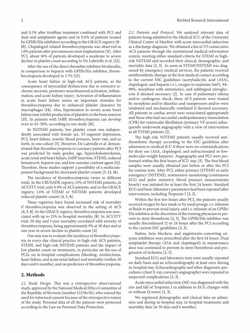
2.2. Patients and Protocol. We analysed relevant data ofpatients being admitted to the Medical ICU of the UniversityClinical Centre of Maribor with acute myocardial infarctionas a discharge diagnosis. We obtained a list of 371 consecutiveACS patients through the institutional medical informationsystem, meeting either standard criteria for STEMI or high-risk NSTEMI and recorded their clinical, demographic, andmortality data [2, 3]. As soon as STEMI/NSTEMI was diag-nosed by emergency medical services, the patients receivedantithrombotic therapy at the first medical contact accordingto the current ESC guidelines (acetylsalicylic acid (ASA),clopidogrel, and heparin i.v.), oxygen to maintain SatO
294–
98%, morphine with antiemetics, and sublingual nitroglyc-erin if deemed necessary [2]. In case of pulmonary edemaand/or cardiogenic shock, these ACS patients were treatedby morphine and/or diuretics and vasopressors and/or wereintubated and mechanically ventilated if deemed necessary.All patients in cardiac arrest were resuscitated appropriatelyand those who had successful cardiopulmonary resuscitation(CPR) for ventricular fibrillation (primary VF-arrest) subse-quently underwent angiography with a view of interventionas all STEMI patients [2].
The high-risk NSTEMI patients usually received anti-thrombotic therapy according to the ESC guidelines afteradmission to medical ICU if there were no contraindicationsfor their use (ASA, clopidogrel, and unfractionated or low-molecular-weight heparin). Angiography and PCI were per-formed within the first hours of ICU stay [3]. The first bloodsamples were usually obtained just before or after the PCIfor routine tests. After PCI, either primary (STEMI) or earlyemergency (NSTEMI), noninvasive monitoring (continuousECG and pulse oximetry, blood pressure measurementshourly) was initiated for at least the first 24 hours. StandardECG and basic laboratory parameters had been repeated afterintervention, including Troponin I [2, 3].
Within the first few hours after PCI, the patients usuallyreceived oxygen by face mask or by nasal prongs, i.v. infusionof fluids to prevent renal injury, and i.v. infusion of an GPIIb/IIIa inhibitor at the discretion of the treating physician to pre-vent in-stent thrombosis [2, 3]. The GPIIb/IIIa inhibitor wasusually discontinued 12 or 24 hours after the PCI accordingto the current ESC guidelines [2, 3].
Statins, beta blockers, and angiotensin-converting en-zyme inhibitors were prescribed after the first 24 hours. Dualantiplatelet therapy (ASA and clopidogrel) in maintenancedose was continued to prevent in-stent thrombosis and pro-gression of ischemia [2, 3].
Standard ECG and laboratory tests were usually repeatedon daily basis and an echocardiography at least once duringin-hospital stay. Echocardiography and other diagnostic pro-cedures (chest X-ray, coronary angiography) were repeated insuspected complications [2, 3].
Acutemyocardial infarction (MI) was diagnosed with therise and fall of Troponin I in addition to ECG changes withor without Q waves [2, 3].
We registered demographic and clinical data on admis-sion and during in-hospital stay, in-hospital treatments andmortality data (at 30 days and 6 months).
BioMed Research International 3
Table 1: Clinical and laboratory data of all ACS patients and nonthrombocytopenic and thrombocytopenic ACS patients.
Clinical and laboratory data(mean ± SD)
All(𝑛 = 371)
Nonthrombocytopenic(𝑛 = 292)
Thrombocytopenic(𝑛 = 79) 𝑝 values
Age (years) 64.1 ± 12.7 63.3 ± 12.8 66.8 ± 11.8 0.032Admission of Troponin I(𝜇g/L) 11.4 ± 23.5 10.6 ± 22.4 14.3 ± 27 ns
Peak Troponin I (𝜇g/L) 41.5 ± 36.2 41.5 ± 35.6 41.3 ± 38.7 nsAdmission CRP (mg/L) 18.9 ± 45.0 16.2 ± 42.3 28.8 ± 53.0 0.030Peak CRP (mg/L) 78.1 ± 88.1 66.0 ± 82.8 117.6 ± 93.8 <0.001ICU stay (days) 3.6 ± 4 2.9 ± 2.3 6.0 ± 6.9 nsIn-hospital stay (days) 10.2 ± 22.7 9.6 ± 25.0 12.4 ± 10.5 <0.001ACS, acute coronary syndrome; SD, standard deviation; CRP, C-reactive protein.
Regarding demographic data, we registered age, gender,comorbidities (arterial hypertension, diabetes, dyslipidemia,prior myocardial infarction, and stroke), and smoking. Fromlaboratory data, we registered platelet count and Troponin Ilevels on admission and peak levels during the hospital stay.
Among in-hospital treatments, we registered PCIs andthe use of antithrombotic therapy (ASA clopidogrel, hepar-ins, and GPIIb/IIIa inhibitors).
In terms of in-hospital complications, we registered acuteheart failure, arrhythmias, reinfarctions, bleeding, and acutekidney injury at any time of in-hospital stay. Arrhythmias,registered by continuous ECGmonitoring and standard ECGrecordings, were defined as atrial or ventricular or conduc-tion disturbances [2].
Heart failure was quantified by the Killip classification asclasses II–IV, including patients with pulmonary congestion,pulmonary edema, and cardiogenic shock [2, 3].
Reinfarctions were classified as recurrence of chest painwith new ECG changes and recurrent rise and fall of serumTroponin I [2].
Bleeding was considered major (cerebral or symptomaticbleeding of other locations with a drop in hemoglobin>50 g/L or the need of≥2 units of blood product transfusions)or minor (symptomatic bleeding with a drop in hemoglobinof 30–50 g/L) or minimal (symptomatic bleeding with a dropin hemoglobin <30 g/L), according to TIMI criteria [2, 3, 17].
Acute kidney injury was defined as an increase of serumcreatinine of more than 50% within 48–72 hours, accordingto Acute Kidney Injury Network (AKIN) criteria [18, 19].
In case of complications, the patients were treated accord-ing to professional protocols (e.g., by vasopressors, inotropicagents, mechanical ventilation, intra-aortic balloon pump(IABP), red blood cells transfusions, antibiotics, antiarrhyth-mic drugs, and pacing) [2, 3].
2.3. Laboratory Tests. Troponin I levels were determined bythe immunochemicalmethod (Boehringer,Germany, normallevels < 0.015 𝜇g/L) [20].
C-reactive protein (CRP), a marker of inflammation, wasmeasured by immunochemical method (Siemens HealthcareDiagnostics, Germany; normal levels were <3.0mg/L).
Platelet count was measured by the Sysmex XE2100automatic analyser, Kobe, Japan (normal levels 140.000/mL–340.000/mL) [6]. Thrombocytopenia in ACS patients was
80.3
19.7
64.4
18.1
51.2
15.16.5
24.3 24.5
83.5
16.5
63.6
16.4
51.7
15
4.1
26.7 26
68.4
34.6
67
24
49.3
15.1 15.1 15.1 18.9
0102030405060708090
Men
Dia
bete
s
Art
eria
lhy
pert
ensio
n
Prio
r MI
Know
ndy
slipi
dem
ia
Smok
ing
(%)
All ACS patientsNonthrombocytopenic ACS patientsThrombocytopenic ACS patients
STEM
I pat
ient
s(p
=0.004)
NST
EMI
patie
nts
(p=0.004)
Prio
r stro
ke(p
=0.001)
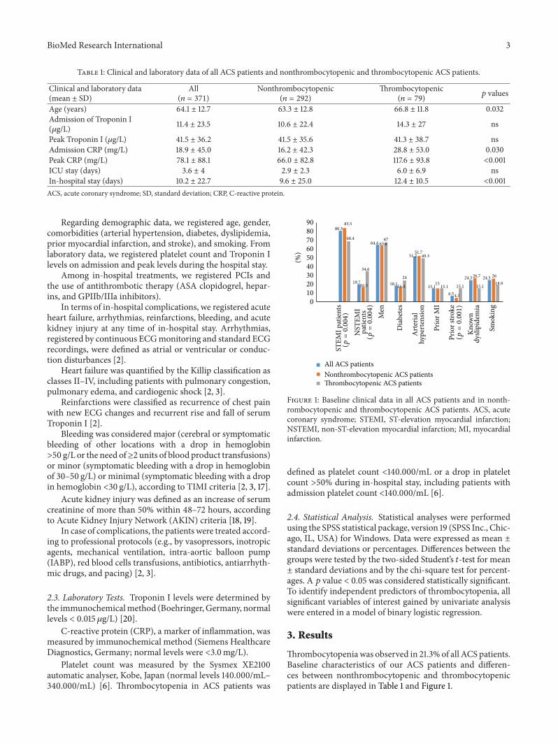
Figure 1: Baseline clinical data in all ACS patients and in nonth-rombocytopenic and thrombocytopenic ACS patients. ACS, acutecoronary syndrome; STEMI, ST-elevation myocardial infarction;NSTEMI, non-ST-elevation myocardial infarction; MI, myocardialinfarction.
defined as platelet count <140.000/mL or a drop in plateletcount >50% during in-hospital stay, including patients withadmission platelet count <140.000/mL [6].
2.4. Statistical Analysis. Statistical analyses were performedusing the SPSS statistical package, version 19 (SPSS Inc., Chic-ago, IL, USA) for Windows. Data were expressed as mean ±standard deviations or percentages. Differences between thegroups were tested by the two-sided Student’s 𝑡-test for mean± standard deviations and by the chi-square test for percent-ages. A 𝑝 value < 0.05 was considered statistically significant.To identify independent predictors of thrombocytopenia, allsignificant variables of interest gained by univariate analysiswere entered in a model of binary logistic regression.
3. Results
Thrombocytopeniawas observed in 21.3% of all ACS patients.Baseline characteristics of our ACS patients and differen-ces between nonthrombocytopenic and thrombocytopenicpatients are displayed in Table 1 and Figure 1.
4 BioMed Research International
8692.2
84.9 85.781.7
6.211.9
28
89.794.8
89 87.684.2
3.87.5
20.5
72.1
82.2
69.678.4
72.1
15.2
27.8
55.7
0102030405060708090
100
(%)
All ACS patientsNonthrombocytopenic ACS patientsThrombocytopenic ACS patients
PCI
urge
nt)
(prim
ary+
(p<0.001)
ASA
(p<0.001)
Clop
idog
rel
(p<0.001)
Hep
arin
s(p
<0.001)
GPI
Ib/II
Iain
hibi
tors
(p<0.001)
IABP
(p=0.001)
Red
bloo
d ce
lltr
ansfu
sions
(p<0.001)
Ant
ibio
tics
(p<0.001)
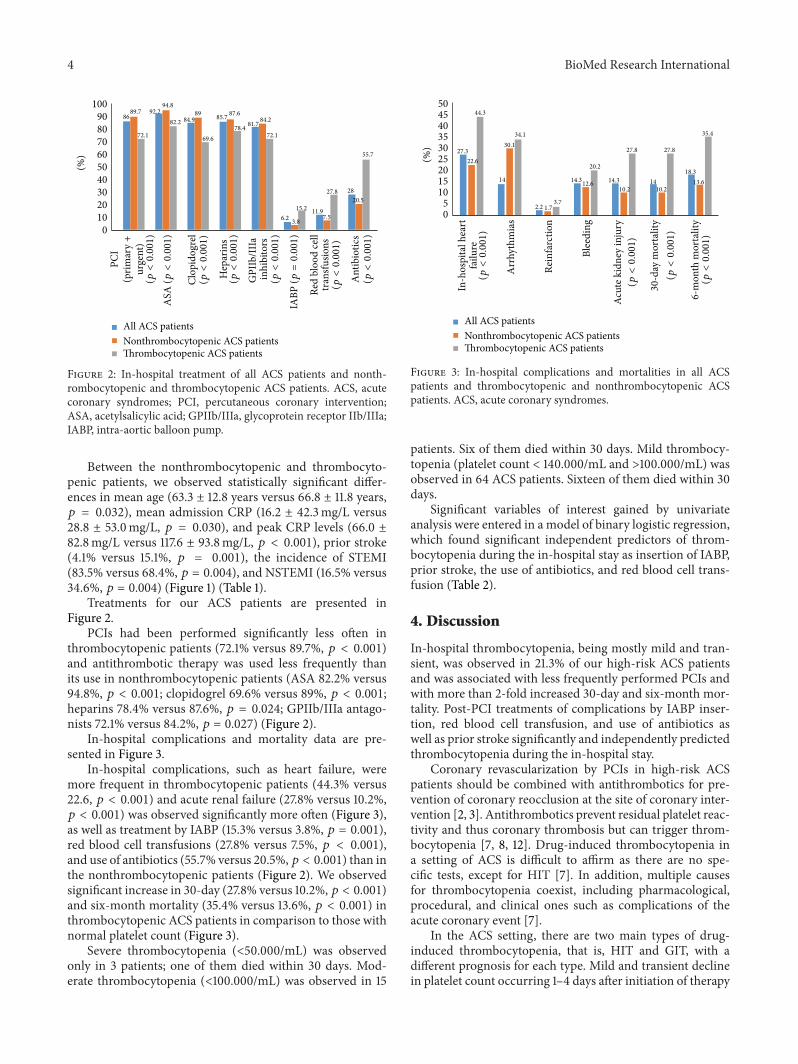
Figure 2: In-hospital treatment of all ACS patients and nonth-rombocytopenic and thrombocytopenic ACS patients. ACS, acutecoronary syndromes; PCI, percutaneous coronary intervention;ASA, acetylsalicylic acid; GPIIb/IIIa, glycoprotein receptor IIb/IIIa;IABP, intra-aortic balloon pump.
Between the nonthrombocytopenic and thrombocyto-penic patients, we observed statistically significant differ-ences in mean age (63.3 ± 12.8 years versus 66.8 ± 11.8 years,𝑝 = 0.032), mean admission CRP (16.2 ± 42.3mg/L versus28.8 ± 53.0mg/L, 𝑝 = 0.030), and peak CRP levels (66.0 ±82.8mg/L versus 117.6 ± 93.8mg/L, 𝑝 < 0.001), prior stroke(4.1% versus 15.1%, 𝑝 = 0.001), the incidence of STEMI(83.5% versus 68.4%, 𝑝 = 0.004), and NSTEMI (16.5% versus34.6%, 𝑝 = 0.004) (Figure 1) (Table 1).
Treatments for our ACS patients are presented inFigure 2.
PCIs had been performed significantly less often inthrombocytopenic patients (72.1% versus 89.7%, 𝑝 < 0.001)and antithrombotic therapy was used less frequently thanits use in nonthrombocytopenic patients (ASA 82.2% versus94.8%, 𝑝 < 0.001; clopidogrel 69.6% versus 89%, 𝑝 < 0.001;heparins 78.4% versus 87.6%, 𝑝 = 0.024; GPIIb/IIIa antago-nists 72.1% versus 84.2%, 𝑝 = 0.027) (Figure 2).
In-hospital complications and mortality data are pre-sented in Figure 3.
In-hospital complications, such as heart failure, weremore frequent in thrombocytopenic patients (44.3% versus22.6, 𝑝 < 0.001) and acute renal failure (27.8% versus 10.2%,𝑝 < 0.001) was observed significantly more often (Figure 3),as well as treatment by IABP (15.3% versus 3.8%, 𝑝 = 0.001),red blood cell transfusions (27.8% versus 7.5%, 𝑝 < 0.001),and use of antibiotics (55.7% versus 20.5%, 𝑝 < 0.001) than inthe nonthrombocytopenic patients (Figure 2). We observedsignificant increase in 30-day (27.8% versus 10.2%, 𝑝 < 0.001)and six-month mortality (35.4% versus 13.6%, 𝑝 < 0.001) inthrombocytopenic ACS patients in comparison to those withnormal platelet count (Figure 3).
Severe thrombocytopenia (<50.000/mL) was observedonly in 3 patients; one of them died within 30 days. Mod-erate thrombocytopenia (<100.000/mL) was observed in 15
27.3
14
2.2
14.3 14.3 1418.3
22.6
30.1
1.7
12.610.2 10.2
13.6
44.3
34.1
3.7
20.2
27.8 27.8
35.4
05
101520253035404550
Arr
hyth
mia
s
Rein
farc
tion
Blee
ding
(%)
All ACS patientsNonthrombocytopenic ACS patientsThrombocytopenic ACS patients
In-h
ospi
tal h
eart
failu
re(p
<0.001)
Acut
e kid
ney
inju
ry(p
<0.001)
30-d
ay m
orta
lity
(p<0.001)
6-m
onth
mor
talit
y(p
<0.001)
Figure 3: In-hospital complications and mortalities in all ACSpatients and thrombocytopenic and nonthrombocytopenic ACSpatients. ACS, acute coronary syndromes.
patients. Six of them died within 30 days. Mild thrombocy-topenia (platelet count < 140.000/mL and >100.000/mL) wasobserved in 64 ACS patients. Sixteen of them died within 30days.
Significant variables of interest gained by univariateanalysis were entered in a model of binary logistic regression,which found significant independent predictors of throm-bocytopenia during the in-hospital stay as insertion of IABP,prior stroke, the use of antibiotics, and red blood cell trans-fusion (Table 2).
4. Discussion
In-hospital thrombocytopenia, being mostly mild and tran-sient, was observed in 21.3% of our high-risk ACS patientsand was associated with less frequently performed PCIs andwith more than 2-fold increased 30-day and six-month mor-tality. Post-PCI treatments of complications by IABP inser-tion, red blood cell transfusion, and use of antibiotics aswell as prior stroke significantly and independently predictedthrombocytopenia during the in-hospital stay.
Coronary revascularization by PCIs in high-risk ACSpatients should be combined with antithrombotics for pre-vention of coronary reocclusion at the site of coronary inter-vention [2, 3]. Antithrombotics prevent residual platelet reac-tivity and thus coronary thrombosis but can trigger throm-bocytopenia [7, 8, 12]. Drug-induced thrombocytopenia ina setting of ACS is difficult to affirm as there are no spe-cific tests, except for HIT [7]. In addition, multiple causesfor thrombocytopenia coexist, including pharmacological,procedural, and clinical ones such as complications of theacute coronary event [7].
In the ACS setting, there are two main types of drug-induced thrombocytopenia, that is, HIT and GIT, with adifferent prognosis for each type. Mild and transient declinein platelet count occurring 1–4 days after initiation of therapy

BioMed Research International 5
Table 2: Binary logistic regression model to identify independent predictors of thrombocytopenia in ACS patients.
Variables OR 95% confidence interval 𝑝 valuesPCIs 2.362 0.695 to 8.033 0.169ASA treatment 0.633 0.138 to 2.900 0.556Clopidogrel treatment 1.739 0.543 to 5.577 0.352Heparin treatment 1.700 0.690 to 4.185 0.249GPIIb/IIIa inhibitors 0.531 0.185 to 1.528 0.241Antibiotics 0.405 0.189 to 0.866 0.020Red blood cell transfusion 0.371 0.143 to 0.963 0.042Acute heart failure 0.822 0.395 to 1.713 0.601Acute kidney injury 1.448 0.559 to 3.750 0.446Stroke 0.292 0.101 to 0.848 0.024IABP 0.232 0.620 to 0.871 0.030STEMI 1.388 0.630 to 3.057 0.415Age 0.995 0.970 to 1.0190 0.671PCI, percutaneous coronary intervention; ASA, acetylsalicylic acid; GPIIb/IIIa, glycoprotein receptor IIb/IIIa; IABP, intra-aortic balloon pump; STEMI, ST-elevation myocardial infarction; OR, odds ratio.
is common and observed in up to 15% of unfractionatedheparin-treated patients. It is not immunomediated andrarely leads to a severe reduction in platelet levels. It mostlyresolves spontaneously, despite continuation of unfraction-ated heparin [7, 8, 12]. The majority of ACS patients in thiscohort with reduced platelet count belonged to this type ofthrombocytopenia.
In our ACS patients, the reversible small molecularGPIIb/IIIa inhibitor, eptifibatide, was used predominantlyand abciximab was used only in individual cases. The timeinterval from the administration of i.v. eptifibatide to theonset of thrombocytopenia was 48–72 hours, suggesting thepossibility of eptifibatide as the cause of thrombocytopenia,but that was not proven as specific tests are lacking [10, 21].
Heparins, either nonfractionated or of low molecularweight, are an important potential cause of immunomediatedthrombocytopenia, HIT. In our ACS patients, HIT antibodieswere demonstrated only in 1 patient, whowas exposed to hep-arin in prior hospitalization. Thrombocytopenia developedwithin the first few days of in-hospital stay in the majorityof our patients, mostly within the first 5 days in contrast toHIT, which generally occurs between 5 and 10 days [7, 8, 12].
On the other hand, PCIs were performed less often inACS patients with reduced platelet count; therefore, anti-thrombotic drugswere also given less frequently to them thanto patients with normal platelet counts.
PCIs were not performed in those presenting late withSTEMI or those with complex multivessel disease in a settingof either NSTEMI or STEMI, if early death occurred orin those who refused the interventional therapy. Regardingcomplex multivessel coronary disease, current ESC STEMIguidelines recommend strategy of “culprit vessel only” pri-mary PCI, followed by further elective revascularisation incase of ongoing ischaemia.This can lead to a substantial delayin reperfusion of the myocardium at risk [2]. This problemwas in particular evident in STEMI patients with apparentlynew left bundle branch block. The advantage of completerevascularization at the time of presentation for STEMIpatients over “culprit vessel only” strategy was demonstratedby the recent Culprit trial but needs further evaluation [22].
In addition, radial arterial access for PCI has been provento offer an advantage in ACS setting with increased risk ofbleeding by decreasing the risk of vascular complications andmortality within 30 days when compared to femoral arterialaccess [2].
The main reasons not to use antithrombotic agents wereearly complications such as early death and renal failure,as well as early CABG. However, PCIs and antithromboticagents were still used in approximately 70% of our thrombo-cytopenic ACS patients.
When early coronary revascularization is delayed orabandoned in the setting of ACS, extensivemyocardial necro-sis develops, which can be followed by severe myocardialdysfunction with subsequent activation of neurohumoralfactors and of inflammation to lead to increased platelet con-sumption and thrombocytopenia. Thus, thrombocytopeniamay act as a marker of the acuity and severity of the inflam-mation in ACS settings. In addition, activated inflammationand neurohumoral system can further impair hemodynamicsand contribute to other organ hypoperfusion, including thatof the kidneys [2, 3, 7, 12]. In our thrombocytopenic ACSpatients, we observed increased incidence of acute heart fail-ure and of kidney injury, increased levels of the inflammatorymarker CRP and, finally, increased short- and long-termmortality.
Several trials demonstrated that thrombocytopenia inaddition to drug-induced antiplatelet effect in ACS patientswas associated with increased risk of bleeding. However,bleeding was not significantly increased in our thrombocy-topenic patients, but red blood cells were significantly moreoften transfused than in the nonthrombocytopenic patients.Red blood cells were transfused in case of major bleeding,but also in case of severe anemia with hemoglobin level <80 g/L irrespective of etiology to improve oxygen delivery inthe setting of coronary ischemia.
No platelets were transfused, what is in accordance withcurrent guidelines, which recommend infusion of plateletrich plasma only in pronounced drop in platelet count(<10.000/mL) in case of active bleeding [2, 3, 7]. We observeda drop in platelet count below 50.000/mL only in 3 patients

6 BioMed Research International
(0.8% of all ACS patients) and the platelet count droppedbelow 100.000/mL only in 15 patients (4% of ACS patients).Other studies observed a drop of platelets below 50.000/mLin approximately 6% of ACS patients, treated by combinationof antiplatelet and anticoagulant agents [4, 5].
We observed that even a mild drop in platelet count inhigh-risk ACS patients was associated with increasedmortal-ity and complication rate, what is consistent with the resultsof other studies [4, 5, 8, 12, 23, 24]. The short- and long-termmortality were twofold increased in our thrombocytopenicACS patients as compared to those with normal plateletcounts. In addition, treatment modalities such as insertion ofIABP, use of antibiotics, and blood transfusions predisposedpatients to thrombocytopenia even more. Our data confirmthat thrombocytopenia in high-risk ACS patients seems to bemultifactorial. It may be associated with drug effects such asGPIIa/IIIb inhibitors, heparins, and even clopidogrel, but alsoprocedures such as PCIs and IABP insertion. An importantpredisposing factor in ACS setting is myocardial pump fail-ure with organ hypoperfusion after delayed percutaneousrevascularization, which can be aggravated by activated neu-rohumoral system and inflammation. Acute kidney injury isan important complication of tissue hypoperfusion, but it canalso be induced by contrast agents during PCI [4, 5, 7, 8].In our ACS patients, acute kidney injury was significantlymore frequent in thrombocytopenic patients, in particular, incombination with heart failure [2, 3, 18, 19].
In recent years, novel antiplatelet and anticoagulant drugshave been developed and tested. Their use seems promisingin preventing drug-induced thrombocytopenia. Novel ADP-receptor antagonists, like prasugrel and ticagrelor, may alsoinduce thrombocytopenia with earlier onset of action thanclopidogrel. It appears prudent to replace the heparin plusGPIIb/IIIa inhibitor regime by one of the novel anticoag-ulants such as bivalirudin in STEMI or fondaparinux inNSTEMI patients presenting with low platelet counts [2, 25].This strategy may help to reduce the risk of drug-inducedthrombocytopenia to some extent [2, 3, 7].
4.1. Conclusions. Our conclusions are as follows: even milddegree of thrombocytopenia, observed in approximately 20%of high-risk ACS patients, is significantly associated withless often performed PCIs and with several complications,including increased short- and long-term mortality. How-ever, thrombocytopenia was independently predicted bypost-PCI procedures, aimed at mastering in-hospital compli-cations. Therefore, early treatment of high-risk ACS patientsshould focus on early PCI to prevent large ischemic necrosis:particularly in the elderly with comorbidities and in patientswith hemodynamic instability or with large anterior infarcts,which can reduce mortality (the higher-risk patients benefitmost from early PCI strategies).The use of novel antiplateletswith reversible platelet inhibition (ticagrelor) and antithrom-botic drugs with direct thrombin inhibition (bivalirudin) canreduce the risk of drug-induced thrombocytopenias duringinterventional therapy and should be preferred.
Regarding PCI procedures multivessel coronary inter-ventions at presentation should be performed in case ofsevere acute heart failure to prevent delays in reperfusion in
particular in STEMI setting. To avoid any further bleedingrisks, radial vascular approach has the priority if possible.
4.2. Limitation of the Study. Our study is a retrospectiveobservational design with limited number of patients, car-rying all the limitations and biases inherent in such studies.However, the data are taken from the “real world” as an obser-vational study and therefore can reflect on the complexity ofmanaging ACS patients with thrombocytopenia during ourdaily practice.
Conflict of Interests
The authors declare that there is no conflict of interestsregarding the publication of this paper.
Acknowledgment
The authors would like to thank Vojko Pogacar for theassistance with the paper.
References
[1] S. M. Lilly and R. L. Wilensky, “Emerging therapies for acutecoronary syndromes,” Frontiers in Pharmacology, vol. 2, article61, 2011.
[2] P. G. Steg, S. K. James, D. Atar et al., “ESC Guidelines for themanagement of acutemyocardial infarction in patients present-ing with ST-segment elevation,” EuropeanHeart Journal, vol. 33,no. 20, pp. 2569–2619, 2012.
[3] C. W. Hamm, J.-P. Bassand, S. Agewall et al., “ESC guidelinesfor the management of acute coronary syndromes in patientspresenting without persistent ST-segment elevation,” EuropeanHeart Journal, vol. 32, no. 23, pp. 2999–3054, 2011.
[4] A. Caixeta, G. D. Dangas, R. Mehran et al., “Incidenceand clinical consequences of acquired thrombocytopenia afterantithrombotic therapies in patients with acute coronary syn-dromes: results from the Acute Catheterization and UrgentIntervention Triage Strategy (ACUITY) trial,” American HeartJournal, vol. 161, no. 2, pp. 298.e1–306.e1, 2011.
[5] T. Y.Wang, F.-S. Ou, M. T. Roe et al., “Incidence and prognosticsignificance of thrombocytopenia developed during acute coro-nary syndrome in contemporary clinical practice,” Circulation,vol. 119, no. 18, pp. 2454–2462, 2009.
[6] S. S. Sekhon and V. Roy, “Thrombocytopenia in adults: a prac-tical approach to evaluation and management,” Southern Medi-cal Journal, vol. 99, no. 5, pp. 491–498, 2006.
[7] W. H.Matthai Jr., “Evaluation of thrombocytopenia in the acutecoronary syndrome,”CurrentOpinion inHematology, vol. 17, no.5, pp. 398–404, 2010.
[8] J. M. Gore, F. A. Spencer, E. P. Gurfinkel et al., “Thrombocy-topenia in patients with an acute coronary syndrome (fromthe Global Registry of Acute Coronary Events [GRACE]),”TheAmerican Journal of Cardiology, vol. 103, no. 2, pp. 175–180,2009.
[9] E. J. Topol, D. J. Moliterno, H. C. Herrmann et al., “Comparisonof two platelet glycoprotein IIb/IIIa inhibitors, tirofiban andabciximab, for the prevention of ischemic events with percu-taneous coronary revascularization,” The New England Journalof Medicine, vol. 344, no. 25, pp. 1888–1894, 2001.

BioMed Research International 7
[10] P. A. Merlini, M. Rossi, A. Menozzi et al., “Thrombocytopeniacaused by abciximab or tirofiban and its association with clin-ical outcome in patients undergoing coronary stenting,” Circu-lation, vol. 109, no. 18, pp. 2203–2206, 2004.
[11] M. Taniuchi, H. I. Kurz, and J. M. Lasala, “Randomized com-parison of ticlopidine and clopidogrel after intracoronary stentimplantation in a broad patient population,” Circulation, vol.104, no. 5, pp. 539–543, 2001.
[12] A. De Labriolle, L. Bonello, G. Lemesle et al., “Decline in plate-let count in patients treated by percutaneous coronary interven-tion: definition, incidence, prognostic importance, and predic-tive factors,” European Heart Journal, vol. 31, no. 9, pp. 1079–1087, 2010.
[13] G. W. Stone, B. Witzenbichler, G. Guagliumi et al., “Bivalirudinduring primary PCI in acute myocardial infarction,” The NewEngland Journal of Medicine, vol. 358, no. 21, pp. 2218–2230,2008.
[14] T. E. Warkentin and M. A. Crowther, “Adverse prognosticsignificance of thrombocytopenia in acute coronary syndrome:can anything be done about it?” Circulation, vol. 119, no. 18, pp.2420–2422, 2009.
[15] T. A. Naqvi, N. Ikhlaque, and M. A. Baumann, “Thrombo-cytopenia due to hypotension unrelated to infection: shockmarrow,” International Journal of Clinical Practice, vol. 59, no.7, pp. 782–784, 2005.
[16] S. K. Roy, E.W.Howard, J. A. Panza, andH. A. Cooper, “Clinicalimplications of thrombocytopenia among patients undergoingintra-aortic balloon pump counterpulsation in the coronarycare unit,” Clinical Cardiology, vol. 33, no. 1, pp. 30–35, 2010.
[17] R. Mehran, S. V. Rao, D. L. Bhatt et al., “Standardized bleedingdefinitions for cardiovascular clinical trials: a consensus reportfrom the bleeding academic research consortium,” Circulation,vol. 123, no. 23, pp. 2736–2747, 2011.
[18] R. L. Mehta, J. A. Kellum, S. V. Shah et al., “Acute Kidney InjuryNetwork: report of an initiative to improve outcomes in acutekidney injury,” Critical Care, vol. 11, no. 2, article R31, 2007.
[19] S. M. Bagshaw, C. George, R. Bellomo, and ANZICS DatabaseManagement Committe, “A comparison of the RIFLE andAKIN criteria for acute kidney injury in critically ill patients,”Nephrology Dialysis Transplantation, vol. 23, no. 5, pp. 1569–1574, 2008.
[20] C. Thygesen, J. S. Alpert, A. S. Jaffe, M. L. Simoons, B. R.Chaitman, andH.D.White, “Third universal definition ofmyo-cardial infarction,” European Heart Journal, vol. 33, no. 20, pp.2551–2567, 2012.
[21] M. W. Tempelhof, K. H. Benzuly, D. Fintel, and M. Z. Kri-chavsky, “Eptifibatide-induced thrombocytopenia: with throm-bosis and disseminated intravascular coagulation immediatelyafter left main coronary artery percutaneous coronary angio-plasty,” Texas Heart Institute Journal, vol. 39, no. 1, pp. 86–91,2012.
[22] A. H. Gershlick, J. N. Khan, D. J. Kelly et al., “Randomizedtrial of complete versus lesion-only revascularization in patientsundergoing primary percutaneous coronary intervention forSTEMI and multivessel disease: the CvLPRIT trial,” Journal ofthe American College of Cardiology, vol. 65, no. 10, pp. 963–972,2015.
[23] D. J. Kereiakes and P. A. Gurbel, “Peri-procedural plateletfunction and platelet inhibition in percutaneous coronary inter-vention,” JACC: Cardiovascular Interventions, vol. 1, no. 2, pp.111–121, 2008.
[24] C. Mueller, F.-J. Neumann,W. Hochholzer et al., “The impact ofplatelet count onmortality in unstable angina/non-ST-segmentelevation myocardial infarction,” American Heart Journal, vol.151, no. 6, pp. 1214.e1–1214.e7, 2006.
[25] NICEClinical GuidelineCenter andRoyal College of Physician,“Myocardial infarction with ST-segment elevation: the acutemanagement of myocardial infarction with ST-segment eleva-tion,” in NICE Clinical Guideline 167, Methods, Evidence andRecommendations, NICE Clinical Guideline Center, Royal Col-lege of Physician, London, UK, 2013, https://www.nice.org.uk/guidance/cg167.

Submit your manuscripts athttp://www.hindawi.com
Stem CellsInternational
Hindawi Publishing Corporationhttp://www.hindawi.com Volume 2014
Hindawi Publishing Corporationhttp://www.hindawi.com Volume 2014
MEDIATORSINFLAMMATION
of
Hindawi Publishing Corporationhttp://www.hindawi.com Volume 2014
Behavioural Neurology
EndocrinologyInternational Journal of
Hindawi Publishing Corporationhttp://www.hindawi.com Volume 2014
Hindawi Publishing Corporationhttp://www.hindawi.com Volume 2014
Disease Markers
Hindawi Publishing Corporationhttp://www.hindawi.com Volume 2014
BioMed Research International
OncologyJournal of
Hindawi Publishing Corporationhttp://www.hindawi.com Volume 2014
Hindawi Publishing Corporationhttp://www.hindawi.com Volume 2014
Oxidative Medicine and Cellular Longevity
Hindawi Publishing Corporationhttp://www.hindawi.com Volume 2014
PPAR Research
The Scientific World JournalHindawi Publishing Corporation http://www.hindawi.com Volume 2014
Immunology ResearchHindawi Publishing Corporationhttp://www.hindawi.com Volume 2014
Journal of
ObesityJournal of
Hindawi Publishing Corporationhttp://www.hindawi.com Volume 2014
Hindawi Publishing Corporationhttp://www.hindawi.com Volume 2014
Computational and Mathematical Methods in Medicine
OphthalmologyJournal of
Hindawi Publishing Corporationhttp://www.hindawi.com Volume 2014
Diabetes ResearchJournal of
Hindawi Publishing Corporationhttp://www.hindawi.com Volume 2014
Hindawi Publishing Corporationhttp://www.hindawi.com Volume 2014
Research and TreatmentAIDS
Hindawi Publishing Corporationhttp://www.hindawi.com Volume 2014
Gastroenterology Research and Practice
Hindawi Publishing Corporationhttp://www.hindawi.com Volume 2014
Parkinson’s Disease
Evidence-Based Complementary and Alternative Medicine
Volume 2014Hindawi Publishing Corporationhttp://www.hindawi.com