Embed Size (px)
Citation preview
Clinical StudyPredictors of Venous Thromboembolic Events Associated withCentral Venous Port Insertion in Cancer Patients
Christine Hohl Moinat,1 Daniel Périard,2 Adrienne Grueber,1 Daniel Hayoz,2
Jean-Luc Magnin,3 Pascal André,4 Marc Kung,1 and Daniel C. Betticher1
1 Department of Medical Oncology, Hopital Cantonal de Fribourg, Chemin des Pensionnats 2,1700 Fribourg, Switzerland
2Department of Angiology, Hopital Cantonal de Fribourg, Chemin des Pensionnats 2, 1700 Fribourg, Switzerland3 Central Laboratory, Hopital Cantonal de Fribourg, Chemin des Pensionnats 2, 1700 Fribourg, Switzerland4 Pharmacy Unit, Hopital Cantonal de Fribourg, Chemin des Pensionnats 2, 1700 Fribourg, Switzerland
Correspondence should be addressed to Daniel Periard; [email protected]
Received 29 August 2013; Revised 2 December 2013; Accepted 16 December 2013; Published 9 February 2014
Academic Editor: Bruno Vincenzi
Copyright © 2014 Christine Hohl Moinat et al. This is an open access article distributed under the Creative Commons AttributionLicense, which permits unrestricted use, distribution, and reproduction in any medium, provided the original work is properlycited.
Insertion of central venous port (CVP) catheter in the cancer population is associated with increased incidence of venousthromboembolic events (VTE). However, trials have shown limited benefit of antithrombotic treatment to prevent catheter-relatedvenous thrombosis. This prospective observational cohort study was designed to assess the incidence of VTE closely related toCVP implantation in patients with cancer and undergoing chemotherapy, and to identify a high risk subgroup of patients. BetweenFebruary 2006 and December 2011, 1097 consecutive cancer patients with first CVP implantation were included. Catheter-relatedVTE were defined as deep venous thrombosis in the arm, with or without pulmonary embolism (PE), or isolated PE.The incidenceof CVP-associated VTE was 5.9% (IC95 4.4–7.3%) at 3 months, and 11.3% (IC95 9.4–13.2%) at 12 months. The incidence of anyVTE was 7.6% (IC95 6.0–9.3%) at 3 months, and 15.3% (IC95 13.1–17.6%) at 12 months. High Khorana risk score and lung cancerwere significant predictors of 3 month VTE. In conclusion, this large cohort study of patients with first CVP catheter implantationconfirms the high incidence of VTE associated with the CVP implantation and allow identifying high risk patients whomay benefitfrom thromboprophylaxis.
1. Introduction
Themajority of patients with cancer undergoing chemother-apy require an efficient venous access for several weeks ormonths. Central venous port (CVP) catheter is widely usedin this setting. The incidence of deep venous thrombosis(DVT) and pulmonary embolism (PE) associated with cen-tral venous catheter has been reported between 2% and 67%[1]. The cancer population combines nonspecific throm-boembolic risk factors (age, malignancy, hypercoagulability,chemotherapy, infections, and bed rest) [2] and specificrisk factors such as catheter material, multiple placementattempts, catheter size and length, number of lumens, andcatheter tip localization [3–5]. Catheter-related VTE may belimited to asymptomatic radiological findings but may also
lead to significant clinical burden with upper limb post-thrombotic syndrome reported in 5 to 28% [6, 7] and res-piratory failure in case of pulmonary embolism. Moreover,catheter thrombosis can also lead to catheter occlusion in 14to 36% and delay chemotherapy [8].
At least eight randomized controlled trials have evaluatedantithrombotic therapy versus placebo in the preventionof central venous catheter-associated thrombosis [9–16]. Asmall study found that fixed dose of warfarin 1mg once dailyreduced the incidence of upper extremity DVT at the 90thday of venography [9]. However, two subsequent trials failedto confirm any benefit with this regimen [10, 11]. In twolarge studies, the administration of a prophylactic dose oflow molecular weight heparin (LMWH) during at least 6weeks after the catheter insertion did not reduce significantly
Hindawi Publishing CorporationJournal of OncologyVolume 2014, Article ID 743181, 6 pageshttp://dx.doi.org/10.1155/2014/743181
2 Journal of Oncology
the incidence of upper limb DVT compared to placebo [14,15]. A systematic thromboprophylaxis is therefore not rec-ommended at the time of CVP implantation, and should beconsidered only for patients with solid tumor and additionalrisk factors for VTE and low bleeding risk [17]. During thelast years, Khorana and colleagues developed and validateda predictive model for chemotherapy-associated thrombosis[18, 19]. This model allows identification of patients at highrisk who may benefit from antithrombotic treatment duringchemotherapy. To the best of our knowledge, there is no riskscore available to evaluate the risk of VTE following CVPinsertion and the Khorana score has not been validated inthis setting.The purpose of this studywas to evaluate the inci-dence of VTE closely associated with the insertion and use ofCVP catheters and to identify high-risk patients amenable tobenefit from a short course of thromboprophylaxis after CVPimplantation.
2. Patients and Method
2.1. Patient Inclusion. FromFebruary 2006 toDecember 2011,all consecutive adult patients suffering from cancer and whowere implanted with a CVP in the Surgery Department of theCantonalHospital, Fribourg, Switzerland (TertiaryCareCen-ter) were screened for inclusion in this prospective cohort.We included only patients older than 18 years with first CVPimplantation. All included patients were then followed upby the department of medical oncology. The study receivedapproval from the institutional ethic committee.
2.2. Surgical Implantation Procedure. Implantation was per-formed under local anaesthesia in the operating roomby ded-icated surgeons. The operator always attempted to find thecephalic-subclavian junction at the right upper limb and toplace a J-curved 0.035 inch guide wire in the superior cavavein. The catheter tip was then placed at the level of the tho-racic rib, under fluoroscopy guidance.The chamber was thenplaced in the pectoral region by tunelisation from the sameskin incision. When the cephalic vein was not accessible,the catheter was implanted in the subclavian vein by directpunction.There was no routine sonography or phlebographyof the subclavian vein prior to the intervention except incase of known previous DVT, previous central line use orfailure to pass the guide wire in a previous attempt. Duringthe study period, the same senior vascular surgeon was incharge of the dedicated CVP surgery team. There was noultrasound guidance during implantation. The first chemo-therapy infusion was allowed at the same day of the CVPimplantation. There was no systematic antithrombotic ther-apy administration at the time of CVP insertion.
2.3. Patient Follow-Up. Patients were regularly followed upclinically during chemotherapy treatment and then every6 months after chemotherapy completion until completeremission, death of any cause, or loss of follow-up. Patientswhose CVP catheter has been removed for lack of use werefollowed up until 6 weeks after removal. At each visit, thesubjects were questioned about any local pain or upper limb
swelling at the CVP side. Any other symptom suggestingupper or lower limb DVT or any thoracic symptom sug-gesting a PE was further investigated by compression venoussonography of the limbs or thoracic CT scan. During follow-up, patient with symptomatic anaemia received blood trans-fusion. Erythropoietin agent was not used in our institution.Granulocyte-colony stimulation factor administration wasallowed to reduce the length of neutropenia.
2.4. Definition and Assessment of Outcome. The main out-come was the 3-month incidence of catheter-related VTE,defined as occlusive DVT in the arm along the catheter, withor without PE, or isolated PE of unknown origin. The sec-ondary outcomes were the 12-month incidence of catheter-related VTE and the 3- and 12-month incidence of any VTErelated or not to the catheter, including DVT of the leg,DVT of the other arm and visceral DVT. Asymptomatic DVTor PE observed on CT scan performed for tumoral stagingwas classified as asymptomatic event. Catheter dysfunctionswere investigated by US-Doppler or phlebography. Catheterdysfunction and small, nonoccluding thrombosis along thecatheter were not considered as event. Complete occlusion ofthe vein along the catheter was considered as asymptomaticevent if it was not associated with local symptoms.
2.5. Khorana Score. The Khorana score is a validated tool forestimation of VTE during chemotherapy [18]. The Khoranapredictive score assigns 2 points to very high-risk cancer sites(pancreatic, gastric, brain) and 1 point to high risk cancersites (lung, ovarian, renal, or bladder). In addition, 1 pointis assigned for each of the following: platelet count ≥ 350× 109/L, hemoglobin < 10 g/dL, or use of erythropoietin-stimulating agents, leukocyte count ≥ 11 × 109/L, and bodymass index ≥ 35 kg/m2. Patients with Khorana score ≥ 3 areconsidered at high risk for VTE.
2.6. Statistical Analysis. Incidences of event were expressedas proportions with 95% confidence intervals, calculated bybinomial Wilson test. Proportions of event were comparedusing Chi2 test, and continuous variables were compared bythe Mann-Whitney rank sum test according to the normalityof their distribution. Statistical significance was consideredfor 𝛼 < 0.05. The contribution of clinical characteristics(age, sex, weight, body mass index, previous VTE, respira-tory failure, renal failure, antithrombotic treatment, cancerlocation, metastatic stage, low performance status, class ofchemotherapy agent, major surgery close to CVP implanta-tion or during follow-up, side of CVP implantation, baselinelaboratory values, and Khorana score) to asymptomatic andsymptomatic VTE was analysed using multivariable logisticregression analysis. Factors were first analyzed individuallyin univariate analysis and then selected for multivariableanalysis based on a 𝑃 value < 0.2 or known confoundingeffect. The statistical analysis was performed using Stata 9.0(Statacorp, College Station, Texas, USA).
Journal of Oncology 3
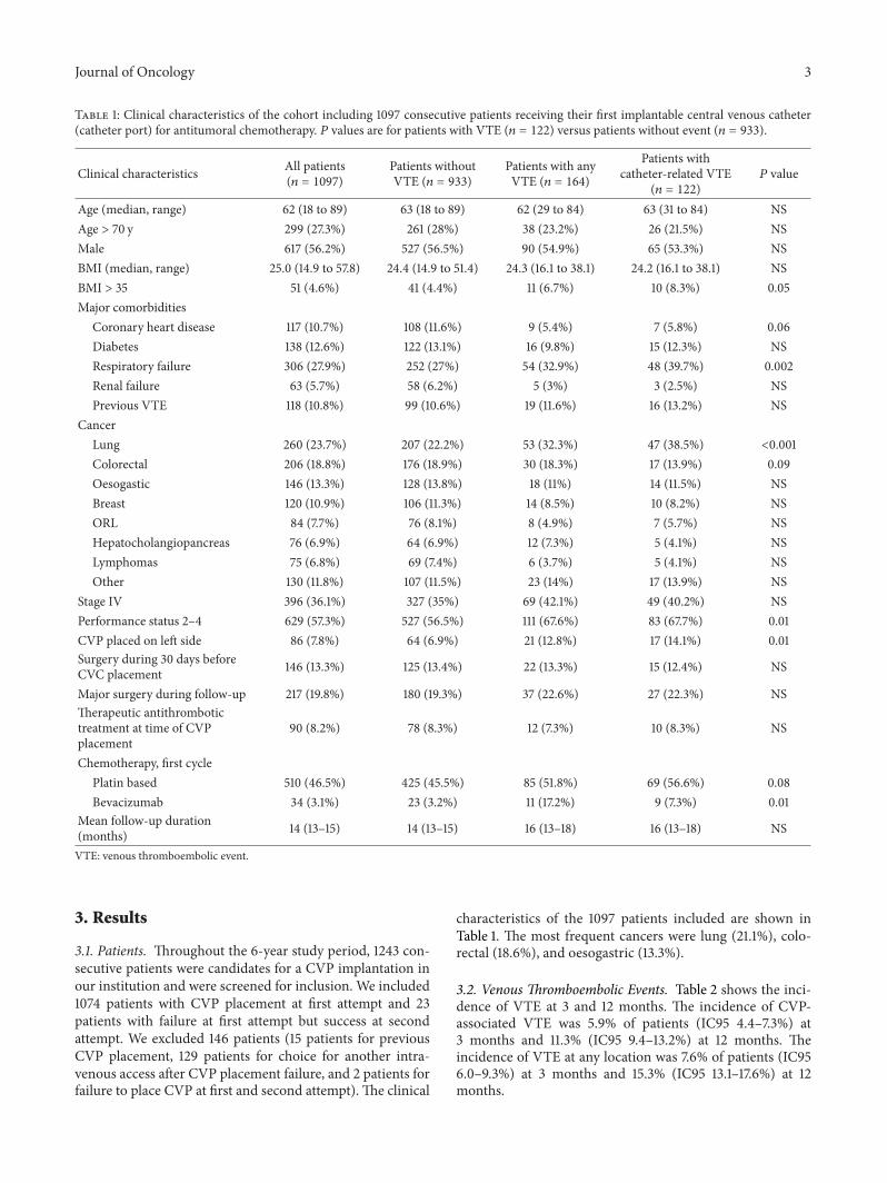
Table 1: Clinical characteristics of the cohort including 1097 consecutive patients receiving their first implantable central venous catheter(catheter port) for antitumoral chemotherapy. 𝑃 values are for patients with VTE (𝑛 = 122) versus patients without event (𝑛 = 933).
Clinical characteristics All patients(𝑛 = 1097)
Patients withoutVTE (𝑛 = 933)
Patients with anyVTE (𝑛 = 164)
Patients withcatheter-related VTE
(𝑛 = 122)𝑃 value
Age (median, range) 62 (18 to 89) 63 (18 to 89) 62 (29 to 84) 63 (31 to 84) NSAge > 70 y 299 (27.3%) 261 (28%) 38 (23.2%) 26 (21.5%) NSMale 617 (56.2%) 527 (56.5%) 90 (54.9%) 65 (53.3%) NSBMI (median, range) 25.0 (14.9 to 57.8) 24.4 (14.9 to 51.4) 24.3 (16.1 to 38.1) 24.2 (16.1 to 38.1) NSBMI > 35 51 (4.6%) 41 (4.4%) 11 (6.7%) 10 (8.3%) 0.05Major comorbidities
Coronary heart disease 117 (10.7%) 108 (11.6%) 9 (5.4%) 7 (5.8%) 0.06Diabetes 138 (12.6%) 122 (13.1%) 16 (9.8%) 15 (12.3%) NSRespiratory failure 306 (27.9%) 252 (27%) 54 (32.9%) 48 (39.7%) 0.002Renal failure 63 (5.7%) 58 (6.2%) 5 (3%) 3 (2.5%) NSPrevious VTE 118 (10.8%) 99 (10.6%) 19 (11.6%) 16 (13.2%) NS
CancerLung 260 (23.7%) 207 (22.2%) 53 (32.3%) 47 (38.5%) <0.001Colorectal 206 (18.8%) 176 (18.9%) 30 (18.3%) 17 (13.9%) 0.09Oesogastic 146 (13.3%) 128 (13.8%) 18 (11%) 14 (11.5%) NSBreast 120 (10.9%) 106 (11.3%) 14 (8.5%) 10 (8.2%) NSORL 84 (7.7%) 76 (8.1%) 8 (4.9%) 7 (5.7%) NSHepatocholangiopancreas 76 (6.9%) 64 (6.9%) 12 (7.3%) 5 (4.1%) NSLymphomas 75 (6.8%) 69 (7.4%) 6 (3.7%) 5 (4.1%) NSOther 130 (11.8%) 107 (11.5%) 23 (14%) 17 (13.9%) NS
Stage IV 396 (36.1%) 327 (35%) 69 (42.1%) 49 (40.2%) NSPerformance status 2–4 629 (57.3%) 527 (56.5%) 111 (67.6%) 83 (67.7%) 0.01CVP placed on left side 86 (7.8%) 64 (6.9%) 21 (12.8%) 17 (14.1%) 0.01Surgery during 30 days beforeCVC placement 146 (13.3%) 125 (13.4%) 22 (13.3%) 15 (12.4%) NS
Major surgery during follow-up 217 (19.8%) 180 (19.3%) 37 (22.6%) 27 (22.3%) NSTherapeutic antithrombotictreatment at time of CVPplacement
90 (8.2%) 78 (8.3%) 12 (7.3%) 10 (8.3%) NS
Chemotherapy, first cyclePlatin based 510 (46.5%) 425 (45.5%) 85 (51.8%) 69 (56.6%) 0.08Bevacizumab 34 (3.1%) 23 (3.2%) 11 (17.2%) 9 (7.3%) 0.01
Mean follow-up duration(months) 14 (13–15) 14 (13–15) 16 (13–18) 16 (13–18) NS
VTE: venous thromboembolic event.
3. Results
3.1. Patients. Throughout the 6-year study period, 1243 con-secutive patients were candidates for a CVP implantation inour institution and were screened for inclusion. We included1074 patients with CVP placement at first attempt and 23patients with failure at first attempt but success at secondattempt. We excluded 146 patients (15 patients for previousCVP placement, 129 patients for choice for another intra-venous access after CVP placement failure, and 2 patients forfailure to place CVP at first and second attempt). The clinical
characteristics of the 1097 patients included are shown inTable 1. The most frequent cancers were lung (21.1%), colo-rectal (18.6%), and oesogastric (13.3%).
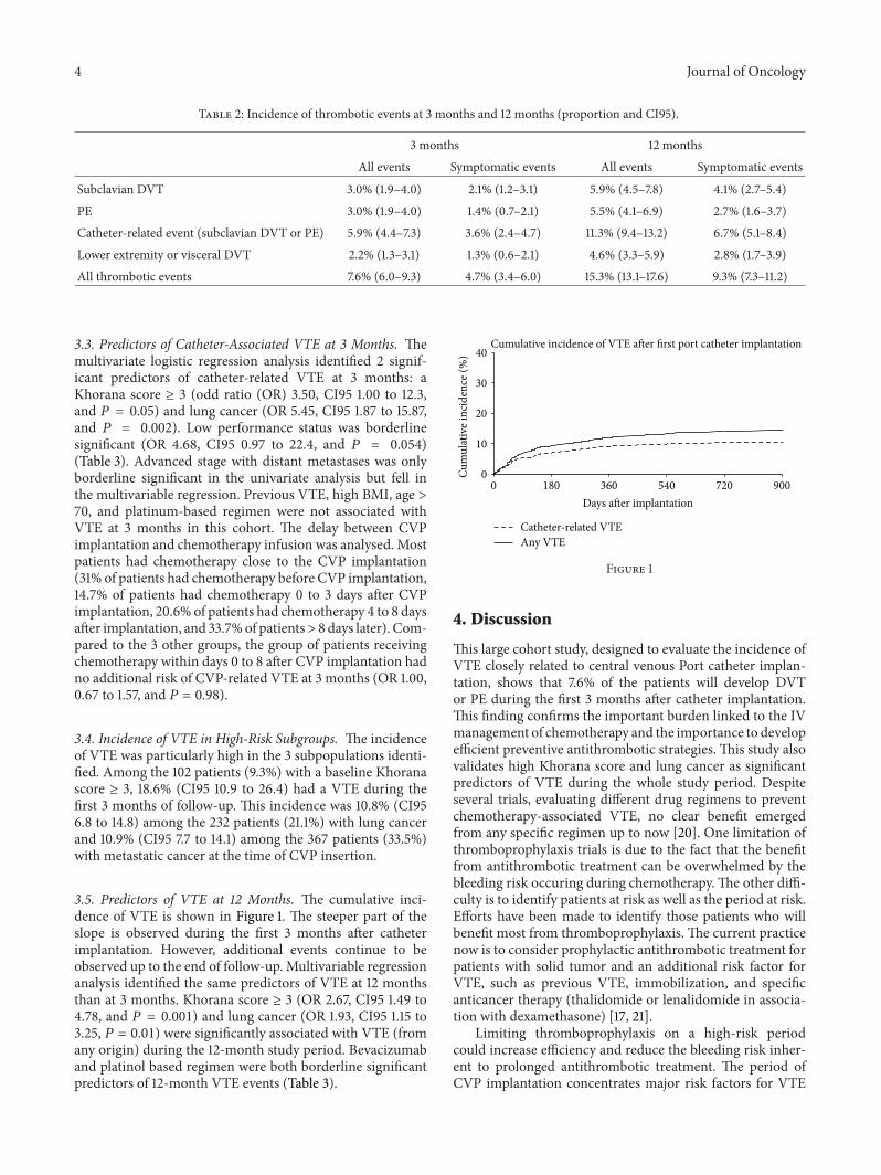
3.2. Venous Thromboembolic Events. Table 2 shows the inci-dence of VTE at 3 and 12 months. The incidence of CVP-associated VTE was 5.9% of patients (IC95 4.4–7.3%) at3 months and 11.3% (IC95 9.4–13.2%) at 12 months. Theincidence of VTE at any location was 7.6% of patients (IC956.0–9.3%) at 3 months and 15.3% (IC95 13.1–17.6%) at 12months.
4 Journal of Oncology
Table 2: Incidence of thrombotic events at 3 months and 12 months (proportion and CI95).
3 months 12 monthsAll events Symptomatic events All events Symptomatic events
Subclavian DVT 3.0% (1.9–4.0) 2.1% (1.2–3.1) 5.9% (4.5–7.8) 4.1% (2.7–5.4)PE 3.0% (1.9–4.0) 1.4% (0.7–2.1) 5.5% (4.1–6.9) 2.7% (1.6–3.7)Catheter-related event (subclavian DVT or PE) 5.9% (4.4–7.3) 3.6% (2.4–4.7) 11.3% (9.4–13.2) 6.7% (5.1–8.4)Lower extremity or visceral DVT 2.2% (1.3–3.1) 1.3% (0.6–2.1) 4.6% (3.3–5.9) 2.8% (1.7–3.9)All thrombotic events 7.6% (6.0–9.3) 4.7% (3.4–6.0) 15.3% (13.1–17.6) 9.3% (7.3–11.2)
3.3. Predictors of Catheter-Associated VTE at 3 Months. Themultivariate logistic regression analysis identified 2 signif-icant predictors of catheter-related VTE at 3 months: aKhorana score ≥ 3 (odd ratio (OR) 3.50, CI95 1.00 to 12.3,and 𝑃 = 0.05) and lung cancer (OR 5.45, CI95 1.87 to 15.87,and 𝑃 = 0.002). Low performance status was borderlinesignificant (OR 4.68, CI95 0.97 to 22.4, and 𝑃 = 0.054)(Table 3). Advanced stage with distant metastases was onlyborderline significant in the univariate analysis but fell inthe multivariable regression. Previous VTE, high BMI, age >70, and platinum-based regimen were not associated withVTE at 3 months in this cohort. The delay between CVPimplantation and chemotherapy infusion was analysed. Mostpatients had chemotherapy close to the CVP implantation(31% of patients had chemotherapy before CVP implantation,14.7% of patients had chemotherapy 0 to 3 days after CVPimplantation, 20.6% of patients had chemotherapy 4 to 8 daysafter implantation, and 33.7%of patients> 8 days later). Com-pared to the 3 other groups, the group of patients receivingchemotherapy within days 0 to 8 after CVP implantation hadno additional risk of CVP-related VTE at 3 months (OR 1.00,0.67 to 1.57, and 𝑃 = 0.98).
3.4. Incidence of VTE in High-Risk Subgroups. The incidenceof VTE was particularly high in the 3 subpopulations identi-fied. Among the 102 patients (9.3%) with a baseline Khoranascore ≥ 3, 18.6% (CI95 10.9 to 26.4) had a VTE during thefirst 3 months of follow-up. This incidence was 10.8% (CI956.8 to 14.8) among the 232 patients (21.1%) with lung cancerand 10.9% (CI95 7.7 to 14.1) among the 367 patients (33.5%)with metastatic cancer at the time of CVP insertion.
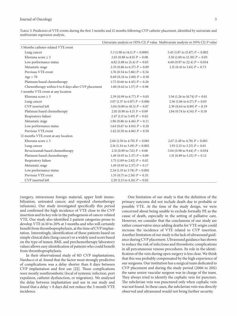
3.5. Predictors of VTE at 12 Months. The cumulative inci-dence of VTE is shown in Figure 1. The steeper part of theslope is observed during the first 3 months after catheterimplantation. However, additional events continue to beobserved up to the end of follow-up. Multivariable regressionanalysis identified the same predictors of VTE at 12 monthsthan at 3 months. Khorana score ≥ 3 (OR 2.67, CI95 1.49 to4.78, and 𝑃 = 0.001) and lung cancer (OR 1.93, CI95 1.15 to3.25, 𝑃 = 0.01) were significantly associated with VTE (fromany origin) during the 12-month study period. Bevacizumaband platinol based regimen were both borderline significantpredictors of 12-month VTE events (Table 3).
0
10
20
30
40
0 180 360 540 720 900
Cum
ulat
ive i
ncid
ence
(%)
Days after implantation
Cumulative incidence of VTE after first port catheter implantation
Catheter-related VTEAny VTE
Figure 1
4. Discussion
This large cohort study, designed to evaluate the incidence ofVTE closely related to central venous Port catheter implan-tation, shows that 7.6% of the patients will develop DVTor PE during the first 3 months after catheter implantation.This finding confirms the important burden linked to the IVmanagement of chemotherapy and the importance to developefficient preventive antithrombotic strategies. This study alsovalidates high Khorana score and lung cancer as significantpredictors of VTE during the whole study period. Despiteseveral trials, evaluating different drug regimens to preventchemotherapy-associated VTE, no clear benefit emergedfrom any specific regimen up to now [20]. One limitation ofthromboprophylaxis trials is due to the fact that the benefitfrom antithrombotic treatment can be overwhelmed by thebleeding risk occuring during chemotherapy.The other diffi-culty is to identify patients at risk as well as the period at risk.Efforts have been made to identify those patients who willbenefit most from thromboprophylaxis. The current practicenow is to consider prophylactic antithrombotic treatment forpatients with solid tumor and an additional risk factor forVTE, such as previous VTE, immobilization, and specificanticancer therapy (thalidomide or lenalidomide in associa-tion with dexamethasone) [17, 21].
Limiting thromboprophylaxis on a high-risk periodcould increase efficiency and reduce the bleeding risk inher-ent to prolonged antithrombotic treatment. The period ofCVP implantation concentrates major risk factors for VTE
Journal of Oncology 5
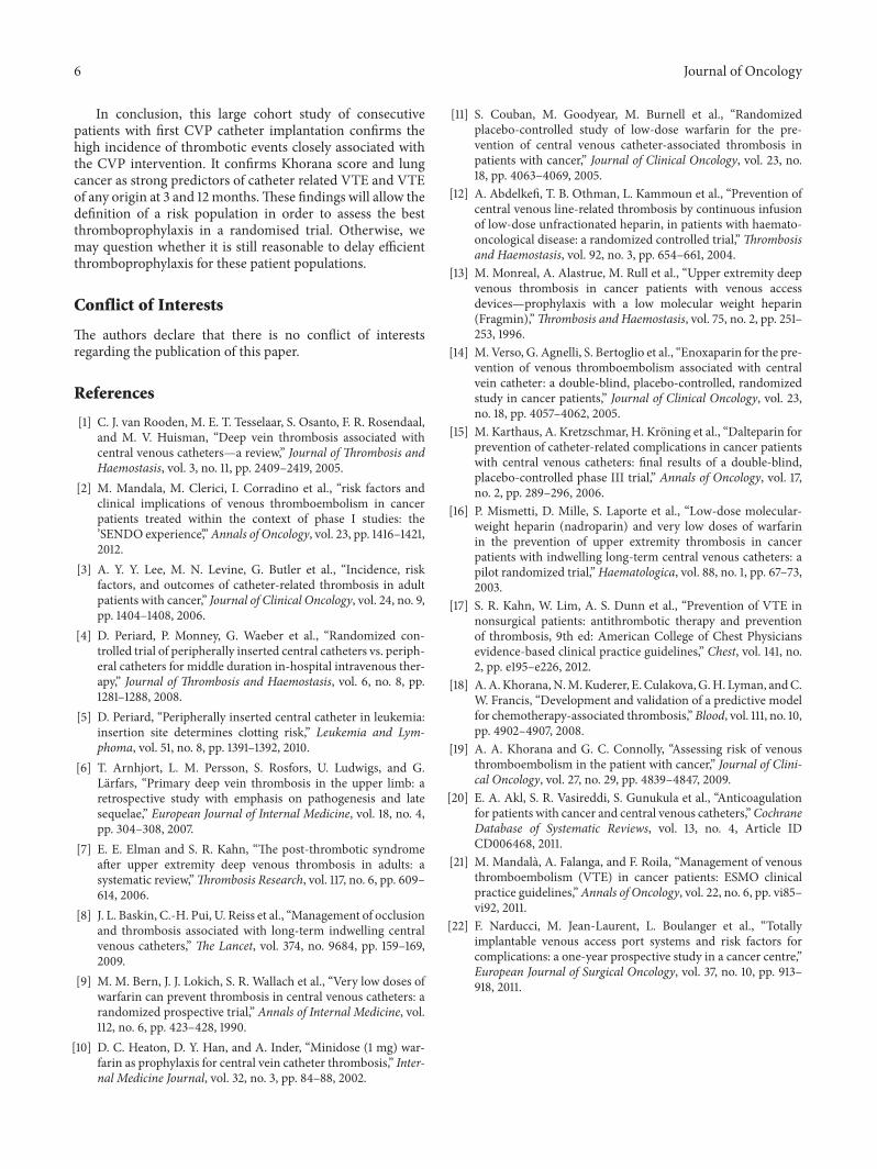
Table 3: Predictors of VTE events during the first 3 months and 12 months following CVP catheter placement, identified by univariate andmultivariate regression analysis.
Univariate analysis or (95% CI) 𝑃 value Multivariate analysis or (95% CI) 𝑃 value3 Months catheter-related VTE event
Lung cancer 5.3 (1.98 to 14.1) 𝑃 = 0.0001 5.45 (1.87 to 15.87) 𝑃 = 0.002Khorana score ≥ 3 2.65 (0.88 to 8.0) 𝑃 = 0.08 3.50 (1.00 to 12.30) 𝑃 = 0.05Low performance status 4.82 (1.08 to 21.4) 𝑃 = 0.03 4.68 (0.97 to 22.4) 𝑃 = 0.054Metastatic stage 2.33 (0.86 to 6.37) 𝑃 = 0.09 1.21 (0.41 to 3.61) 𝑃 = 0.73Previous VTE event 1.76 (0.54 to 5.86) 𝑃 = 0.34Age > 70 0.69 (0.24 to 2.00) 𝑃 = 0.50Platinum based chemotherapy 1.72 (0.66 to 4.45) 𝑃 = 0.26Chemotherapy within 0 to 8 days after CVP placement 1.00 (0.62 to 1.57) 𝑃 = 0.98
3 months VTE event at any locationKhorana score ≥ 3 2.59 (0.99 to 6.77) 𝑃 = 0.05 3.56 (1.26 to 10.74) 𝑃 = 0.01Lung cancer 3.07 (1.37 to 6.87) 𝑃 = 0.006 2.56 (1.06 to 6.17) 𝑃 = 0.03CVP inserted left 3.04 (0.89 to 10.3) 𝑃 = 0.07 2.39 (0.64 to 8.89) 𝑃 = 0.19Platinum based chemotherapy 2.01 (0.90 to 4.5) 𝑃 = 0.09 1.84 (0.74 to 4.54) 𝑃 = 0.18Respiratory failure 2.47 (1.11 to 5.49) 𝑃 = 0.02Metastatic stage 1.96 (0.86 to 4.46) 𝑃 = 0.11Low performance status 1.64 (0.67 to 4.04) 𝑃 = 0.28Previous VTE event 1.42 (0.50 to 4.06) 𝑃 = 0.50
12 months VTE event at any locationKhorana score ≥ 3 2.66 (1.50 to 4.70) 𝑃 = 0.001 2.67 (1.49 to 4.78) 𝑃 = 0.001Lung cancer 2.16 (1.34 to 3.49) 𝑃 = 0.002 1.93 (1.15 to 3.25) 𝑃 = 0.01Bevacizumab based chemotherapy 2.51 (0.89 to 7.11) 𝑃 = 0.08 3.04 (0.98 to 9.44) 𝑃 = 0.054Platinum based chemotherapy 1.49 (0.93 to 2.37) 𝑃 = 0.09 1.51 (0.89 to 3.25) 𝑃 = 0.12Respiratory failure 1.75 (1.09 to 2.82) 𝑃 = 0.02Metastatic stage 1.49 (0.93 to 2.37) 𝑃 = 0.17Low performance status 2.24 (1.33 to 3.78) 𝑃 = 0.002Previous VTE event 1.35 (0.71 to 2.56) 𝑃 = 0.35CVP inserted left 2.20 (1.13 to 4.26) 𝑃 = 0.02
(surgery, intravenous foreign material, upper limb immo-bilization, untreated cancer, and repeated chemotherapyinfusions). Our study investigated specifically this periodand confirmed the high incidence of VTE close to the CVPinsertion and its key role in the pathogenesis of cancer-relatedVTE. Our study also identified 2-patient categories prone todevelop VTE in 10 to 20% at 3 months and who will certainlybenefit from thromboprophylaxis, at the time ofCVP implan-tation. Interestingly, identification of these patients based onsimple clinical data (lung cancer) or a widely used score basedon the type of tumor, BMI, and prechemotherapy laboratoryvalues allows easy identification of patients who could benefitfrom thromboprophylaxis.
In their observational study of 815 CVP implantations,Narducci et al. found that the factor most strongly predictiveof complications was a delay shorter than 8 days betweenCVP implantation and first use [22]. These complicationswere mostly nonthrombotic (local of systemic infection, portexpulsion, catheter dysfunction, or migration). We analyzedthe delay between implantation and use in our study andfound that a delay > 8 days did not reduce the 3-month VTEincidence.
One limitation of our study is that the definition of theprimary outcome did not include death due to probable orpossible VTE. At the time of the study design, we wereconcerned about being unable to exclude formally PE as thecause of death, especially in the setting of palliative care.However, we consider that the conclusions of our study arerather conservative since adding deaths for VTE origin couldincrease the incidence of VTE related to CVP insertion.Another limitation of our study is the lack of ultrasound guid-ance duringCVPplacement. Ultrasound guidance has shownto reduce the risk of infectious and thrombotic complicationsin all percutaneous venous procedures. Its role in the identi-fication of the vein during open surgery is less clear.We thinkthat this was probably compensated by the high experience ofour surgeons. Our institution has a surgical team dedicated toCVP placement and during the study period (2006 to 2011)the same senior vascular surgeon was in charge of the team.They always tried to identify the cephalic vein for puncture.The subclavian vein was punctured only when cephalic veinwas not found. In these cases, the subclavian vein was directlyobserved and ultrasound would not bring further security.
6 Journal of Oncology
In conclusion, this large cohort study of consecutivepatients with first CVP catheter implantation confirms thehigh incidence of thrombotic events closely associated withthe CVP intervention. It confirms Khorana score and lungcancer as strong predictors of catheter related VTE and VTEof any origin at 3 and 12 months.These findings will allow thedefinition of a risk population in order to assess the bestthromboprophylaxis in a randomised trial. Otherwise, wemay question whether it is still reasonable to delay efficientthromboprophylaxis for these patient populations.
Conflict of Interests
The authors declare that there is no conflict of interestsregarding the publication of this paper.
References
[1] C. J. van Rooden, M. E. T. Tesselaar, S. Osanto, F. R. Rosendaal,and M. V. Huisman, “Deep vein thrombosis associated withcentral venous catheters—a review,” Journal of Thrombosis andHaemostasis, vol. 3, no. 11, pp. 2409–2419, 2005.
[2] M. Mandala, M. Clerici, I. Corradino et al., “risk factors andclinical implications of venous thromboembolism in cancerpatients treated within the context of phase I studies: the’SENDO experience’,”Annals of Oncology, vol. 23, pp. 1416–1421,2012.
[3] A. Y. Y. Lee, M. N. Levine, G. Butler et al., “Incidence, riskfactors, and outcomes of catheter-related thrombosis in adultpatients with cancer,” Journal of Clinical Oncology, vol. 24, no. 9,pp. 1404–1408, 2006.
[4] D. Periard, P. Monney, G. Waeber et al., “Randomized con-trolled trial of peripherally inserted central catheters vs. periph-eral catheters for middle duration in-hospital intravenous ther-apy,” Journal of Thrombosis and Haemostasis, vol. 6, no. 8, pp.1281–1288, 2008.
[5] D. Periard, “Peripherally inserted central catheter in leukemia:insertion site determines clotting risk,” Leukemia and Lym-phoma, vol. 51, no. 8, pp. 1391–1392, 2010.
[6] T. Arnhjort, L. M. Persson, S. Rosfors, U. Ludwigs, and G.Larfars, “Primary deep vein thrombosis in the upper limb: aretrospective study with emphasis on pathogenesis and latesequelae,” European Journal of Internal Medicine, vol. 18, no. 4,pp. 304–308, 2007.
[7] E. E. Elman and S. R. Kahn, “The post-thrombotic syndromeafter upper extremity deep venous thrombosis in adults: asystematic review,”Thrombosis Research, vol. 117, no. 6, pp. 609–614, 2006.
[8] J. L. Baskin, C.-H. Pui, U. Reiss et al., “Management of occlusionand thrombosis associated with long-term indwelling centralvenous catheters,” The Lancet, vol. 374, no. 9684, pp. 159–169,2009.
[9] M. M. Bern, J. J. Lokich, S. R. Wallach et al., “Very low doses ofwarfarin can prevent thrombosis in central venous catheters: arandomized prospective trial,” Annals of Internal Medicine, vol.112, no. 6, pp. 423–428, 1990.
[10] D. C. Heaton, D. Y. Han, and A. Inder, “Minidose (1 mg) war-farin as prophylaxis for central vein catheter thrombosis,” Inter-nal Medicine Journal, vol. 32, no. 3, pp. 84–88, 2002.
[11] S. Couban, M. Goodyear, M. Burnell et al., “Randomizedplacebo-controlled study of low-dose warfarin for the pre-vention of central venous catheter-associated thrombosis inpatients with cancer,” Journal of Clinical Oncology, vol. 23, no.18, pp. 4063–4069, 2005.
[12] A. Abdelkefi, T. B. Othman, L. Kammoun et al., “Prevention ofcentral venous line-related thrombosis by continuous infusionof low-dose unfractionated heparin, in patients with haemato-oncological disease: a randomized controlled trial,”Thrombosisand Haemostasis, vol. 92, no. 3, pp. 654–661, 2004.
[13] M. Monreal, A. Alastrue, M. Rull et al., “Upper extremity deepvenous thrombosis in cancer patients with venous accessdevices—prophylaxis with a low molecular weight heparin(Fragmin),”Thrombosis and Haemostasis, vol. 75, no. 2, pp. 251–253, 1996.
[14] M. Verso, G. Agnelli, S. Bertoglio et al., “Enoxaparin for the pre-vention of venous thromboembolism associated with centralvein catheter: a double-blind, placebo-controlled, randomizedstudy in cancer patients,” Journal of Clinical Oncology, vol. 23,no. 18, pp. 4057–4062, 2005.
[15] M. Karthaus, A. Kretzschmar, H. Kroning et al., “Dalteparin forprevention of catheter-related complications in cancer patientswith central venous catheters: final results of a double-blind,placebo-controlled phase III trial,” Annals of Oncology, vol. 17,no. 2, pp. 289–296, 2006.
[16] P. Mismetti, D. Mille, S. Laporte et al., “Low-dose molecular-weight heparin (nadroparin) and very low doses of warfarinin the prevention of upper extremity thrombosis in cancerpatients with indwelling long-term central venous catheters: apilot randomized trial,”Haematologica, vol. 88, no. 1, pp. 67–73,2003.
[17] S. R. Kahn, W. Lim, A. S. Dunn et al., “Prevention of VTE innonsurgical patients: antithrombotic therapy and preventionof thrombosis, 9th ed: American College of Chest Physiciansevidence-based clinical practice guidelines,” Chest, vol. 141, no.2, pp. e195–e226, 2012.
[18] A.A.Khorana,N.M.Kuderer, E. Culakova,G.H. Lyman, andC.W. Francis, “Development and validation of a predictive modelfor chemotherapy-associated thrombosis,” Blood, vol. 111, no. 10,pp. 4902–4907, 2008.
[19] A. A. Khorana and G. C. Connolly, “Assessing risk of venousthromboembolism in the patient with cancer,” Journal of Clini-cal Oncology, vol. 27, no. 29, pp. 4839–4847, 2009.
[20] E. A. Akl, S. R. Vasireddi, S. Gunukula et al., “Anticoagulationfor patients with cancer and central venous catheters,”CochraneDatabase of Systematic Reviews, vol. 13, no. 4, Article IDCD006468, 2011.
[21] M. Mandala, A. Falanga, and F. Roila, “Management of venousthromboembolism (VTE) in cancer patients: ESMO clinicalpractice guidelines,”Annals of Oncology, vol. 22, no. 6, pp. vi85–vi92, 2011.
[22] F. Narducci, M. Jean-Laurent, L. Boulanger et al., “Totallyimplantable venous access port systems and risk factors forcomplications: a one-year prospective study in a cancer centre,”European Journal of Surgical Oncology, vol. 37, no. 10, pp. 913–918, 2011.

Submit your manuscripts athttp://www.hindawi.com
Stem CellsInternational
Hindawi Publishing Corporationhttp://www.hindawi.com Volume 2014
Hindawi Publishing Corporationhttp://www.hindawi.com Volume 2014
MEDIATORSINFLAMMATION
of
Hindawi Publishing Corporationhttp://www.hindawi.com Volume 2014
Behavioural Neurology
EndocrinologyInternational Journal of
Hindawi Publishing Corporationhttp://www.hindawi.com Volume 2014
Hindawi Publishing Corporationhttp://www.hindawi.com Volume 2014
Disease Markers
Hindawi Publishing Corporationhttp://www.hindawi.com Volume 2014
BioMed Research International
OncologyJournal of
Hindawi Publishing Corporationhttp://www.hindawi.com Volume 2014
Hindawi Publishing Corporationhttp://www.hindawi.com Volume 2014
Oxidative Medicine and Cellular Longevity
Hindawi Publishing Corporationhttp://www.hindawi.com Volume 2014
PPAR Research
The Scientific World JournalHindawi Publishing Corporation http://www.hindawi.com Volume 2014
Immunology ResearchHindawi Publishing Corporationhttp://www.hindawi.com Volume 2014
Journal of
ObesityJournal of
Hindawi Publishing Corporationhttp://www.hindawi.com Volume 2014
Hindawi Publishing Corporationhttp://www.hindawi.com Volume 2014
Computational and Mathematical Methods in Medicine
OphthalmologyJournal of
Hindawi Publishing Corporationhttp://www.hindawi.com Volume 2014
Diabetes ResearchJournal of
Hindawi Publishing Corporationhttp://www.hindawi.com Volume 2014
Hindawi Publishing Corporationhttp://www.hindawi.com Volume 2014
Research and TreatmentAIDS
Hindawi Publishing Corporationhttp://www.hindawi.com Volume 2014
Gastroenterology Research and Practice
Hindawi Publishing Corporationhttp://www.hindawi.com Volume 2014
Parkinson’s Disease
Evidence-Based Complementary and Alternative Medicine
Volume 2014Hindawi Publishing Corporationhttp://www.hindawi.com