Embed Size (px)
Citation preview
Clinical StudyOsteoid Osteoma Treated with Radiofrequency Ablation
Murat Çakar,1 Cem Zeki Esenyel,1 Metin Seyran,1 Ali ÇaLrJ Tekin,1 Müjdat AdaG,1
Mehmet KürGad Bayraktar,1 and Ünsal CoGkun2
1Orthopaedics and Traumatology Clinic, Okmeydanı Training and Education Hospital, Sisli, 34834 Istanbul, Turkey2Radiology Clinic, Okmeydanı Training and Education Hospital, Sisli, 34834 Istanbul, Turkey
Correspondence should be addressed to Murat Cakar; [email protected]
Received 7 January 2015; Accepted 14 January 2015
Academic Editor: Panagiotis Korovessis
Copyright © 2015 Murat Cakar et al. This is an open access article distributed under the Creative Commons Attribution License,which permits unrestricted use, distribution, and reproduction in any medium, provided the original work is properly cited.
Purpose. Our aim is to evaluate the results of treatment with computed tomography (CT) guided percutaneous radiofrequencyablation for osteoid osteomas which were localized in a difficult area for operation. Materials and Methods. Glenoid, distal tibia,humerus shaft, proximal humerus, and in third finger of the hand proximal phalanx were involved in one patient. Proximal femurwas involved in three patients, distal femur was involved in three patients, and proximal tibia was involved in two patients. 9 malesand 4 females were aged 4 to 34 years (mean age: 18.5 years). All patients had pain and were evaluated with X-rays, CT, bonescintigraphy, and MRI. In all patients, RF ablation was performed with local anesthesia. The lesion heated to 90∘C for 6 minutes.Results. All of the patients achieved complete pain relief after ablation and were fully weight bearing without any support. In allpatients, there was soft tissue edema after the procedure. During follow-up, all patients were free from the pain and there was nosign about the tumor. There was no other complication after the process. Conclusion. CT guided RFA is a minimally invasive, safe,and cost-effective treatment for osteoid osteoma placed in difficult area for surgery.
1. Introduction
Osteoid osteoma, first described by Jaffe in 1935, is a benignosteoblastic lesion characterized by a nidus of osteoid tissue,constituting 10% of all benign bone tumors [1, 2]. Osteoidosteoma occurs in the young, usually between the ages of10–35 with a male predominance [3, 4]. In over 50% ofcases, they are centered on the cortex of the diaphysis of thefemur or tibia with the proximal femur beingmost frequentlyaffected [5]. Other common sites include the spine, hands,and feet. The most important clinical symptom is pain thatis more severe at night and responds to salicylates or othernonsteroidal anti-inflammatory drugs well [2]. This is dueto the secretion of prostaglandins in varying degrees [6, 7].Other possible symptoms include growth disturbances, bonydeformity, and painful scoliosis, and if located within thecapsule of a joint, they include swelling, synovitis, restrictedmovement, and contracture [8]. Osteoid osteoma was diag-nosed clinically and radiologically. Radiographs characteris-tically show a circular or ovoid lucency representing the nidus(usually less than 1.5 cm in diameter) with a variable degree
of surrounding sclerosis [9]. Radionuclide bone scanningis sensitive but has low specificity; computed tomography(CT) is more effective than magnetic resonance imaging formaking diagnosis and localizing the tumors [10].
There are different treatment options for osteoid osteomaincluding surgical, conservative, and percutaneous tech-niques [11]. The purpose of this study is to report our experi-ence of the technique and clinical problems in patients withosteoid osteomas treated with CT guided radiofrequencyablation.
2. Materials and Methods
Between June 2010 and October 2011, 13 patients (9 malesand 4 females) with osteoid osteoma were treated withpercutaneous CT guided radiofrequency ablation technique.The mean age at presentation was 18.5 years (range 4–34years). The diagnosis is based on severe pain, relief of painafter administration of NSAID, and radiological features.The duration of pain before application to the outpatientsdepartment varied from 3 months to 42 months. All patients
Hindawi Publishing CorporationAdvances in OrthopedicsVolume 2015, Article ID 807274, 5 pageshttp://dx.doi.org/10.1155/2015/807274
2 Advances in Orthopedics
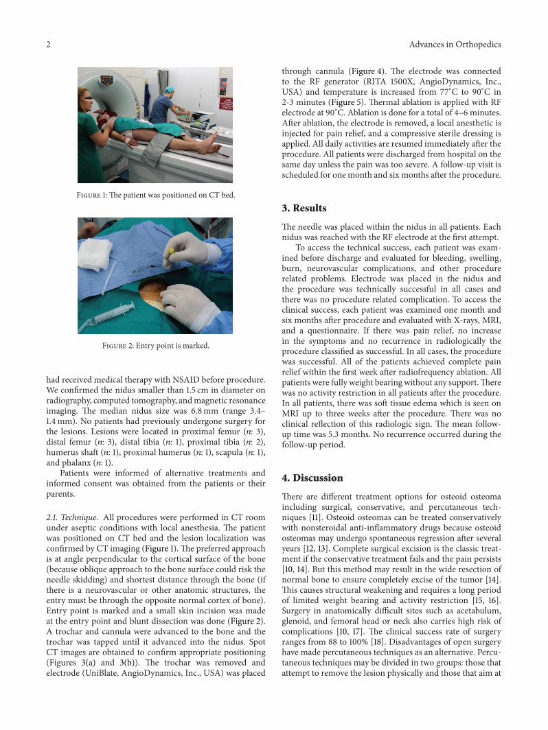
Figure 1: The patient was positioned on CT bed.
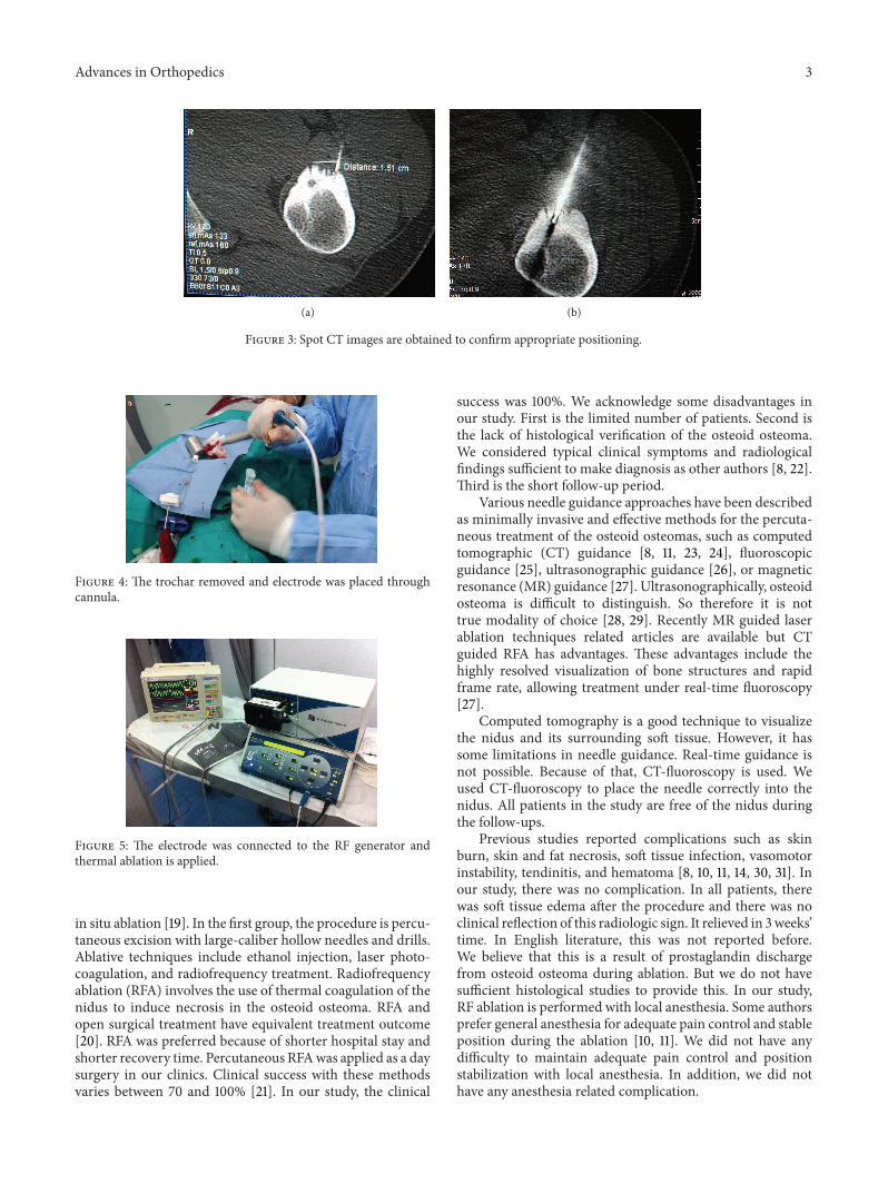
Figure 2: Entry point is marked.
had received medical therapy with NSAID before procedure.We confirmed the nidus smaller than 1.5 cm in diameter onradiography, computed tomography, andmagnetic resonanceimaging. The median nidus size was 6.8mm (range 3.4–1.4mm). No patients had previously undergone surgery forthe lesions. Lesions were located in proximal femur (𝑛: 3),distal femur (𝑛: 3), distal tibia (𝑛: 1), proximal tibia (𝑛: 2),humerus shaft (𝑛: 1), proximal humerus (𝑛: 1), scapula (𝑛: 1),and phalanx (𝑛: 1).
Patients were informed of alternative treatments andinformed consent was obtained from the patients or theirparents.
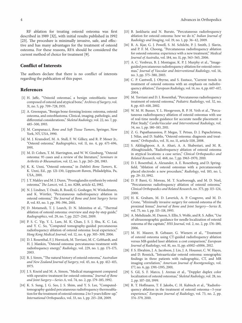
2.1. Technique. All procedures were performed in CT roomunder aseptic conditions with local anesthesia. The patientwas positioned on CT bed and the lesion localization wasconfirmed by CT imaging (Figure 1). The preferred approachis at angle perpendicular to the cortical surface of the bone(because oblique approach to the bone surface could risk theneedle skidding) and shortest distance through the bone (ifthere is a neurovascular or other anatomic structures, theentry must be through the opposite normal cortex of bone).Entry point is marked and a small skin incision was madeat the entry point and blunt dissection was done (Figure 2).A trochar and cannula were advanced to the bone and thetrochar was tapped until it advanced into the nidus. SpotCT images are obtained to confirm appropriate positioning(Figures 3(a) and 3(b)). The trochar was removed andelectrode (UniBlate, AngioDynamics, Inc., USA) was placed
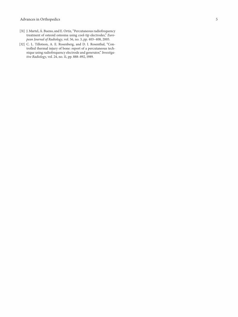
through cannula (Figure 4). The electrode was connectedto the RF generator (RITA 1500X, AngioDynamics, Inc.,USA) and temperature is increased from 77∘C to 90∘C in2-3 minutes (Figure 5). Thermal ablation is applied with RFelectrode at 90∘C. Ablation is done for a total of 4–6 minutes.After ablation, the electrode is removed, a local anesthetic isinjected for pain relief, and a compressive sterile dressing isapplied. All daily activities are resumed immediately after theprocedure. All patients were discharged from hospital on thesame day unless the pain was too severe. A follow-up visit isscheduled for one month and six months after the procedure.
3. Results
The needle was placed within the nidus in all patients. Eachnidus was reached with the RF electrode at the first attempt.
To access the technical success, each patient was exam-ined before discharge and evaluated for bleeding, swelling,burn, neurovascular complications, and other procedurerelated problems. Electrode was placed in the nidus andthe procedure was technically successful in all cases andthere was no procedure related complication. To access theclinical success, each patient was examined one month andsix months after procedure and evaluated with X-rays, MRI,and a questionnaire. If there was pain relief, no increasein the symptoms and no recurrence in radiologically theprocedure classified as successful. In all cases, the procedurewas successful. All of the patients achieved complete painrelief within the first week after radiofrequency ablation. Allpatients were fully weight bearing without any support.Therewas no activity restriction in all patients after the procedure.In all patients, there was soft tissue edema which is seen onMRI up to three weeks after the procedure. There was noclinical reflection of this radiologic sign. The mean follow-up time was 5.3 months. No recurrence occurred during thefollow-up period.
4. Discussion
There are different treatment options for osteoid osteomaincluding surgical, conservative, and percutaneous tech-niques [11]. Osteoid osteomas can be treated conservativelywith nonsteroidal anti-inflammatory drugs because osteoidosteomas may undergo spontaneous regression after severalyears [12, 13]. Complete surgical excision is the classic treat-ment if the conservative treatment fails and the pain persists[10, 14]. But this method may result in the wide resection ofnormal bone to ensure completely excise of the tumor [14].This causes structural weakening and requires a long periodof limited weight bearing and activity restriction [15, 16].Surgery in anatomically difficult sites such as acetabulum,glenoid, and femoral head or neck also carries high risk ofcomplications [10, 17]. The clinical success rate of surgeryranges from 88 to 100% [18]. Disadvantages of open surgeryhave made percutaneous techniques as an alternative. Percu-taneous techniques may be divided in two groups: those thatattempt to remove the lesion physically and those that aim at
Advances in Orthopedics 3
(a) (b)
Figure 3: Spot CT images are obtained to confirm appropriate positioning.
Figure 4: The trochar removed and electrode was placed throughcannula.
Figure 5: The electrode was connected to the RF generator andthermal ablation is applied.
in situ ablation [19]. In the first group, the procedure is percu-taneous excision with large-caliber hollow needles and drills.Ablative techniques include ethanol injection, laser photo-coagulation, and radiofrequency treatment. Radiofrequencyablation (RFA) involves the use of thermal coagulation of thenidus to induce necrosis in the osteoid osteoma. RFA andopen surgical treatment have equivalent treatment outcome[20]. RFA was preferred because of shorter hospital stay andshorter recovery time. Percutaneous RFAwas applied as a daysurgery in our clinics. Clinical success with these methodsvaries between 70 and 100% [21]. In our study, the clinical
success was 100%. We acknowledge some disadvantages inour study. First is the limited number of patients. Second isthe lack of histological verification of the osteoid osteoma.We considered typical clinical symptoms and radiologicalfindings sufficient to make diagnosis as other authors [8, 22].Third is the short follow-up period.
Various needle guidance approaches have been describedas minimally invasive and effective methods for the percuta-neous treatment of the osteoid osteomas, such as computedtomographic (CT) guidance [8, 11, 23, 24], fluoroscopicguidance [25], ultrasonographic guidance [26], or magneticresonance (MR) guidance [27]. Ultrasonographically, osteoidosteoma is difficult to distinguish. So therefore it is nottrue modality of choice [28, 29]. Recently MR guided laserablation techniques related articles are available but CTguided RFA has advantages. These advantages include thehighly resolved visualization of bone structures and rapidframe rate, allowing treatment under real-time fluoroscopy[27].
Computed tomography is a good technique to visualizethe nidus and its surrounding soft tissue. However, it hassome limitations in needle guidance. Real-time guidance isnot possible. Because of that, CT-fluoroscopy is used. Weused CT-fluoroscopy to place the needle correctly into thenidus. All patients in the study are free of the nidus duringthe follow-ups.
Previous studies reported complications such as skinburn, skin and fat necrosis, soft tissue infection, vasomotorinstability, tendinitis, and hematoma [8, 10, 11, 14, 30, 31]. Inour study, there was no complication. In all patients, therewas soft tissue edema after the procedure and there was noclinical reflection of this radiologic sign. It relieved in 3weeks’time. In English literature, this was not reported before.We believe that this is a result of prostaglandin dischargefrom osteoid osteoma during ablation. But we do not havesufficient histological studies to provide this. In our study,RF ablation is performed with local anesthesia. Some authorsprefer general anesthesia for adequate pain control and stableposition during the ablation [10, 11]. We did not have anydifficulty to maintain adequate pain control and positionstabilization with local anesthesia. In addition, we did nothave any anesthesia related complication.
4 Advances in Orthopedics
RF ablation for treating osteoid osteoma was firstdescribed in 1989 [32], with initial results published in 1992[23]. The procedure is minimally invasive, safe, and effec-tive and has many advantages for the treatment of osteoidosteoma. For these reasons, RFA should be considered thecurrent method of choice for treatment [9].
Conflict of Interests
The authors declare that there is no conflict of interestsregarding the publication of this paper.
References
[1] H. Jaffe, “‘Osteoid osteomas’, a benign osteoblastic tumorcomposed of osteoid and atypical bone,”Archives of Surgery, vol.31, no. 5, pp. 709–728, 1935.
[2] A. Greenspan, “Benign bone-forming lesions: osteoma, osteoidosteoma, and osteoblastoma. Clinical, imaging, pathologic, anddifferential considerations,” Skeletal Radiology, vol. 22, no. 7, pp.485–500, 1993.
[3] M. Campanacci, Bone and Soft Tissue Tumors, Springer, NewYork, NY, USA, 1990.
[4] M. J. Kransdorf, M. A. Stull, F. W. Gilkey, and R. P. Moser Jr.,“Osteoid osteoma,” Radiographics, vol. 11, no. 4, pp. 671–696,1991.
[5] M. D. Cohen, T. M. Harrington, and W. W. Ginsburg, “Osteoidosteoma: 95 cases and a review of the literature,” Seminars inArthritis & Rheumatism, vol. 12, no. 3, pp. 265–281, 1983.
[6] K. K. Unni, “Osteoid osteoma,” in Dahlin’s Bone Tumors, K.K. Unni, Ed., pp. 121–130, Lippincott-Raven, Philadelphia, Pa,USA, 2001.
[7] J. T.Makley andM. J. Dunn, “Prostaglandin synthesis by osteoidosteoma,”The Lancet, vol. 2, no. 8288, article 42, 1982.
[8] N. J. Lindner, T. Ozaki, R. Roedl, G. Gosheger,W.Winkelmann,and K. Wortler, “Percutaneous radiofrequency ablation inosteoid osteoma,” The Journal of Bone and Joint Surgery SeriesB, vol. 83, no. 3, pp. 391–396, 2001.
[9] D. Motamedi, T. J. Learch, D. N. Ishimitsu et al., “Thermalablation of osteoid osteoma: overview and step-by-step guide,”Radiographics, vol. 29, no. 7, pp. 2127–2141, 2009.
[10] P. S. C. Yip, Y. L. Lam, M. K. Chan, J. S. J. Shu, K. C. Lai,and Y. C. So, “Computed tomography-guided percutaneousradiofrequency ablation of osteoid osteoma: local experience,”Hong Kong Medical Journal, vol. 12, no. 4, pp. 305–309, 2006.
[11] D. I. Rosenthal, F. J. Hornicek,M. Torriani, M. C. Gebhardt, andH. J. Mankin, “Osteoid osteoma: percutaneous treatment withradiofrequency energy,” Radiology, vol. 229, no. 1, pp. 171–175,2003.
[12] R. J. Simm, “The natural history of osteoid osteoma,”Australianand New Zealand Journal of Surgery, vol. 45, no. 4, pp. 412–415,1975.
[13] J. S. Kneisl and M. A. Simon, “Medical management comparedwith operative treatment for osteoid-osteoma,” Journal of Boneand Joint Surgery—Series A, vol. 74, no. 2, pp. 179–185, 1992.
[14] K. S. Sung, J. G. Seo, J. S. Shim, and Y. S. Lee, “Computed-tomography-guided percutaneous radiofrequency thermoabla-tion for the treatment of osteoid osteoma-2 to 5 years follow-up,”International Orthopaedics, vol. 33, no. 1, pp. 215–218, 2009.
[15] B. Jankharia and N. Burute, “Percutaneous radiofrequencyablation for osteoid osteoma: how we do it,” Indian Journal ofRadiology and Imaging, vol. 19, no. 1, pp. 36–42, 2009.
[16] R. A. Kjar, G. J. Powell, S. M. Schilcht, P. J. Smith, J. Slavin,and P. F. M. Choong, “Percutaneous radiofrequency ablationfor osteoid osteoma: experience with a new treatment,”MedicalJournal of Australia, vol. 184, no. 11, pp. 563–565, 2006.
[17] A. C. Venbrux, B. J. Montague, K. P. J. Murphy et al., “Image-guided percutaneous radiofrequency ablation for osteoid osteo-mas,” Journal of Vascular and Interventional Radiology, vol. 14,no. 3, pp. 375–380, 2003.
[18] C. P. Cantwell, J. Obyrne, and S. Eustace, “Current trends intreatment of osteoid osteoma with an emphasis on radiofre-quency ablation,” European Radiology, vol. 14, no. 4, pp. 607–617,2004.
[19] M. Torriani and D. I. Rosenthal, “Percutaneous radiofrequencytreatment of osteoid osteoma,” Pediatric Radiology, vol. 32, no.8, pp. 615–618, 2002.
[20] W. M. H. Busser, Y. L. Hoogeveen, R. P. H. Veth et al., “Percu-taneous radiofrequency ablation of osteoid osteomas with useof real-time needle guidance for accurate needle placement: aPilot Study,” CardioVascular and Interventional Radiology, vol.34, no. 1, pp. 180–183, 2011.
[21] Z. G. Papathanassiou, P. Megas, T. Petsas, D. J. Papachristou,J. Nilas, and D. Siablis, “Osteoid osteoma: diagnosis and treat-ment,” Orthopedics, vol. 31, no. 11, article 1118, 2008.
[22] S. Akhlaghpoor, A. A. Ahari, A. A. Shabestari, and M. R.Alinaghizadeh, “Radiofrequency ablation of osteoid osteomain atypical locations: a case series,” Clinical Orthopaedics andRelated Research, vol. 468, no. 7, pp. 1963–1970, 2010.
[23] D. I. Rosenthal, A. Alexander, A. E. Rosenberg, and D. Spring-field, “Ablation of osteoid osteomas with a percutaneouslyplaced electrode: a new procedure,” Radiology, vol. 183, no. 1,pp. 29–33, 1992.
[24] D. P. Barei, G. Moreau, M. T. Scarborough, and M. D. Neel,“Percutaneous radiofrequency ablation of osteoid osteoma,”Clinical Orthopaedics and Related Research, no. 373, pp. 115–124,2000.
[25] H. K. Graham, M. D. Laverick, A. P. Cosgrove, and M. D.Crone, “Minimally invasive surgery for osteoid osteoma of theproximal femur,” Journal of Bone and Joint Surgery—Series B,vol. 75, no. 1, pp. 115–118, 1993.
[26] A.Mehdizade,M.Danon, S. Ellis, S.Wolfe, and R. S. Adler, “Useof ultrasonographic guidance for needle localization of osteoidosteoma of the capitate,” HSS Journal, vol. 2, no. 2, pp. 176–180,2006.
[27] M. H. Maurer, B. Gebauer, G. Wieners et al., “Treatmentof osteoid osteoma using CT-guided radiofrequency ablationversus MR-guided laser ablation: a cost comparison,” EuropeanJournal of Radiology, vol. 81, no. 11, pp. e1002–e1006, 2012.
[28] F. S. Ebrahim, J. A. Jacobson, J. Lin, J. A. Housner, C. W. Hayes,and D. Resnick, “Intraarticular osteoid osteoma: sonographicfindings in three patients with radiographic, CT, and MRimaging correlation,” American Journal of Roentgenology, vol.177, no. 6, pp. 1391–1395, 2001.
[29] S. Gil, S. F. Marco, J. Arenas et al., “Doppler duplex colorlocalization of osteoid osteomas,” Skeletal Radiology, vol. 28, no.2, pp. 107–110, 1999.
[30] R. T. Hoffmann, T. F. Jakobs, C. H. Kubisch et al., “Radiofre-quency ablation in the treatment of osteoid osteoma—5-yearexperience,” European Journal of Radiology, vol. 73, no. 2, pp.374–379, 2010.
Advances in Orthopedics 5
[31] J.Martel, A. Bueno, and E. Ortiz, “Percutaneous radiofrequencytreatment of osteoid osteoma using cool-tip electrodes,” Euro-pean Journal of Radiology, vol. 56, no. 3, pp. 403–408, 2005.
[32] C. L. Tillotson, A. E. Rosenberg, and D. I. Rosenthal, “Con-trolled thermal injury of bone: report of a percutaneous tech-nique using radiofrequency electrode and generator,” Investiga-tive Radiology, vol. 24, no. 11, pp. 888–892, 1989.

Submit your manuscripts athttp://www.hindawi.com
Stem CellsInternational
Hindawi Publishing Corporationhttp://www.hindawi.com Volume 2014
Hindawi Publishing Corporationhttp://www.hindawi.com Volume 2014
MEDIATORSINFLAMMATION
of
Hindawi Publishing Corporationhttp://www.hindawi.com Volume 2014
Behavioural Neurology
EndocrinologyInternational Journal of
Hindawi Publishing Corporationhttp://www.hindawi.com Volume 2014
Hindawi Publishing Corporationhttp://www.hindawi.com Volume 2014
Disease Markers
Hindawi Publishing Corporationhttp://www.hindawi.com Volume 2014
BioMed Research International
OncologyJournal of
Hindawi Publishing Corporationhttp://www.hindawi.com Volume 2014
Hindawi Publishing Corporationhttp://www.hindawi.com Volume 2014
Oxidative Medicine and Cellular Longevity
Hindawi Publishing Corporationhttp://www.hindawi.com Volume 2014
PPAR Research
The Scientific World JournalHindawi Publishing Corporation http://www.hindawi.com Volume 2014
Immunology ResearchHindawi Publishing Corporationhttp://www.hindawi.com Volume 2014
Journal of
ObesityJournal of
Hindawi Publishing Corporationhttp://www.hindawi.com Volume 2014
Hindawi Publishing Corporationhttp://www.hindawi.com Volume 2014
Computational and Mathematical Methods in Medicine
OphthalmologyJournal of
Hindawi Publishing Corporationhttp://www.hindawi.com Volume 2014
Diabetes ResearchJournal of
Hindawi Publishing Corporationhttp://www.hindawi.com Volume 2014
Hindawi Publishing Corporationhttp://www.hindawi.com Volume 2014
Research and TreatmentAIDS
Hindawi Publishing Corporationhttp://www.hindawi.com Volume 2014
Gastroenterology Research and Practice
Hindawi Publishing Corporationhttp://www.hindawi.com Volume 2014
Parkinson’s Disease
Evidence-Based Complementary and Alternative Medicine
Volume 2014Hindawi Publishing Corporationhttp://www.hindawi.com